What is a pancreatic pseudocyst
Pancreatic pseudocyst is best defined as localized fluid collection arising from the pancreas that are rich in pancreatic juice containing amylase, lipase and zymogens or, if no communication with the pancreatic ducts exists, with protease-free serous fluid, that have a nonepithelialized wall consisting of fibrous and inflammatory tissue, and that usually appear several weeks after the onset of acute pancreatitis or chronic pancreatitis
1. Pancreatic pseudocysts are to be distinguished from acute fluid collections, organized necrosis, and abscesses 2. Pancreatic pseudocyst belongs to a large and heterogeneous group of cystic pancreatic lesions and represent a complication of acute or chronic pancreatitis 3.
Several classification systems of pancreatic pseudocysts have been proposed addressing either the pathogenesis of pseudocyst formation, as in the Atlanta classification, or morphological features such as pancreatic duct anatomy and communication of the pseudocyst with the ducts. The latter are less frequently used. The Atlanta classification system 4 subdivides four entities: a) acute fluid collection, occurring early in the course of acute pancreatitis and lacking a wall of granulomatous or fibrous tissue; b) acute pseudocysts, a cavity surrounded by fibrous or granulomatous tissue that is a consequence of acute pancreatitis or trauma; c) chronic pseudocysts, arising in chronic pancreatitis and without a preceding episode of acute pancreatitis; and d) pancreatic abscess, an intra-abdominal collection of pus in the proximity of the pancreas with little or no necrosis resulting from acute or chronic pancreatitis or trauma. The diagnosis of an acute pseudocyst can be made if an acute fluid collection persists for 4–6 weeks and is enveloped by a distinct wall 5. Another classification system offered by D’Egidio and Schein in 1991 6 is also based on the underlying disease (acute, acute-on-chronic or chronic pancreatitis), but takes the duct anatomy (normal, diseased, strictured) and the pseudocyst–duct communication (rare, sometimes, always) into account.
The following are the latest terms according to the updated Atlanta classification to describe fluid collections associated with acute pancreatitis 7:
- fluid collections in interstitial edematous pancreatitis
- acute peripancreatic fluid collections: in the first 4 weeks: non-encapsulated peripancreatic fluid collections
- pseudocysts: develop after 4 weeks; encapsulated peripancreatic or remote fluid collections
- fluid collections in necrotizing pancreatitis
- acute necrotic collection: in the first 4 weeks; non-encapsulated heterogeneous non-liquefied material
- walled-off necrosis: develop after 4 weeks; encapsulated heterogeneous non-liquefied material
Nealon and Walser 8 classified pancreatic pseudocysts according to the duct anatomy and the presence or absence of communication with the pseudocyst cavity.
The incidence of pancreatic pseudocysts in both acute and chronic pancreatitis has been assessed in large series of clinical studies. The relative proportion of acute and chronic pancreatic pseudocysts varies between reports and depends on how pancreatic pseudocysts are defined and by what means they are detected 9. The incidence of pancreatic pseudocysts ranges from 5% to 16% in acute pancreatitis 10, whereas in chronic pancreatitis the numbers are higher and incidence rates of 20–40% have been published even in cohorts where advanced imaging techniques were not employed 11.
The highest incidence of pancreatic pseudocysts can be found in patients with chronic pancreatitis due to alcohol abuse. In a study of 97 patients 12 with pseudocysts, alcohol consumption was found to be the causative factor in 64% of patients with chronic pancreatitis and in 26% of patients with acute pancreatitis.
Other studies also revealed alcohol-related pancreatitis preceding pancreatic pseudocysts in about 56–78% of patients 13. Besides this, as far as cause of pancreatitis is concerned 6–36% of pancreatic pseudocysts arise in gallstone-induced pancreatitis, 3–8% in post-surgical or traumatic pancreatitis, rarely after hyperlipidemia-induced pancreatitis and in 6–20% no cause is found (idiopathic pancreatitis) 3.
Chronic pancreatic pseudocysts over 8 weeks are less likely to resolve spontaneously and as the risk of complications increases with time, treatment of large pseudocysts (>5 cm) should not be postponed 14. Surgery is the traditional modality for treating pancreatic pseudocysts, with high success rates and low morbidity and mortality, and it still plays an important role in therapy. Laparoscopic management has been reported with very encouraging results, but long-term follow-up has still to show equivalence to open surgery. Endoscopic therapy is a reasonable alternative to surgery, particularly for chronic pseudocysts, displaying an even lower morbidity and mortality rate. Failure of transpapillary or transmural drainage may make subsequent surgery necessary 15. Nonetheless, initial endoscopic drainage should be considered as a valuable tool and the method of choice in patients with chronic pancreatitis-associated large pseudocysts 16.
Rupture of the pancreatic pseudocyst is a medical emergency. Go to the emergency room or call the local emergency number if you develop symptoms of bleeding or shock, such as:
- Fainting
- Fever and chills
- Rapid heartbeat
- Severe abdominal pain
Pancreatic pseudocyst causes
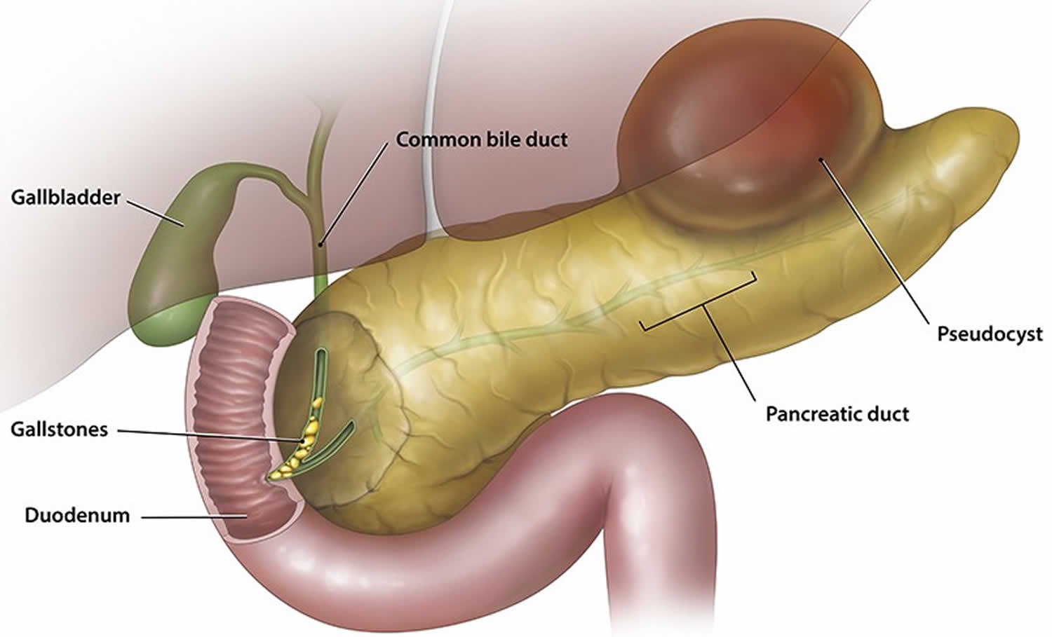
The pancreas is an organ located behind the stomach. It produces chemicals (called enzymes) needed to digest food. It also produces the hormones insulin and glucagon.
Pancreatic pseudocysts most often develop after an episode of severe pancreatitis (acute or chronic pancreatitis). Pancreatitis happens when your pancreas get inflamed. There are many causes of pancreatitis.
Acute pancreatitis affects men more often than women. Certain diseases, surgeries, and habits make you more likely to develop acute pancreatitis.
- Alcohol use is responsible for up to 70% of cases in the United States. About 5 to 8 drinks per day for 5 or more years can damage the pancreas.
- Gallstones are the next most common cause (up to 15% develop pancreatitis). When the gallstones travel out of the gallbladder into the bile ducts, they block the opening that drains bile and enzymes. The bile and enzymes “back up” into the pancreas and cause swelling.
- Genetics may be a factor in some cases.
- Idiopathic (cause is not known): 20% (range 10%-30%) of cases of acute pancreatitis, evidence suggests that most cases are associated with congenital duct abnormalities
Other conditions that have been linked to pancreatitis are:
- Autoimmune problems (when the immune system attacks the body)
- Damage to the ducts or pancreas during surgery
- High blood levels of a fat called triglycerides — most often above 1,000 mg/dL
- Injury to the pancreas from an accident
Other causes include:
- After certain procedures used to diagnose gallbladder and pancreas problems (ERCP) or ultrasound guided biopsy
- After trauma to the belly, more often in children
- Iatrogenic, e.g. post partial gastrectomy 17
- Cystic fibrosis
- Overactive parathyroid gland
- Reye syndrome
- Use of certain medicines (especially estrogens, corticosteroids, sulfonamides, thiazides, and azathioprine)
- Certain infections, such as mumps, that involve the pancreas
The major risk factors for the development of chronic pancreatitis may be categorized according to the TIGAR-O system 18:
- T: toxic-metabolic (e.g. alcohol)
- I: idiopathic, recent guidelines recommend that cystic fibrosis needs to be ruled out in these patients before calling it idiopathic 19
- G: genetic, more commonly seen in the pediatric population
- A: autoimmune
- R: recurrent
- O: obstructive (e.g. choledocholithiasis, pancreatic head tumor)
Pancreatic pseudocyst happens when the pancreatic ducts (tubes) in the pancreas are damaged with resulting leakage and accumulation of pancreatic juice resulting in hemorrhagic fat necrosis. A severe inflammatory reaction that is incited by this results in encapsulation of the cyst by fibrous tissue. This usually takes 4-6 weeks 20. In approximately 50% of cases the pancreatic pseudocyst retains a communication with the pancreatic duct 21. Such pancreatic pseudocysts are more problematic to treat, and are more likely to recur.
Pancreatic pseudocyst prevention
The way to prevent pancreatic pseudocysts is by preventing pancreatitis. If pancreatitis is caused by gallstones, the provider will perform surgery to remove the gallbladder (cholecystectomy).
When pancreatitis occurs due to alcohol abuse, you must stop drinking alcohol to prevent future attacks.
When pancreatitis occurs due to high blood triglycerides, this condition should be treated.
Pancreatic pseudocyst symptoms
Pancreatic pseudocysts are frequently found on imaging follow-up of pancreatitis, and may in themselves be asymptomatic for some time. Symptoms can occur within days to months after an attack of pancreatitis. They include:
- mass effect
- biliary obstruction
- gastric outlet obstruction
- secondary infection
- bloating of the abdomen
- constant pain or deep ache in the abdomen, which may also be felt in the back
- nausea and vomiting
- loss of appetite
- difficulty eating and digesting food
- jaundice or sepsis from an infected pseudocyst (rare)
Pancreatic pseudocyst possible complications
Pancreatic pseudocyst complications may include:
- A pancreatic abscess can develop if the pseudocyst becomes infected.
- The pseudocyst can break open (rupture). This can be a serious complication because shock and excess bleeding (hemorrhage) may develop.
- The pseudocyst may press down on (compress) nearby organs.
Bleeding is the most feared complication and is caused by the erosion of the pseudocyst into a vessel. Consider the possibility of bleeding in any patient who has a sudden increase in abdominal pain coupled with a drop in hematocrit level or a change in vital signs. Therapy is emergent surgery or angiography with embolization of the bleeding vessel.
Do not perform a percutaneous or endoscopic drainage procedure under any circumstances in patients with suspected bleeding into a pseudocyst. Consider the possibility of infection of the pseudocyst in patients who develop fever or an elevated white blood cell count. Treat infection with antibiotics and urgent drainage.
Gastrointestinal obstruction, manifesting as nausea and vomiting, is an indication for drainage.
The pseudocyst can also rupture. A controlled rupture into an enteric organ occasionally causes gastrointestinal bleeding. On rare occasions, a profound rupture into the peritoneal cavity causes peritonitis and death.
Pancreatic pseudocyst diagnosis
Your health care provider may feel your abdomen for a pseudocyst. It will feel like a lump in the middle or left upper abdomen.
Laboratory studies that may be considered include the following:
- Serum amylase and lipase levels (limited utility) – Often elevated but may be within the reference ranges
- Serum bilirubin and liver function tests (limited utility) – Sometimes elevated if the biliary tree is involved
- Cyst fluid analysis – Carcinoembryonic antigen (CEA) and CEA-125 (low in pseudocysts and elevated in tumors); fluid viscosity (low in pseudocysts and elevated in tumors); amylase (usually high in pseudocysts and low in tumors)
Imaging studies that may be helpful include the following:
- Abdominal ultrasonography – Not the study of choice for establishing a diagnosis
- Abdominal computed tomography (CT) – The criterion standard for pancreatic pseudocysts
- Endoscopic retrograde cholangiopancreatography (ERCP) – Not necessary for diagnosis but useful in planning drainage strategy.
- Magnetic resonance imaging (MRI) – Not necessary for diagnosis but useful in detecting a solid component to the cyst and in differentiating between organized necrosis and a pseudocyst
- Endoscopic ultrasonography – Not necessary for diagnosis but very important in planning therapy, particularly if endoscopic drainage is contemplated
Employing imaging techniques, pancreatic pseudocyst characteristics like size, location, wall thickness and septa can be detected. However, approximately 10% of pancreatic pseudocysts can have ill-defined features that overlap with the characteristics of cystic tumors 22.
Pancreatic pseudocyst treatment
Treatment depends on the size of the pancreatic pseudocyst and whether it is causing symptoms. Many pancreatic pseudocysts go away on their own 21. Those that remain for more than 6 weeks and are larger than 5 cm in diameter often need treatment.
No medications are specific to the treatment of pancreatic pseudocysts. Antibiotics and octreotide may be useful adjuncts in some cases.
Possible treatments include:
- Drainage through the skin using a needle, most often guided by a CT scan.
- Endoscopic-assisted drainage using an endoscope. In this, a tube containing a camera and a light is passed down into the stomach)
- Surgical drainage of the pseudocyst. A connection is made between the cyst and the stomach or small intestine. This may be done using a laparoscope.
Drainage options are as follows:
- Percutaneous catheter drainage – The procedure of choice for infected pseudocysts; although recurrence and failure rates are high, it may be a good temporizing measure
- Endoscopic drainage, either transpapillary (via ERCP) or transmural – The complication rate appears to decrease and efficacy to increase with experience
- Surgical drainage – The criterion standard; internal drainage is the procedure of choice, but laparoscopic drainage has yielded good results in some cases
Table 1. Indications for therapeutic intervention of pancreatic pseudocysts
| Complicated pancreatic pseudocysts (one criterion sufficient) |
|---|
|
Symptomatic pancreatic pseudocyst:
|
| Asymptomatic pancreatic pseudocyst: |
Pancreatic pseudocyst prognosis
The outcome is generally good with treatment. It is important to make sure that it is not a pancreatic cancer that starts in a cyst, which has a worse outcome.
- Kloppel G. Pseudocysts and other non-neoplastic cysts of the pancreas. Semin Diagn Pathol. 2000;17:7–15.[↩]
- Pancreatic pseudocysts. https://emedicine.medscape.com/article/184237-overview[↩]
- Aghdassi AA, Mayerle J, Kraft M, Sielenkämper AW, Heidecke CD, Lerch MM. Pancreatic pseudocysts–when and how to treat?. HPB (Oxford). 2006;8(6):432-41. https://www.ncbi.nlm.nih.gov/pmc/articles/PMC2020756/[↩][↩][↩]
- Bradley EL., 3rd A clinically based classification system for acute pancreatitis. Summary of the International Symposium on Acute Pancreatitis, Atlanta, Ga, September 11 through 13, 1992. Arch Surg. 1993;128:586–90[↩]
- Pitchumoni CS, Agarwal N. Pancreatic pseudocysts. When and how should drainage be performed? Gastroenterol Clin North Am. 1999;28:615–39.[↩]
- D’Egidio A, Schein M. Pancreatic pseudocysts: a proposed classification and its management implications. Br J Surg. 1991;78:981–4[↩]
- Banks PA, Bollen TL, Dervenis C et-al. Classification of acute pancreatitis-2012: revision of the Atlanta classification and definitions by international consensus. Gut. 2012;62 (1): 102-11. doi:10.1136/gutjnl-2012-302779[↩]
- Nealon WH, Walser E. Main pancreatic ductal anatomy can direct choice of modality for treating pancreatic pseudocysts (surgery versus percutaneous drainage) Ann Surg. 2002;235:751–8.[↩]
- Grace PA, Williamson RC. Modern management of pancreatic pseudocysts. Br J Surg. 1993;80:573–81[↩]
- Maringhini A, Uomo G, Patti R, Rabitti P, Termini A, Cavallera A, et al. Pseudocysts in acute nonalcoholic pancreatitis: incidence and natural history. Dig Dis Sci. 1999;44:1669–73[↩]
- Barthet M, Bugallo M, Moreira LS, Bastid C, Sastre B, Sahel J. Management of cysts and pseudocysts complicating chronic pancreatitis. A retrospective study of 143 patients. Gastroenterol Clin Biol. 1993;17:270–6[↩]
- Sanfey H, Aguilar M, Jones RS. Pseudocysts of the pancreas, a review of 97 cases. Am Surg. 1994;60:661–8.[↩]
- Usatoff V, Brancatisano R, Williamson RC. Operative treatment of pseudocysts in patients with chronic pancreatitis. Br J Surg. 2000;87:1494–9[↩]
- Grace PA, Williamson RC. Modern management of pancreatic pseudocysts. Br J Surg. 1993;80:573–81.[↩]
- Rosso E, Alexakis N, Ghaneh P, Lombard M, Smart HL, Evans J, et al. Pancreatic pseudocyst in chronic pancreatitis: endoscopic and surgical treatment. Dig Surg. 2003;20:397–406.[↩]
- Heider R, , Meyer AA, , Galanko Ja, , Behrns KE. Percutaneous drainage of pancreatic pseudocysts is associated with a higher failure rate than surgical treatment in unselected patients. Ann Surg 1999;229:781–7; discussion 787–9.[↩]
- Surgical Pathology of the GI Tract, Liver, Biliary Tract and Pancreas: Expert Consult. Saunders. ISBN:1416040595.[↩]
- Etemad B, Whitcomb DC. Chronic pancreatitis: diagnosis, classification, and new genetic developments. Gastroenterology. 2001;120 (3): 682-707.[↩]
- Löhr JM, Dominguez-Munoz E, Rosendahl J, Besselink M, Mayerle J, Lerch MM, Haas S, Akisik F, Kartalis N, Iglesias-Garcia J, Keller J, Boermeester M, Werner J, Dumonceau JM, Fockens P, Drewes A, Ceyhan G, Lindkvist B, Drenth J, Ewald N, Hardt P, de Madaria E, Witt H, Schneider A, Manfredi R, Brøndum FJ, Rudolf S, Bollen T, Bruno M. United European Gastroenterology evidence-based guidelines for the diagnosis and therapy of chronic pancreatitis (HaPanEU). United European gastroenterology journal. 5 (2): 153-199. doi:10.1177/2050640616684695[↩]
- Kim YH, Saini S, Sahani D et-al. Imaging diagnosis of cystic pancreatic lesions: pseudocyst versus nonpseudocyst. Radiographics. 25 (3): 671-85. doi:10.1148/rg.253045104[↩]
- Lucey BC, Kuligowska E. Radiologic management of cysts in the abdomen and pelvis. AJR Am J Roentgenol. 2006;186 (2): 562-73. doi:10.2214/AJR.04.1051[↩][↩]
- Diagnosis of pancreatic cystic neoplasms: a report of the cooperative pancreatic cyst study. Brugge WR, Lewandrowski K, Lee-Lewandrowski E, Centeno BA, Szydlo T, Regan S, del Castillo CF, Warshaw AL. Gastroenterology. 2004 May; 126(5):1330-6.[↩]
- Bradley EL., 3rd A clinically based classification system for acute pancreatitis. Summary of the International Symposium on Acute Pancreatitis, Atlanta, Ga, September 11 through 13, 1992. Arch Surg. 1993;128:586–90.[↩]
- Gouyon B, Levy P, Ruszneiwski P, Zins M, Hammel P, Vilgrain V, et al. Predictive factors in the outcome of pseudocysts complicating alcoholic chronic pancreatitis. Gut. 1997;41:821–5.[↩]
- Ridder GJ, Maschek H, Klempnauer J. Favourable prognosis of cystadeno- over adenocarcinoma of the pancreas after curative resection. Eur J Surg Oncol. 1996;22:232–6.[↩]


