Pupillary light reflex
The pupillary light reflex is an autonomic reflex that constricts the pupil in response to light, thereby adjusting the amount of light that reaches the retina 1. Pupillary constriction occurs via innervation of the iris sphincter muscle, which is controlled by the parasympathetic system 1.
Testing of the pupillary light reflex is useful to identify a relative afferent pupillary defect due to asymmetric afferent output from a lesion anywhere along the afferent pupillary pathway as described above 2. In patients with an relative afferent pupillary defect, when light is shined in the affected eye, there will be dilation of both pupils due to an abnormal afferent arm 3. When the examiner swings the light to the unaffected eye, both pupils constrict. Detection of an relative afferent pupillary defect requires two eyes but only one functioning pupil; if the second pupil is unable to constrict, such as due to a third nerve palsy, a “reverse relative afferent pupillary defect” test can be performed using the swinging flashlight test 3. Direct and consensual responses should be compared in the reactive pupil. If the reactive pupil constricts more with the direct response than with the consensual response, then the relative afferent pupillary defect is in the unreactive pupil. Alternatively, if the reactive pupil constricts more with the consensual response than with the direct response, then the relative afferent pupillary defect is in the reactive pupil. An relative afferent pupillary defect can occur due to downstream lesions in the pupillary light reflex pathway (such as in the optic tract or pretectal nuclei) 3. A transient relative afferent pupillary defect can occur secondary to local anesthesia[4]. “Pupillary escape” is an abnormal pupillary response to a bright light, in which the pupil initially constricts to light and then slowly redilates to its original size 3. Pupillary escape can occur on the side of a diseased optic nerve or retina, most often in patients with a central field defect.
Pupillary light reflex pathway
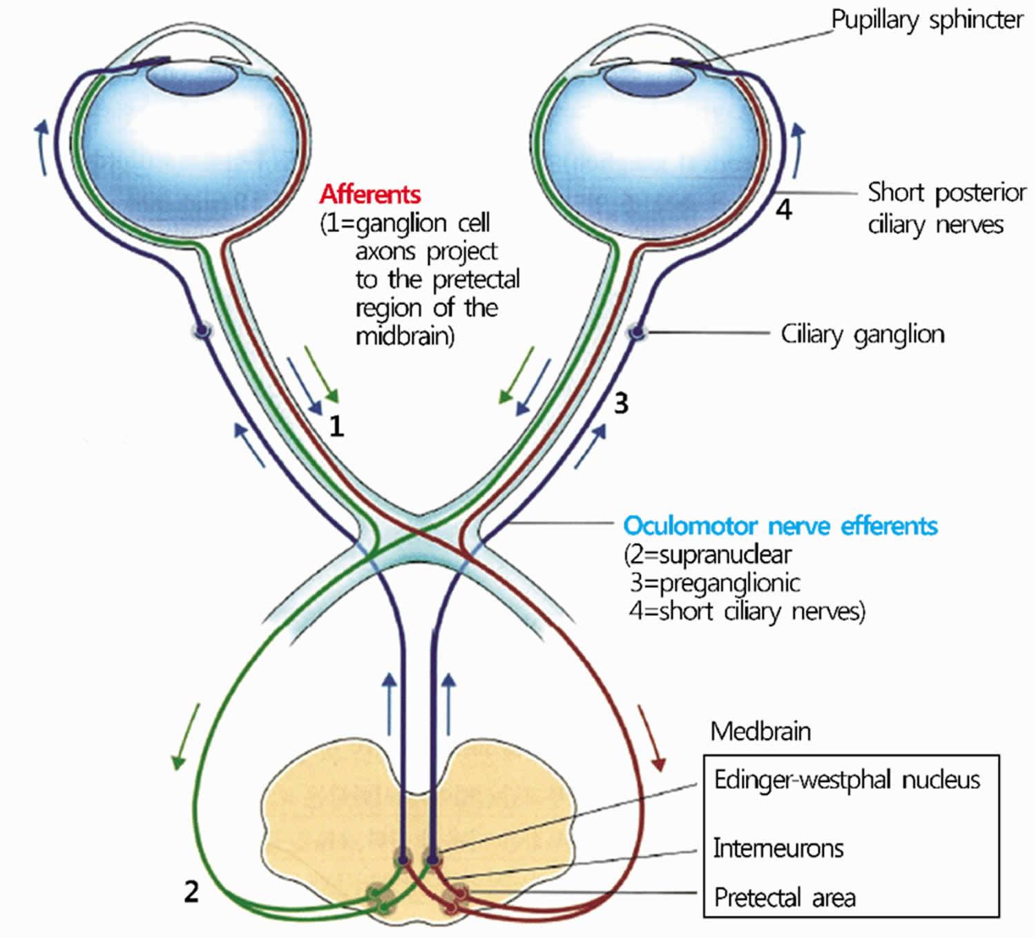
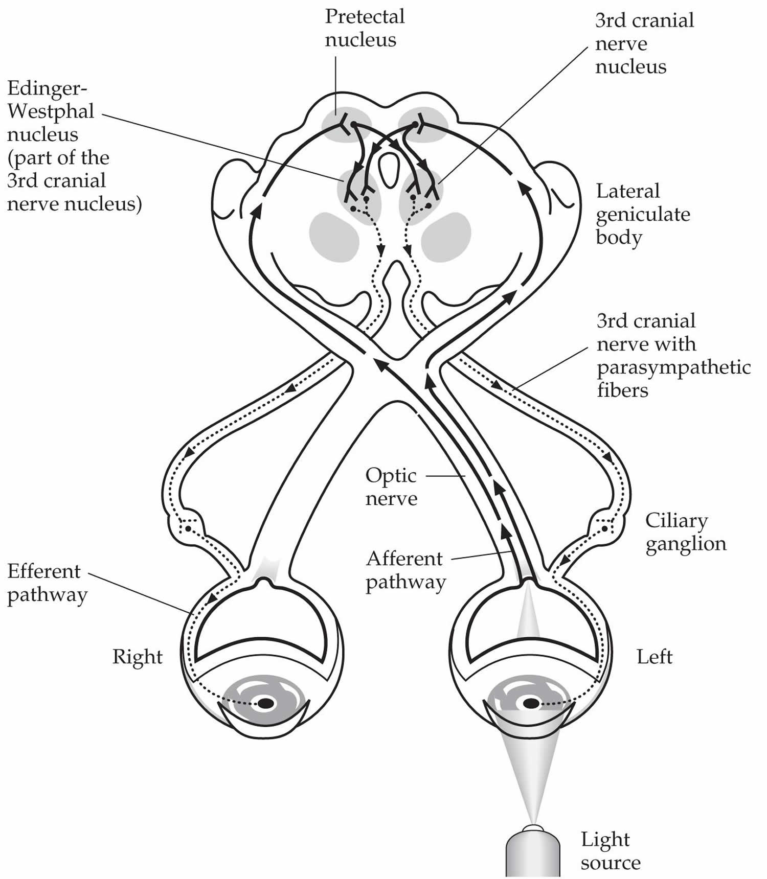
Light travels through the cornea, anterior chamber, pupil, lens, and the posterior chamber, eventually reaching the retina. Photoreceptor cells in the outer layers of the retina, which are called rods and cones, convert light stimuli into neuronal impulses. These signals are then relayed to the bipolar cells, which interact with ganglion cells, which in turn coalesce to form the optic disc and optic nerve (CN II). The optic nerve sends impulses to the brain for further processing and image recognition 4. These are the first steps of the pupillary light reflex afferent pathway. The optic nerve then forms the optic chiasm, which diverges into a left and right optic tract. At the optic chiasm, nasal retinal fibers will cross to the contralateral side of the optic tract, and the temporal retinal fibers continue on the ipsilateral side. Thus, the right optic tract will contain temporal retinal fibers from the right eye, as well as nasal retinal fibers from the left eye. The optic tracts join the brachium of the superior colliculus, and then signals travel to the pretectal area of the midbrain. Each pretectal area sends bilateral signals to the preganglionic parasympathetic nuclei in the midbrain called Edinger-Westphal nuclei of the oculomotor complex 5. From the Edinger-Westphal nucleus, efferent pupillary parasympathetic preganglionic fibers travel on the oculomotor nerve to synapse in the ciliary ganglion, which sends parasympathetic postganglionic axons in the short ciliary nerve to innervate the iris sphincter smooth muscle via M3 muscarinic receptors 2. The contraction of the iris sphincter muscles leads to pupillary constriction (miosis) 6. This extensive pathway is being tested when a light is shined in the eyes. And, because of the crossing fibers, there is not only a direct pupillary reflex but also a consensual pupillary light reflex. Of note, the pupillary dark reflex involves a separate pathway, which ends with sympathetic fibers from long ciliary nerves innervating the dilator pupillae muscle.
Pupillary light reflex is used to assess the brain stem function. Abnormal pupillary light reflex can be found in optic nerve injury, oculomotor nerve damage, brain stem lesions, such as tumors, and medications like barbiturates 7.
Figure 1. Pupillary light reflex pathway
Pupillary light reflex abnormalities
Pupillary light reflexes are measured based on a 0 to 4+ gradient that considers the magnitude and speed of the light response 7. A normal, healthy adult patient is expected to have a 4+ response, which indicates a brisk, large response. A 3+ grading indicates a moderate response, 2+ is a small, slowed response, 1+ represents a tiny/just visible response, and a 0 indicates unresponsive pupils. Commonly, clinicians document pupils that are equal, round and reactive to light (PERRL)–saying the pupils are equal, round, and reactive to light.
In standard clinical testing conditions, the diameter of the pupils will usually range from two to five millimeters. Per decade of aging that occurs, there is a 0.3 mm decrease in the standard pupil diameter that has been associated with iris stiffening. The pupillary light response exhibits varying sensitivity to the chromic spectrum, indicating that the process of light recognition is significantly complex; it is not as simple as a binary response with detection of “light” versus “no light.” While there is a baseline fluctuation in the steady-state conditions for pupillary dilation, a concern for neurological abnormalities is considered in cases of marked pupillary changes, whether with constriction or dilation. One such condition is anisocoria, and it is estimated at 4% of the general population has anisocoria of greater than 1 millimeter, in which case neurological compromise must be ruled out. Pupillary latency occurs when the reaction time of the pupil is inversely related to the increase in light intensity from the stimulus; this can serve as a cue to a potential neurologic cause. Latency increases by approximately 1 millisecond per year with aging. Overall, normal pupillary response times are about one second for initial constriction and 5 seconds for dilation 8.
Direct and consensual pupillary light reflexes test for appropriate neurological pathway connections and functioning of both cranial nerve II and III. Light entering the eye is processed through the pupillary light reflex, and signals directed to the iris sphincter muscle to adjust the amount of light that reaches the retina. While there are other reasons for variation in pupillary dilation and constriction, such as arousal leading to changes in the balance of the sympathetic and parasympathetic nervous systems, here we will focus on its relation to light exposure. Pupils can become mydriatic, or dilate, in response to potential disease, drug toxicity, trauma, increased intracranial pressure, brainstem damage, or nerve damage to cranial nerve II and/or III 9.
Abnormalities also depend on where in the track the damage has been done. In the event of optic nerve damage, visual field defects or complete vision loss can occur. If this damage is before the optic chiasm, in the optic nerve, then there are deficits noted to bilateral ipsilateral monocular vision loss. This damage leads to a relative afferent pupillary defect (RAPD), known as a Marcus Gunn pupil, which is examined using the swinging flashlight test. Causes of a Marcus Gunn pupil include ischemic optic neuropathy, optic neuritis, nerve compression, trauma or through asymmetric glaucoma.
Optic nerve damage on one side:
- The ipsilateral direct reflex is lost
- The contralateral consensual reflex is intact
- The ipsilateral direct reflex is lost (because light shone into the eye on the damaged side cannot signal to the brain)
- The contralateral consensual reflex is intact (because light shone into the opposite eye can signal to the brain, causing constriction of both pupils via the normal oculomotor nerves)
Oculomotor nerve damage on one side:
- The ipsilateral direct reflex is LOST
- The contralateral consensual reflex is lost
- The ipsilateral direct reflex is LOST (because light shone into the damaged eye can still signal to the brain via the normal optic nerve, causing attempted constriction of both pupils; the contralateral pupil constricts via its normal oculomotor nerve, but the ipsilateral pupil is unable to constrict due to its damaged oculomotor nerve)
- The contralateral consensual reflex is lost (because although light shone into the opposite eye can still signal to the brain, causing attempted constriction of both pupils, the pupil on the damaged side is unable to constrict due to its damaged oculomotor nerve; the pupil on the undamaged side will still be able to constrict via its normal oculomotor nerve)
Unilateral optic neuropathies, most notably optic neuritis, can cause relative afferent pupillary defects. Optic neuritis is an anterior or posterior inflammatory demyelination of the optic nerve, leading to atrophy of optic nerve fibers and a relative afferent pupillary defect. A relative afferent pupillary defect can be detected in 96% of acute unilateral cases of optic neuritis 8. Ischemic optic neuropathies, such as NAION and AION, can cause relative afferent pupillary defects via optic nerve ischemia and infarction secondary to optic nerve edema 10. Asymmetric glaucoma can result in an relative afferent pupillary defect, secondary to retinal nerve fiber layer loss.
Relative afferent pupillary defects can occur due to ischemic retinal diseases such as branch retinal vein occlusion, central retinal vein occlusion, branch retinal artery occlusion, and central retinal artery occlusion secondary to death of photoreceptors and viable retina, ultimately leading to an uneven pupillary response 5.. Via the same mechanism of significant retinal cell death, retinal detachments can cause relative afferent pupillary defects. In 1987, a prediction model quantified the correlation between the sizes of relative afferent pupillary defect to the amount of retina detached. Detachment of each peripheral quadrant correlated to 0.36 log units of pupillary defect. The detachment of the macula caused 0.68 log units of pupillary defect.
Argyll Robertson pupil, noted in tabes dorsalis from neurosyphilis, is the notable weak-to-absent pupillary light reflex bilaterally, though the pupils will still constrict for the near response. With the near response (accommodation) intact it can be assumed that the afferent and efferent pathways are grossly intact and that the deficit is related to degeneration in bilateral olivary pretectal nuclei or their projections 10.
Compression damage to the optic chiasm leads to bitemporal hemianopia and is frequently related to a pituitary adenoma. Downstream to the optic chiasm, damage to the optic tract will produce contralateral homonymous hemianopia; for example, if there is damage to the left optic tract, there are right visual field deficits to both eyes. In the case of comatose patients, it has been noted that a majority of the patients had non-reactive, dilated pupils, and the one patient that had pinpoint pupils became vegetative 11. Uncal herniation, in which the uncus protrudes over the edge of the tentorium, can lead to compression of CN III, suggesting current or impending brainstem compromise. Lesions within the efferent pathway, specifically the preganglionic fibers of the oculomotor nerve, can cause ipsilateral mydriasis and accommodation paralysis. One syndrome noted to have this finding is Weber Syndrome. If there is damage to the postganglionic fibers, tonic dilated pupil or Adie syndrome develops so that the constrictor muscles are hypersensitive to a cholinergic stimulus. If there is a disruption between the balance of parasympathetic and sympathetic innervation, such as in Horner syndrome where there is a loss of sympathetic stimulation, leading to miosis of the ipsilateral pupil 10.
Transient mydriasis can be associated with tricyclic antidepressants, typical antipsychotics, and selective serotonin reuptake inhibitors, but usually these are not long-term consequences. Topiramate, used for migraines, has been associated with acquired myopia and angle-closure glaucoma 12. The fixed dilation of pupils noted in coma patients was related to the increased intracranial pressure (ICP) in which an association was noted in 1866 from Von Leyden’s animal experiments. Through continued research over the next 50 years or so, it was noted that the fixed dilation of pupils was noted to be a sign of acute mass effect in relation to in the intracranial pressure 13.
Tumors in the retina, optic nerve, and brain can also cause relative afferent pupillary defects. In children, the most common intraocular tumors are benign, developmental cysts. The most common malignant intraocular tumor is retinoblastoma 13. Tumors or lesions affecting the optic chiasm or midbrain can cause decreased signals reaching the Edinger-Westphal nuclei, leading to pupillary constriction. In children, the most common intracranial tumor detected is a glioma. They account for 75% of intracranial tumors in children. Also common in children are astrocytomas, medulloblastomas, and ependymomas.
Another cause of relative afferent pupillary defect is severe amblyopia, characterized as amblyopia of 20/100 to 20/400. Clinically, relative afferent pupillary defects are found in severe amblyopia with best corrected visual acuity 20/400 or worse. While the etiology of a relative afferent pupillary defects in amblyopia is poorly understood, significant risk factors include anisometropia, early age of onset with a history of strabismus, level of visual acuity at the conclusion of treatment, and extended periods of occlusion therapy 14.
Pupillary escape is a phenomenon that can occur in the setting of a diseased optic nerve or retina. When light is shone onto the affected pupil, there will be a transient pupillary constriction and then a slow dilation to the original size 15.
In cases in which one pupil is unable to constrict (such as due to a third nerve palsy), the “reverse relative afferent pupillary defect test” can be performed, with direct and consensual responses compared in the reactive pupil. If the reactive pupil constricts more during the direct response, then the relative afferent pupillary defect is in the unreactive eye. If the reactive pupil constricts more during the consensual response, then the relative afferent pupillary defect is in the unreactive eye 16. Emergency clinicians often encounter patients with the triad of pinpoint pupils, respiratory depression, and coma related to opioid overuse. Opioids are used for pain relief by interacting with opioid receptors, including mu, delta, and kappa. With significant respiratory depression resulting in hypoxia, pupils can become dilated. Oxygenation causes the pupils to revert to the original pinpoint presentation caused by the opioid. For stabilization, one of the medications given to these patients is naloxone, an opioid antagonist, which has a peak effect at approximately 10 minutes. Repeated dosing is frequently required and can be given up to 5 mg per hour. If there is a dilation of the pupil with the administration of naloxone, this also eliminates organophosphate poisoning, which can present similarly. Pupillary changes are used to recognize when the effects of naloxone are waning because of how the pupils will begin to constrict again, indicating that the opioid has not yet been metabolized out of the body. There is also the concern in the event of minimal reaction that the patient may be affected by other central nervous system depressants or have hypoxic brain damage 14.
Accommodation pupillary reflex
Accommodative reflex or near response is a three-component reflex that assist in the redirection of gaze from a distant to a nearby object 1. It consists of a pupillary accommodation reflex, lens accommodation reflex, and convergence reflex. The optic nerve is the afferent limb of this reflex. Unlike in the pupillary light reflex, an afferent stimulus is required to be relayed through the visual pathway reaching the primary visual cortex and visual association areas 5. The neurons in the visual association areas initiate the efferent limb of the reflex by stimulating the Edinger-Westphal nucleus and oculomotor nucleus located in the midbrain. Similar to the pupillary light reflex, pupillary constriction results from the activation of the two neuron system previously described. In addition to the stimulation of the sphincter pupillae, the short ciliary nerves also innervate the ciliary muscle. Contraction of the ciliary muscle causes the zonular fibers attached to the lens to relax. This relaxation results in the axial thickness to increase resulting in higher refractive power. Finally, the somatic fibers of the oculomotor nerves are responsible for stimulation of the medial rectus muscle. Contraction of the medial rectus muscles will cause adduction of the eyes, and when this coincides, convergence is achieved. Thus, the accommodation reflex is a combination of miosis, an increase in lens refractive power, and convergence.
Afferent pathway for pupillary constriction, lens accommodation, and convergence
Afferent input from the retina is sent to the lateral geniculate nucleus via the optic tract 1. Fibers from the lateral geniculate nucleus then project to the visual cortex.
Efferent pathway for pupillary constriction
Efferent parasympathetic fibers from the Edinger-Westphal nucleus project via the oculomotor nerve to the ciliary ganglion and then short ciliary nerves to innervate the iris sphincter muscle to cause pupillary constriction 1.
Efferent pathway for lens accommodation
Efferent parasympathetic fibers from the Edinger-Westphal nucleus project via the oculomotor nerve to the ciliary ganglion and then short ciliary nerves to innervate the ciliary muscle to cause contraction 1. Contraction of the ciliary muscle allows the lens zonular fibers to relax and the lens to become more round, increasing its refractive power.
Efferent pathway for convergence
Efferent fibers from the medial rectus subnucleus of the oculomotor complex in the midbrain innervate the bilateral medial rectus muscles to cause convergence 1.
Ophthalmologic considerations
Deficits in accommodation are usually acquired due to aging and presbyopia 17. Isolated accommodation deficits can occur in healthy persons or in patients with neurological or systemic conditions (such as in children after a viral illness and in women before or after childbirth). Accommodation insufficiency is also less commonly associated with primary ocular disorders (e.g. glaucoma in children and young adults causing secondary atrophy of the ciliary body, metastases in the suprachoroidal space damaging the ciliary neural plexus, ocular trauma), neuromuscular disorders (e.g. myasthenia gravis, botulism toxin, tetanus), focal or generalized neurologic disease (e.g. supranuclear lesions, encephalitis, obstructive hydrocephalus, pineal tumors, Wilson disease), trauma, pharmacologic agents, and various other conditions. Light-near dissociation describes constriction of the pupils during the accommodative response that is stronger than the light response, and it is the primary feature of Argyll Robertson pupils in patients with neurosyphilis 17. Light-near dissociation can also occur in patients with pregeniculate blindness, mesencephalic lesions, and damage to the parasympathetic innervation of the iris sphincter, as in Adie’s tonic pupil 17.
Adie’s tonic pupil syndrome is a relatively common, idiopathic condition caused by an acute postganglionic neuron denervation followed by appropriate and inappropriate reinnervation of the ciliary body and iris sphincter 17. Immediately following denervation injury, there is a dilated pupil that is unresponsive to light or near stimulation. Ciliary muscle dysfunction gradually improves over several months as injured axons regenerate and reinnervate the ciliary muscle, and the pupil becomes smaller over time. While the near response of the pupil begins to improve, the light response remains impaired, causing light-near dissociation.
- Dragoi, Valentin. “Chapter 7: Ocular Motor System”. Neuroscience Online: An Electronic Textbook for the Neurosciences. Department of Neurobiology and Anatomy, The University of Texas Medical School at Houston.[↩][↩][↩][↩][↩][↩][↩]
- Hunyor, AP. Reflexes and the Eye. Aust N Z J Ophthalmol. 1994;22(3):155-159. doi:10.1111/j.1442-9071.1994.tb01709.x[↩][↩]
- Felten, DL, O’Banion, MK, Maida, MS. “Chapter 14: Sensory Systems”. Netter’s Atlas of Neuroscience, 3rd ed. 2016;353-389.[↩][↩][↩][↩]
- Shumway CL, Motlagh M, Wade M. Anatomy, Head and Neck, Eye Extraocular Muscles. [Updated 2019 Jun 26]. In: StatPearls [Internet]. Treasure Island (FL): StatPearls Publishing; 2019 Jan-. Available from: https://www.ncbi.nlm.nih.gov/books/NBK519565[↩]
- Smith AM, Czyz CN. Neuroanatomy, Cranial Nerve 2 (Optic) [Updated 2019 Jan 20]. In: StatPearls [Internet]. Treasure Island (FL): StatPearls Publishing; 2019 Jan-. Available from: https://www.ncbi.nlm.nih.gov/books/NBK507907[↩][↩][↩]
- Nozaki K, Kamiya K, Matsue Y, Hamazaki N, Matsuzawa R, Tanaka S, Maekawa E, Kishi T, Matsunaga A, Masuda T, Izumi T, Ako J. Pupillary Light Reflex as a New Prognostic Marker in Patients With Heart Failure. J. Card. Fail. 2019 Mar;25(3):156-163.[↩]
- Belliveau AP, Somani AN, Dossani RH. Pupillary Light Reflex. [Updated 2019 Aug 15]. In: StatPearls [Internet]. Treasure Island (FL): StatPearls Publishing; 2019 Jan-. Available from: https://www.ncbi.nlm.nih.gov/books/NBK537180[↩][↩]
- Ciuffreda KJ, Joshi NR, Truong JQ. Understanding the effects of mild traumatic brain injury on the pupillary light reflex. Concussion. 2017 Nov;2(3):CNC36[↩][↩]
- Lynch G. Using Pupillometry to Assess the Atypical Pupillary Light Reflex and LC-NE System in ASD. Behav Sci (Basel). 2018 Nov 21;8(11).[↩]
- Bloom J, Motlagh M, Czyz CN. Anatomy, Head and Neck, Eye Iris Sphincter Muscle. [Updated 2019 Jun 6]. In: StatPearls [Internet]. Treasure Island (FL): StatPearls Publishing; 2019 Jan-. Available from: https://www.ncbi.nlm.nih.gov/books/NBK532252[↩][↩][↩]
- Kongpolprom N, Cholkraisuwat J. Neurological Prognostications for the Therapeutic Hypothermia among Comatose Survivors of Cardiac Arrest. Indian J Crit Care Med. 2018 Jul;22(7):509-518.[↩]
- Richa S, Yazbek JC. Ocular adverse effects of common psychotropic agents: a review. CNS Drugs. 2010 Jun;24(6):501-26.[↩]
- Koehler PJ, Wijdicks EF. Fixed and dilated: the history of a classic pupil abnormality. J. Neurosurg. 2015 Feb;122(2):453-63.[↩][↩]
- Henry J, Volans G. ABC of poisoning. Analgesics: opioids. Br Med J (Clin Res Ed). 1984 Oct 13;289(6450):990-3.[↩][↩]
- Cox TA. Pupillary escape. Ophthalmology. 2003 Feb;110(2):245; author reply, 245-8.[↩]
- Broadway DC. How to test for a relative afferent pupillary defect (RAPD). Community Eye Health. 2016;29(96):68-69.[↩]
- Miller NR, Newman NJ, Biousse, V, Kerrison, JB, et al. Walsh and Hoyt’s Clinical Neuro-Ophthalmology Sixth edition. 2005;1(6).[↩][↩][↩][↩]


