What is PVNS
PVNS short for pigmented villonodular synovitis, is a disease in which the tissue lining the joints and tendons in your body (synovium) grows abnormally and thickens. The mass or tumor that results from this overgrowth is not cancerous and does not spread (metastasize) to other areas of the body. PVNS is a progressive disease that slowly worsens and can lead to bone damage and arthritis. There are two types of PVNS: the localized or nodular form (where the tumor involves the tendons that support the joint, or in one area of the joint) and the diffuse form (where the entire lining of the joint is involved).
Localized PVNS
- When the tumor involves the tendons that support the joint, or occurs in just one area of the joint, it is called localized PVNS. This type usually responds well to treatment.
Diffuse PVNS
- When the condition is more widespread and involves an entire joint, it is called diffuse PVNS. It tends to be more destructive and is more difficult to treat.
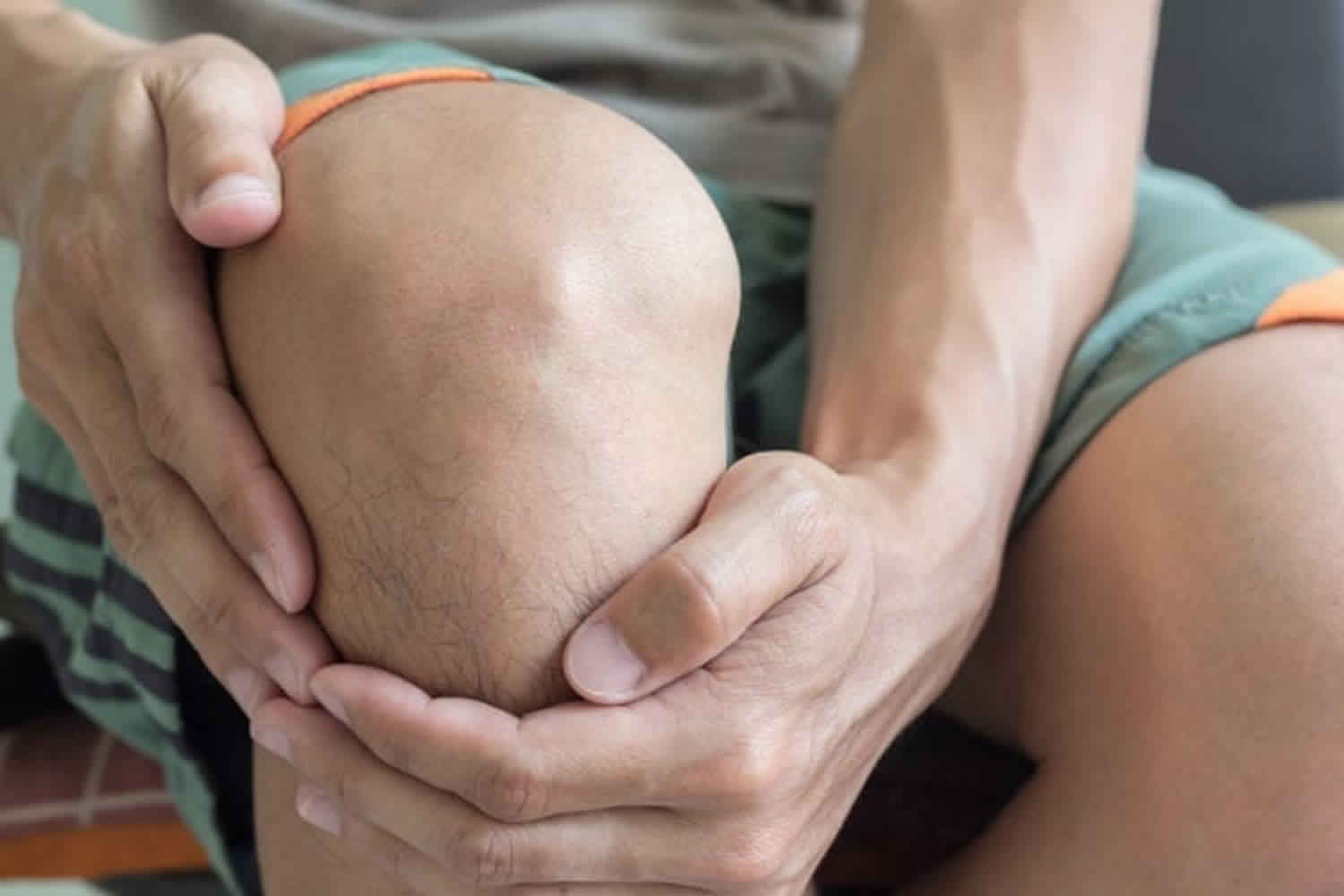
PVNS typically involves only one joint. PVNS most commonly affects the knee (80% of patients) although it can affect other joints such as the hip, shoulder, elbow, ankle, wrist, and rarely the jaw. PVNS can affect people of all ages, but it occurs most often in young adults in their 30s and 40s. The average age of diagnosis for pigmented villonodular synovitis is 35 years. Symptoms might include: pain, limitation of movement, and locking of the joint. In some cases, the normal joint structure can be destroyed. The cause of PVNS is unknown. Treatment involves surgery to remove the tumor and damaged portions of the synovium 1. Pigmented villonodular synovitis is first treated with surgery to remove as much of the abnormal tissue growth as possible. The type of surgery depends on the location and extent of the disease within the joint. Radiation therapy is sometimes used to treat this condition if surgery is not an option, or if the condition returns (recurs) after an initial surgery 2.
PVNS causes
The cause of PVNS is not known. Genetic changes associated with PVNS have been identified, but the evidence for a genetic cause is not conclusive.
PVNS symptoms
Localized PVNS causes pain and swelling in the affected joint. The swelling can be quite dramatic. Other symptoms may include locking, catching, and instability in the joint.
In diffuse PVNS, there is often a gradual onset of symptoms including joint pain, swelling, and stiffness.
In both types, the symptoms may come and go over time.
PVNS diagnosis
PVNS can look like arthritis and some other joint conditions. Your doctor will perform a physical examination and use imaging studies and other tests to diagnose PVNS.
Tests
X-rays. X-rays create clear pictures of dense structures such as bone. If PVNS has not damaged or caused changes in your bone, it may not appear in an x-ray. However, x-ray images may help your doctor rule out other causes for your pain.
Magnetic resonance imaging (MRI) scan. An MRI provides clear images of soft tissues and can be helpful in diagnosing PVNS. In localized PVNS, an MRI will show a nodular mass. In diffuse PVNS, it will show extensive thickening of the joint lining or an extensive mass, possibly with destructive bone changes and cartilage damage.
Joint aspiration. In this procedure, fluid is removed from the joint with a syringe and analyzed. In many cases of PVNS, the joint fluid is bloody.
Biopsy. A biopsy may be necessary to confirm the diagnosis of PVNS. In a biopsy, a tissue sample of the tumor is taken and examined under a microscope. A small operation is required to obtain a tissue sample.
PVNS treatment
Because PVNS destroys healthy bone and can grow to a large size, treatment involves surgery to remove the tumor and the damaged portions of the joint lining. When localized PVNS has also damaged a tendon, your surgeon will repair it during the procedure to remove the tumor. Your doctor will discuss the various surgical options with you.
Surgical procedures
Arthroscopy. In many cases of both localized and diffuse PVNS, the tumor and damaged joint lining are removed arthroscopically. During arthroscopy the surgeon makes a few small incisions around the joint and inserts a small camera called an arthroscope. The camera displays pictures on a television screen, and the surgeon uses these images to guide miniature surgical instruments.
Open surgery. Diffuse PVNS that affects both the front and back of the knee requires removal of the entire joint lining to reduce the chances of the tumor returning. In many cases, this is best achieved with traditional “open” surgery. A single larger incision provides the surgeon with full access to the joint, and facilitates removal of the mass and the entire joint lining.
Combined arthroscopic and open surgery. When most of the mass is in the back of the knee, a combined surgical approach can be undertaken. The back of the knee is treated with open surgery to remove the mass and posterior joint lining, and the front of the knee is treated with arthroscopic removal of the anterior joint lining. This combined method decreases the scale of surgery, allowing for an easier recovery.
Total joint replacement. In its end stages, diffuse PVNS can cause extensive joint destruction. Once the joint has been significantly damaged, the best option to relieve pain and improve function is a total joint replacement. Total joint replacement is a procedure in which parts of a damaged joint are removed and replaced with metal, plastic, or ceramic components. The tumor does not usually return after the joint has been replaced.
Recovery
After surgery, physical therapy will be extremely important in helping you return to your daily activities. Specific exercises will help you regain strength and range of motion in the affected joint.
Recovery from arthroscopic surgery usually requires a short course of physical therapy, after which you may return to normal activity.
Open surgery is more extensive, however, so there is an increased risk of postoperative stiffness. A more regimented and extensive physical therapy program is often required for patients recovering from open surgery to treat diffuse PVNS. In this case, the return to normal activity will take longer—possibly several months.
Localized PVNS rarely recurs after surgery. The recurrence rate for diffuse PVNS is usually around 10%, but can be as high as 30%. Patients with diffuse PVNS will require physician follow-up for several years after surgery. During these visits your doctor may order tests such as an MRI to check for recurrence of PVNS.
Radiation
Radiation therapy can shrink tumors and is sometimes used to treat widespread diffuse PVNS. It is usually reserved for patients in whom standard surgery has not been successful.
In the past, radiation therapy has been given via an external beam that is directed from outside the skin to the inside of the affected joint. While often successful, this method can cause significant complications.
A newer method called intra-articular radiation has also been used successfully. During this procedure, radioactive fluid is injected into the joint with a needle.
Living with PVNS
Even with treatment, PVNS comes back about half the time. If the pain comes back again and again, radiation therapy may help. Sometimes, the only real remedy comes by completely replacing the joint.
- Guo-ping Xie Nan Jiang, Chang-xiang Liang, Jian-chun Zeng, Zhi-yuan Chen, Qian Xu, Rui-zhen Qi, Yi-rong Chen, Bin Yu. Pigmented Villonodular Synovitis: A Retrospective Multicenter Study of 237 Cases. PLoS One. March 23, 2015; 10(3):http://www.ncbi.nlm.nih.gov/pmc/articles/PMC4370558[↩]
- Mendenhall WM, Mendenhall CM, Reith JD, Scarborough MT, Gibbs CP, Mendenhall NP. Pigmented Villonodular Synovitis. American Journal of Clinical Oncology. 2006; 29:548-550. http://www.ncbi.nlm.nih.gov/pubmed/17148989[↩]


