What is a sebaceous adenoma
Sebaceous adenoma is defined as a benign epithelial neoplasm composed of sebaceous gland (skin oil gland) like structures or tumors with well-recognized sebaceous differentiation by microscopic examination. Sebaceous adenoma is the most common benign sebaceous gland tumor associated with Muir-Torre syndrome. Sebaceous adenomas frequently appear on the face or scalp of middle-aged and older individuals, after age 50 years. The mean age at onset is 60 years. Sebaceous adenomas affect men and women equally.
Sebaceous adenoma and sebaceous epithelioma are the most characteristic cutaneous markers of Torre-Muir syndrome.
- Sebaceous adenoma are benign tumours that present as yellow papules or nodules.
- Sebaceous adenoma can grow on any part the body but are mostly located on the head, particularly on the face, scalp and eyelids.
Muir-Torre syndrome also known as ‘Torre-Muir syndrome’ and ‘sebaceous neoplasia/visceral carcinoma, is a rare inherited condition in which there are sebaceous (oil gland) skin tumors in association with internal cancer. The gastrointestinal tract is the most common affected organ, with almost one-half of patients having colorectal cancer. The second most common site is cancer of the genitourinary tract. Muir-Torre syndrome is observed in 9.2% of patients with hereditary nonpolyposis colorectal cancer (Lynch syndrome).
In most cases of Muir-Torre syndrome, the sebaceous neoplasms and associated viscera malignancies are due to abnormalities of mismatch repair proteins (MSH2, MSH6, MLH1, PMS2) and microsatellite instability. In contrast, sporadic sebaceous neoplasms are usually due to derangements in the Wnt/beta-catenin signaling pathway. Lymphoid enhancer–binding factor (LEF-1) gene mutations are typical of sebaceomas and sebaceous adenomas, while sebaceous carcinomas may have complete silencing of the LEF1 gene 1.
Patients with Torre-Muir syndrome need to undergo medical and physical examinations and appropriate laboratory and radiographic tests regularly to check for internal malignancies, particularly of the gastrointestinal and genitourinary tracts. Patients need a regular colonoscopy for early detection of colorectal tumors. Cancers should be managed with appropriate anti-cancer therapies.
In some families, a genetic diagnosis can identify asymptomatic carriers of the mutation. All first-degree relatives, especially mutation carriers, should be referred from the age of 20 years for routine follow-up and early treatment.
What is the cause of Muir-Torre syndrome?
Muir-Torre syndrome is an autosomal dominant condition due to genetic mutations, meaning half of an affected person’s children also have the Muir-Torre syndrome. Children of an individual with Muir-Torre syndrome, therefore, may have a 50% risk of inheriting the cancer predisposition. In families in which the germline mutation can be identified, those individuals who have inherited the mutation should undergo regular screening examinations, particularly of the gastrointestinal tract, colorectum, genitourinary tract, and female genital tract.
The genes most commonly mutated in Muir-Torre syndrome include MLH1, MSH2, MSH6, and PMS2 2. These mostly involving the MSH2 gene 3, located on chromosome 2p. Some patients have mutations in the MLH1 gene on chromosome 3p.
More recently, some autosomal recessive cases of Muir-Torre syndrome have been described which do not display microsatellite instability and are due to defects in the base excision repair gene known as MYH 2. It is estimated that these cases account for roughly 35% of Muir-Torre syndrome cases and have been named Muir-Torre syndrome 2. Sporadic cases also have been described, and these are usually seen in the setting of immunosuppression with tacrolimus and cyclosporine being the main offenders 4.
How is Muir-Torre syndrome diagnosed?
Torre-Muir syndrome is usually suspected because of the clinical features. However, if a sebaceous adenoma, a sebaceous carcinoma or colonic polyp is removed surgically, the pathologist may examine the cells to see whether the MSH gene products have microsatellite instability, a characteristic of the syndrome. Immunohistochemistry testing shows loss of MMR protein expression, indicating a mutation in MSH2 or another associated gene.
MMR-deficiency is present in 30–45% of sebaceous lesions but not all of these result from the syndrome. If the patient’s personal or family history or clinical features are suggestive of Torre-Muir or Lynch syndrome, the patient may undergo further genetic testing by whole gene or whole exome sequencing.
Muir-Torre syndrome treatment
Once the diagnosis has been established, Muir-Torre syndrome patients should undergo annual surveillance for visceral and cutaneous malignancy. Upper and lower gastrointestinal endoscopy should be performed. Colonoscopy may begin as early as 18 years while upper endoscopy is recommended around 25 to 30 years. Men should undergo annual testicular and prostate exams, and women should have annual breast and pelvic exams along with transvaginal ultrasound and endometrial sampling for suspicious findings. Other potentially beneficial tests would include chest x-ray, cervical and urine cytology, carcinoembryonic antigen levels, fecal occult blood testing, liver function tests, and complete blood counts.
The cutaneous manifestations of Muir-Torre syndrome can be difficult to treat due to the potential number and disfigurement that numerous sebaceous neoplasms can cause. Reassurance should be offered for benign lesions. Local excision and cryotherapy may be performed for patients desiring removal of benign lesions. Keratoacanthomas should also be managed conservatively with local excision. Sebaceous carcinomas have the potential for local and distant metastasis and should be managed more aggressively with wide local excision with 5 to 6 millimeter margins or Mohs micrographic surgery. Radiation may also be employed but only as adjuvant therapy after excision of a recurrent sebaceous carcinoma, or regional metastasis. Radiation as monotherapy for primary cutaneous sebaceous carcinoma has been associated with higher rates of mortality and recurrence. Additionally, interferon-alpha in conjunction with oral isotretinoin has been reported to decrease the development of cutaneous and visceral malignancies.
Sebaceous adenoma signs and symptoms
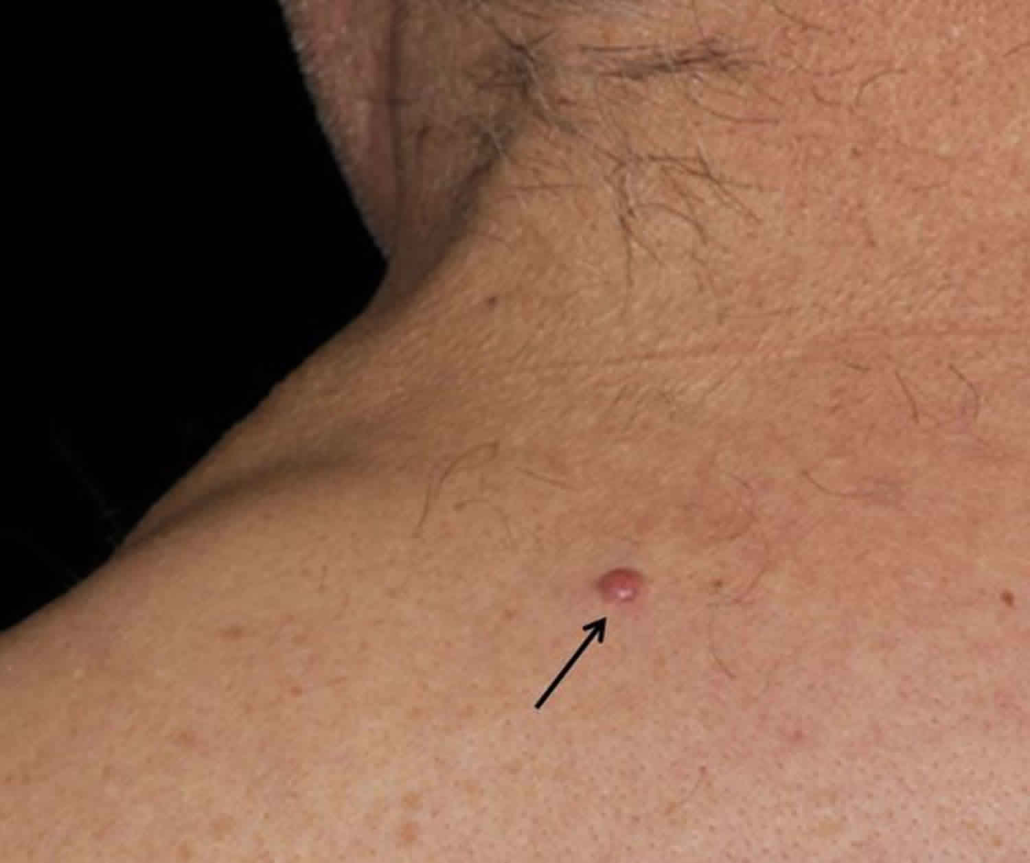
Patients with sebaceous adenomas typically experience a gradual onset of small, usually less than 0.5 cm in diameter (2-4 mm) to greater than 5 cm in maximum dimension. Sebaceous adenomas most frequently appear as smooth-surfaced, yellow, sometimes speckled papules that may feature crusting and/or central umbilication. Typically, they grow on the skin of the face or scalp over the course of a few months and are asymptomatic.
Tumors are commonly located on the face, the scalp, and the neck. Occasionally, tumors may be seen at other sites, including the trunk and the legs. Sebaceous adenomas have been described on the caruncle of the eye, eyelid, bulbar conjunctiva and buccal mucosa 5.
Identification of sebaceous adenomas is crucial because of their association with Muir-Torre syndrome. In Muir-Torre syndrome, germline mutations in mismatch repair genes result in regions of DNA microsatellite instability and subsequent increased risk of developing internal malignancies, commonly colorectal and genitourinary carcinomas.
Sebaceous adenoma diagnosis
A biopsy of skin tumors performed for histopathologic examination provides an accurate diagnosis of sebaceous neoplasms, including sebaceous adenomas 6. Sebaceous adenoma and sebaceous hyperplasia are often physically and histologically similar in appearance. However, it is important to distinguish between the two, as sebaceous adenoma is the most common sebaceous tumor in Muir-Torre syndrome, whereas sebaceous hyperplasia is not associated with Muir-Torre syndrome and is a common occurrence in the general population 1.
Appropriate laboratory testing for possible occult internal malignancies, such as gastrointestinal tract, hematologic, or laryngeal carcinomas, is indicated. The following laboratory tests can be of diagnostic value if patients present with cutaneous signs of Muir-Torre syndrome:
- Sigmoidoscopy may be performed to screen for colonic polyposis and colonic carcinoma.
- Perform endoscopy to check for an occult gastric carcinoma.
- Serum carcinoembryonic antigen (CEA) values are frequently increased in patients with colonic carcinomas.
- A complete blood cell count assists in detecting hematologic malignancies.
- Bone marrow examination may be needed to further delineate a hematologic malignancy.
- Laryngoscopy with biopsy examination of any suspicious lesions can rule out an occult laryngeal carcinoma.
Imaging Studies
Abdominal CT scanning and MRI assist in detecting an occult internal malignancy, such as kidney and urothelial cancers, in patients with Muir-Torre syndrome.
Sebaceous adenoma histology
Sebaceous adenoma is comprised of predominantly sebaceous lobules with a rim of basaloid germinative cells. The sebaceous component forms the majority of the tumor here.
Sebaceous adenomas are typically multilobulated tumors located in the upper dermis with frequent connections to the epidermis, and they often bear a strong resemblance to normal sebaceous glands. At a low-power view, sebaceous adenomas are well-circumscribed and sharply demarcated from the surrounding tissue, with a proliferation of variously sized sebaceous lobules consisting of central, larger, mature sebaceous cells (sebocytes); peripheral, smaller, undifferentiated, germinative basaloid cells; and transitional cells.
The sebocytes contain pale-staining, foamy-to-bubbly cytoplasm, and central, crenated, hyperchromatic nuclei. The smaller, immature germinative sebocytes contain round-to-oval, vesicular nuclei and basophilic cytoplasm. The transitional cells show more eosinophilic cytoplasm.
Sebaceous adenoma treatment
Sebaceous adenoma may be treated surgically with complete excision or cryotherapy. Surgical treatment of sebaceous adenomas is aimed at completely removing the tumor and preventing regrowth of the tumorous tissue. Solitary tumors are treated by complete surgical removal with a 100% cure rate. Incomplete removal has occasionally resulted in local recurrence.
Sebaceous adenoma prognosis
Sebaceous adenomas are benign tumor growths that derive from sebaceous glands. Solitary tumors are treated by complete surgical removal with a 100% cure rate. Incomplete removal has occasionally resulted in local recurrence.
When multiple sebaceous adenomas are associated with Muir-Torre syndrome, visceral carcinomas, including adenocarcinomas of the colon, stomach, duodenum, hematologic system, genitourinary tract, endometrium, and larynx (in decreasing order of frequency) may also be present. The most significant feature of Muir-Torre syndrome is the favorable prognosis of each of the associated carcinomas 7.
However, some of these malignancies have metastatic potential; deaths due to internal malignancies have been reported. The Mayo clinic has established a scoring system to determine the risk of associated disease. The scoring system includes age at presentation of the initial sebaceous neoplasm, the total number of sebaceous neoplasms, any personal history of a Lynch-related cancer, and family history of Lynch-related cancers. Patients with a score of 3 or more were more likely to have Muir-Torre syndrome 8.
- Flux K. Sebaceous Neoplasms. Surg Pathol Clin. 2017 Jun. 10 (2):367-382.[↩][↩]
- Gay JT, Gross GP. Muir-Torre Syndrome. [Updated 2019 Jun 4]. In: StatPearls [Internet]. Treasure Island (FL): StatPearls Publishing; 2019 Jan-. Available from: https://www.ncbi.nlm.nih.gov/books/NBK513271[↩][↩]
- Suspiro A, Fidalgo P, Cravo M, et al. The Muir-Torre syndrome: a rare variant of hereditary nonpolyposis colorectal cancer associated with hMSH2 mutation. Am J Gastroenterol. 1998 Sep. 93(9):1572-4.[↩]
- Chen Q, Wang M, Xu Z, Wang M, Jin S, Tian S, Xiao S. Muir-Torre Syndrome With a Frame-shift Mutation in the MSH2 Gene: A Rare Case Report and Literature Review. Int. J. Gynecol. Pathol. 2018 Dec 03[↩]
- Ferguson JW, Geary CP, MacAlister AD. Sebaceous cell adenoma. Rare intra-oral occurrence of a tumour which is a frequent marker of Torre’s syndrome. Pathology. 1987 Apr. 19 (2):204-8.[↩]
- Marques-da-Costa J, Campos-do-Carmo G, Ormiga P, Ishida C, Cuzzi T, Ramos-E-Silva M. Sebaceous adenoma: clinics, dermatoscopy, and histopathology. Int J Dermatol. 2014 Oct 14[↩]
- Svec J, Schwarzová L, Janošíková B, Stekrová J, Mandys V, Kment M, et al. Synchronous gastric and sebaceous cancers, a rare manifestation of MLH1-related Muir-Torre syndrome. Int J Clin Exp Pathol. 2014. 7 (8):5196-202.[↩]
- Roberts ME, Riegert-Johnson DL, Thomas BC, Rumilla KM, Thomas CS, Heckman MG, et al. A clinical scoring system to identify patients with sebaceous neoplasms at risk for the Muir-Torre variant of Lynch syndrome. Genet Med. 2014 Sep. 16 (9):711-6.[↩]


