What is syringohydromyelia
Syringohydromyelia is also called syringomyelia or hydromyelia, is a condition characterized by an abnormal widening of the central canal of the spinal cord that creates a cavity or cyst known as a syrinx, in which cerebrospinal fluid (commonly known as spinal fluid) can accumulate. Hydromyelia is sometimes used interchangeably with syringomyelia, the name for a condition that also involves cavitation in the spinal cord. In hydromyelia, the cavity that forms is connected to the fourth ventricle in the brain, and is almost always associated in infants and children with hydrocephalus or birth defects such as Chiari Malformation II and Dandy-Walker syndrome. Syringomyelia, however, features a closed cavity and occurs primarily in adults, the majority of whom have Chiari Malformation type 1 or have experienced spinal cord trauma. The distinction between the two is an academic exercise at best 1. The amalgamated term syringohydromyelia is used in neurosurgical parlance commonly, since there is no consensus on the correct terminology 1. Symptoms, which may occur over time, include weakness of the hands and arms, stiffness in the legs; and sensory loss in the neck and arms. Some individuals have severe pain in the neck and arms. Diagnosis is made by magnetic resonance imaging (MRI), which reveals abnormalities in the anatomy of the spinal cord.
Syringohydromyelia is a chronic condition and a syrinx can expand over time compressing or destroying the surrounding nerve tissue. A wide variety of symptoms can potentially be associated with syringomyelia depending upon the size and exact location of the syrinx. Common signs/symptoms include pain in the neck and shoulders, muscle weakness, pain and stiffness in the legs, numbness or decreased sensation, especially to hot and cold, abnormal curvature of the spine (scoliosis), muscle contractions, and uncoordinated movements (ataxia). The majority of cases of syringohydromyelia are associated with a complex brain abnormality known as a Chiari malformation. Additional known causes of syringohydromyelia include tethered cord syndrome, meningitis (arachnoiditis), certain tumors of the spinal cord, and trauma. In some patients, no underlying cause can be identified (idiopathic).
Syringohydromyelia most commonly presents in young adults between 20 and 40 years of age, but can also develop in young children or older adults. Some reports suggest that syringohydromyelia is slightly more common in males than females. One estimate places the incidence at 8.4 individuals per 100,000 in the general population in the United States.
Figure 1. Syringohydromyelia
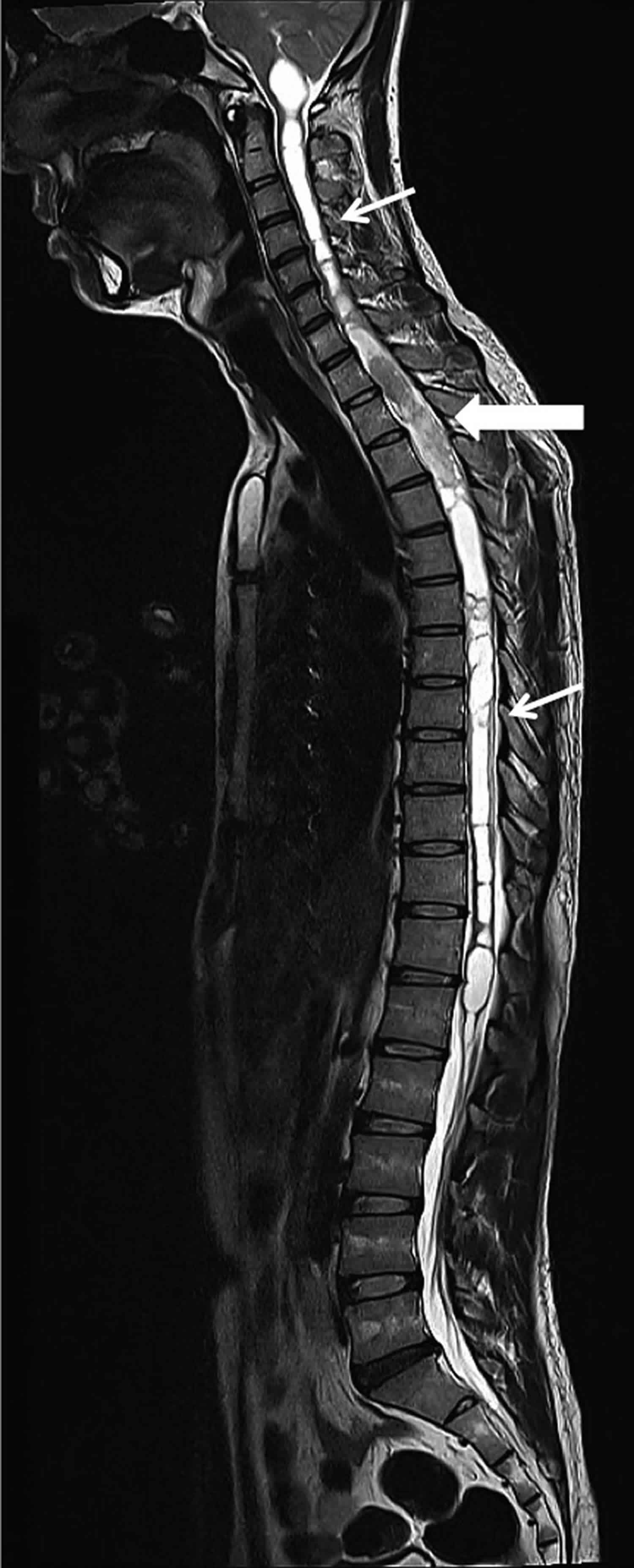
Footnote: T2-weighted MRI in sagittal plane showing extensive syringohydromyelia involving the entire spinal cord (thin arrow) in association with the spinal neurocytoma (thick arrow).
[Source 2 ]Syringohydromyelia vs Syringomyelia
Syringohydromyelia is the amalgamated term of syringomyelia and hydromyelia, since there is no consensus on the correct terminology 1. The terminology used in the medical literature to describe syringomyelia can be confusing. Numerous attempts to classify syringomyelia have existed in literature the most frequently quoted being that of Milhorat et al. 3 who classified syringomyelia as communicating (anatomically in continuum with the fourth ventricle), noncommunicating (which is separated from the fourth ventricle by a segment of normal spinal cord) and atrophic syrinx (associated with myelomalacia). Recently, Batzdorf 4 proposed a simpler classification whereby syringomyelia was classified into those which had problems at the craniovertebral junction abnormalities and those due to problems lower down in the spine. Oi et al.5 have recently proposed a new clinical category of hydrocephalus wherein central canal dilation is classified as a type 4 hydromyelic hydrocephalus – with an isolated central canal dilation.
Syringomyelia is often associated with an abnormality of the craniovertebral junction, usually a Chiari malformation, and these cases may be referred to as congenital syringomyelia. Secondary syringomyelia refers to cases that have a known cause. The term primary syringomyelia has been used to refer to cases of unknown cause or to cases that occur because of specific known causes that damage the spinal cord. Posttraumatic syringomyelia refers to cases that result from trauma to the spinal cord (these cases are sometimes sub-classified as primary syringomyelia).
Some individuals have a condition related to syringomyelia known as hydro(syringo)myelia, which is characterized by abnormal widening of the central canal of the spinal cord (the normal small canal running through the center of the spinal cord). Some physicians use the terms syringomyelia or hydromyelia interchangeably. Hydromyelia may also be present in infants and young children with or without brain abnormalities, such as Chiari malformation type II that is seen in patients with spina bifida. The fluid-filled cavities in cases of syringomyelia often do not connect to any other fluid-filled areas or spaces and occur more often in adults than children.
Syringohydromyelia causes
The exact, underlying reason for the formation of a syrinx is unknown. Most theories implicate the obstruction or disruption of the flow of cerebrospinal fluid (CSF) as the most common cause. CSF is a clear fluid that surrounds and is within the brain and surrounds the spinal cord. CSF has several functions including protecting and cushioning the brain and giving the brain buoyancy. CSF is also found in the central canal, a small canal that runs through the center of the spinal cord in infants. The central canal eventually collapses over time.
A variety of conditions that block or impair the normal flow cerebrospinal fluid have been associated with syringohydromyelia. The condition most commonly associated with syringohydromyelia is the Chiari malformations, a group of complex brain abnormalities that affect the area in the lower back of the skull where the brain and spinal cord connect (craniovertebral junction).
Syringohydromyelia can also develop following spinal cord injury. These cases are sometimes referred to as posttraumatic syringohydromyelia. The development of posttraumatic syringohydromyelia may occur many years after the initial traumatic injury.
Additional conditions that are associated with syringohydromyelia include certain spinal cord tumors, meningitis, inflammation of the arachnoid membrane (arachnoiditis), one of the membranes that surrounds and protects the spinal cord, and a tethered spinal cord, which is usually a stretch-induced functional disorder associated with the fixation (tethering) effect of inelastic tissue (filum terminale usually infiltrated with fat) found at the caudal end of the spinal cord, limiting its movement.
Spinal dysraphism (spina bifida occulta), which is characterized by malformations of the spinal canal and its contents, can also be associated with syringohydromyelia. Spinal dysraphism is often associated with a tethered spinal cord.
In some individuals, syringohydromyelia can develop without any known cause. These cases are referred to as idiopathic syringohydromyelia.
Syringohydromyelia symptoms
The specific symptoms and severity of syringohydromyelia can vary greatly from one person to another. Some individuals may not have any noticeable symptoms (asymptomatic); others may have a variety of symptoms that can progress to cause significant disability. It is important to note the highly variable nature of syringohydromyelia and to realize that affected individuals can have different sets of symptoms and a different rate of progression (or no progression of the disease at all).
Syringohydromyelia is usually slowly progressive, but rapid onset can occur. Common symptoms include pain in the neck and shoulders. Pain may also affect the arms and hands and may be described as a burning, tingling or piercing sensation. Some affected individuals also experience numbness or decreased sensation, especially to hot and cold. Muscle weakness and wasting, especially of the hands, arms, and eventually the shoulders, may also develop. The upper (cervical and thoracic) portions of the spinal cord are often affected in syringohydromyelia. Affected individuals may first notice a loss of feeling for pain and temperature in their fingers, hands, arms, and upper chest. In the early stages, a sense of touch is still present. A loss of feeling may spread over the shoulders and back, described as a “cape-like” distribution.
Affected individuals may also develop pain and stiffness (spasticity) in the legs and uncoordinated movements (ataxia), eventually affecting the ability to walk. In severe cases, paralysis of the arms or legs can occur. Some affected individuals may develop muscle contractions such as small, involuntary muscle contractions or “twitches” (fasciculations).
Skeletal abnormalities can develop including the abnormal side-to-side curvature of the spine (scoliosis). In some children, scoliosis may be the only symptom. Some individuals may develop Charcot joints, in which chronic, progressive degeneration of the joint occurs because of damage to the nerves that supply the joint. Charcot joints are initially seen as swelling and redness of the affected areas. Without treatment, deformity of the affected joints can occur.
Some affected individuals develop symptoms associated with damage to the autonomic nervous system, which is the part of the nervous system that controls involuntary functions. Such symptoms include loss of bowel and bladder control, excessive sweating (hyperhidrosis), and fluctuating blood pressure levels. Horner’s syndrome, a rare condition that develops because of damage to one of the nerves that supplies the eyes and face, may also occur. Horner’s syndrome usually affects one side of the face and is characterized by a droopy eyelid, narrowing of the opening between the eyelids, decreased pupil size, and decreased sweating on the affected side of the face.
Individuals with syringohydromyelia associated with a Chiari malformation may also have a condition called hydrocephalus, in which there is an abnormal accumulation of cerebrospinal fluid in the brain. In infancy, hydrocephalus can cause a variety of symptoms including an abnormally enlarged head, vomiting, headache, sleepiness, irritability, seizures, and downward deviation of the eyes.
Syringohydromyelia diagnosis
A diagnosis of syringohydromyelia is based upon identification of characteristic symptoms, a detailed patient history, a thorough clinical evaluation and a variety of specialized tests. In some cases, syringohydromyelia is discovered incidentally when a person is being evaluated for another reason.
Clinical Testing and Workup
A specialized imaging technique called magnetic resonance imaging (MRI) is used to diagnose syringohydromyelia. An MRI uses a magnetic field and radio waves to produce cross-sectional images of particular organs and bodily tissues such as the brain and spinal cord. An MRI can reveal a syrinx or another condition related to syringohydromyelia such as an intraspinal tumor or Chiari malformation. The location and extent of the syrinx is much more accurately determined and treatment can begin, when appropriate, earlier than in the past with older diagnostic techniques (e.g., myelogram).
Syringohydromyelia treatment
The treatment of syringohydromyelia is directed toward the specific symptoms that are apparent in each individual. Treatment may require the coordinated efforts of a team of specialists. Pediatricians, neurologists, neurosurgeons, surgeons, eye specialists (ophthalmologists) and other healthcare professionals may need to systematically and comprehensively plan an affected child’s treatment.
Specific therapeutic procedures and interventions may vary, depending upon numerous factors, such as disease progression; the presence or absence of certain symptoms; the underlying cause; the impact of symptoms on quality of life; an individual’s age and general health; and/or other elements. Decisions concerning the use of particular drug regimens and/or other treatments should be made by physicians and other members of the health care team in careful consultation with the patient based upon the specifics of his or her case; a thorough discussion of the potential benefits and risks, including possible side effects and long-term effects; patient preference; and other appropriate factors.
Some individuals with syringohydromyelia who do not have any symptoms may not require treatment, but should be regularly monitored to see whether the disorder progresses.
General therapeutic options include pain medications (analgesics), physical therapy, and a reduction in activities, especially those that require straining such as heavy lifting. The goal of treatment for syringohydromyelia is to restore the proper flow of cerebrospinal fluid and to remove the pressure that a syrinx places on the spinal cord. Initial treatment is usually targeted at the underlying cause of syringohydromyelia.
There is no specific, agreed-upon therapy or treatment regimen for the most common cause of a syrinx, a Chiari malformation. Neurosurgeons and other physicians may disagree as to the best approach to treat a Chiari malformation. Different neurosurgeons may recommend different surgical techniques or treatment regimens.
Like syringohydromyelia, individuals with a Chiari malformation who do not have symptoms are generally not treated, but monitored to see whether the disorder progresses. If mild or nonspecific symptoms are present, such as neck pain or headaches, physicians may recommend conservative treatment. Symptomatic Chiari malformations are most often treated by surgery. There are no specific criteria or objective tests that can be used to determine when to undergo surgery or the best procedures to choose. The most common surgery is known as posterior fossa decompression. With this procedure, a surgeon creates room by removing small pieces of bone in the back of the skull, thereby enlarging the foramen magnum. This relieves pressure and reduces compression of the brainstem. The surgeon may also choose to open the covering (dura mater) of the brain in this region and explore the herniated tissue and then sew in a graft (duraplasty).
If hydrocephalus is associated with the Chiari malformation-related syrinx, then the surgeon will usually treat this first. The most common surgical treatment is to place a shunt that connects the dilated ventricles of the brain to another cavity of the body. The most common cavity chosen is the abdomen. Once the hydrocephalus is treated, then the Chiari malformation is addressed.
Surgery may be used to treat other conditions that cause syringohydromyelia including surgery to remove a tumor. Tethered spinal cord may require surgery to release the tension of the cord.
In some cases, a tiny tube called a shunt/stent may be placed into the syrinx. The shunt allows the fluid within the syrinx to drain to an area outside of the spinal column. Shunting can stop the progression of the disorder and relieve some symptoms such as pain and headaches. However, shunts can be associated with significant side effects including spinal cord injury or infection, bleeding (hemorrhaging), and blockage.
Posttraumatic syringohydromyelia may be difficult to treat. Surgery is recommended for individuals with neurological deterioration and/or intractable pain. Surgery is aimed at expanding the area around the spinal cord by the site of trauma or injury and to decrease fluid volume. The insertion of shunt may also be used to treat posttraumatic syringohydromyelia. Shunting in posttraumatic syringohydromyelia carries risks including further spinal cord injury and may need to be replaced if the shunt becomes clogged or defective. Many physicians consider shunts a last resort for individuals with posttraumatic syringohydromyelia.
Surgery for syringohydromyelia can often lead to an improvement of symptoms and stabilization of the disorder in many individuals. However, syringohydromyelia can recur after successful treatment, thereby necessitating more operations.
After surgery, the physician will evaluate the syrinx to make sure that it stabilizes or decreases in size. These evaluations will entail obtaining an MRI.
- Rao KS, Balasubramaniam C, Subramaniam K. Acute onset of postoperative syringohydromyelia. J Pediatr Neurosci. 2015;10(3):240–243. doi:10.4103/1817-1745.165671 https://www.ncbi.nlm.nih.gov/pmc/articles/PMC4611893[↩][↩][↩]
- Hanafiah M, Low SF, Sridharan R, Young B. Spinal neurocytoma with extensive syringohydromyelia. BMJ Case Rep. 2013;2013:bcr2013201285. Published 2013 Oct 16. doi:10.1136/bcr-2013-201285 https://www.ncbi.nlm.nih.gov/pmc/articles/PMC3822201/[↩]
- Milhorat TH, Johnson RW, Milhorat RH, Capocelli AL, Jr, Pevsner PH. Clinicopathological correlations in syringomyelia using axial magnetic resonance imaging. Neurosurgery. 1995;37:206–13[↩]
- Batzdorf U, Klekamp J, Johnson JP. A critical appraisal of syrinx cavity shunting procedures. J Neurosurg. 1998;89:382–8.[↩]
- Oi S, Kudo H, Yamada H, Kim S, Hamano S, Urui S, et al. Hydromyelic hydrocephalus. Correlation of hydromyelia with various stages of hydrocephalus in postshunt isolated compartments. J Neurosurg. 1991;74:371–9[↩]


