Traumatic asphyxia
Traumatic asphyxia also called Perthes syndrome or Perte’s syndrome, results from severe crush injury of the chest causing sudden compression of the thorax 1. Commonly associated injuries include pulmonary contusion, hemothorax, and pneumothorax. Traumatic asphyxia should always be kept in mind as a possible complication of injuries of the chest and abdomen. Traumatic asphyxia is a clinical syndrome associated with craniocervical cyanosis, subconjunctival hemorrhage, multiple petechiae, and neurological symptoms 2. Traumatic asphyxia or Perte’s syndrome occurs as a result of sudden or severe compression of the thorax or upper abdomen, or both. A Valsalva maneuver (deep inspiration against a closed glottis) is necessary before thoracic compression for the development of Perte’s syndrome 3. The increased thoracic pressure compresses the right atrium, precluding blood return from the superior vena cava, and resulting in rupture of venules and capillaries of the face and head 4. Patients will exhibit conjunctival hemorrhages, facial swelling, and petechial hemorrhages on the face and upper chest. Although severe cases may result in loss of vision or other permanent neurologic complications, the morbidity and mortality associated with traumatic asphyxia is generally related to associated injuries.
Compression against a closed glottis (Valsalva maneuver) may contribute to this increased intrathoracic pressure 4. The backpressure within the venous system causes the capillaries of the head and neck to become engorged with blood, resulting in the classic subconjunctival hemorrhages and petechiae, as well as soft tissue edema of the face. This engorgement ultimately results in stagnation of blood flow and subsequent localized blood desaturation (loss of oxygen), causing the characteristic craniocervical cyanosis. The duration and magnitude of the compressive force are both key factors in the development of traumatic asphyxia and potential death 5.
The extent of the signs and symptoms depend on the duration and severity of the compression that thorax and upper abdomen are exposed to 6. Many signs may accompany the characteristic findings. Confusion, amnesia, disorientation, uneasiness, agitation, hypoxia, cerebral edema, and hemorrhage are the most common ones 7. Other than subconjunctival hemorrhage, decreased vision, blurred vision, papillary changes, optic nerve atrophy, diplopia, and exophthalmia are the most frequent ocular findings. Epistaxes due to capillary rupture, hearing loss due to the edema of the Eustachian tubes, or hemotympanum are the other probable findings 8. Severe life-threatening conditions may coexist since the traumatic asphyxia develops as a result of major trauma to thorax, mediastinum, and upper abdomen. These are thoracic and extrathoracic injuries as pulmonary contusion, hemothorax, pneumothorax, flail chest, and hepatic laceration 9. Signs of pulmonary injury like dyspnea, tachypnea, and hemoptysis may be observed. In rare cases, cardiac injury may be encountered 8. An increase in the abdominal pressure may lead to organ injury, hematemesis, and hematuria. Apnea and hypoxemia associated with prolonged thoracic compression may be life threatening and give rise to increased mortality. The results may be such severe as shock, cerebral anoxia, neurological sequelae, and sudden death 10.
The presence of traumatic asphyxia points to a serious mechanism of injury and is an indicator of severe trauma. Potentially life-threatening pulmonary injuries are common, and may include hemothoraces and/or pneumothoraces, pulmonary contusions or lacerations, and flail chest. There may be associated intra-abdominal or pelvic injuries, diaphragmatic rupture and skeletal fractures.
Cardiac injuries may also occur, but they’re less commonly seen in those who survive the initial crush injury. Symptoms reported by survivors often include chest pain, shortness of breath, sore throat, hoarseness, epistaxis, loss of consciousness and transient confusion, temporary visual changes or blindness, and hearing loss.
Prompt treatment with attention to the reestablishment of oxygenation and perfusion may result in a good outcome. The patients’ recoveries were related to the severity of the injuries and the associated injuries. Fortunately, survivors of traumatic asphyxia are often reported to have full recovery at 12 months without long-term complications, other than the morbidity associated with the crush injury itself 11.
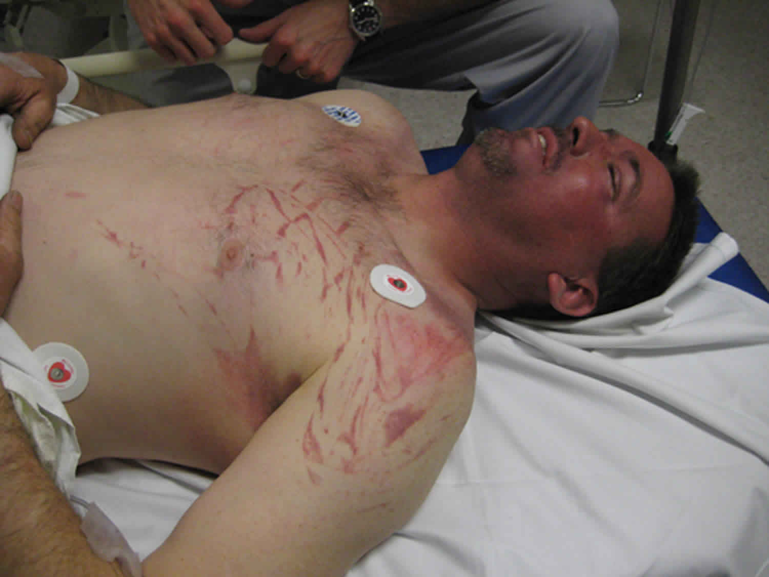
Figure 1. Traumatic asphyxia
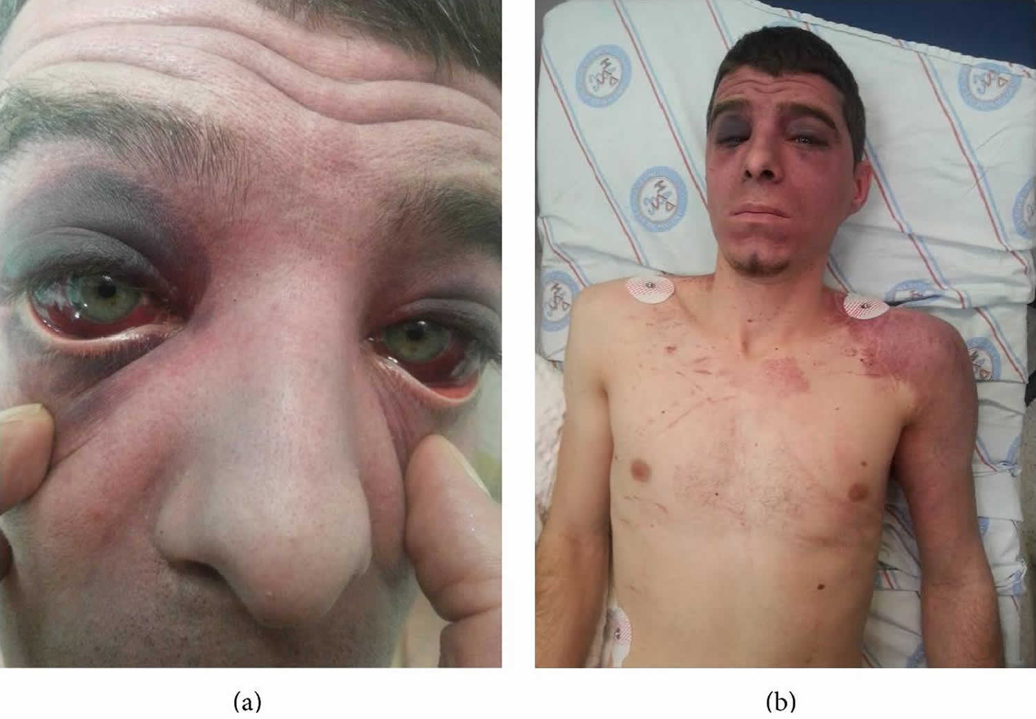
Footnote: (a) The patient had bilateral subconjunctival hemorrhage. (b) The patient had facial cyanosis, petechial eruptions on the anterior surface of the thoracic cage and on left upper extremity.
[Source 12 ]Traumatic asphyxia causes
Traumatic asphyxia or Perte’s syndrome occurs as a result of sudden or severe compression of the thorax or upper abdomen, or both. The mechanism of injury in traumatic asphyxia is usually a crush by an object that compresses the chest or upper abdomen. Traumatic asphyxia or Perthes syndrome most commonly seen in motor vehicle collisions in which victims are ejected from a vehicle that subsequently rolls over their torso.
Another common cause includes individuals being pinned underneath an object. This may inadvertently occur while working under a vehicle. Traumatic asphyxia can also be seen in those who work around heavy equipment in industrial, farming and construction settings, as well as in trench collapses 13. Other causes include injuries from machines and furniture, blast injury, a python tightened around the thorax and, rarely, deep-sea diving, weightlifting, epileptic seizures, difficult obstetric delivery and asthmatic attack 14. The typical range of the duration of compression is between two and five minutes 14. The duration and the amount of pressure affect the outcome after traumatic asphyxia. Significant weight can be tolerated for a short time, whereas a relatively modest weight applied for a longer period may result in death 14.
The exact pathophysiologic mechanism of traumatic asphyxia is still not fully understood and remains controversial. However, the prevailing theory is that the acute compression of the chest results in increased intrathoracic pressure, driving blood from the right atrium and superior vena cava into the innominate (brachiocephalic) and jugular veins, which have valves that are unable to prevent backflow when encountering excessive pressure. It is generally considered that a compressive force to the thoracoabdominal region together with the ‘fear response’ (deep breath and closing of the glottis [Valsalva maneuver]) cause a huge increase in the central venous pressure. This induces reversal of venous blood flow from the heart through the superior vena cava into the innominate (brachiocephalic) and jugular veins of the head and neck. The back transmission of the elevated central venous pressure to the head and neck venules and capillaries, while arterial flow is continued, results into capillary stasis and rupture, producing the characteristic upper body petechial and subconjunctival hemorrhages 15. These features are often more prominent on the eyelids, nose and lips 16. The lack of petechiae in the lower body may be due to the compressive obstruction of the inferior vena cava in the chest or abdomen. Furthermore, the fact that the lower part of the body is protected from back transmission of venous pressure by a series of valves could be another mechanism, since the superior vena cava, innominate and jugular veins have no valves 16.
Traumatic asphyxia signs and symptoms
Traumatic asphyxia also called Perte’s syndrome, results from severe crush injury of the chest causing sudden compression of the thorax 1. Traumatic asphyxia is characterized by craniocervical cyanosis, subconjunctival hemorrhage, edema, neurological symptoms and petechial eruptions on the face, neck, upper parts of the thoracic cage, and the upper extremities 17. According to Perthes, neurological injury in traumatic asphyxia includes cerebral hypoxia or anoxia, ischemia, venous hypertension, cerebral vascular congestion, rupture of small vessels, petechial hemorrhages and hydrostatic edema 18. Commonly associated injuries include pulmonary contusion, hemothorax, and pneumothorax 9. Traumatic asphyxia should always be kept in mind as a possible complication of injuries of the chest and abdomen.
The vision may be affected with the same mechanism: retinal hemorrhage, retrobulbar hemorrhage and vitreous exudates (Purtscher’s retinopathy) 19. A hearing deficit can be caused by edema of the Eustachian tubes, or a hemotympanum. Other neurologic manifestations of the syndrome are loss of consciousness, prolonged but self-limiting confusion, disorientation, agitation, restlessness, seizures, visual disturbances, blurred vision, papillary changes, optic nerve atrophy, exophthalmos, diplopia and hearing loss 19. Often, the neurologic status improves during transfer to the emergency room 14. The suggested mechanism for loss of consciousness and prolonged confusion associated with traumatic asphyxia includes cerebral hypoxia, ischemia and venous hypertension, which lead to cortical dysfunction. This dysfunction resolves within the following 24 to 48 hours. Intracranial hemorrhage has seldom ever been evident in a patient 14. CT scans of the brain are usually normal, whereas in fatal cases, autopsy shows only petechiae and congestion, suggesting brain injury at the cellular level 15.
Despite the dramatic appearance of the ‘ecchymotic mask’, mortality in crush asphyxia is low. However, it may be influenced by the severity, nature and duration of the compressive force and the presence of concomitant injuries, which can be useful markers of the severity of compression 18.
Traumatic asphyxia diagnosis
Traumatic asphyxia also called Perte’s syndrome diagnosis is reached from the physical appearance, clinical examination, history and trauma mechanism 20. Although traumatic asphyxia can be diagnosed easily through short anamnesis and physical examination, other reasons like superior vena cava syndrome and skull base fractures leading to subconjunctival hemorrhage and periorbital ecchymosis must be evaluated 10. Superior vena cava (SVC) obstruction and basilar skull fracture have features that closely resemble the appearance of traumatic asphyxia. Yet, the history of traumatic injury should rule out superior vena cava obstruction, while skull fractures are rare in traumatic asphyxia, unless the force of compression is applied to the head 21.
Careful and thorough examination is necessary to detect the thoracic and extrathoracic injuries that need urgent intervention 12.
Traumatic asphyxia treatment
Optimal management of traumatic asphyxia must focus on early recognition of this entity based upon the classic physical signs and the mechanism of injury. Resuscitation efforts should include rapid administration of oxygen with effective ventilation and fluid resuscitation, and must focus on reversing hypoxia and prevent further tissue damage.
Traumatic asphyxia cases must be monitored after securing the airway and fixing the cervical spine. Oxygen inhalation therapy and intravenous fluid replacement need to be initiated and the patient shall be intubated and followed on mechanical ventilation as needed 8. The intubation may be difficult because of the airway edema, which is seen rarely 22. Uncomplicated cases are treated conservatively. The head of the bed is preferred to be elevated at 30 degrees and oxygen administration is essential. This procedure decreases the intracranial pressure. Continuous monitorization of the patient, arterial blood gas, oxygen saturation level examinations at certain intervals, and supportive oxygen therapy are the key factors for the management throughout the hospitalization period 6.
The prognosis is good if the patient survives the initial few hours following injury, although a prolonged thoracic compression could lead to cerebral anoxia and permanent neurological sequelae 20.
Traumatic asphyxia EMT treatment
The proposed algorithm for the management of all trauma patients on arrival and during the initial phases of treatment is the ABCDE (Airway, Breath, Circulation, Disability, Environment) algorithm, described in the Advanced Trauma Life Support guidelines by the American College of Surgeons Committee on Trauma. The outcome is improved by airway control and cervical spine protection, rapid restoration of ventilation, oxygenation and circulation by thoracic decompression, fluid resuscitation and prevention of renal complications secondary to rhabdomyolysis and other secondary causes 15. Management of these patients may be complicated by severe upper airway edema, and the possibility of a difficult intubation should thus be considered early.
- Williams JS, Minken SL, Adams JT. Traumatic asphyxia—reappraised. Ann Surg. 1968;167:384. doi: 10.1097/00000658-196803000-00012[↩][↩]
- Karamustafaoglu YA, Yavasman I, Tiryaki S, Yoruk Y. Traumatic asphyxia. Int J Emerg Med. 2010;3(4):379–380. Published 2010 Aug 25. doi:10.1007/s12245-010-0204-x https://www.ncbi.nlm.nih.gov/pmc/articles/PMC3047851[↩]
- Barakat M, Belkhadir ZH, Belkrezia R, Faroudy M, Ababou A, Lazreq C, Sbihi A. Traumatic asphyxia or Perte’s syndrome. Six case reports. Ann Fr Anesth Rèanim. 2004;23:59–62. doi: 10.1016/j.annfar.2003.10.011[↩]
- Ashcraft’s Pediatric Surgery 5th Ed. ISBN 978-1-4160-6127-4 https://doi.org/10.1016/B978-1-4160-6127-4.X0001-8[↩][↩]
- Richards CE, Wallis DN. Asphyxiation: A review. Trauma. 2005;7:37–45.[↩]
- Ertok I., Kurtoglu Çelik G., Ercan Haydar G., Karakayali O., Yılmaz M., Erşen T. Review of traumatic asphyxia syndrome with a case presentation. Journal of Academic Emergency Medicine Case Reports. 2013;4(2):58–61. doi: 10.5505/jaemcr.2013.32448[↩][↩]
- Hurtado T. R., Della-Giustina D. A. Traumatic asphyxia in a 6-year-old boy. Pediatric Emergency Care. 2003;19(3):167–168. doi: 10.1097/00006565-200306000-00007[↩]
- Sertaridou E., Papaioannou V., Kouliatsis G., Theodorou V., Pneumatikos I. Traumatic asphyxia due to blunt chest trauma: a case report and literature review. Journal of Medical Case Reports. 2012;6, article 257 doi: 10.1186/1752-1947-6-257[↩][↩][↩]
- Kamali S., Kesici S., Gunduz I., Kesici U. A case of traumatic asphyxia due to motorcycle accident. Case Reports in Emergency Medicine. 2013;2013:3. doi: 10.1155/2013/857131.857131[↩][↩]
- Dunne J. R., Shaked G., Golocovsky M. Traumatic asphyxia: an indicator of potentially severe injury in trauma. Injury. 1996;27(10):746–749. doi: 10.1016/s0020-1383(96)00113-1[↩][↩]
- Dunne JR, Shaked G, Golocovsky. Traumatic asphyxia: An indicator of potentially severe injury in trauma. Injury. 1996;27:746–749.[↩]
- Gulbahar G, Kaplan T, Gundogdu AG, et al. A Rare and Serious Syndrome That Requires Attention in Emergency Service: Traumatic Asphyxia. Case Rep Emerg Med. 2015;2015:359814. doi:10.1155/2015/359814 https://www.ncbi.nlm.nih.gov/pmc/articles/PMC4458292[↩][↩]
- Jongewaard WR, Cogbill TH, Landercasper J. Neurologic consequences of traumatic asphyxia. J Trauma. 1992;32:28–31.[↩]
- Jongewaard WR, Cogbill TH, Landercasper J: Neurologic consequences of traumatic asphyxia. J Trauma. 1992, 32: 28-31. 10.1097/00005373-199201000-00006[↩][↩][↩][↩][↩]
- Richards EC, Wallis ND: Asphyxiation: a review. Trauma. 2005, 7: 37-45. 10.1191/1460408605ta330oa[↩][↩][↩]
- Cenker E, Ozlem Y: Traumatic asphyxia: a rare syndrome in trauma patients. Int J Emerg Med. 2009, 2: 255-256. 10.1007/s12245-009-0115-x[↩][↩]
- Eken C., Yiğit Ö. Traumatic asphyxia: a rare syndrome in trauma patients. International Journal of Emergency Medicine. 2009;2(4):255–256. doi: 10.1007/s12245-009-0115-x[↩]
- Eren B, Türkmen N, Fedakar R: An unusual case of thorax compression. J Ayub Med Coll Abbottabad. 2008, 20 (1): 134-135.[↩][↩]
- Choi JY, Lee JS, Kim JH, Yim JH: Bilateral retrobulbar hemorrhage and visual loss following traumatic asphyxia. Korean J Opthalmol. 2010, 24: 380-383. 10.3341/kjo.2010.24.6.380[↩][↩]
- Karamustafaoglu AY, Yavasman I, Tiryaki S, Yoruk Y: Traumatic asphyxia. Int J Emerg Med. 2010, 3: 379-380. 10.1007/s12245-010-0204-x[↩][↩]
- Byard WR, Wick R, Simpson R, Gilbert JD: The pathological features and circumstances of death of lethal crush/traumatic asphyxia in adults – a 25-year study. Foren Scien Inter. 2006, 159: 200-205. 10.1016/j.forsciint.2005.08.003[↩]
- Ibarra P., Capan L. M., Wahlander S., Sutin K. M. Difficult airway management in a patient with traumatic asphyxia. Anesthesia & Analgesia. 1997;85(1):216–218. doi: 10.1097/00000539-199707000-00039[↩]


