Urticaria multiforme
Urticaria multiforme is also known as acute annular urticaria or acute urticarial hypersensitivity syndrome, is a benign cutaneous hypersensitivity reaction primarily mediated by histamine release that seen in pediatric patients that is characterized by the acute and transient onset of blanchable, annular, polycyclic, erythematous wheals with dusky, ecchymotic centers in association with angioedema of the face, hands, and feet 1. Itch (pruritus) is an almost universal complaint and systemic symptoms include a mild fever that usually resolves within 1 to 3 days of eruption 2.
Urticaria multiforme represents an allergic hypersensitivity reaction that may be IgE dependent or independent 3. Urticaria multiforme is observed in the pediatric population, most often in those between 4 months and 4 years old, urticaria multiforme is characterized by a transient cutaneous erythema and dermal edema 3. Typical lesions are initially characterized by small erythematous macules, papules, and plaques that rapidly expand to form polycyclic and annular blanchable wheals with ecchymotic, dusky centers usually affecting the face, trunk, and extremities 1. In most cases there is also associated acral and facial angioedema. Affecting the trunk, extremities, and face, the evanescent lesions of urticaria multiforme are characteristic of any urticarial eruption that may last only minutes to hours, but in some instances persist beyond an acute time span 1. The course of disease is self-limited and usually resolves within 2 to 12 days and a favorable response to antihistamines 4, leaving normal skin after its resolution 5. Dermal edema can commonly involve the hands, feet, and face without associated laryngoedema 5. Dermatographism secondary to mast cell-mediated cutaneous dermal hypersensitivity at sites of skin trauma is a common part of the clinical picture in affected patients that can manifest in geometric or linear patterns 2.
Urticaria multiforme is most commonly misdiagnosed as erythema multiforme, a serum-sickness-like reaction, or urticarial vasculitis 6. Table 1 provides differential diagnoses to consider when evaluating a patient for possible urticaria multiforme 1.
Table 1. Distinguishing features of urticarial multiforme, erythema multiforme, serum-sickness-like reaction, and urticarial vasculitis
| FEATURES | URTICARIA MULTIFORME | ERYTHEMA MULTIFORME | SERUM-SICKNESS-LIKE REACTION | URTICARIA VASCULITIS |
|---|---|---|---|---|
| Population affected | Infants and small children | All ages: 50% under 20 years | Adults | |
| Appearance of individual lesions | Annular and polycyclic wheals with dusky, ecchymotic centers Classic “target” lesion with purpuric or dusky, violaceous center that may blister. Middle ring of pallor and edema with an outer ring of erythema or blisters | Classic “target” lesion with purpuric or dusky, violaceous center that may blister. Middle ring of pallor and edema with an outer ring of erythema or blisters | Urticarial, polycyclic wheal with central clearing that may be ecchymotic-appearing | Hives with dusky, purpuric centers |
| Location | Trunk, extremities, face | Dorsum of the hands, palms and soles, forearms, feet, face, elbows and knees, penis (50%) and vulva | Face, trunk, extremities, and lateral borders of hands and feet | Trunk, extremities, face, lateral borders of hands and feet |
| Lesion duration | <24 hours | Days to weeks | Days to weeks | Days to weeks |
| Fixed lesions | No | Yes | Yes | Yes |
| Total rash duration | 2-12 days | 2-3 weeks | 1-6 weeks | 1-6 weeks |
| Mucous membrane involvement | Oral edema common, no erosions or blisters | Erosions with fibrin membranes, occasionally ulcerations, lips, oropharynx, nasal, conjunctival, vulvar and anal | Edema of the oral cavity is common with no erosions or blisters | Oral erosions with blistering of the lips, buccal mucosa, and tongue. May involve several sites, including the conjunctiva, nasal, and/or urogenital mucosa |
| Facial or acral edema | Common | Rare | Less common | Common |
| Dermatographism | Yes | No | No | No |
| Fever | Occasional low grade | Occasional low grade | High grade | Variable |
| Arthralgias | No | No | Yes | Yes |
| Common triggers | Viral illness, antibiotics, immunizations | Herpes simplex virus, other viral illness | Antibiotics | Infections, autoimmune processes, neoplastic processes, drugs |
| Associated symptoms | Pruritus | Mild pruritus or burning | Myalgias, arthralgias, lymphadenopathy | Variable |
| Laboratory findings | CBC, ESR, C-reactive protein, throat swab normal or negative IgE levels raised in 20% of cases | Elevations in ESR, WBC, and liver enzymes may be detected | Frequent elevations in ESR and C-reactive protein. Frequent neutropenia with development of reactive, plasmacytoid lymphocytosis. Mild thrombocytopenia as well as eosinophilia may be seen | Elevated ESR, hypocomplementemia (70%), microhematuria, proteinuria |
| Histology | Indistinct from other subtypes of acute urticaria: Dermal edema with a perivascular lymphocytic infiltrate with few intermingled eosinophils | Exocytosis as well as spongiosis are apparent in conjunction with varying degrees of epidermal necrosis. Necrotic keratinocytes are generally present at all epidermal levels with an edematous, papillary dermis with dilated capillaries | Features consistent with urticarial without evidence of vasculitis | Nuclear debris or fibrinoid alteration of the microvasculature with or without extravasation of erythrocytes |
| Treatment | Discontinue offending agents, such as any new or unnecessary medications and antibiotics. Systemic H1 and H2 antihistamines are effective. Systemic steroids should only be given in severe cases refractory to antihistamine therapy | Systemic glucocorticoids consisting of prednisone 50-80 mg/d in divided doses should be given and quickly tapered. To prevent development of recurrent EM, control of herpes simplex with oral valacyclovir or famciclovir may be considered | Discontinue offending agents, such as new medications and antibiotics as well as supportive care and H1 and H2 antihistamines. Systemic steroids may be considered in severe cases | First-line therapy consists of H1 and H2 blockers plus nonsteroidal anti-inflammatory agents |
The diagnosis of urticaria multiforme is largely clinical and can be obtained through a careful history and physical examination 7. A few hallmarks of the rash are the resolution of individual lesions within 24 hours and the presence of dermatographism, a wheal-and-flare reaction after minor skin trauma. Antecedent viral illnesses, antipyretics, and antibiotics, especially amoxicillin and cephalosporins, have all been listed as culprits.
A detailed history and physical examination are invaluable parts of the medical workup to arrive at the correct diagnosis. Of note, the true target lesions seen in erythema multiforme are typically not seen in patients with urticaria multiforme. Although initial skin lesions may all appear similar, they will progress to a unique clinical entity relatively quickly. Skin necrosis and blistering as well as mucous membrane involvement, arthralgias, and/or arthritis are absent in urticaria multiforme 3. Pruritus frequently accompanies urticaria multiforme, but not erythema multiforme and urticiaral vasculitis, where pain and burning are more commonly reported. While the lesions associated with erythema multiforme, serum-sickness-like reactions, or urticarial vasculitis are fixed and last days to weeks, the skin changes of urticaria multiforme are transient and last less than 24 hours, similar to acute urticaria or juvenile idiopathic arthritis (Still’s disease). While dermatographism commonly makes up part of the clinical picture in urticaria multiforme and urticaria, it is classically absent in patients with erythema multiforme or serum-sickness-like reactions. While lesions characteristic of urticarial vasculitis and erythema multiforme can manifest with dusky, purpuric centers, these skin lesions usually resolve with postinflammatory dyspigmentation, which is not seen in urticarial multiforme 8.
Treatment is largely symptomatic, as episodes of urticaria multiforme are self-resolving within 8 to 10 days. Suggested treatment for urticaria multiforme involves complete histamine receptor blockage 9. Long-duration H1blockers (such as cetirizine hydrochloride) should be given in association with H2 blockers (such as ranitidine) with breakthrough relief as needed by diphenhydramine or hydroxyzine hydrochloride. Most patients have symptom relief with this combination within a few days to 2 weeks 10. Steroids should be reserved for patients who remain severely symptomatic despite antihistamine therapy.
Figure 1. Urticaria multiforme
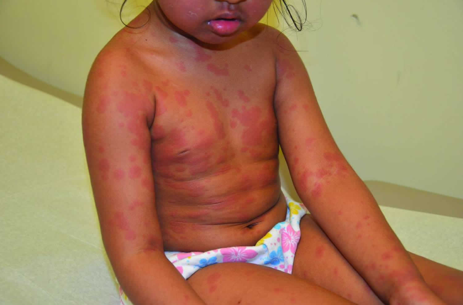
Footnote: Trunk and extremities of child on presentation. Annular papules and plaques with dusky centers.
[Source 6 ]Figure 2. Urticaria multiforme (up close of back lesions of patient from figure 1)
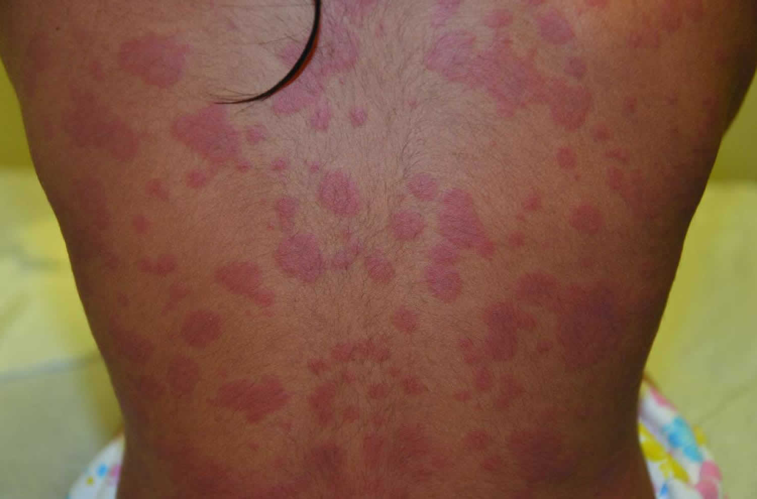
Figure 3. Urticaria multiforme rash feet
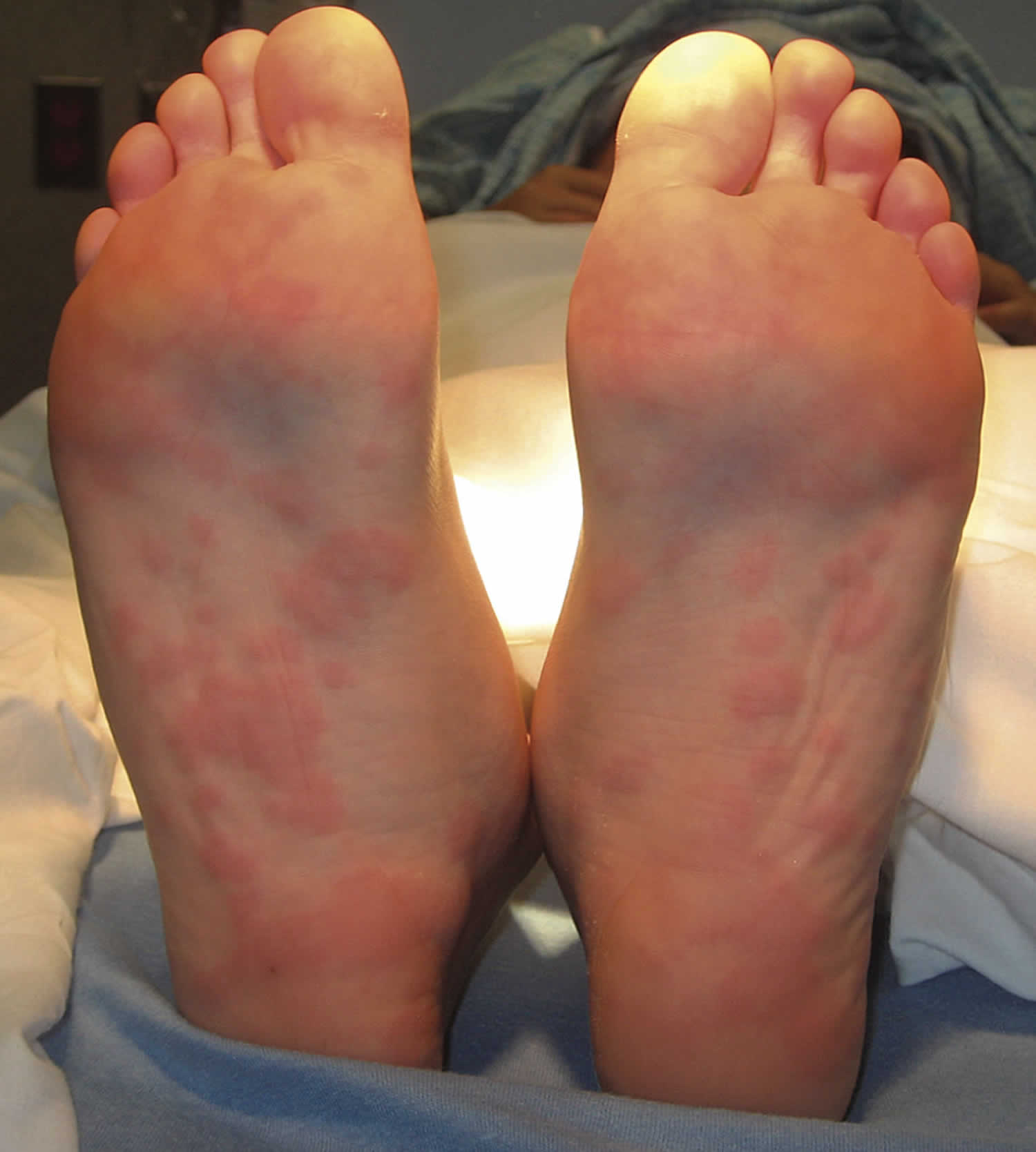
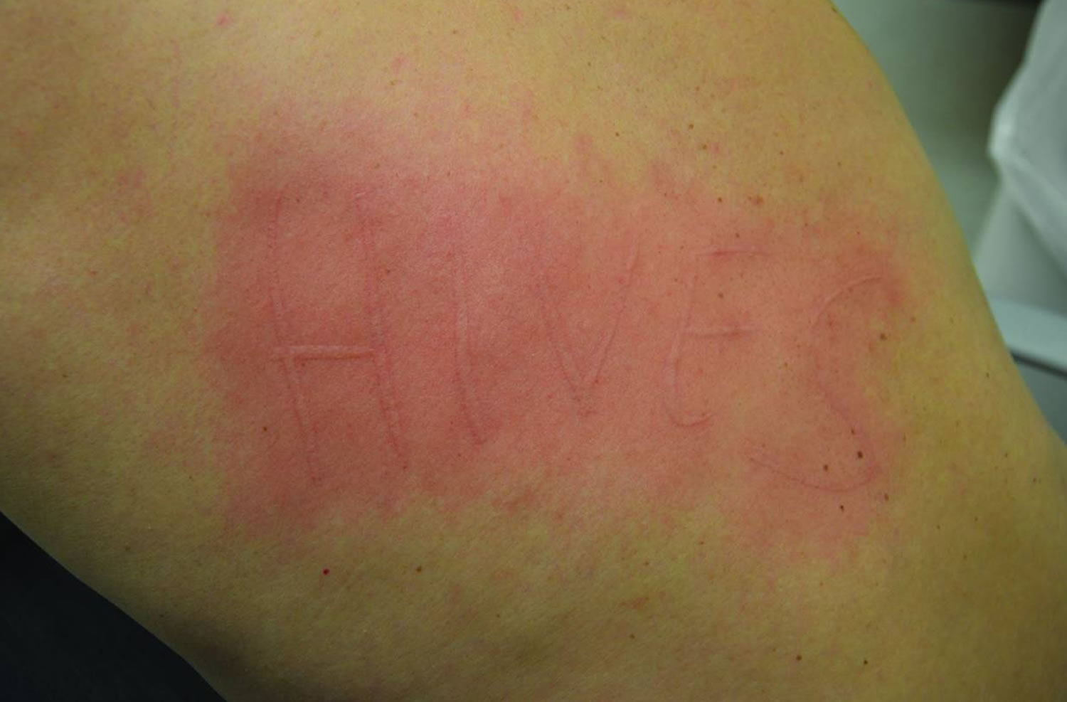
Figure 4. Dermatographism
Urticaria multiforme causes
Urticaria multiforme represents an allergic hypersensitivity reaction that may be IgE dependent or independent 3. Many children with urticaria have a history of infection or recent use of a systemic medication, often an antibiotic or an antipyretic. Viral infection is the more frequently associated pathogen at all ages 11. Urticaria among children is more commonly associated with infectious rather than allergic causes 12. In 1 retrospective study, 17% of patients with urticaria multiforme had laboratory-confirmed infection, including 1 patient with Mycoplasma pneumoniae pneumonia 13.
Importantly, the angioedema present in urticaria multiforme has not been associated with laryngoedema and has not been reported to be associated with food allergy 10.
Urticaria multiforme symptoms
Urticaria multiforme, a distinct morphological subtype of urticaria, is a benign cutaneous hypersensitivity reaction primarily mediated by histamine release characterized by transient erythema and angioedema 4. Pruritus is an almost universal complaint and systemic symptoms include a mild fever that usually resolves within 1 to 3 days of eruption 14. It is observed in the pediatric population, most often in those between 4 months and 4 years old. It is characterized by the presence of erythematous macules, papules, and plaques that quickly coalesce into large, blanchable, annular, polycyclic, violaceous wheals with dusky centers usually affecting the face, trunk, and extremities. In most cases there is also associated acral and facial angioedema. The rash usually persists for 2 to 12 days with full resolution, leaving normal skin after its resolution and a favorable response to antihistamines 4. Dermal edema can commonly involve the hands, feet, and face without associated laryngoedema 10. Dermatographism secondary to mast cell-mediated cutaneous dermal hypersensitivity at sites of skin trauma is a common part of the clinical picture in affected patients that can manifest in geometric or linear patterns 3.
Urticaria multiforme diagnosis
The diagnosis of urticaria multiforme can be made on history and physical examination grounds, thus detailed laboratory testing and/or skin biopsy is not routinely required in affected patients 1. A few hallmarks of the rash are the resolution of individual lesions within 24 hours and the presence of dermatographism, a wheal-and-flare reaction after minor skin trauma. Antecedent viral illnesses, antipyretics, and antibiotics, especially amoxicillin and cephalosporins, have all been listed as culprits 15. While an elevation in acute phase reactants (ESR and/or CRP) may be seen in some patients, a completely normal CBC, ESR, blood and lesional bacterial cultures, stool sample, antistreptolysin O (ASO), and throat swabs may be found positive in many cases. Although unnecessary in the medical workup for a patient with urticaria multiforme, skin biopsies reported in the literature are indistinct from other subtypes of acute urticaria, which demonstrate dermal edema with a perivascular lymphocytic infiltrate with few intermingled eosinophils 1. Serum-sickness-like reaction has been similarly described as demonstrating features consistent with urticaria without evidence of vasculitis. However, the histopathology of serum-sicknesslike reaction has been described as under-reported and possibly misunderstood 16. The histopathological spectrum of urticarial vasculitis ranges from minimal vascular damage associated with swollen endothelial cells and a sparse infiltrate of neutrophils, eosinophils, and lymphocytes to severe leukocytoclastic vasculitis. Nuclear debris or fibrinoid alteration of the microvasculature with or without extravasation of erythrocytes are the minimal essential criteria for a histopathological diagnosis of urticarial vasculitis 17. Erythema multiforme is characterized by histopathology distinct from urticaria multiforme. Exocytosis as well as spongiosis are apparent in conjunction with varying degrees of epidermal necrosis. Necrotic keratinocytes are generally present at all epidermal levels with an edematous papillary dermis with dilated capillaries 18.
Urticaria multiforme treatment
Treatment of urticaria multiforme is largely symptomatic since episodes are self-resolving within two weeks in the vast majority of cases 19. Any medications suspected of precipitating urticaria multiforme should be discontinued. Most patients will require systemic therapy with an H1 antihistamine, such as cetirizine, diphenhydramine, or hydroxyzine with or without an H2 antihistamine, such as ranitidine, in order to achieve optimal symptomatic relief 8. Combination therapy of oral antihistamines has been shown to be effective in a majority of cases, with symptoms resolving in most patients within 24 to 48 hours 1. Treatment with systemic corticosteroids should be reserved for only the most severe cases refractory to combination antihistamine therapy. Topical therapy with antipruritic agents, such as topical corticosteroids, calamine, pramoxine, colloidal oatmeal, and/or menthol can be used. Exhaustive laboratory evaluations for an infectious etiology is not recommended since these tests rarely yield clinically important information 3.
References- Starnes L, Patel T, Skinner RB. Urticaria multiforme—a case report. PediatrDermatol. 2011;28(4):436–438.
- Shah KN, Honig PJ, Yan AC. Urticaria multiforme: a case series and review of acute annular urticarial hypersensitivity syndromes in children. Pediatrics. 2007;119:e1177–e1183. doi: 10.1542/peds.2006-1553
- Shah KN, Honig PJ, Yan AC. “Urticaria multiforme”: a case series and review of acute annular urticarial hypersensitivity syndromes in children. Pediatrics. 2007;119(5):e1177–e1183.
- Starnes L, Patel T, Skinner RB. Urticaria multiforme: a case report. Pediatr Dermatol. 2011;28:436–438. doi: 10.1111/j.1525-1470.2011.01311.x
- Tamayo-Sanchez L, Ruiz-Maldonado R, Laterza A. Acute annular urticaria in infants and children. Pediatr Dermatol. 1997;14(3):231–234.
- Emer JJ, Bernardo SG, Kovalerchik O, Ahmad M. Urticaria multiforme. J Clin Aesthet Dermatol. 2013;6(3):34–39. https://www.ncbi.nlm.nih.gov/pmc/articles/PMC3613272
- Emer JJ, Bernardo SG, Kovalerchik O, Ahmad M. Urticaria multiforme. J Clin Aesthet Dermatol. 2013;6:34–39.
- Guerrier G, Daronat JM, Deltour R. Unusual presentation of acute annular urticaria: a case report. Case Rep Dermatol Med. 2011;2011:604390. doi:10.1155/2011/604390 https://www.ncbi.nlm.nih.gov/pmc/articles/PMC3505924
- Picture of the Month—Diagnosis. Arch Pediatr Adolesc Med. 2009;163(12):1158. doi:10.1001/archpediatrics.2009.221-b
- Shah KN, Honig PJ, Yan AC. “Urticaria multiforme”: a case series and review of acute annular urticarial hypersensitivity syndromes in children. Pediatrics. 2007;119(5):e1177–e1183. doi:10.1542/peds.2006-1553
- Novembre E, Cianferoni A, Mori F, et al. Urticaria and urticaria related skin condition/disease in children. European Annals of Allergy and Clinical Immunology. 2008;40(1):5–13.
- Sackesen C, Sekerel BE, Orhan F, Kocabas CN, Tuncer A, Adalioglu G. The etiology of different forms of urticaria in childhood. Pediatr Dermatol. 2004;21(2):102–108. doi:10.1111/j.0736-8046.2004.21202.x
- Shah KNHonig PJYan AC Urticaria multiforme: a case series and review of acute annular urticarial hypersensitivity syndromes in children. Pediatrics 2007;119 (5) e1177- e1183
- Shah KN, Honig PJ, Yan AC. Urticaria multiforme: a case series and review of acute annular urticarial hypersensitivity syndromes in children. Pediatrics. 2007;119:e1177–e1183. doi: 10.1542/peds.2006-1553.
- Gavin M, Sharp L, Stetson CL. Urticaria multiforme in a 2-year-old girl. Proc (Bayl Univ Med Cent). 2019;32(3):427–428. Published 2019 Jun 3. doi:10.1080/08998280.2019.1608627 https://www.ncbi.nlm.nih.gov/pmc/articles/PMC6650249
- Tolpinrud WL, Bunick CG, King BA. Serum-sickness-like reaction: histopathology and case report. J Am Acad Dermatol. 2011;65(3):e83–e85.
- Barksdale SK, Barnhill RL. Vasculitis and related disorders. In: Barnhill RL, Crowson AN, Magro CM, et al., editors. Dermatopathology. 3rd ed. China: McGraw Hill Medical; 2010. pp. 202–204.
- Ramos-Ceballos FI, Horn TD. Interface dermatitis. In: Barnhill RL, Crowson AN, Magro CM, et al., editors. Dermatopathology. 3rd ed. China: McGraw Hill Medical; 2010. pp. 342–344.
- Myers SR, Lavelle J. Picture of the month—quiz case. Pneumonia with associated urticaria multiforme rash. Arch Pediatr Adolesc Med. 2009;163(12):1157.


