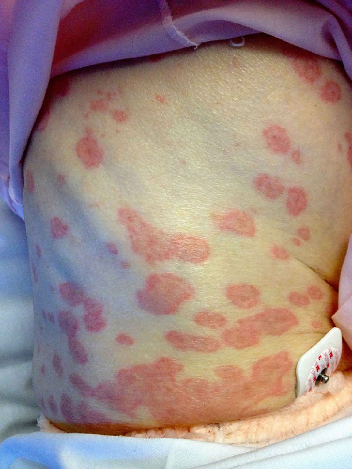
Target lesion
Target lesion is a round skin lesion with three concentric color zones:
- A darker center with a blister or crust
- A ring around this that is paler pink and raised due to edema (fluid swelling)
- A bright red outermost ring.
Target lesions typically occur in erythema multiforme. Target lesions can arise on any body site, including face, upper chest, back, arms, legs, hands, feet and mucous membranes (such as the lips). A target lesion is also called a bulls-eye lesion or a cockade (a rosette pattern of concentric rings).
An iris lesion represents an early target lesion and has two parts:
- A central dusky zone
- A red outer zone.
Single-component red plaques are also typical of erythema multiforme.
Target lesions appear within the first 3 days of an episode of erythema multiforme and once one lesion has appeared, it stays in the same location for 7 days or more until the skin heals.
Atypical target lesions
Atypical target lesions show just two zones and/or an indistinct border. In erythema multiforme, these lesions are raised (papular). In Stevens-Johnson syndrome / toxic epidermal necrolysis, they are flat (macular).
Target like lesions
Target like lesions commonly known as targetoid lesions have concentric zones and look similar to target lesions but are not due to erythema multiforme. They may evolve over a different time frame. Several skin conditions cause target like lesions.
Melanocytic naevus
A targetoid nevus (mole) is also called a cockade nevus (nevus en cocade). They are sometimes multiple. They are usually found on the trunk of a fair-skinned individual but may also be seen in the scalp, particularly in children. They are harmless and tend to remain unchanged long-term. Sometimes the outer, darker rim fades or disappears later in life.
Urticaria
Urticaria is characterized by weals, which are raised plaques with a smooth surface that change location and/or size within a 24 hour period. Weals may be large and have unusual shapes. Target like lesions in urticaria have an area of normal skin in the middle surrounded by a raised circular weal. Dermographism (formation of a weal on light stroking of the skin) may be present.
Fixed drug eruption
Fixed drug eruption is an adverse drug reaction that usually occurs in the same site/s on the skin every time the medication is taken. A fresh lesion is a well defined targetoid plaque, with a dusky red raised area of skin and sometimes a central blister.
Fixed drug eruption is not usually very extensive. Examination of a skin biopsy by a pathologist may be helpful in making the diagnosis.
Polymorphous light eruption
Polymorphous light eruption occurs on skin a few hours after it has been exposed to sunlight. As its name suggests, it can take many forms, but generally consists of small or large raised red spots. Target like lesions are less common.
Erythema annulare centrifugum
Erythema annulare centrifugum begins as a small raised pink spot that enlarges and forms a ring shape, while the central area flattens and clears. There may be an inner rim of scale, resulting in the 3 zones characterizing a target like lesion.
Subacute cutaneous lupus erythematosus
Subacute cutaneous lupus erythematosus occurs on the upper back and chest, often following sun exposure. It often has a ring-shaped appearance (annular or polycyclic), and occasionally develops a target like lesion with 3 concentric zones.
Rowell syndrome
Rowell syndrome is a rare form of lupus erythematosus with target like lesions and positive blood tests for lupus antibodies (a speckled pattern of antinuclear antibodies, and sometimes positive anti-La/anti-Ro or rheumatoid factor). The skin lesions are large, raised and ring-shaped. Patients may also have chilblains.
Polymorphic eruption of pregnancy
Polymorphic eruption of pregnancy occurs in the last 3 months of pregnancy. In most women, pink papules develop within the stretch marks on the abdomen. These may be surrounded by a pale halo or evolve to target like lesions with 3 rings. Polymorphic eruption of pregnancy is characteristically very itchy.
Immunobullous disorders
Target like lesions may occur in several bullous (blister-forming) disorders, including:
- Paraneoplastic pemphigus
- Bullous pemphigoid
- Pemphigoid gestationis
- Linear IgA bullous dermatosis
The target like lesions of linear IgA bullous dermatosis have peripheral blisters, in contrast to target lesions in erythema multiforme, which have central blisters.
Vasculitis
Some forms of vasculitis may present with target like lesions on the skin. These include:
- Kawasaki disease, which occurs in children generally under the age of 5. The skin lesions may range from a measles-like rash to target-like skin lesions.
- Acute hemorrhagic edema of infancy, which occurs in children under the age of 2, is a form of urticarial vasculitis. The rash begins as a red raised lesion and then changes to the typical targetoid appearance, with a purplish color.
Trauma
The impact of a ball, such as a squash ball or a table tennis ball, hitting the skin at high speed can also produce a targetoid bruise.
Hobnail hemangioma
Another name for a hobnail hemangioma is targetoid hemosiderotic hemangioma. It is a benign (non-cancerous) overgrowth of blood vessels, generally occurring on an arm, leg or trunk of a young to middle-aged person. It typically has a small red/purple raised center surrounded by a purple or brown ring that can expand or disappear altogether.
Target lesion causes
Target lesions typically occur in erythema multiforme.
Erythema multiforme is a hypersensitivity reaction usually triggered by infections, most commonly herpes simplex virus (HSV). It presents with a skin eruption characterised by a typical target lesion. There may be mucous membrane involvement. It is acute and self-limiting, usually resolving without complications.
Erythema multiforme is divided into major and minor forms and is now regarded as distinct from Stevens–Johnson syndrome (SJS) and toxic epidermal necrolysis (TEN).
What triggers erythema multiforme?
Infections
Infections are probably associated with at least 90% of cases of erythema multiforme. The single most common trigger for developing erythema multiforme is herpes simplex virus (HSV) infection, usually herpes labialis (cold sore on the lip) and less often genital herpes. HSV type 1 is more commonly associated than type 2. The herpes infection usually precedes the skin eruption by 3–14 days.
Mycoplasma pneumonia (a lung infection caused by the bacteria Mycoplasma pneumoniae) is the next most common trigger.
Many different virus infections have been reported to trigger erythema multiforme including:
- Parapoxvirus (orf and milkers’ nodules)
- Herpes varicella-zoster (chickenpox, shingles)
- Adenovirus
- Hepatitis viruses
- Human immunodeficiency virus (HIV)
- Cytomegalovirus
- Viral vaccines
Dermatophyte fungal infections (tinea) have also been reported in association with erythema multiforme.
Drugs
Medications are probably an uncommon cause (<10%) of erythema multiforme. If this diagnosis is being seriously considered then alternative drug eruptions should be excluded, such as Stevens–Johnson syndrome or toxic epidermal necrolysis, generalized fixed drug eruption, polymorphic exanthematous drug eruption and urticaria.
Many drugs have been reported to trigger erythema multiforme, including barbiturates, non-steroidal anti-inflammatory drugs, penicillins, sulphonamides, nitrofurantoin, phenothiazines, and anticonvulsants.
Clinical features of erythema multiforme
General symptoms
There are usually no prodromal symptoms in erythema multiforme minor. However, erythema multiforme major may be preceded by mild symptoms such as fever or chills, weakness or painful joints.
Skin lesions
Typically in erythema multiforme, few to hundreds of skin lesions erupt within a 24-hour period. The lesions are first seen on the backs of hands and/or tops of feet and then spread down the limbs towards the trunk. The upper limbs are more commonly affected than the lower. Palms and soles may be involved. The face, neck and trunk are common sites. Skin lesions are often grouped on elbows and knees. There may be an associated mild itch or burning sensation.
The initial lesions are sharply demarcated, round, red/pink and flat (macules), which become raised (papules/palpable) and gradually enlarge to form plaques (flat raised patches) up to several centimeters in diameter. The center of the papule/plaque darkens in color and develops surface (epidermal) changes such as blistering or crusting. Lesions usually evolve over 72 hours.
The typical target lesion (also called iris lesion) of erythema multiforme has a sharp margin, regular round shape and three concentric color zones:
- The center is dusky or dark red with a blister or crust
- Next ring is a paler pink and is raised due to edema (fluid swelling)
- The outermost ring is bright red.
Atypical target lesions show just two zones and/or an indistinct border.
The eruption is polymorphous (many forms), hence the ‘multiforme’ in the name. Lesions may be at various stages of development with both typical and atypical targets present at the same time. A full skin examination may be required to find typical targets, as these may be few in number.
Lesions show the Köbner (isomorphic) phenomenon, meaning they can develop at sites of preceding (but not concurrent or subsequent) skin trauma.
There is no associated swelling of face, hands or feet, despite these being common sites of rash distribution. However, the lips are often swollen, especially in erythema multiforme major.
Erythema multiforme diagnosis
Erythema multiforme is a clinical diagnosis, although skin biopsy may be required to exclude other conditions. The histology of erythema multiforme is characteristic but not diagnostic. It varies with the age of the lesion, its appearance, and which part is biopsied.
Other tests may be done looking for infections commonly seen in association with erythema multiforme, such as mycoplasma.
Target lesion treatment
Target lesion treatment involves treating the underlying cause.
Treatment of erythema multiforme
For the majority of cases, no treatment is required, as the rash settles by itself over several weeks without complications.
Treatment directed to any possible cause may be required such as oral aciclovir (not topical) for HSV or antibiotics (eg, erythromycin) for Mycoplasma pneumoniae. If a drug cause is suspected then the possible offending drug should be ceased.
Supportive/symptomatic treatment may be necessary:
- Itch — oral antihistamines and/or topical corticosteroids may help.
- Oral pain — mouthwashes containing local anesthetic and antiseptic reduce pain and secondary infection.
- Eye involvement should be assessed and treated by an ophthalmologist.
- Erythema multiforme major may require hospital admission for supportive care, particularly if severe oral involvement restricts drinking.
The role of oral corticosteroids remains controversial, as no controlled studies have shown any benefit. However for severe disease 0.5–1 mg/kg/d prednisone or prednisolone is often used early in the disease process.
Recurrent erythema multiforme is usually treated initially with continuous oral aciclovir for 6 months at a dose of 10 mg/kg/d in divided doses (eg, 400 mg twice daily), even if HSV has not been an obvious trigger for the patient’s erythema multiforme. This has been shown to be effective in placebo-controlled double-blind studies. However, erythema multiforme may recur when the aciclovir is ceased. Other antiviral drugs such as valaciclovir (500–1000 mg/day) and famciclovir (250 mg twice daily) should be tried if aciclovir has not helped.
Other treatments (used continuously) that have been reported to help suppress recurrent erythema multiforme include:
- Dapsone 100–150 mg/day
- Antimalarial drugs (eg, hydroxychloroquine)
- Azathioprine 100–150 mg/day
- Others: thalidomide, ciclosporin, mycophenolate mofetil, photochemotherapy (PUVA)
What is the outlook for erythema multiforme?
Erythema multiforme minor usually resolves spontaneously without scarring over 2–3 weeks. Erythema multiforme major can take up to 6 weeks to resolve. Erythema multiforme does not progress to Stevens-Johnson syndrome / toxic epidermal necrolysis.
There may be residual mottled skin discoloration. Significant eye involvement in erythema multiforme major may rarely result in serious problems, including blindness.


