What is atypical fibroxanthoma
Atypical fibroxanthoma is a dermal spindle-cell tumor that occurs primarily in older individuals after the skin of the head and neck has been damaged significantly by sun exposure and/or therapeutic radiation. Less common risk factors for atypical fibroxanthoma include immunosuppression, previous history of radiation therapy in the region of the tumor, and a history of xeroderma pigmentosa. Clinically, atypical fibroxanthoma lesions usually are suggestive of malignancy because they arise rapidly (over just a few weeks or months) in skin in which other skin cancers have been found and treated. Atypical fibroxanthoma tumor-like growth should be considered a type of skin cancer but it may behave in a benign fashion. When this clinical impression is combined with highly anaplastic pathology, misdiagnosis can result in unnecessary and extensive surgery and radiation. A review of systems is most commonly negative, due to the very small risk for metastasis.
A rare type of atypical fibroxanthoma occurs in younger patients on parts of the body that are not normally overexposed to the sun. These tumors are usually found on the trunk and extremities and tend to be larger and slower growing.
Atypical fibroxanthoma causes
The etiology of atypical fibroxanthomas is poorly understood. The development of atypical fibroxanthomas is associated with ageing, sun exposure (ultraviolet radiation) and/or ionizing radiation (X-rays) 1. Ultraviolet light is believed to play an important role since most lesions appear on the sun-exposed head and neck of Caucasian patients. Both sun exposure (ultraviolet radiation) and/or ionizing radiation (X-rays) can cause abnormal growth of atypical spindle cells. These are believed to come from fibrous cells in the dermis or from epidermal keratinocytes.
Genetic factors also contribute to the development of atypical fibroxanthoma, since it shares genetic alterations with undifferentiated pleomorphic sarcomas including chromosome 9p and 13q deletions 2. However, undifferentiated pleomorphic sarcoma has a statistically significant larger amount of genomic alterations and a more aggressive clinical course. Other possible risk factors for atypical fibroxanthoma include trauma to the skin, radiation therapy, and immunosuppression (e.g., diabetes, HIV, organ transplant) 3. Reports have shown an increased incidence of atypical fibroxanthoma in patients with acquired immune deficiency syndrome (AIDS) and in patients who are immune suppressed, for example because of organ transplantation 4.
Atypical fibroxanthoma affects both sexes equally, with a mean age of 69 years at diagnosis.
Atypical fibroxanthomas most commonly are seen on the head and neck of elderly Caucasian patients. Other reported sites of atypical fibroxanthoma include the trunk, shoulders, upper extremities, and dorsum of the hands. Trunk and limb lesions are believed to occur more commonly in younger patients (mean age 39) compared to head and neck lesions in the elderly (mean age 69). A review of 171 cases in Western Australia found a patient age-range of 41 to 97 years old (median age 74) with 76% of the tumors occurring in men. Atypical fibroxanthoma also has been reported in patients as young as 13 with predisposing disorders such as Li Fraumeni Syndrome and Xeroderma Pigmentosum. The incidence is increased in immunosuppressed populations, with an estimated incidence of 78 per 100,000 transplant patients.
While no direct association has been made, researchers hypothesize that arsenic exposure may predispose patients to atypical fibroxanthoma 2.
Atypical fibroxanthoma pathology
Hematoxylin and eosin stains of atypical fibroxanthoma lesions show a dermally-based tumor with pleomorphism, atypical mitotic figures, and a spindly architecture. Atypical fibroxanthoma lesions occasionally extend to the subcutaneous tissue. Note that atypical fibroxanthoma is a dermal-based process while spindle cell squamous cell carcinoma variants (on the differential diagnosis) connect with the epidermis and show signs of keratinization. Multinucleated giant cells and solar elastosis are often present in atypical fibroxanthoma biopsy specimens, while a mixed inflammatory infiltrate may be found at the edge of the tumor. Immunohistochemistry is required to rule out other neoplasms including spindloid squamous cell carcinoma, melanoma, and undifferentiated pleomorphic sarcoma. Since atypical fibroxanthoma stains positive for several non-specific stains, including CD10, p53, S100A6, vimentin, and procollagen-1, it is considered a diagnosis of exclusion histologically. Of note, atypical fibroxanthoma stains negative for HMB-45, p40 (often positive in squamous cell carcinoma), desmin, pan-cytokeratin stains, CD31, and has sparse staining for S100, which may help differentiate it from melanoma. Since poorly differentiated sarcomatoid carcinomas may lose cytokeratin expression, a variety of cytokeratin stains should be used. Severe variants of atypical fibroxanthoma have been described based on various histologic findings, including pigmented atypical fibroxanthoma due to hemosiderin deposit and not actual melanin pigment, granular cell atypical fibroxanthoma, sclerotic atypical fibroxanthoma, spindle cell nonpleomorphic atypical fibroxanthoma, osteoid atypical fibroxanthoma, chondroid atypical fibroxanthoma, myxoid atypical fibroxanthoma, keloidal atypical fibroxanthoma, osteoclast-like giant-cell rich atypical fibroxanthoma, and clear cell atypical fibroxanthoma.
Atypical fibroxanthoma prevention
Most cases of atypical fibroxanthoma could be prevented by avoiding excessive sun exposure. Patients are advised to follow sun protection methods when outdoors.
Atypical fibroxanthoma symptoms
Atypical fibroxanthoma often appears in areas that have received excessive sun exposure, usually around the scalp, ears, nose, cheeks, and back of the neck, or in areas where individuals may have previously received radiotherapy treatment. They have also been reported to occur on the trunk, extremities, and in sun-protected areas. More than 90% of atypical fibroxanthoma tumors occur on the head and neck, while the remaining cases typically present on the extremities and trunk.
Atypical fibroxanthoma typically presents with the sudden onset of an asymptomatic, solitary, growth of the head and neck. Bleeding and ulceration are common. Patients are commonly elderly and have a history of extensive sun exposure or previous skin cancers.
- A solitary tumor or multiple tumors may occur.
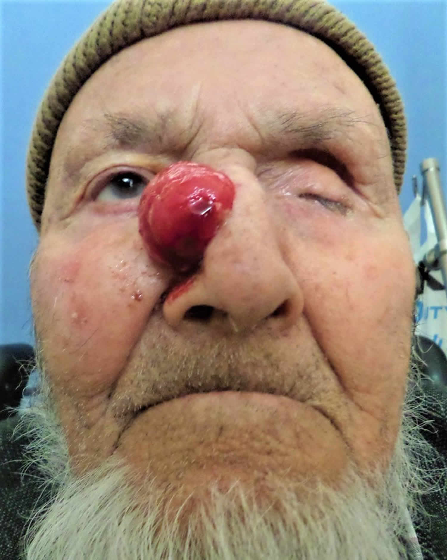
- Typically, atypical fibroxanthoma is a red, juicy, dome-shaped nodule that may be bleeding, ulcerated or crusted.
- The tumor starts off as a small nodule that grows quickly over 6 months to a size of about 2–3 cm.
- Four times as many tumors are diagnosed on the head and neck as are diagnosed in other body sites.
Atypical fibroxanthoma has a nonspecific clinical appearance on physical examination, characterized by pink or red papules or nodules with a firm texture, located most commonly on the head or neck, and less commonly on the trunk or extremities. Rare locations include the eyelid, cornea, and ocular surface. Most lesions are smaller than 2 cm in diameter, with the average size being 1.1-1.5 cm in diameter (range: 0.3-0 cm). Ulceration (up to 45% of cases) with oozing or bleeding is common.
Although rare, cutaneous metastases from atypical fibroxanthoma have been reported.
Atypical fibroxanthoma diagnosis
Because atypical fibroxanthoma can look like other skin cancers, it is usually diagnosed by a pathologist after a skin biopsy or excision.
The diagnosis depends on finding large, pleomorphic, fibrocytic, spindle-shaped, anaplastic tumour cells haphazardly arranged in the dermis. Immunohistochemistry stains should be undertaken but may be nonspecific. Cytokeratin and melanoma stains are negative.
When atypical fibroxanthoma lesions have metastasized or are poorly accessible (e.g., subungual lesions), magnetic resonance imaging (MRI) may be helpful, which shows intermediate T1 and T2-weighted signal intensity compared to the high signal intensity of squamous cell carcinoma and melanoma.
Atypical fibroxanthoma treatment
Atypical fibroxanthoma is treated by complete surgical excision. Small lesions may be removed by curettage. Mohs micrographic surgery is becoming the treatment of choice for large or recurrent lesions, as it reliably removes the complete tumor while sparing surrounding normal healthy tissue.
Atypical fibroxanthoma prognosis
Atypical fibroxanthoma rarely recurs after complete excision with clear margins. Recurrence and metastasis are more likely in people that are immunosuppressed.
Fortunately, atypical fibroxanthoma rarely metastasizes and recurs in only 6% to 10% of cases 2. Risk factors for metastases include immunocompromised patients, tumor depth, vascular or perineural invasion, and the presence of tumor necrosis. The most commonly reported locations for metastases include the parotid gland, lymph nodes, and subcutaneous tissue 2. Metastases, when present, are typically evident 12 to 24 months after the initial diagnosis of atypical fibroxanthoma.
- Atypical fibroxanthoma. https://www.dermnetnz.org/topics/atypical-fibroxanthoma/[↩]
- Kolb L, Schmieder GJ. Atypical Fibroxanthoma. [Updated 2019 Jan 27]. In: StatPearls [Internet]. Treasure Island (FL): StatPearls Publishing; 2019 Jan-. Available from: https://www.ncbi.nlm.nih.gov/books/NBK459342[↩][↩][↩][↩]
- Tolkachjov SN, Kelley BF, Alahdab F, Erwin PJ, Brewer JD. Atypical fibroxanthoma: Systematic review and meta-analysis of treatment with Mohs micrographic surgery or excision. J. Am. Acad. Dermatol. 2018 Nov;79(5):929-934.e6[↩]
- Moscarella E, Piana S, Specchio F, Kyrgidis A, Nazzaro G, Eliceche ML, Savoia F, Bugatti L, Filosa G, Zalaudek I, Scarfi F, Inskip M, Rosendahl C, Pyne JH, Siggs G, Toğral AK, Cabo H, Drlik L, Lallas A, Longo C, Argenziano G. Dermoscopy features of atypical fibroxanthoma: A multicenter study of the International Dermoscopy Society. Australas. J. Dermatol. 2018 Nov;59(4):309-314[↩]


