What is Buruli ulcer
Buruli ulcer is also called Bairnsdale ulcer, Searles ulcer, Daintree ulcer, and Sik-belonga-sepik, is a disease caused by the bacterium Mycobacterium ulcerans 1. Buruli ulcer mainly affects the skin but can also affect the bone. Cases are generally seen in the tropics, primarily in West Africa, Asia, Mexico, South America and Australia. Mycobacterium ulcerans infection often leads to ulcers on the arms or legs, which can also destroy skin or soft tissue. When not properly treated, Buruli ulcer disease can cause irreversible deformity or long-term functional disability.
Buruli ulcer disease traditionally presents as a single painless nodule under the skin’s surface and swelling in this region may or may not be present. Fever may accompany disease forms where diffuse swelling is present. In the absence of treatment, the lesion may progress into an ulcer where underlying structures such as muscle or tendons could become exposed. If the disease reaches this stage, the infection may spread to the deeper surrounding organs or bones. Disfiguring scars are not uncommon in the absence of treatment. Buruli ulcer tends to favor the arms and legs. A greater risk of infection exists for both children and the elderly.
See a doctor if you experience the symptoms of Buruli ulcer. Your doctor can perform tests to check for the Buruli ulcer disease.
In endemic areas, Buruli ulcer is often diagnosed and treated based on clinical findings.
- A direct smear is taken from the necrotic base of the ulcer, stained with Ziehl-Neelsen stain to reveal clumps of acid-fast bacilli (mycobacteria) under a microscope.
- Polymerase chain reaction (PCR) can rapidly confirm Mycobacterium ulcerans in swabs of an ulcer or tissue biopsy.
- Biopsy of the lesion can reveal characteristic microscopic changes and clumps of acid-fast bacilli.
- Mycobacterium ulcerans can also be cultured from swabs taken from an ulcer or fresh tissue biopsy, but the result takes 6 to 8 weeks or more to be reported.
If you have the Buruli ulcer disease, the doctor will give you antibiotics (medicine that can help stop the disease).
Be sure to follow the doctor’s instructions for taking the antibiotics.
In most cases, Buruli ulcer can be treated without hospitalization.
In rare cases, surgery may be necessary to:
- Remove dead skin
- Cover skin defects
- Correct deformities
Buruli ulcer transmission
It is not known how people get Buruli ulcer. Buruli ulcer is found in at least 33 countries with tropical, subtropical, and temperate climates. In 15 of these 33 countries, between 5,000 and 6,000 cases are reported every year.
Overall, most cases occur in rural communities in sub-Saharan Africa. Nearly half of those affected in Africa are children under 15 years of age.
One possibility of transmission is that Buruli ulcer is passed to humans from some insects that are found in water. While no proven link exists between human and animal infection, some animals can get the disease.
For example, laboratory tests from Victoria, Australia, confirmed the disease in several animals, including:
- Horses
- Dogs
- Alpacas
- Koalas
- Opossums
The incubation period ranges from a few weeks to months. Buruli ulcer begins as a firm, painless nodule (swelling) in the skin, which is around one to two cm in diameter. M. ulcerans produces a toxin, called mycolactone, which is directly toxic to cells and also dampens the immune system. The toxin causes extensive tissue destruction, without any systemic symptoms (such as fever, malaise, or enlarged lymph nodes).
Over the following weeks, the nodule breaks down to form a painless necrotic ulcer with undermined edges (tissue destruction underlying intact skin). The necrosis may extend several centimetres beyond the edges of the ulcer, making the lesion appear smaller than its actual size. The ulcer can extend down into deeper tissues destroying nerves, blood vessels, muscles, and occasionally bone. The limbs, particularly the lower limbs, are most commonly involved.
What causes Buruli ulcer?
Buruli ulcer is caused by bacteria called Mycobacterium ulcerans. These bacteria are atypical mycobacteria and come from the same family of organisms that cause leprosy and tuberculosis.
Buruli ulcer is found in more than 33 tropical, subtropical, and temperate countries. The majority of cases occur in central and western Africa. Cases have also been reported from Australia, South East Asia, and Central and South America. Over the last 2 decades, the incidence of Buruli ulcer has increased, despite significant underreporting of cases. In 1999 there were 6000 new cases in Ghana; in Australia, there were 25 cases in 2004, 47 in 2005 and 72 in 2006. In 2018, there were more than 340 cases in the state of Victoria alone.
Buruli ulcer predominantly affects poor rural communities living near swampy terrain. Although the exact mode of transmission is unknown, M. ulcerans most likely cause infection through inoculation or contamination of a traumatic wound. In Victoria, Australia, the infection has been found in wild animals such as ring-tailed possums, and in mosquitoes. It has been speculated that the mycobacterial infection may follow an infected mosquito bite.
People of any age can be affected, but most cases are among children aged less than 15 years.
Signs and symptoms of buruli ulcer
The incubation period ranges from a few weeks to months. Buruli ulcer begins as a firm, painless nodule (swelling) in the skin, which is around one to two cm in diameter. Mycobacterium ulcerans produces a toxin, called mycolactone, which is directly toxic to cells and also dampens the immune system. The toxin causes extensive tissue destruction, without any systemic symptoms (such as fever, malaise, or enlarged lymph nodes).
Over the following weeks, the nodule breaks down to form a painless necrotic ulcer with undermined edges (tissue destruction underlying intact skin). The necrosis may extend several centimetres beyond the edges of the ulcer, making the lesion appear smaller than its actual size. The ulcer can extend down into deeper tissues destroying nerves, blood vessels, muscles, and occasionally bone. The limbs, particularly the lower limbs, are most commonly involved.
The symptoms of Buruli ulcer include:
- Swelling of the skin
- Destroyed skin and soft tissue
- One or more slow growing, generally painless ulcers
People who are sick should see a doctor and get antibiotics (medicine that can help stop the disease).
If these antibiotics are not given soon after getting sick, the disease can sometimes lead to:
- Deformity
- Functional disability (such as limited joint movement)
- Bone infection
- Secondary bacterial infection of skin ulcer lesions
The World Health Organization (WHO) clinical case definition for Buruli ulcer divides the disease into two stages: active and inactive.
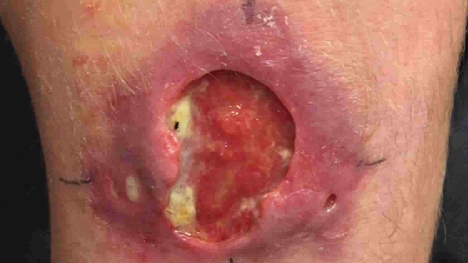
The active form is characterized by non-ulcerative (papules, nodules, plaques, and edema) and ulcerative disease. The distinctive features of a Buruli ulcer include:
- undermining edges
- white cotton wool-like appearance
- thickening and darkening of the skin surrounding the lesion
The ulcers are generally painless and progressive; 85% are found on the limbs, with lower limb lesions twice as common as upper limb lesions.
The inactive form is characterized by evidence of previous infection with a depressed stellate scar with or without complications.
Buruli ulcer complications
Tissue destruction can be extensive (involving up to 15% of the patient’s skin surface) and secondary infection may occur. Other complications include osteomyelitis (infection of the bone) and metastatic lesions (the spread of the wounds to distant sites). Extensive lesions heal with scarring, which may cause irreversible deformity, secondary lymphedema (swelling due to fluid retention), and restriction of joint movement. Few people die from Buruli ulcer, but it often causes long-term disability, disfigurement, and a significant socioeconomic burden.
Treatment with antibiotics can sometimes cause a paradoxical inflammatory reaction and enlarging ulceration.
Buruli ulcer diagnosis
Buruli ulcer disease is diagnosed on the basis of the WHO clinical case definition.
Confirmation of disease is achieved through the use of two or more of the following laboratory tests:
- acid-fast bacilli (AFB) identified on microscopic smear stained by Ziehl-Neelsen technique
- polymerase chain reaction (PCR)
- histopathology
- culture
With the exception of acid-fast bacilli (AFB) smear, these tests are not suited for use in the remote rural areas where Buruli ulcer disease occurs most frequently. Confirmation, if accomplished, typically occurs well after treatment has begun.
Depending on the patient’s age, location of lesions, pain, and geographic area, other conditions should be excluded from the diagnosis. These include
- tropical phagedenic ulcers
- chronic lower leg ulcers due to arterial and venous insufficiency (often in the older and elderly populations)
- diabetic ulcer
- cutaneous leishmaniasis
- extensive yaws
Early nodular lesions are occasionally confused with:
- boils
- lipomas
- ganglions
- lymph node tuberculosis
- onchocerciasis nodules
- other subcutaneous infections (such as fungal infection).
In Australia, papular lesions may initially be confused with an insect bite. Cellulitis may look like edema caused by Mycobacterium ulcerans infection but in the case of cellulitis, the lesions are painful and the patient is ill and febrile.
Buruli ulcer treatment
Current WHO recommendations for treatment are as follows:
- Different combinations of antibiotics given for eight weeks are used to treat the Buruli ulcer:
- a combination of oral rifampicin (10 mg/kg once daily) and intravenous streptomycin (15mg/kg once daily); or
- a combination of oral rifampicin (10 mg/kg once daily) and oral clarithromycin (7.5 mg/kg twice daily) has been used though effectiveness has not been proven by a randomized trial. Since streptomycin is contraindicated in pregnancy, the combination of rifampicin and clarithromycin is also considered the safer option for this group of patients; or
- a combination of oral rifampicin (10 mg/kg once daily) and oral moxifloxacin (400 mg once daily) has also been used though effectiveness has not been proven by randomized trial
- Complementary treatment, such as wound care, surgery (mainly debridement and skin grafting) and interventions to minimize or prevent disabilities, is often necessary depending on the stage of the disease.
BCG vaccination appears to offer some short-term protection (less than one year) from the Buruli ulcer disease.
References


