Nervous System

Lateral hypothalamus

lateral hypothalamus

Lateral hypothalamus

Lateral hypothalamus also called the lateral hypothalamic area 1 is considered to be a small (6 × 6 × 3.5 mm) target within the hypothalamus located in the midbrain, lying inferior to the fornix and superoposterior to the optic nerve and chiasm 2, 3. Lateral hypothalamus is divided into three rostro-caudal zones – the anterior lateral hypothalamus, the tuberal lateral hypothalamus, and the posterior lateral hypothalamus 4. The anterior lateral hypothalamus is continuous rostrally with the lateral preoptic area, and extends caudally to the level of the rostral pole of the venteromedial nucleus (VMN). While the tuberal lateral hypothalamus is coextensive with the venteromedial nucleus (VMN), the posterior lateral hypothalamus follows the tuberal division at the level of the mamillary complex 5. The lateral hypothalamus is home to a heterogeneous population of neurons that include both gamma-aminobutyric acid (GABA)-ergic and glutamatergic neurons as well as subpopulations of neurons expressing neuropeptides that have been linked to the modulation of motivated behaviors such as orexin (hypocretin) 6, 7, melanin-concentrating hormone (MCH) 8, cocaine- and amphetamine-regulated transcript (CART)9, neurotensin (NT) 10, leptin receptor 11 and galanin 12, 13. These neurons connect to other brain structures via efferent projections from the lateral hypothalamus to multiple structures including the amygdala, hippocampal formation, thalamus, the pons, brainstem and spinal cord, as well as intra-structural projections within the lateral hypothalamus to other hypothalamic subnuclei 14, 15, 16, 17. The lateral hypothalamus also projects densely onto the ventral tegmental area (VTA) 18, 19.

The large number of inputs, outputs, neuron types and functions present within the lateral hypothalamus suggest that this structure is very complex and hosts an extremely diverse population of neurons, with many neurons co-expressing multiple neuropeptides and projecting to numerous target neural structures 20, 21, moving forward it will be important to gain a more precise understanding of these neurons, projections and functions to better disentangle the many roles of the lateral hypothalamus 20. Interestingly, neurons in the lateral hypothalamus are the largest in the hypothalamus and are topographically well organized 5. Chief among them are the orexin neurons that project widely to the neuraxis and undertake many important functions. A growing body of evidence suggests that orexin neurons play a key role in regulating wakefulness 22, sleep 23, food intake 24, autonomic and endocrine functions 25, reward-related behaviors 26 and pain-related behaviors 27. However, despite significant progress in research, a more refined understanding of the detailed functions of orexin neurons of the lateral hypothalamus in the regulation of inflammatory pain is needed 28.

The lateral hypothalamus has been implicated in numerous functions including sleep-wake transitions 29, 30, 31, feeding 32, energy balance 33, stress 34 and reward 35, 36 and plays a critical role in maintaining physiological and behavioral homeostasis. As well as being dubbed a “feeding center” or “hunger center” by Anand and Brobeck 32, the lateral hypothalamus has also been labeled as a “pleasure center” 37 after it was shown that electrode implantation into the medial forebrain bundle in the lateral hypothalamus resulted in persistent intracranial self-stimulation 38. It has been suggested that this behavior is a result of stimulation of the descending fibers in the medial forebrain bundle that feed into the ventral tegmental area (VTA), likely triggering a reward response 39.

The lateral hypothalamus was discovered to be the source of extensive projections of neurons containing 2 newly discovered neuropeptides: melanin-concentrating hormone and orexin 40, 41. Both neuronal populations have broad projections throughout the central nervous system (CNS), and direct injection of either melanin-concentrating hormone or orexin into the ventricles will cause rats to feed, and these levels increase in rats during periods of starvation 42, 40. Multiple early animal studies demonstrated that low-frequency stimulation of the lateral hypothalamus resulted in an excitatory stimulation of these fibers, with animals demonstrating food-seeking and food-hoarding behavior, increased gastrointestinal blood flow, and activation of vagal pathways 43, 44, 45. In 2007, Sani et al. 46 reported on the use of high-frequency deep brain stimulation (DBS) of the bilateral lateral hypothalamus in 16 rats that resulted in a 2.3% weight loss in stimulated rats and a 13.8% weight gain in unstimulated controls. After lateral hypothalamic damage in rats only simple automatisms (such as grooming, chewing, licking) are present, but intense stimuli can activate more complex actions (walking, orientation, swimming) 47. In the anorexic stage, tactile stimuli dominate in steering locomotion and “spontaneous” locomotion depends on activation from the empty stomach 47.

Anatomical and electrophysiological studies have shown strong neuronal connections between the olfactory system and the hypothalamus 48. Anterograde and retrograde axonal tracing studies have revealed that projections from the olfactory system are more prominent to the lateral hypothalamus than to the thalamus 48. Furthermore, four primary areas in the posterior lateral hypothalamus (anterior olfactory nucleus, olfactory tubercle, piriform cortex, and anterior cortical nucleus of amygdala) were shown to receive this input from the olfactory bulb 48.

The lateral hypothalamus became classically known as the “feeding center” or “hunger center” of the brain after multiple early animal studies demonstrated that bilateral lateral hypothalamus lesions in rats resulted in decreased food intake, weight loss, and decreased food-seeking behavior leading to anorexia, somnolence, akinesia, and sensory neglect combine to produce complete aphagia, indicating the vast importance of lateral hypothalamus circuitry in appetite and/or eating or drinking properties of feeding behavior 49, 50, 51, 52, 53, 54, 55, 56.

Targeted photostimulation of projections from the lateral hypothalamus to the paraventricular hypothalamus in mice elicited voracious feeding and repetitive self-grooming behavior 57. Compared with sham-operated controls, rats with bilateral lateral hypothalamus lesions gained significantly less weight with equal quantities of digested food, and they exhibited increased core body temperature 58, 59.

The first pilot study of bilateral lateral hypothalamus deep brain stimulation (DBS) for obesity in humans was performed in 2013 by Whiting et al. 60. Three patients who met the criteria for morbid obesity, who had had multiple unsuccessful attempts at lifestyle modification, and in whom weight loss surgery (bariatric surgery) had failed underwent stereotactic deep brain stimulation (DBS) electrode placement bilaterally in the lateral hypothalamus 60. The study was primarily focused on safety outcomes, and no serious adverse effects were observed during the mean follow-up of 35 months 60. Transient nausea, anxiety, and temperature change sensations were noted during programming changes but lasted less than 5 minutes. Throughout most of this pilot study, stimulation parameters were set at a frequency of 185 Hz and a pulse width of 90 µsec. The resting metabolic rate was tested systematically using monopolar stimulation at different voltages and electrode contacts to find the optimized stimulation parameters and contacts. Although the study was primarily focused on safety, early data on weight change showed a trend toward weight loss in 2 of the 3 patients at optimized parameters 60. Other studies are currently being completed that examine the effect of different frequencies and pulse widths on resting metabolic rate and sleep energy expenditure, and long-term studies are examining weight changes, effects on comorbidities, and the durability of resting metabolic rate changes 3.

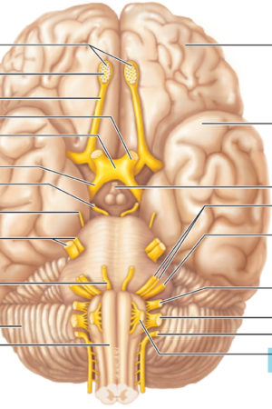
Figure 1. Hypothalamus

hypothalamus

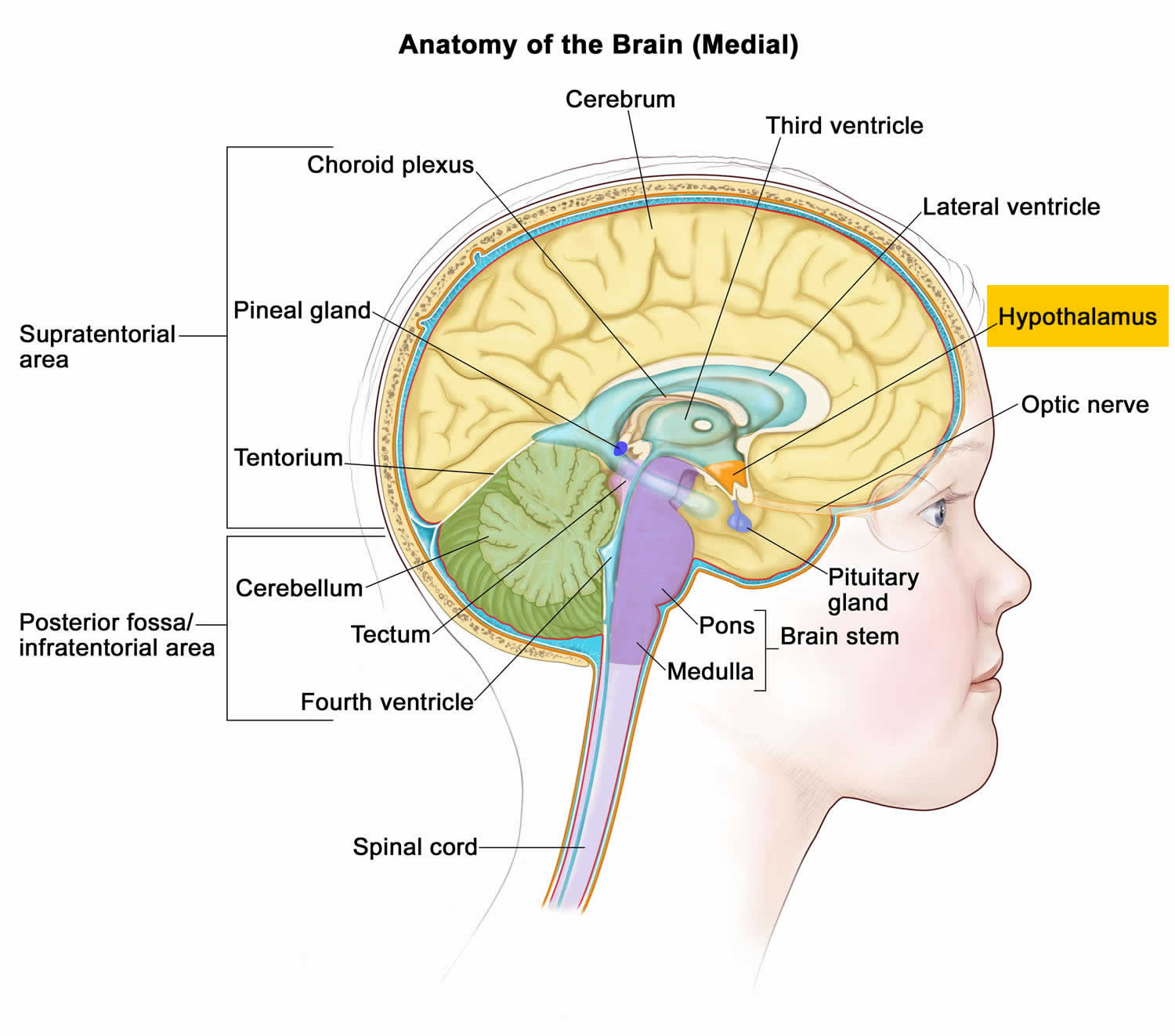
Figure 2. Lateral hypothalamus

lateral hypothalamus

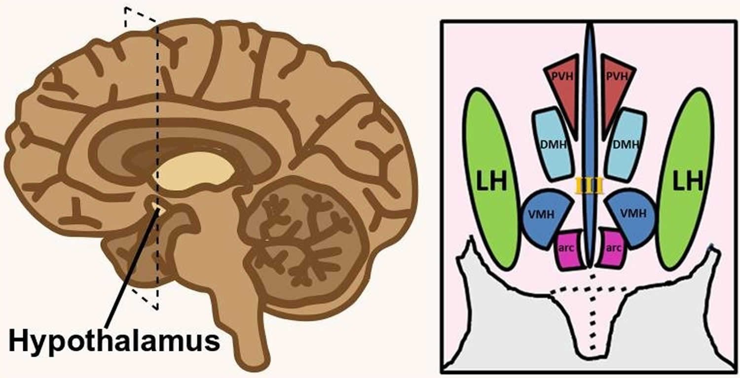
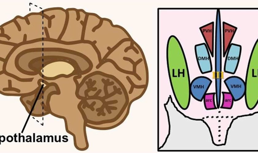
Footnote: Hypothalamus is composed of several spatially clustered neural populations. (Left) Location of the hypothalamus at the base of the forebrain. Dashed lines indicate a coronally-cut section of the hypothalamus shown on the right. (Right) Simplified diagram of hypothalamic nuclei involved in feeding behavior and energy metabolism.

Abbreviations: III = third ventricle; arc = arcuate nucleus; DMH = dorsomedial hypothalamus; LH = lateral hypothalamus; PVH = paraventricular hypothalamus; VMH = ventromedial hypothalamus.

[Source 57 ]

Figure 3. Lateral hypothalamus neurons and functions

Lateral hypothalamus neurons and functions

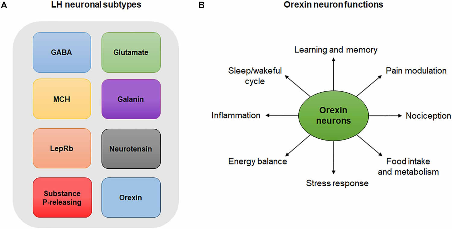
Footnotes: (A) Lateral hypothalamus neuronal subtypes. Simplistic diagram showing existing neuronal populations in the lateral hypothalamus. Neuronal populations in the lateral hypothalamus include, but are not limited to, GABA neurons, glutamate neurons, melanin-concentrating hormone (MCH)-expressing neurons, galanin-expressing neurons, leptin-receptor (LepRb)-expressing neurons, neurotensin-releasing neurons, substance P-releasing neurons and orexin neurons. The degree to which these neuronal populations overlap is not represented in this diagram. (B) Orexin neuron functions. Orexin neurons are involved in numerous physiological and behavioral processes including sleep/wakeful cycles, learning, memory, pain, nociception, food intake, metabolism, stress, energy balance and inflammation.

[Source 28 ]

Lateral hypothalamus function

The lateral hypothalamus is generally known as the hunger center or feeding center, and two of its main functions are the stimulation of feeding behavior and arousal 61, 3, 62. Electrical stimulation of the lateral hypothalamus results in ravenous eating behavior, and animals are extremely motivated to work for a food reward 56. The neurons of the lateral hypothalamus are mainly orexin expressing neurons and they respond to both melanocortins and neuropeptide Y 63, 64. Orexin neurons stimulate wakefulness, and a loss of orexin neurons causes narcolepsy 65, which in turn is associated with increased risk on the development of type 2 diabetes 66. Together, these properties of orexin promote alertness in a fasted state, which is crucial for food-seeking behavior 67. Recent animal studies indicated that the lateral hypothalamus plays a key role in eliciting violent forms of aggression 68, 69, 70, 71, 72

The lateral hypothalamus is also a site for integration of autonomic and endocrine responses, and a crucial regulator of pituitary function and homeostatic balance 73, 74. The lateral hypothalamus plays a key role in regulating autonomic functions and relays information to all major parts of the brain including the major hypothalamic nuclei 75, 56, 76, 77, 78. Converging evidence from functional, structural, and behavioral studies confirmed the importance of this region not only in regulating metabolism and feeding behavior, but also in serving as a motivation-cognition interface 56, 62.

Lateral hypothalamus neurons control feeding, blood pressure, heart rate, water intake and sodium excretion largely through the activation of adrenergic receptors 79, 80, 81. In addition, they receive inhibitory noradrenergic input from the locus coeruleus, which helps prevent excessive activity in the arousal pathway during the waking cycle 82. Also, beta (β)-adrenoceptors activation by noradrenaline in the lateral hypothalamus appears to be involved in the suppression of feeding behavior 83. On the other hand, activation of alpha 1 (α1)-adrenoceptors of the lateral hypothalamus has been linked to behavioral activation and exploration, despite the insignificant number of these receptors in the lateral hypothalamus 84.

The lateral hypothalamus also plays an important role in the brain reward system. This was demonstrated by studies using intracranial self-stimulation in rodents showing that animals will willingly perform an operant response to receive rewarding pulses of electrical stimulation within the lateral hypothalamus 85, 86. The rewarding effect of lateral hypothalamus self-simulation is largely influenced by the dopamine and opioid systems as alterations in these systems were shown to either suppress or disrupt the self-stimulation behavior 87, 86. The lateral hypothalamus, through GABAergic neurons, also plays an important role in learning to respond to cues that predict the delivery of a reward 88. In addition, GABAergic neurons of the lateral hypothalamus highly project to the ventral tegmental area (VTA) 88, a center rich in dopaminergic neurons that is known to be crucial for learning, reward processes and feeding behavior 89, 90.

Recently, many reports have implicated the lateral hypothalamus in the regulation of inflammatory pain 91, 92, 93. Studies have shown that stimulation of the lateral hypothalamus produces analgesic and anti-nociceptive effects in an animal model of inflammatory pain 92 and that this effect is largely due to the activation of α-adrenoceptors in the dorsal horn of the spinal cord 94, 92, and to the involvement of lateral hypothalamic orexin neurons 95.

Ventral tegmental area structure and reward function

The ventral tegmental area (VTA) is a semi-circular nucleus which lies along the midline in the midbrain, it is home to a heterogeneous population of neurons containing multiple neurotransmitters including neurotensin (NT) 96, cholecystokinin (CCK) 97 and dopamine 98. The ventral tegmental area (VTA) dopaminergic system has been implicated in brain-stimulation reward and food reward, psychomotor stimulation, learning and memory formation 99, 100, 101, 102, 103 and it has been shown that goal-directed behavior is promoted by dopamine release from ventral tegmental area (VTA) dopaminergic neurons 104, 105, 106. Studies have shown that both the synaptic connections and intrinsic excitability of dopaminergic neurons are highly plastic dependent on the experiences of the animals 107, 108, 109, 102, 110. This suggests the possibility for experience/outcome-based modulation of behavioral motivation to be mediated via ventral tegmental area (VTA) dopaminergic neurons and a “directing” role for dopamine in goal-oriented behaviors.

Ventral tegmental area (VTA) dopaminergic neuron involvement in reward processing has been studied extensively in an attempt to understand how these neurons code for rewards and the mechanisms through which they are able to modulate animal behaviors. However the complexity of the ventral tegmental area (VTA), as well as the lateral hypothalamus inputs into the ventral tegmental area (VTA), require equally complex methods to investigate specific neuron populations within such heterogeneous neuron populations. Eshel et al. 111 carried out a complex set of experiments using a multi-method approach combining computational modeling, extra-cellular recordings, optogenetics and viral injections to investigate the computational mechanisms by which ventral tegmental area (VTA) dopaminergic neurons calculate reward prediction error. Performing extra-cellular recordings of dopaminergic neurons while delivering expected and unexpected rewards, and using subsequent optogenetic manipulations to investigate the importance of ventral tegmental area (VTA) GABAergic neurons to normal ventral tegmental area (VTA) dopaminergic function. They found that as the size of the reward the animal receives increases, so does the dopaminergic neuron response, which was consistent with previous results 112, 113, they also found that expectation of a reward resulted in a suppression of the dopaminergic neuron response, and that this response fit to a subtractive computational model better than an alternative divisive model 111. Then, Eshel et al. 111 investigated the role of ventral tegmental area (VTA) GABAergic neurons in their subtraction model of ventral tegmental area (VTA) dopaminergic neuron suppression in expected rewards by optogenetically mimicking normal ventral tegmental area (VTA) GABAergic neuron firing patterns and observing ventral tegmental area (VTA) dopaminergic activity. They found that ventral tegmental area (VTA) GABA stimulation resulted in the suppression of dopamine responses to unexpected rewards in a similar pattern to that seen in animals receiving expected rewards. This ventral tegmental area (VTA) GABA-induced suppression of  dopaminergic responses also fit with a subtractive computational model. Additionally, they showed that inhibition of ventral tegmental area (VTA) GABAergic neurons partially reversed the expectation-dependent suppression of ventral tegmental area (VTA) dopamine reward responses. Taken together this suggests that ventral tegmental area (VTA) dopaminergic neurons calculate reward-error using a subtractive model, and that ventral tegmental area (VTA) GABAergic neurons play a role in the temporal expectation modulation of dopamine responses in a manner that is consistent with the ramping expectation function in some models of prediction error computational models 114, 115. This modulation of reward response in the ventral tegmental area (VTA) may play an important role in directing motivated behaviors to rewards that are less predictable over rewards that are more regularly available. This also suggests a mechanism by which ventral tegmental area (VTA) dopaminergic neurons can rationalize between multiple rewards within an environment by modulating the reward value of more reliable rewards to be less rewarding than unpredictable rewards to shift the animals drive to focus on less readily-available rewards. This series of experiments shows the multitude of benefits that can be gained by using a multi-method approach to investigate neural circuits, by using behavioral protocols, optogenetics, electrophysiology and computational modeling (as well as investigating the role of both dopamine and GABA activity for comparison) these researchers were able to gain a deeper understanding of ventral tegmental area (VTA) dopaminergic activity by observing it from multiple angles.

The development of new techniques and biomarkers has allowed a closer look at the roles of different populations of lateral hypothalamus neurons in ventral tegmental area (VTA) function. For example, optogenetic stimulation of lateral hypothalamus GABA inputs to the ventral tegmental area (VTA) results in conditioned place preference 116, reduces ventral tegmental area (VTA) GABA activity, and drives nucleus accumbens dopamine release 117. Nieh et al. 117 additionally showed that optogenetic stimulation of glutamatergic lateral hypothalamus inputs to the ventral tegmental area (VTA) results in conditioned place aversion. Interestingly, the behavioral response to optogenetic stimulation of ventral tegmental area (VTA)-innervating lateral hypothalamus GABAergic neurons differed depending on the stimulation frequency, with low frequency stimulation (5–10 Hz) resulting in increased feeding, and high frequency stimulation (40 Hz) appeared to trigger reward, resulting in a place preference 116. It is possible that these two functions may be being mediated by two different neuropeptides co-expressed within lateral hypothalamus GABAergic neurons, or that this stimulation triggers a general “drive” and the stimulation frequencies result in the release of different neuropeptides in the ventral tegmental area (VTA), resulting in a target for the “drive” response. This could suggest that the lateral hypothalamus and the ventral tegmental area (VTA) are the “drive” and “focus” sources for motivated behaviors, respectively, with lateral hypothalamus activation producing a general energizing of the animal to perform a behavior, and the ventral tegmental area (VTA) then directing that energy to a specific goal-oriented behavior—depending on which neurotransmitters are released, or the stimulation frequency, or some other determining factor. Additionally, the development of this gene-targeting methodology also opens up the possibility to investigate the multiple other neuron types in the lateral hypothalamus that are known to project to the ventral tegmental area (VTA) to better understand how these neuron types differentiate between multiple input signals and determine which environmental goals to pursue 20.

GABA Neurons

Lateral hypothalamus neurons are composed of many overlapping neuronal populations that play distinct functions in the central nervous system (CNS) (Figure 3) 28. Studies over the past few decades have largely focused on the functions of GABAergic neurons of the lateral hypothalamus and their projections in reward and feeding behavior 118. Evidence suggests that these neurons encode information necessary for associating specific cues with reward delivery. In experiments employing optogenetics, a highly specific technique that involves the use of light to activate or inhibit neurons, inhibition of lateral hypothalamus GABAergic neurons was shown to reduce responding to a cue predicting a food reward, indicating that these neurons encode information pertaining to reward prediction 88. On the other hand, optogenetic inhibition of lateral hypothalamus GABA neurons that project to the ventral tegmental area (VTA) increased responding to the food reward-paired cue, suggesting that these neurons may play a role in relaying reward-predictive information to other neuronal structures 88. Interestingly, a study evaluating the role of lateral hypothalamus GABA neurons projecting to the ventral tegmental area (VTA) showed that optogenetic activation of this pathway can either induce a feeding or rewarding effect depending on the frequency of the stimulation used 119. Last but not least, a recent study by Giardino et al. 120 found that the bed nucleus of the stria terminalis sends two non-overlapping GABAergic projections to the lateral hypothalamus that express several neuropeptides including corticotropin-releasing factor (CRF) and cholecystokinin (CCK).

Glutamate Neurons

Glutamate neurons of the lateral hypothalamus mediate important physiological processes in the central nervous system (CNS). Studies have shown that lateral hypothalamus glutamate neurons produce behavioral functions opposite to those of lateral hypothalamus GABAergic neurons. In mice, optogenetic activation of putative glutamate neurons of the lateral hypothalamus suppressed feeding and produced aversion-related phenotypes 121, while the opposite effect was observed following optogenetic or chemogenetic activation of lateral hypothalamus GABA neurons 122. The opposite functions of lateral hypothalamus glutamate and GABA neurons in feeding and reward-related processes are mainly explained by differences in their projection pattern. Glutamate neurons of the lateral hypothalamus send dense projections to the lateral habenula (LHb), a region involved in processing aversive stimuli 123, in contrast to lateral hypothalamus GABA neurons, whose projections mainly target the ventral tegmental area (VTA) 119. Besides its role in the regulation of feeding and aversion-related behaviors, lateral hypothalamus glutamate neurons have been implicated in compulsive 124 and hyperkinetic behaviors 125.

Melanin-Concentrating Hormone (MCH)-Expressing Neurons

Neurons expressing melanin-concentrating hormone (MCH) are also widely present in the lateral hypothalamus. Functional and anatomical studies showed that melanin-concentrating hormone (MCH) neurons co-express the vesicular glutamate transporter 2 (VGLUT2), indicating a glutamatergic identity 125 and project to several regions of the central nervous system (CNS) ranging from the cortex to the spinal cord 126. Melanin-concentrating hormone (MCH) is an appetite stimulant (orexigenic) hypothalamic peptide that exerts inhibitory effects on lateral hypothalamic neurons 127. Although the mechanism of action of melanin-concentrating hormone (MCH) is yet to be fully determined, its inhibitory effect on lateral hypothalamus neurons is largely mediated by the attenuation of excitatory glutamate transmission presynaptically 127. Melanin-concentrating hormone (MCH) was also shown to depress synaptic activity of lateral hypothalamus GABA neurons, suggesting a substantial level of complexity in its modulation of lateral hypothalamus neuron activity 128. Mounting evidence suggests that MCH neurons directly regulate feeding behavior. Both administration of melanin-concentrating hormone (MCH) 129, 130 and activation of MCH receptors 131 increases food intake and facilitates body weight gain in rodents. Conversely, genetic knockout of the MCH gene 132 or pharmacological blockade of MCH receptors 131 leads to substantial decreases in food intake in mice. In addition, through their direct projections to gonadotropin-releasing hormone (GnRH) synthesizing neurons, melanin-concentrating hormone (MCH) neurons convey critical homeostatic signals to the reproductive axis, and contribute considerably to the functional connection between the regulation of food intake and reproduction 128, 133.

Galanin and Leptin-Receptor Expressing Neurons

Another type of neuron found in the lateral hypothalamus is the galanin-containing neuron 134. These neurons represent a GABAergic subpopulation of lateral hypothalamus neurons with a distinct molecular phenotype and projection pattern. Unlike GABAergic neurons of the lateral hypothalamus, which project to the ventral tegmental area (VTA), galanin neurons of the lateral hypothalamus lack direct ventral tegmental area (VTA) innervation 134. Instead, galanin neurons of the lateral hypothalamus strongly innervate the locus coeruleus 135, a site involved in the control of arousal 136, 137 and reward processing 138, 139. Galanin is a 29 amino acid neuropeptide widely distributed in the brain 140, 141, 142 that acts as an inhibitor of synaptic transmission in the hypothalamus 143. Galanin also acts in the hypothalamus to produce behavioral hyperalgesia through activation of two descending pronociceptive pathways; one that involves the medullary dorsal reticular nucleus, and another one that involves serotonin neurons acting on the spinal cord 144. Galanin has also been reported to promote feeding behavior. Chemogenetic activation of lateral hypothalamus galanin neurons 134 or central injection of galanin 145 enhances food-seeking behavior, while targeted knockout of the galanin gene 146 or the galanin receptor 147 reduces dietary fat intake.

Another neuronal population of the lateral hypothalamus involved in the regulation of feeding behaviors is the leptin-receptor (LepRb) expressing neuron. LepRb-expressing neurons are widely expressed in the brain, but are particular enriched within the hypothalamus and the brainstem 148. In mice, leptin acts on leptin-receptor (LepRb)-expressing neurons of the lateral hypothalamus to decrease feeding and body weight 149. Leptin-receptor (LepRb)-expressing neurons of the lateral hypothalamus also innervate the ventral tegmental area (VTA), and leptin action on these neurons increases ventral tegmental area (VTA) dopaminergic neuron activity, suggesting a link between the anorexic effect of leptin and the mesolimbic dopaminergic system 149. Interestingly, leptin-receptor (LepRb)-expressing neurons in the lateral hypothalamus are thought to be GABAergic 149, and a subpopulation of these neurons in the lateral hypothalamus was shown to co-express the inhibitory acting neuropeptide galanin 150, suggesting that the anorexic effect of leptin is likely due to its interaction with other neuropeptidergic receptors in the lateral hypothalamus.

Substance P and Neurotensin-Releasing Neurons

Substance P and neurotensin-releasing neurons are also found in the lateral hypothalamus 151, 152. Substance P is a member of the tachykinin neuropeptide family and is associated with multiple physiological processes including wound healing, neurogenic inflammation and tissue homeostasis 153. In the lateral hypothalamus, substance P-containing neurons have been proposed to exert antinociceptive functions by activating spinally projecting serotonin neurons in the rostral ventromedial medulla (RVM) 91. These cells activate spinally projecting serotonin neurons either through direct contact, or indirectly through the innervation of interneurons in the rostral ventromedial medulla (RVM), thereby altering nociceptive responses in the dorsal horn of the spinal cord 91.

On the other hand, neurotensin neurons of the lateral hypothalamus are involved in the regulation of the sleep/wake cycle 154 and are implicated in feeding behavior 155 and reward processes 156. Studies exploring the role of neurotensin in reward and feeding have indicated that this neuropeptide promotes reward by enhancing glutamate transmission in the mesolimbic dopaminergic system 156 and promotes weight loss by suppressing the increased appetitive drive through activation of the G-protein-coupled neurotensin receptor-1 155. Finally, neurotensin neurons of the lateral hypothalamus have been implicated in a number of other physiological processes, including hyperthermia and energy balance, though the central mechanisms by which these processes are mediated remain to be fully elucidated 157, 158.

Orexin Neurons and Other Neuronal Populations of the lateral hypothalamus

By being an extensively researched population of cells in the past recent years, orexinergic neurons, and especially those of the lateral hypothalamus, have been shown to have different roles in inflammatory pain and in the balance of psychological functions (Figure 3B). Orexinergic neurons synthesize two neuropeptides (Orexin A and B) from the precursor prepro-orexin 159. Furthermore, inhibition of orexin neurons by local GABAergic neurons of the lateral hypothalamus is thought to disrupt the sleep cycle (Ferrari et al., 2018). On the other hand, inhibition of orexin neurons through activation of acetylcholine and dynorphin promotes wakefulness 160.

Other neuronal populations in the lateral hypothalamus not mentioned above include neurons that express cocaine- and amphetamine-regulated transcript, thyrotropin-releasing hormone, encephalin, urocortin-3 and corticotropin-releasing hormone 161, 162.

Orexin Neurons

Orexin also known as hypocretin, is a neuropeptide secreted by orexin neurons in the lateral hypothalamic area. They are two types of orexin; orexin A (hypocretin-1) and orexin B (hypocretin-2). These neuropeptides originate from the same precursor known as prepro-orexin 159. Orexin-A is a 33-amino-acid peptide while orexin-B is a 28-amino-acid peptide 159. Both orexin A and orexin B bind to the G-protein coupled receptors orexin receptor 1 (OX-1) and 2 (OX-2) (also known as hypocretin receptors type 1 and 2) 163. Orexin A binds to both OX-1 and OX-2 with the same affinity while orexin B has a higher affinity for OX-2 over OX-1 receptors 164.

In the central nervous system (CNS), orexin (hypocretin) is co-localized with other transmitters, some of which include dynorphin 165, glutamate 166, 167, galanin 168 and prolactin 169. Experiments employing in situ hybridization and immunohistochemical techniques indicate that orexin neurons in the lateral hypothalamus mostly express the vesicular glutamate transporters, VGLUT1 and VGLUT2, suggesting that they are glutamatergic 170.

Orexin neurons project their axons to most parts in the brain and spinal cord, especially to areas that are involved in the modulation of pain 171. In addition, orexin neurons in the lateral hypothalamus send projections to multiple sites related to arousal including the serotonergic dorsal raphe 172. Orexin neurons also project to the tuberomammillary nucleus (TMN) 167, a center involved in the control of arousal, learning and memory 173, 174. Pre-synaptically, orexin increases the release of glutamate and GABA in the hypothalamus, while post-synaptically, it increases Ca2+ levels, thus leading to the depolarization, hence activation, of tuberomammillary nucleus (TMN neurons by glutamatergic orexin terminals 167.

Last but not least, orexin neurons have been shown to directly interact with neuropeptide Y (NPY), a peptide that plays a role in the regulation of feeding behavior, metabolism and energy balance 175. This neuropeptide is primarily synthesized by neurons in the arcuate nucleus (ARC) and is present in different areas of the brain including the cortex, hippocampus, hindbrain and hypothalamus 175. Through its heavy projections to the arcuate nucleus (ARC) 176, orexin neurons interact with neuropeptide Y (NPY) to regulate numerous physiological processes and behaviors including food intake and calcium signaling 177, 178.

The distribution of OX-1 and OX-2 receptors has been established in different species including rats and mice. Studies employing in situ hybridization, immunohistochemistry and quantitative reverse transcription–polymerase chain reaction in rodents found that these receptors are widely distributed throughout the brain and spinal cord 179, 180. Although some overlap exist in the distribution pattern of OX-1 and OX-2 receptors, these receptors are differentially expressed in the central nervous system (CNS) 179, 181.

OX-1 receptors are primarily expressed in the ventromedial hypothalamic nucleus, prefrontal and infralimbic cortex, hippocampus, paraventricular thalamic nucleus, dorsal raphe, and locus coeruleus 179, 181, 180, and to a lesser extent in the medial preoptic area, lateroanterior and dorsomedial hypothalamic nuclei, lateral mammillary nucleus and posterior hypothalamic area 179. They are also found in the periaqueductal gray and dorsal root ganglia, which suggests a role in the regulation of pain 180, 182, and in the spinal cord, which suggests a role in the regulation of the parasympathetic and sympathetic system 180. On the other hand, OX-2 receptors are predominantly expressed in the tuberomammillary nucleus (TMN), paraventricular nucleus (PVN), cerebral cortex, nucleus accumbens (NAc), subthalamic and paraventricular thalamic nuclei, septal nuclei, raphe nuclei, and anterior pretectal nucleus, and to a lesser extent in the ventromedial/dorsomedial hypothalamic nuclei and the posterior and lateral hypothalamic areas 179, 181.

Interestingly, a study examining the expression of OX-1 and OX-2 receptors mRNA with in situ hybridization in rats and mice found some species-specific differences 183. For instance, OX-1 receptors are expressed in the caudate putamen and ventral tuberomammillary nucleus (TMN) in rats only, while they are detected in the bed nucleus of the stria terminalis, medial division, posteromedial part in mice only 183. On the other hand, OX-2 receptors show similar pattern of expression between the two species, though they are more widely expressed in the ventral tuberomammillary nucleus (TMN) of rats compared to mice 183. This differential distribution of orexin receptors is consistent with the proposed multifaceted roles of orexin in regulating homeostasis and other functions in the central nervous system (CNS).

Orexin A and B neuropeptides, as demonstrated in the literature, are also widely expressed in different regions of the brain and spinal cord. Findings from immunohistochemical and radioimmunoassay techniques indicate that orexin A fibers are found throughout the hypothalamus, septum, thalamus, locus coeruleus and spinal cord, and in the paraventricular and supraoptic nucleus 184, 185, 186, 187. In addition, orexin A fibers colocalize with substance P positive afferents of dorsal root ganglia neurons, which further strengthens its confirmed role in the regulation of pain 188. On the other hand, orexin B fibers are distributed sparsely in the hypothalamus and the spinal cord 184, 185, but are absent in the paraventricular and supraoptic nucleus 187. Interestingly, a study investigating the distribution of orexin A and orexin B in the brain of nocturnal and diurnal rodents found striking differences among species, in particular in the lateral mammillary nucleus, ventromedial hypothalamic nucleus and flocculus 187.

  1. Lateral hypothalamic area. http://braininfo.rprc.washington.edu/centraldirectory.aspx?type=h&ID=409[↩]
  2. Schaltenbrand G, Wahren W, Hassler R: Atlas for stereotaxy of the human brain, in: Atlas for Stereotaxy of the Human Brain, ed 2. Stuttgart: Thieme, 1977, p 84[↩]
  3. Whiting AC, Oh MY, Whiting DM. Deep brain stimulation for appetite disorders: a review. Neurosurg Focus. 2018 Aug;45(2):E9. https://doi.org/10.3171/2018.4.FOCUS18141[↩][↩][↩]
  4. Berthoud, H. R., and Munzberg, H. (2011). The lateral hypothalamus as integrator of metabolic and environmental needs: from electrical self-stimulation to opto-genetics. Physiol. Behav. 104, 29–39. doi: 10.1016/j.physbeh.2011.04.051[↩]
  5. Bernardis, L. L., and Bellinger, L. L. (1993). The lateral hypothalamic area revisited: neuroanatomy, body weight regulation, neuroendocrinology and metabolism. Neurosci. Biobehav. Rev. 17, 141–193. doi: 10.1016/s0149-7634(05)80149-6[↩][↩]
  6. de Lecea, L., Kilduff, T. S., Peyron, C., Gao, X., Foye, P. E., Danielson, P. E., et al. (1998). The hypocretins: hypothalamus-specific peptides with neuroexcitatory activity. Proc. Natl. Acad. Sci. U S A 95, 322–327. doi: 10.1073/pnas.95.1.322[↩]
  7. Sakurai, T., Amemiya, A., Ishii, M., Matsuzaki, I., Chemelli, R. M., Tanaka, H., et al. (1998). Orexins and orexin receptors: a family of hypothalamic neuropeptides and G protein-coupled receptors that regulate feeding behavior. Cell 92, 573–585. doi: 10.1016/s0092-8674(00)80949-6[↩]
  8. Qu, D., Ludwig, D. S., Gammeltoft, S., Piper, M., Pelleymounter, M. A., Cullen, M. J., et al. (1996). A role for melanin-concentrating hormone in the central regulation of feeding behaviour. Nature 380, 243–247. doi: 10.1038/380243a0[↩]
  9. Kristensen, P., Judge, M. E., Thim, L., Ribel, U., Christjansen, K. N., Wulff, B. S., et al. (1998). Hypothalamic CART is a new anorectic peptide regulated by leptin. Nature 393, 72–76. doi: 10.1038/29993[↩]
  10. Luttinger, D., King, R. A., Sheppard, D., Strupp, J., Nemeroff, C. B., and Prange, A. J. (1982). The effect of neurotensin on food consumption in the rat. Eur. J. Pharmacol. 81, 499–503. doi: 10.1016/0014-2999(82)90116-9[↩]
  11. Leinninger, G. M., Opland, D. M., Jo, Y.-H., Faouzi, M., Christensen, L., Cappellucci, L. A., et al. (2011). Leptin action via neurotensin neurons controls orexin, the mesolimbic dopamine system and energy balance. Cell Metab. 14, 313–323. doi: 10.1016/j.cmet.2011.06.016[↩]
  12. Skofitsch, G., Jacobowitz, D. M., and Zamir, N. (1985). Immunohistochemical localization of a melanin concentrating hormone-like peptide in the rat brain. Brain Res. Bull. 15, 635–649. doi: 10.1016/0361-9230(85)90213-8[↩]
  13. Melander, T., Hökfelt, T., and Rökaeus, A. (1986). Distribution of galaninlike immunoreactivity in the rat central nervous system. J. Comp. Neurol. 248, 475–517. doi: 10.1002/cne.902480404[↩]
  14. Ricardo, J. A., and Koh, E. T. (1978). Anatomical evidence of direct projections from the nucleus of the solitary tract to the hypothalamus, amygdala, and other forebrain structures in the rat. Brain Res. 153, 1–26. doi: 10.1016/0006-8993(78)91125-3[↩]
  15. Berk, M. L., and Finkelstein, J. A. (1982). Efferent connections of the lateral hypothalamic area of the rat: an autoradiographic investigation. Brain Res. Bull. 8, 511–526. doi: 10.1016/0361-9230(82)90009-0[↩]
  16. Ter Horst, G. J., and Luiten, P. G. M. (1987). Phaseolus vulgaris leuco-agglutinin tracing of intrahypothalamic connections of the lateral, ventromedial, dorsomedial and paraventricular hypothalamic nuclei in the rat. Brain Res. Bull. 18, 191–203. doi: 10.1016/0361-9230(87)90190-0[↩]
  17. Ter Horst, G. J., De Boer, P., Luiten, P. G. M., and Van Willigen, J. D. (1989). Ascending projections from the solitary tract nucleus to the hypothalamus. A Phaseolus vulgaris lectin tracing study in the rat. Neuroscience 31, 785–797. doi: 10.1016/0306-4522(89)90441-7[↩]
  18. Phillipson, O. T. (1979). Afferent projections to the ventral tegmental area of Tsai and interfascicular nucleus: a horseradish peroxidase study in the rat. J. Comp. Neurol. 187, 117–143. doi: 10.1002/cne.901870108[↩]
  19. Watabe-Uchida, M., Zhu, L., Ogawa, S. K., Vamanrao, A., and Uchida, N. (2012). Whole-brain mapping of direct inputs to midbrain dopamine neurons. Neuron 74, 858–873. doi: 10.1016/j.neuron.2012.03.017[↩]
  20. Tyree SM, de Lecea L. Lateral Hypothalamic Control of the Ventral Tegmental Area: Reward Evaluation and the Driving of Motivated Behavior. Front Syst Neurosci. 2017 Jul 6;11:50. doi: 10.3389/fnsys.2017.00050[↩][↩][↩]
  21. Bonnavion, P., Mickelsen, L. E., Fujita, A., de Lecea, L., and Jackson, A. C. (2016). Hubs and spokes of the lateral hypothalamus: cell types, circuits and behaviour. J. Physiol. 594, 6443–6462. doi: 10.1113/jp271946[↩]
  22. Eggermann, E., Bayer, L., Serafin, M., Saint-Mleux, B., Bernheim, L., Machard, D., et al. (2003). The wake-promoting hypocretin-orexin neurons are in an intrinsic state of membrane depolarization. J. Neurosci. 23, 1557–1562. doi: 10.1523/jneurosci.23-05-01557.2003[↩]
  23. Inutsuka, A., and Yamanaka, A. (2013). The physiological role of orexin/hypocretin neurons in the regulation of sleep/wakefulness and neuroendocrine functions. Front. Endocrinol. 4:18. doi: 10.3389/fendo.2013.00018[↩]
  24. Barson, J. R., and Leibowitz, S. F. (2017). Orexin/hypocretin system: role in food and drug overconsumption. Int. Rev. Neurobiol. 136, 199–237. doi: 10.1016/bs.irn.2017.06.006[↩]
  25. Grimaldi, D., Silvani, A., Benarroch, E. E., and Cortelli, P. (2014). Orexin/hypocretin system and autonomic control: new insights and clinical correlations. Neurology 82, 271–278. doi: 10.1212/WNL.0000000000000045[↩]
  26. Espana, R. A. (2012). Hypocretin/orexin involvement in reward and reinforcement. Vitam. Horm. 89, 185–208. doi: 10.1016/B978-0-12-394623-2.00010-X[↩]
  27. Inutsuka, A., Yamashita, A., Chowdhury, S., Nakai, J., Ohkura, M., Taguchi, T., et al. (2016). The integrative role of orexin/hypocretin neurons in nociceptive perception and analgesic regulation. Sci. Rep. 6:29480. doi: 10.1038/srep29480[↩]
  28. Fakhoury M, Salman I, Najjar W, Merhej G, Lawand N. The Lateral Hypothalamus: An Uncharted Territory for Processing Peripheral Neurogenic Inflammation. Front Neurosci. 2020 Feb 12;14:101. https://doi.org/10.3389/fnins.2020.00101[↩][↩][↩]
  29. Adamantidis A. R., Zhang F., Aravanis A. M., Deisseroth K., de Lecea L. (2007). Neural substrates of awakening probed with optogenetic control of hypocretin neurons. Nature 450, 420–424. 10.1038/nature06310[↩]
  30. Adamantidis A. R., Carter M. C., de Lecea L. (2010). Optogenetic deconstruction of sleep-wake circuitry in the brain. Front. Mol. Neurosci. 2:31. 10.3389/neuro.02.031.2009[↩]
  31. Carter M. E., Adamantidis A., Ohtsu H., Deisseroth K., de Lecea L. (2009). Sleep homeostasis modulates hypocretin-mediated sleep-to-wake transitions. J. Neurosci. 29, 10939–10949. 10.1523/JNEUROSCI.1205-09.2009[↩]
  32. Anand B. K., Brobeck J. R. (1951). Localization of a “feeding center” in the hypothalamus of the rat. Proc. Soc. Exp. Biol. Med. 77, 323–324. 10.3181/00379727-77-18766[↩][↩]
  33. BROBECK JR. Mechanism of the development of obesity in animals with hypothalamic lesions. Physiol Rev. 1946 Oct;26(4):541-59. doi: 10.1152/physrev.1946.26.4.541[↩]
  34. Bonnavion P., Jackson A. C., Carter M. E., de Lecea L. (2015). Antagonistic interplay between hypocretin and leptin in the lateral hypothalamus regulates stress responses. Nat. Commun. 6:6266. 10.1038/ncomms7266[↩]
  35. Olds J., Milner P. (1954). Positive reinforcement produced by electrical stimulation of septal area and other regions of rat brain. J. Comp. Physiol. Psychol. 47, 419–427. 10.1037/h0058775[↩]
  36. Hoebel B. G., Teitelbaum P. (1962). Hypothalamic control of feeding and self-stimulation. Science 135, 375–377. 10.1126/science.135.3501.375[↩]
  37. Olds J. (1970). Pleasure centers in the brain. Eng. Sci. 33, 22–31.[↩]
  38. Olds J., Olds M. E. (1965). Drives, rewards, and the brain. New Direct. Psychol. 2, 327–410.[↩]
  39. Bielajew C, Shizgal P. Evidence implicating descending fibers in self-stimulation of the medial forebrain bundle. J Neurosci. 1986 Apr;6(4):919-29. doi: 10.1523/JNEUROSCI.06-04-00919.1986[↩]
  40. Sakurai T, Amemiya A, Ishii M, Matsuzaki I, Chemelli RM, Tanaka H, Williams SC, Richardson JA, Kozlowski GP, Wilson S, Arch JR, Buckingham RE, Haynes AC, Carr SA, Annan RS, McNulty DE, Liu WS, Terrett JA, Elshourbagy NA, Bergsma DJ, Yanagisawa M. Orexins and orexin receptors: a family of hypothalamic neuropeptides and G protein-coupled receptors that regulate feeding behavior. Cell. 1998 Feb 20;92(4):573-85. doi: 10.1016/s0092-8674(00)80949-6[↩][↩]
  41. Bittencourt JC, Presse F, Arias C, Peto C, Vaughan J, Nahon JL, Vale W, Sawchenko PE. The melanin-concentrating hormone system of the rat brain: an immuno- and hybridization histochemical characterization. J Comp Neurol. 1992 May 8;319(2):218-45. doi: 10.1002/cne.903190204[↩]
  42. Qu D, Ludwig DS, Gammeltoft S, Piper M, Pelleymounter MA, Cullen MJ, Mathes WF, Przypek R, Kanarek R, Maratos-Flier E. A role for melanin-concentrating hormone in the central regulation of feeding behaviour. Nature. 1996 Mar 21;380(6571):243-7. doi: 10.1038/380243a0[↩]
  43. Folkow B, Rubinstein EH. Behavioural and autonomic patterns evoked by stimulation of the lateral hypothalamic area in the cat. Acta Physiol Scand. 1965 Dec;65(4):292-9. doi: 10.1111/j.1748-1716.1965.tb04276.x[↩]
  44. Herberg LJ, Blundell JE. Lateral hypothalamus: hoarding behavior elicited by electrical stimulation. Science. 1967 Jan 20;155(3760):349-50. doi: 10.1126/science.155.3760.349[↩]
  45. MENDELSON J, CHOROVER SL. LATERAL HYPOTHALAMIC STIMULATION IN SATIATED RATS: T-MAZE LEARNING FOR FOOD. Science. 1965 Jul 30;149(3683):559-61. doi: 10.1126/science.149.3683.559[↩]
  46. Sani S, Jobe K, Smith A, Kordower JH, Bakay RA. Deep brain stimulation for treatment of obesity in rats. J Neurosurg. 2007 Oct;107(4):809-13. doi: 10.3171/JNS-07/10/0809[↩]
  47. Levitt DR, Teitelbaum P. Somnolence, akinesia, and sensory activation of motivated behavior in the lateral hypothalamic syndrome. Proc Natl Acad Sci U S A. 1975 Jul;72(7):2819-23. https://www.ncbi.nlm.nih.gov/pmc/articles/PMC432863/pdf/pnas00050-0353.pdf[↩][↩]
  48. Price J.L., Slotnick B.M., Revial M.F. Olfactory projections to the hypothalamus. J. Comp. Neurol. 1991;306:447–461. doi: 10.1002/cne.903060309[↩][↩][↩]
  49. Taghva A, Corrigan JD, Rezai AR. Obesity and brain addiction circuitry: implications for deep brain stimulation. Neurosurgery. 2012 Aug;71(2):224-38. doi: 10.1227/NEU.0b013e31825972ab[↩]
  50. ANAND BK, BROBECK JR. Localization of a “feeding center” in the hypothalamus of the rat. Proc Soc Exp Biol Med. 1951 Jun;77(2):323-4. doi: 10.3181/00379727-77-18766[↩]
  51. Hetherington AM, Ranson SW: Hypothalamic lesions and adiposity in the rat. Anat Rec 78:149–172, 1940[↩]
  52. MONTEMURRO DG, STEVENSON JA. The localization of hypothalamic structures in the rat influencing water consumption. Yale J Biol Med. 1955 Dec-1956 Feb;28(3-4):396-403. https://www.ncbi.nlm.nih.gov/pmc/articles/PMC2603397/pdf/yjbm00310-0233.pdf[↩]
  53. Grossman SP, Dacey D, Halaris AE, Collier T, Routtenberg A. Aphagia and adipsia after preferential destruction of nerve cell bodies in hypothalamus. Science. 1978 Nov 3;202(4367):537-9. doi: 10.1126/science.705344[↩]
  54. Grossman SP, Grossman L. Iontophoretic injections of kainic acid into the rat lateral hypothalamus: effects on ingestive behavior. Physiol Behav. 1982 Sep;29(3):553-9. doi: 10.1016/0031-9384(82)90281-5[↩]
  55. Stricker EM, Swerdloff AF, Zigmond MJ. Intrahypothalamic injections of kainic acid produce feeding and drinking deficits in rats. Brain Res. 1978 Dec 15;158(2):470-3. doi: 10.1016/0006-8993(78)90692-3[↩]
  56. Stuber GD, Wise RA. Lateral hypothalamic circuits for feeding and reward. Nat Neurosci. 2016 Feb;19(2):198-205. doi: 10.1038/nn.4220[↩][↩][↩][↩]
  57. Mangieri, Leandra. (2018). HYPOTHALAMIC CIRCUITS IN THE CONTROL OF FEEDING AND EMOTIONAL BEHAVIORS. https://www.researchgate.net/publication/329341331_HYPOTHALAMIC_CIRCUITS_IN_THE_CONTROL_OF_FEEDING_AND_EMOTIONAL_BEHAVIORS[↩][↩]
  58. Harrell LE, Decastro JM, Balagura S. A critical evaluation of body weight loss following lateral hypothalamic lesions. Physiol Behav. 1975 Jul;15(1):133-6. doi: 10.1016/0031-9384(75)90292-9[↩]
  59. Keesey RE, Powley TL. Self-stimulation and body weight in rats with lateral hypothalamic lesions. Am J Physiol. 1973 Apr;224(4):970-8. doi: 10.1152/ajplegacy.1973.224.4.970[↩]
  60. Whiting DM, Tomycz ND, Bailes J, de Jonge L, Lecoultre V, Wilent B, Alcindor D, Prostko ER, Cheng BC, Angle C, Cantella D, Whiting BB, Mizes JS, Finnis KW, Ravussin E, Oh MY. Lateral hypothalamic area deep brain stimulation for refractory obesity: a pilot study with preliminary data on safety, body weight, and energy metabolism. J Neurosurg. 2013 Jul;119(1):56-63. doi: 10.3171/2013.2.JNS12903[↩][↩][↩][↩]
  61. Kalsbeek MJT, Yi CX. The infundibular peptidergic neurons and glia cells in overeating, obesity, and diabetes. Handb Clin Neurol. 2021;180:315-325. https://doi.org/10.1016/B978-0-12-820107-7.00019-7[↩]
  62. Petrovich, G. D. (2018). Lateral hypothalamus as a motivation-cognition interface in the control of feeding behavior. Front. Syst. Neurosci. 12:14. doi: 10.3389/fnsys.2018.00014[↩][↩]
  63. Campbell RE, Smith MS, Allen SE, Grayson BE, Ffrench-Mullen JM, Grove KL. Orexin neurons express a functional pancreatic polypeptide Y4 receptor. J Neurosci. 2003 Feb 15;23(4):1487-97. doi: 10.1523/JNEUROSCI.23-04-01487.2003[↩]
  64. Morgan DA, McDaniel LN, Yin T, Khan M, Jiang J, Acevedo MR, Walsh SA, Ponto LL, Norris AW, Lutter M, Rahmouni K, Cui H. Regulation of glucose tolerance and sympathetic activity by MC4R signaling in the lateral hypothalamus. Diabetes. 2015 Jun;64(6):1976-87. doi: 10.2337/db14-1257[↩]
  65. Shan L, Dauvilliers Y, Siegel JM. Interactions of the histamine and hypocretin systems in CNS disorders. Nat Rev Neurol. 2015 Jul;11(7):401-13. doi: 10.1038/nrneurol.2015.99[↩]
  66. Mohammadi S, Dolatshahi M, Zare-Shahabadi A, Rahmani F. Untangling narcolepsy and diabetes: Pathomechanisms with eyes on therapeutic options. Brain Res. 2019 Sep 1;1718:212-222. doi: 10.1016/j.brainres.2019.04.013[↩]
  67. Tsujino N, Sakurai T. Role of orexin in modulating arousal, feeding, and motivation. Front Behav Neurosci. 2013 Apr 18;7:28. doi: 10.3389/fnbeh.2013.00028[↩]
  68. Woodworth C. H. (1971). Attack elicited in rats by electrical stimulation of the lateral hypothalamus. Physiol. Behav. 6, 345–353. 10.1016/0031-9384(71)90166-1[↩]
  69. Koolhaas J. M. (1978). Hypothalamically induced intraspecific aggressive behaviour in the rat. Exper. Brain Res. 32, 365–375. 10.1007/BF00238708[↩]
  70. Tulogdi A., Biro L., Barsvari B., Stankovic M., Haller J., Toth M. (2015). Neural mechanisms of predatory aggression in rats-implications for abnormal intraspecific aggression. Behav. Brain Res. 283, 108–115. 10.1016/j.bbr.2015.01.030[↩]
  71. Biro L., Sipos E., Bruzsik B., Farkas I., Zelena D., Balazsfi D., et al.. (2018). Task division within the prefrontal cortex: distinct neuron populations selectively control different aspects of aggressive behavior via the hypothalamus. J. Nerosci. 38, 3234–17. 10.1523/JNEUROSCI.3234-17.2018[↩]
  72. Haller J. The Role of the Lateral Hypothalamus in Violent Intraspecific Aggression-The Glucocorticoid Deficit Hypothesis. Front Syst Neurosci. 2018 Jun 8;12:26. doi: 10.3389/fnsys.2018.00026[↩]
  73. Ferguson, A. V., and Samson, W. K. (2003). The orexin/hypocretin system: a critical regulator of neuroendocrine and autonomic function. Front. Neuroendocrinol. 24, 141–150. doi: 10.1016/S0091-3022(03)00028-1[↩]
  74. Timper, K., and Bruning, J. C. (2017). Hypothalamic circuits regulating appetite and energy homeostasis: pathways to obesity. Dis. Model. Mech. 10, 679–689. doi: 10.1242/dmm.026609[↩]
  75. Seoane-Collazo, P., Ferno, J., Gonzalez, F., Dieguez, C., Leis, R., Nogueiras, R., et al. (2015). Hypothalamic-autonomic control of energy homeostasis. Endocrine 50, 276–291. doi: 10.1007/s12020-015-0658-y[↩]
  76. Szymusiak R., McGinty D. (2008). Hypothalamic regulation of sleep and arousal. Ann. N. Y. Acad. Sci. 1129 275–286. 10.1196/annals.1417.027[↩]
  77. Seoane-Collazo P., Ferno J., Gonzalez F., Dieguez C., Leis R., Nogueiras R., et al. (2015). Hypothalamic-autonomic control of energy homeostasis. Endocrine 50 276–291. 10.1007/s12020-015-0658-y[↩]
  78. Gerashchenko D, Shiromani PJ. Different neuronal phenotypes in the lateral hypothalamus and their role in sleep and wakefulness. Mol Neurobiol. 2004 Feb;29(1):41-59. doi: 10.1385/MN:29:1:41[↩]
  79. Shiraishi T. (1991). Noradrenergic neurons modulate lateral hypothalamic chemical and electrical stimulation-induced feeding by sated rats. Brain Res. Bull. 27 347–351. 10.1016/0361-9230(91)90123-2[↩]
  80. Saad W. A., Guarda I. F., Ferreira A. C., de Arruda Camargo L. A., Neto A. F., dos Santos T. A. (2000). Participation of alpha-1 and alpha-2 adrenoceptors of the lateral hypothalamic area in water intake, and renal sodium, potassium and urinary volume excretion induced by central administration of angiotensin II. Brain Res. Bull. 52 491–497. 10.1016/s0361-9230(00)00285-289[↩]
  81. Mendonca M. M., Santana J. S., da Cruz K. R., Ianzer D., Ghedini P. C., Nalivaiko E., et al. (2018). Involvement of GABAergic and adrenergic neurotransmissions on paraventricular nucleus of hypothalamus in the control of cardiac function. Front. Physiol. 9:670 10.3389/fphys.2018.00670[↩]
  82. Breton-Provencher V., Sur M. (2019). Active control of arousal by a locus coeruleus GABAergic circuit. Nat. Neurosci. 22 218–228. 10.1038/s41593-018-0305-z[↩]
  83. Leibowitz S. F. (1970). Reciprocal hunger-regulating circuits involving alpha- and beta-adrenergic receptors located, respectively, in the ventromedial and lateral hypothalamus. Proc. Natl. Acad. Sci. U.S.A. 67 1063–1070. 10.1073/pnas.67.2.1063[↩]
  84. Lin Y., Quartermain D., Dunn A. J., Weinshenker D., Stone E. A. (2008). Possible dopaminergic stimulation of locus coeruleus alpha1-adrenoceptors involved in behavioral activation. Synapse 62 516–523. 10.1002/syn.20517[↩]
  85. Fakhoury M., Rompre P. P., Boye S. M. (2016). Role of the dorsal diencephalic conduction system in the brain reward circuitry. Behav. Brain Res. 296 431–441. 10.1016/j.bbr.2015.10.038[↩]
  86. Ide S., Takahashi T., Takamatsu Y., Uhl G. R., Niki H., Sora I., et al. (2017). Distinct roles of opioid and dopamine systems in lateral hypothalamic intracranial self-stimulation. Int. J. Neuropsychopharmacol. 20 403–409. 10.1093/ijnp/pyw113[↩][↩]
  87. Koob G. F., Fray P. J., Iversen S. D. (1978). Self-stimulation at the lateral hypothalamus and locus coeruleus after specific unilateral lesions of the dopamine system. Brain Res. 146 123–140. 10.1016/0006-8993(78)90222-6[↩]
  88. Sharpe M. J., Marchant N. J., Whitaker L. R., Richie C. T., Zhang Y. J., Campbell E. J., et al. (2017). Lateral hypothalamic GABAergic neurons encode reward predictions that are relayed to the ventral tegmental area to regulate learning. Curr. Biol. 27 2089–2100.e5. 10.1016/j.cub.2017.06.024[↩][↩][↩][↩]
  89. Hommel J. D., Trinko R., Sears R. M., Georgescu D., Liu Z. W., Gao X. B., et al. (2006). Leptin receptor signaling in midbrain dopamine neurons regulates feeding. Neuron 51 801–810. 10.1016/j.neuron.2006.08.023[↩]
  90. Ranaldi R. (2014). Dopamine and reward seeking: the role of ventral tegmental area. Rev. Neurosci. 25 621–630. 10.1515/revneuro-2014-0091[↩]
  91. Holden J. E., Pizzi J. A. (2008). Lateral hypothalamic-induced antinociception may be mediated by a substance P connection with the rostral ventromedial medulla. Brain Res. 1214 40–49. 10.1016/j.brainres.2008.03.051[↩][↩][↩]
  92. Jeong Y., Holden J. E. (2009a). Lateral hypothalamic-induced alpha-adrenoceptor modulation occurs in a model of inflammatory pain in rats. Biol. Res. Nurs. 10 331–339. 10.1177/1099800408325053[↩][↩][↩]
  93. Wardach J., Wagner M., Jeong Y., Holden J. E. (2016). Lateral hypothalamic stimulation reduces hyperalgesia through spinally descending orexin-a neurons in neuropathic pain. West J. Nurs. Res. 38 292–307. 10.1177/0193945915610083[↩]
  94. Aimone L. D., Gebhart G. F. (1987). Spinal monoamine mediation of stimulation-produced antinociception from the lateral hypothalamus. Brain Res. 403 290–300. 10.1016/0006-8993(87)90066-7[↩]
  95. Inutsuka A., Yamashita A., Chowdhury S., Nakai J., Ohkura M., Taguchi T., et al. (2016). The integrative role of orexin/hypocretin neurons in nociceptive perception and analgesic regulation. Sci. Rep. 6:29480. 10.1038/srep29480[↩]
  96. Kalivas P. W., Miller J. S. (1984). Neurotensin neurons in the ventral tegmental area project to the medial nucleus accumbens. Brain Res. 300, 157–160. 10.1016/0006-8993(84)91351-9[↩]
  97. Studler J. M., Simon H., Cesselin F., Legrand J. C., Glowinski J., Tassin J. P. (1981). Biochemical investigation on the localization of the cholecystokinin octapeptide in dopaminergic neurons originating from the ventral tegmental area of the rat. Neuropeptides 2, 131–139. 10.1016/0143-4179(81)90062-7[↩]
  98. Oades R. D., Halliday G. M. (1987). Ventral tegmental (A10) system: neurobiology. 1. Anatomy and connectivity. Brain Res. Rev. 12, 117–165. 10.1016/0165-0173(87)90011-7[↩]
  99. Yokel R. A., Wise R. A. (1975). Increased lever pressing for amphetamine after pimozide in rats: implications for a dopamine theory of reward. Science 187, 547–549. 10.1126/science.1114313[↩]
  100. De Wit H., Wise R. A. (1977). Blockade of cocaine reinforcement in rats with the dopamine receptor blocker pimozide, but not with the noradrenergic blockers phentolamine or phenoxybenzamine. Can. J. Psychol. 31, 195–203. 10.1037/h0081662[↩]
  101. Berridge K. C. (2007). The debate over dopamine’s role in reward: the case for incentive salience. Psychopharmacology 191, 391–431. 10.1007/s00213-006-0578-x[↩]
  102. Friedman A. K., Walsh J. J., Juarez B., Ku S. M., Chaudhury D., Wang J., et al.. (2014). Enhancing depression mechanisms in midbrain dopamine neurons achieves homeostatic resilience. Science 344, 313–319. 10.1126/science.1249240[↩][↩]
  103. Popescu A. T., Zhou M. R., Poo M.-M. (2016). Phasic dopamine release in the medial prefrontal cortex enhances stimulus discrimination. Proc. Natl. Acad. Sci. U S A 113, E3169–E3176. 10.1073/pnas.1606098113[↩]
  104. Phillips P. E. M., Stuber G. D., Heien M. L. A. V., Wightman R. M., Carelli R. M. (2003). Subsecond dopamine release promotes cocaine seeking. Nature 422, 614–618. 10.1038/nature01476[↩]
  105. Grace A. A., Floresco S. B., Goto Y., Lodge D. J. (2007). Regulation of firing of dopaminergic neurons and control of goal-directed behaviors. Trends Neurosci. 30, 220–227. 10.1016/j.tins.2007.03.003[↩]
  106. Gallistel CR, Gomita Y, Yadin E, Campbell KA. Forebrain origins and terminations of the medial forebrain bundle metabolically activated by rewarding stimulation or by reward-blocking doses of pimozide. J Neurosci. 1985 May;5(5):1246-61. doi: 10.1523/JNEUROSCI.05-05-01246.1985[↩]
  107. Stuber G. D., Klanker M., De Ridder B., Bowers M. S., Joosten R. N., Feenstra M. G., et al.. (2008). Reward-predictive cues enhance excitatory synaptic strength onto midbrain dopamine neurons. Science 321, 1690–1692. 10.1126/science.1160873[↩]
  108. Mao D., Gallagher K., McGehee D. S. (2011). Nicotine potentiation of excitatory inputs to ventral tegmental area dopamine neurons. J. Neurosci. 31, 6710–6720. 10.1523/JNEUROSCI.5671-10.2011[↩]
  109. Collo G., Cavalleri L., Spano P. (2014). Structural plasticity in mesencephalic dopaminergic neurons produced by drugs of abuse: critical role of BDNF and dopamine. Front. Pharmacol. 5:259. 10.3389/fphar.2014.00259[↩]
  110. Gore B. B., Soden M. E., Zweifel L. S. (2014). Visualization of plasticity in fear-evoked calcium signals in midbrain dopamine neurons. Learn. Mem. 21, 575–579. 10.1101/lm.036079.114[↩]
  111. Eshel N., Bukwich M., Rao V., Hemmelder V., Tian J., Uchida N. (2015). Arithmetic and local circuitry underlying dopamine prediction errors. Nature 525, 243–246. 10.1038/nature14855[↩][↩][↩]
  112. Tobler P. N., Fiorillo C. D., Schultz W. (2005). Adaptive coding of reward value by dopamine neurons. Science 307, 1642–1645. 10.1126/science.1105370[↩]
  113. Cohen J. Y., Haesler S., Vong L., Lowell B. B., Uchida N. (2012). Neuron-type-specific signals for reward and punishment in the ventral tegmental area. Nature 482, 85–88. 10.1038/nature10754[↩]
  114. Hazy T. E., Frank M. J., O’Reilly R. C. (2010). Neural mechanisms of acquired phasic dopamine responses in learning. Neurosci. Biobehav. Rev. 34, 701–720. 10.1016/j.neubiorev.2009.11.019[↩]
  115. Rivest F., Kalaska J. F., Bengio Y. (2014). Conditioning and time representation in long short-term memory networks. Biol. Cybern. 108, 23–48. 10.1007/s00422-013-0575-1[↩]
  116. Barbano M. F., Wang H.-L., Morales M., Wise R. A. (2016). Feeding and reward are differentially induced by activating GABAergic lateral hypothalamic projections to VTA. J. Neurosci. 36, 2975–2985. 10.1523/JNEUROSCI.3799-15.2016[↩][↩]
  117. Nieh E. H., Vander Weele C. M., Matthews G. A., Presbrey K. N., Wichmann R., Leppla C. A., et al.. (2016). Inhibitory input from the lateral hypothalamus to the ventral tegmental area disinhibits dopamine neurons and promotes behavioral activation. Neuron 90, 1286–1298. 10.1016/j.neuron.2016.04.035[↩][↩]
  118. Suyama S., Yada T. (2018). New insight into GABAergic neurons in the hypothalamic feeding regulation. J. Physiol. Sci. 68 717–722. 10.1007/s12576-018-0622-8[↩]
  119. Barbano M. F., Wang H. L., Morales M., Wise R. A. (2016). Feeding and reward are differentially induced by activating GABAergic lateral hypothalamic projections to VTA. J. Neurosci. 36 2975–2985. 10.1523/JNEUROSCI.3799-15.2016[↩][↩]
  120. Giardino W., Eban-Rothschild A., Christoffel D., Li S. B., Malenka R., Lecea L. (2018). Parallel circuits from the bed nuclei of stria terminalis to the lateral hypothalamus drive opposing emotional states. Nat. Neurosci. 21 1084–1095. 10.1038/s41593-018-0198-x[↩]
  121. Jennings J. H., Rizzi G., Stamatakis A. M., Ung R. L., Stuber G. D. (2013). The inhibitory circuit architecture of the lateral hypothalamus orchestrates feeding. Science 341 1517–1521. 10.1126/science.1241812[↩]
  122. Jennings J. H., Ung R. L., Resendez S. L., Stamatakis A. M., Taylor J. G., Huang J., et al. (2015). Visualizing hypothalamic network dynamics for appetitive and consummatory behaviors. Cell 160 516–527. 10.1016/j.cell.2014.12.026[↩]
  123. Stamatakis A. M., Van Swieten M., Basiri M. L., Blair G. A., Kantak P., Stuber G. D. (2016). Lateral hypothalamic area glutamatergic neurons and their projections to the lateral habenula regulate feeding and reward. J. Neurosci. 36 302–311. 10.1523/JNEUROSCI.1202-15.2016[↩]
  124. Mangieri L. R., Lu Y., Xu Y., Cassidy R. M., Xu Y., Arenkiel B. R., et al. (2018). A neural basis for antagonistic control of feeding and compulsive behaviors. Nat. Commun. 9:52. 10.1038/s41467-017-02534-9[↩]
  125. Schneeberger M., Tan K., Nectow A. R., Parolari L., Caglar C., Azevedo E., et al. (2018). Functional analysis reveals differential effects of glutamate and MCH neuropeptide in MCH neurons. Mol. Metab. 13 83–89. 10.1016/j.molmet.2018.05.001[↩][↩]
  126. Bittencourt J. C., Elias C. F. (1998). Melanin-concentrating hormone and neuropeptide EI projections from the lateral hypothalamic area and zona incerta to the medial septal nucleus and spinal cord: a study using multiple neuronal tracers. Brain Res. 805 1–19. 10.1016/s0006-8993(98)00598-8[↩]
  127. Gao X. B. (2009). Electrophysiological effects of MCH on neurons in the hypothalamus. Peptides 30 2025–2030. 10.1016/j.peptides.2009.05.006[↩][↩]
  128. Gao X. B., van den Pol A. N. (2001). Melanin concentrating hormone depresses synaptic activity of glutamate and GABA neurons from rat lateral hypothalamus. J. Physiol. 533(Pt 1), 237–252. 10.1111/j.1469-7793.2001.0237b.x[↩][↩]
  129. Qu D., Ludwig D. S., Gammeltoft S., Piper M., Pelleymounter M. A., Cullen M. J., et al. (1996). A role for melanin-concentrating hormone in the central regulation of feeding behaviour. Nature 380 243–247. 10.1038/380243a0[↩]
  130. Clegg D. J., Air E. L., Benoit S. C., Sakai R. S., Seeley R. J., Woods S. C. (2003). Intraventricular melanin-concentrating hormone stimulates water intake independent of food intake. Am. J. Physiol. Regul. Integr. Comp. Physiol. 284 R494–R499. 10.1152/ajpregu.00399.2002[↩]
  131. Shearman L. P., Camacho R. E., Sloan Stribling D., Zhou D., Bednarek M. A., Hreniuk D. L., et al. (2003). Chronic MCH-1 receptor modulation alters appetite, body weight and adiposity in rats. Eur. J. Pharmacol. 475 37–47. 10.1016/s0014-2999(03)02146-2140[↩][↩]
  132. Shimada M., Tritos N. A., Lowell B. B., Flier J. S., Maratos-Flier E. (1998). Mice lacking melanin-concentrating hormone are hypophagic and lean. Nature 396 670–674. 10.1038/25341[↩]
  133. Skrapits K., Kanti V., Savanyu Z., Maurnyi C., Szenci O., Horvath A., et al. (2015). Lateral hypothalamic orexin and melanin-concentrating hormone neurons provide direct input to gonadotropin-releasing hormone neurons in the human. Front. Cell Neurosci. 9:348. 10.3389/fncel.2015.00348[↩]
  134. Qualls-Creekmore E., Yu S., Francois M., Hoang J., Huesing C., Bruce-Keller A., et al. (2017). Galanin-expressing GABA neurons in the lateral hypothalamus modulate food reward and noncompulsive locomotion. J. Neurosci. 37 6053–6065. 10.1523/JNEUROSCI.0155-17.2017[↩][↩][↩]
  135. Laque A., Yu S., Qualls-Creekmore E., Gettys S., Schwartzenburg C., Bui K., et al. (2015). Leptin modulates nutrient reward via inhibitory galanin action on orexin neurons. Mol. Metab. 4 706–717. 10.1016/j.molmet.2015.07.002[↩]
  136. Gonzalez M. M., Aston-Jones G. (2006). Circadian regulation of arousal: role of the noradrenergic locus coeruleus system and light exposure. Sleep 29 1327–1336. 10.1093/sleep/29.10.1327[↩]
  137. Sara S. J., Bouret S. (2012). Orienting and reorienting: the locus coeruleus mediates cognition through arousal. Neuron 76 130–141. 10.1016/j.neuron.2012.09.011[↩]
  138. Bouret S., Richmond B. J. (2015). Sensitivity of locus ceruleus neurons to reward value for goal-directed actions. J. Neurosci. 35 4005–4014. 10.1523/JNEUROSCI.4553-14.2015[↩]
  139. Hofmeister J., Sterpenich V. (2015). A role for the locus ceruleus in reward processing: encoding behavioral energy required for goal-directed actions. J. Neurosci. 35 10387–10389. 10.1523/JNEUROSCI.1734-15.2015[↩]
  140. Skofitsch G., Jacobowitz D. M. (1986). Quantitative distribution of galanin-like immunoreactivity in the rat central nervous system. Peptides 7 609–613. 10.1016/0196-9781(86)90035-5[↩]
  141. Gentleman S. M., Falkai P., Bogerts B., Herrero M. T., Polak J. M., Roberts G. W. (1989). Distribution of galanin-like immunoreactivity in the human brain. Brain Res. 505 311–315. 10.1016/0006-8993(89)91458-3[↩]
  142. Perez S. E., Wynick D., Steiner R. A., Mufson E. J. (2001). Distribution of galaninergic immunoreactivity in the brain of the mouse. J. Comp. Neurol. 434 158–185. 10.1002/cne.1171[↩]
  143. Kinney G. A., Emmerson P. J., Miller R. J. (1998). Galanin receptor-mediated inhibition of glutamate release in the arcuate nucleus of the hypothalamus. J. Neurosci. 18 3489–3500. 10.1523/jneurosci.18-10-03489.1998[↩]
  144. Amorim D., Viisanen H., Wei H., Almeida A., Pertovaara A., Pinto-Ribeiro F. (2015). Galanin-mediated behavioural hyperalgesia from the dorsomedial nucleus of the hypothalamus involves two independent descending pronociceptive pathways. PLoS One 10:e0142919. 10.1371/journal.pone.0142919[↩]
  145. Kyrkouli S. E., Stanley B. G., Seirafi R. D., Leibowitz S. F. (1990). Stimulation of feeding by galanin: anatomical localization and behavioral specificity of this peptide’s effects in the brain. Peptides 11 995–1001. 10.1016/0196-9781(90)90023-x[↩]
  146. Adams A. C., Clapham J. C., Wynick D., Speakman J. R. (2008). Feeding behaviour in galanin knockout mice supports a role of galanin in fat intake and preference. J. Neuroendocrinol. 20 199–206. 10.1111/j.1365-2826.2007.01638.x[↩]
  147. Zorrilla E. P., Brennan M., Sabino V., Lu X., Bartfai T. (2007). Galanin type 1 receptor knockout mice show altered responses to high-fat diet and glucose challenge. Physiol. Behav. 91 479–485. 10.1016/j.physbeh.2006.11.011[↩]
  148. Elmquist J. K., Bjorbaek C., Ahima R. S., Flier J. S., Saper C. B. (1998). Distributions of leptin receptor mRNA isoforms in the rat brain. J. Comp. Neurol. 395 535–547. 10.1002/(sici)1096-9861(19980615)395:4<535::aid-cne9>3.0.co;2-2[↩]
  149. Leinninger G. M., Jo Y. H., Leshan R. L., Louis G. W., Yang H., Barrera J. G., et al. (2009). Leptin acts via leptin receptor-expressing lateral hypothalamic neurons to modulate the mesolimbic dopamine system and suppress feeding. Cell Metab. 10 89–98. 10.1016/j.cmet.2009.06.011[↩][↩][↩]
  150. Laque A., Zhang Y., Gettys S., Nguyen T. A., Bui K., Morrison C. D., et al. (2013). Leptin receptor neurons in the mouse hypothalamus are colocalized with the neuropeptide galanin and mediate anorexigenic leptin action. Am. J. Physiol. Endocrinol. Metab. 304 E999–E1011. 10.1152/ajpendo.00643.2012[↩]
  151. Ljungdahl A., Hokfelt T., Nilsson G. (1978). Distribution of substance P-like immunoreactivity in the central nervous system of the rat–I. Cell bodies and nerve terminals. Neuroscience 3 861–943. 10.1016/0306-4522(78)90116-90111[↩]
  152. Yamano M., Inagaki S., Kito S., Tohyama M. (1986). A substance P-containing pathway from the hypothalamic ventromedial nucleus to the medial preoptic area of the rat: an immunohistochemical analysis. Neuroscience 18 395–402. 10.1016/0306-4522(86)90161-2[↩]
  153. Suvas S. (2017). Role of substance P Neuropeptide in inflammation, wound healing, and tissue homeostasis. J. Immunol. 199 1543–1552. 10.4049/jimmunol.1601751[↩]
  154. Gerashchenko D., Shiromani P. J. (2004). Different neuronal phenotypes in the lateral hypothalamus and their role in sleep and wakefulness. Mol. Neurobiol. 29 41–59. 10.1385/MN:29:1,41[↩]
  155. Woodworth H. L., Beekly B. G., Batchelor H. M., Bugescu R., Perez-Bonilla P., Schroeder L. E., et al. (2017). Lateral hypothalamic neurotensin neurons orchestrate dual weight loss behaviors via distinct mechanisms. Cell Rep. 21 3116–3128. 10.1016/j.celrep.2017.11.068[↩][↩]
  156. Kempadoo K. A., Tourino C., Cho S. L., Magnani F., Leinninger G. M., Stuber G. D., et al. (2013). Hypothalamic neurotensin projections promote reward by enhancing glutamate transmission in the VTA. J. Neurosci. 33 7618–7626. 10.1523/JNEUROSCI.2588-12.2013[↩][↩]
  157. Brown J., Sagante A., Mayer T., Wright A., Bugescu R., Fuller P. M., et al. (2018). Lateral hypothalamic area neurotensin neurons are required for control of orexin neurons and energy balance. Endocrinology 159 3158–3176. 10.1210/en.2018-0031[↩]
  158. Naganuma F., Kroeger D., Bandaru S. S., Absi G., Madara J. C., Vetrivelan R. (2019). Lateral hypothalamic neurotensin neurons promote arousal and hyperthermia. PLoS Biol. 17:e3000172. 10.1371/journal.pbio.3000172[↩]
  159. Chieffi S., Carotenuto M., Monda V., Valenzano A., Villano I., Precenzano F., et al. (2017). Orexin System: the key for a healthy life. Front. Physiol. 8:357 10.3389/fphys.2017.00357[↩][↩][↩]
  160. Ferrari L. L., Park D., Zhu L., Palmer M. R., Broadhurst R. Y., Arrigoni E. (2018). Regulation of lateral hypothalamic orexin activity by local GABAergic neurons. J. Neurosci. 38 1588–1599. 10.1523/JNEUROSCI.1925-17.2017[↩]
  161. Bonnavion P., Mickelsen L. E., Fujita A., de Lecea L., Jackson A. C. (2016). Hubs and spokes of the lateral hypothalamus: cell types, circuits and behaviour. J. Physiol. 594 6443–6462. 10.1113/JP271946[↩]
  162. Tyree S. M., de Lecea L. (2017). Lateral hypothalamic control of the ventral tegmental area: reward evaluation and the driving of motivated behavior. Front. Syst. Neurosci. 11:50 10.3389/fnsys.2017.00050[↩]
  163. Smart D., Jerman J. (2002). The physiology and pharmacology of the orexins. Pharmacol. Ther. 94 51–61. 10.1016/s0163-7258(02)00171-7[↩]
  164. Sakurai T., Amemiya A., Ishii M., Matsuzaki I., Chemelli R. M., Tanaka H., et al. (1998). Orexins and orexin receptors: a family of hypothalamic neuropeptides and G protein-coupled receptors that regulate feeding behavior. Cell 92:696 10.1016/s0092-8674(02)09256-5[↩]
  165. Chou TC, Lee CE, Lu J, Elmquist JK, Hara J, Willie JT, Beuckmann CT, Chemelli RM, Sakurai T, Yanagisawa M, Saper CB, Scammell TE. Orexin (hypocretin) neurons contain dynorphin. J Neurosci. 2001 Oct 1;21(19):RC168. https://www.ncbi.nlm.nih.gov/pmc/articles/PMC6762880/pdf/ns2119rc168.pdf[↩]
  166. Abrahamson E. E., Leak R. K., Moore R. Y. (2001). The suprachiasmatic nucleus projects to posterior hypothalamic arousal systems. Neuroreport 12 435–440. 10.1097/00001756-200102120-00048[↩]
  167. Torrealba F., Yanagisawa M., Saper C. B. (2003). Colocalization of orexin a and glutamate immunoreactivity in axon terminals in the tuberomammillary nucleus in rats. Neuroscience 119 1033–1044. 10.1016/s0306-4522(03)00238-0[↩][↩][↩]
  168. Barson J. R., Chang G. Q., Poon K., Morganstern I., Leibowitz S. F. (2011). Galanin and the orexin 2 receptor as possible regulators of enkephalin in the paraventricular nucleus of the hypothalamus: relation to dietary fat. Neuroscience 193 10–20. 10.1016/j.neuroscience.2011.07.057[↩]
  169. Risold P. Y., Griffond B., Kilduff T. S., Sutcliffe J. G., Fellmann D. (1999). Preprohypocretin (orexin) and prolactin-like immunoreactivity are coexpressed by neurons of the rat lateral hypothalamic area. Neurosci. Lett. 259 153–156. 10.1016/s0304-3940(98)00906-909[↩]
  170. Rosin D. L., Weston M. C., Sevigny C. P., Stornetta R. L., Guyenet P. G. (2003). Hypothalamic orexin (hypocretin) neurons express vesicular glutamate transporters VGLUT1 or VGLUT2. J. Comp. Neurol. 465 593–603. 10.1002/cne.10860[↩]
  171. Watanabe S., Kuwaki T., Yanagisawa M., Fukuda Y., Shimoyama M. (2005). Persistent pain and stress activate pain-inhibitory orexin pathways. Neuroreport 16 5–8. 10.1097/00001756-200501190-00002[↩]
  172. Brown R. E., Sergeeva O. A., Eriksson K. S., Haas H. L. (2002). Convergent excitation of dorsal raphe serotonin neurons by multiple arousal systems (orexin/hypocretin, histamine and noradrenaline). J. Neurosci. 22 8850–8859. 10.1523/jneurosci.22-20-08850.2002[↩]
  173. Huston J. P., Wagner U., Hasenohrl R. U. (1997). The tuberomammillary nucleus projections in the control of learning, memory and reinforcement processes: evidence for an inhibitory role. Behav. Brain Res. 83 97–105. 10.1016/s0166-4328(97)86052-4[↩]
  174. Sakai K., Takahashi K., Anaclet C., Lin J. S. (2010). Sleep-waking discharge of ventral tuberomammillary neurons in wild-type and histidine decarboxylase knock-out mice. Front. Behav. Neurosci. 4:53. 10.3389/fnbeh.2010.00053[↩]
  175. Beck B. (2006). Neuropeptide Y in normal eating and in genetic and dietary-induced obesity. Philos. Trans. R. Soc. Lond. B Biol. Sci. 361 1159–1185. 10.1098/rstb.2006.1855[↩][↩]
  176. Guan J. L., Saotome T., Wang Q. P., Funahashi H., Hori T., Tanaka S., et al. (2001). Orexinergic innervation of POMC-containing neurons in the rat arcuate nucleus. Neuroreport 12 547–551. 10.1097/00001756-200103050-00023[↩]
  177. Yamanaka A., Kunii K., Nambu T., Tsujino N., Sakai A., Matsuzaki I., et al. (2000). Orexin-induced food intake involves neuropeptide Y pathway. Brain Res. 859 404–409. 10.1016/s0006-8993(00)02043-6[↩]
  178. Muroya S., Funahashi H., Yamanaka A., Kohno D., Uramura K., Nambu T., et al. (2004). Orexins (hypocretins) directly interact with neuropeptide Y, POMC and glucose-responsive neurons to regulate Ca 2+ signaling in a reciprocal manner to leptin: orexigenic neuronal pathways in the mediobasal hypothalamus. Eur. J. Neurosci. 19 1524–1534. 10.1111/j.1460-9568.2004.03255.x[↩]
  179. Trivedi P., Yu H., MacNeil D. J., Van der Ploeg L. H., Guan X. M. (1998). Distribution of orexin receptor mRNA in the rat brain. FEBS Lett. 438 71–75. 10.1016/s0014-5793(98)01266-6[↩][↩][↩][↩][↩]
  180. Hervieu G. J., Cluderay J. E., Harrison D. C., Roberts J. C., Leslie R. A. (2001). Gene expression and protein distribution of the orexin-1 receptor in the rat brain and spinal cord. Neuroscience 103 777–797. 10.1016/s0306-4522(01)00033-31[↩][↩][↩][↩]
  181. Marcus J. N., Aschkenasi C. J., Lee C. E., Chemelli R. M., Saper C. B., Yanagisawa M., et al. (2001). Differential expression of orexin receptors 1 and 2 in the rat brain. J. Comp. Neurol. 435 6–25. 10.1002/cne.1190[↩][↩][↩]
  182. Ho Y. C., Lee H. J., Tung L. W., Liao Y. Y., Fu S. Y., Teng S. F., et al. (2011). Activation of orexin 1 receptors in the periaqueductal gray of male rats leads to antinociception via retrograde endocannabinoid (2-arachidonoylglycerol)-induced disinhibition. J. Neurosci. 31 14600–14610. 10.1523/JNEUROSCI.2671-11.2011[↩]
  183. Ikeno T., Yan L. (2018). A comparison of the orexin receptor distribution in the brain between diurnal Nile grass rats (Arvicanthis niloticus) and nocturnal mice (Mus musculus). Brain Res. 1690 89–95. 10.1016/j.brainres.2018.04.002[↩][↩][↩]
  184. Cutler D. J., Morris R., Sheridhar V., Wattam T. A., Holmes S., Patel S., et al. (1999). Differential distribution of orexin-A and orexin-B immunoreactivity in the rat brain and spinal cord. Peptides 20 1455–1470. 10.1016/s0196-9781(99)00157-6[↩][↩]
  185. Date Y., Mondal M. S., Matsukura S., Nakazato M. (2000). Distribution of orexin-A and orexin-B (hypocretins) in the rat spinal cord. Neurosci. Lett. 288 87–90. 10.1016/s0304-3940(00)01195-1192[↩][↩]
  186. Bingham S., Davey P. T., Babbs A. J., Irving E. A., Sammons M. J., Wyles M., et al. (2001). Orexin-A, an hypothalamic peptide with analgesic properties. Pain 92 81–90. 10.1016/s0304-3959(00)00470-x[↩]
  187. Nixon J. P., Smale L. (2007). A comparative analysis of the distribution of immunoreactive orexin A and B in the brains of nocturnal and diurnal rodents. Behav. Brain Funct. 3:28. 10.1186/1744-9081-3-28[↩][↩][↩]
  188. Colas D., Manca A., Delcroix J. D., Mourrain P. (2014). Orexin A and orexin receptor 1 axonal traffic in dorsal roots at the CNS/PNS interface. Front. Neurosci. 8:20 10.3389/fnins.2014.00020[↩]
read more

Encephalitis in children

Encephalitis in children

Encephalitis in children

Encephalitis is a term used to describe inflammation of the brain. The inflammation causes the brain to swell, which leads to changes in the child’s neurological condition, including mental confusion and seizures. Encephalitis can be life threatening and requires urgent treatment in hospital.

It’s not always clear what causes encephalitis, but it can be caused by:

  • viral infections – several common viruses can spread to the brain and cause encephalitis in rare cases, including the herpes simplex virus (which causes cold sores and genital herpes) and the chickenpox virus
  • a problem with the immune system, the body’s defence against infection – sometimes something goes wrong with the immune system and it mistakenly attacks the brain, causing it to become inflamed
  • bacterial or fungal infections – these are much rarer causes of encephalitis than viral infections

Some types of encephalitis are spread by mosquitoes (such as Japanese encephalitis), ticks (such as tick-borne encephalitis) and mammals (such as rabies).

You cannot catch encephalitis from someone else.

Encephalitis often causes only mild flu-like signs and symptoms such as a fever or headache or no symptoms at all. Sometimes the flu-like symptoms are more severe. Encephalitis can also cause confused thinking, seizures, or problems with movement or with senses such as sight or hearing.

In some cases, encephalitis can be life-threatening. Timely diagnosis and treatment are important because it’s difficult to predict how encephalitis will affect each individual.

Encephalitis needs to be treated in a hospital. The earlier treatment is started, the more successful it’s likely to be.

Specific treatment for encephalitis will be determined by your child’s doctor based on:

  • Your child’s age, overall health, and medical history
  • The extent of the condition
  • Your child’s tolerance for specific medications, procedures, or therapies
  • Expectations for the course of the condition
  • Your opinion or preference

The key to treating encephalitis is early detection and treatment. A child with encephalitis requires immediate hospitalization and close monitoring. Sometimes, depending on what doctors think the specific cause of the encephalitis is, certain medications can be used to fight infections that may cause it.

The goal of treatment is to reduce the swelling in the head and to prevent other related complications. Medications to control the infection, seizures, fever, or other conditions may be used.

The extent of the problem is dependent on the severity of the encephalitis and the presence of other organ system problems that could affect the child. In severe cases, a breathing machine may be required to help the child breathe easier.

Treatment depends on the underlying cause, but may include:

  • antiviral medicines
  • steroid injections
  • treatments to help control the immune system
  • antibiotics or antifungal medicines
  • painkillers to reduce discomfort or a high temperature
  • medicine to control seizures or fits
  • support with breathing, such as oxygen through a face mask or a breathing machine (ventilator)

As the child recovers, physical, occupational, or speech therapy may be necessary to help the child regain muscle strength and/or speech skills.

How long someone with encephalitis needs to stay in hospital can range from a few days to several weeks or even months.

Your health care team will educate you and your family after hospitalization on how to best care for your child at home and outlines specific clinical problems that require immediate medical attention by his or her doctor. A child with encephalitis requires frequent medical evaluations following hospitalization.

When to see a doctor

Get immediate care if you are experiencing any of the more-severe symptoms associated with encephalitis. A severe headache, fever and altered consciousness require urgent care.

Infants and young children with any signs or symptoms of encephalitis should receive urgent care.

Is encephalitis contagious?

Brain inflammation itself is not contagious. But the viruses that cause encephalitis can be. Of course, getting a virus does not mean that someone will develop encephalitis.

Encephalitis in children causes

There are more than 100 different recognized causes that can lead to encephalitis in children and many of these differ with respect to the season, the area of the country, and the exposure of the child. According to a new review of medical records 1, viral, bacterial and autoimmune causes account for most cases of encephalitis in children, but more than four in 10 have no recognized cause.

Viruses are the leading cause of encephalitis. Although vaccines for many viruses, including measles, mumps, rubella, and chickenpox have greatly lowered the rate of encephalitis from these diseases, other viruses can cause encephalitis. These include herpes simplex virus (HSV), human herpesvirus 6 (HHV-6), West Nile virus (carried by mosquitoes), varicella zoster virus and rabies (carried by a number of different animals).

Encephalitis can also occur following a bacterial infection, such as Bartonella henselae (the cause of cat scratch fever), Lyme disease (carried by ticks), Streptococcus pneumoniae, Rickettsia rickettsii (the cause of Rocky Mountain spotted fever), tuberculosis and syphilis, and by parasites, such as toxoplasmosis (carried by cats).

Autoimmune and immune-mediated causes of encephalitis represented 45% of all patients with an identified etiology. The most frequently identified single cause in this group was anti-N-methyl-D-aspartate receptor (NMDAR) encephalitis 2. Second most common was acute disseminated encephalomyelitis (ADEM) 2.

Male patients were more likely to present with infectious causes, whereas female patients were more likely to have autoimmune causes. The proportion of autoimmune cases relative to infectious cases increased with increasing age.

Compared with autoimmune encephalitis, infectious encephalitis was more likely to occur in immunocompromised patients and to be associated with abnormal brain MRI findings.

Common viral causes

The viruses that can cause encephalitis include:

  • Herpes simplex virus (HSV). Both HSV type 1 — associated with cold sores and fever blisters around your mouth — and HSV type 2 — associated with genital herpes — can cause encephalitis. Encephalitis caused by HSV type 1 is rare but can result in significant brain damage or death.
  • Other herpes viruses. These include the Epstein-Barr virus, which commonly causes infectious mononucleosis, and the varicella-zoster virus, which commonly causes chickenpox and shingles.
  • Enteroviruses. These viruses include the poliovirus and the coxsackievirus, which usually cause an illness with flu-like symptoms, eye inflammation and abdominal pain.
  • Mosquito-borne viruses. These viruses can cause infections such as West Nile, La Crosse, St. Louis, western equine and eastern equine encephalitis. Symptoms of an infection might appear within a few days to a couple of weeks after exposure to a mosquito-borne virus.
  • Tick-borne viruses. The Powassan virus is carried by ticks and causes encephalitis in the Midwestern United States. Symptoms usually appear about a week after a bite from an infected tick.
  • Rabies virus. Infection with the rabies virus, which is usually transmitted by a bite from an infected animal, causes a rapid progression to encephalitis once symptoms begin. Rabies is a rare cause of encephalitis in the United States.
  • Childhood infections. Common childhood infections — such as measles (rubeola), mumps and German measles (rubella) — used to be fairly common causes of secondary encephalitis. These causes are now rare in the United States due to the availability of vaccinations for these diseases.

Encephalitis in children prevention

It’s not always possible to prevent encephalitis, but some of the infections that cause it can be prevented with vaccinations. Keep your own and your children’s vaccinations current. Before traveling, talk to your doctor about recommended vaccinations for different destinations.

Vaccinations include the:

  • measles, mumps and rubella (MMR) vaccine – a routine vaccination offered to all children in England
  • Japanese encephalitis vaccine – recommended for travelers to at-risk areas, such as parts of Asia
  • tick-borne encephalitis vaccine – recommended for travelers to certain parts of Europe (but not the UK) and Asia
  • rabies vaccination – recommended for travelers to at-risk areas where access to medical care is likely to be limited

Speak to a doctor if you’re not sure whether your vaccinations are up to date, or you’re planning to travel abroad and do not know if you need any vaccinations.

The best way to prevent viral encephalitis is to take precautions to avoid exposure to viruses that can cause the disease.

  • Practice good hygiene. Wash hands frequently and thoroughly with soap and water, particularly after using the toilet and before and after meals.
  • Don’t share utensils. Don’t share tableware and beverages.
  • Teach your children good habits. Make sure they practice good hygiene and avoid sharing utensils at home and school.

Protection against mosquitoes and ticks

To minimize your exposure to mosquitoes and ticks:

  • Dress to protect yourself. Wear long-sleeved shirts and long pants if you’re outside between dusk and dawn when mosquitoes are most active, and when you’re in a wooded area with tall grasses and shrubs where ticks are more common.
  • Apply mosquito repellent. Chemicals such as DEET can be applied to both the skin and clothes. To apply repellent to your face, spray it on your hands and then wipe it on your face. If you’re using both sunscreen and a repellent, apply sunscreen first.
  • Use insecticide. The Environmental Protection Agency recommends the use of products containing permethrin, which repels and kills ticks and mosquitoes. These products can be sprayed on clothing, tents and other outdoor gear. Permethrin shouldn’t be applied to the skin.
  • Avoid mosquitoes. Refrain from unnecessary activity in places where mosquitoes are most common. If possible, avoid being outdoors from dusk till dawn, when mosquitoes are most active. Repair broken windows and screens.
  • Get rid of water sources outside your home. Eliminate standing water in your yard, where mosquitoes can lay their eggs. Common problems include flowerpots or other gardening containers, flat roofs, old tires and clogged gutters.
  • Look for outdoor signs of viral disease. If you notice sick or dying birds or animals, report your observations to your local health department.

Protection for young children

Insect repellents aren’t recommended for use on infants younger than 2 months of age. Instead, cover an infant carrier or stroller with mosquito netting.

For older infants and children, repellents with 10% to 30% DEET are considered safe. Products containing both DEET and sunscreen aren’t recommended for children because reapplication — which might be necessary for the sunscreen component — will expose the child to too much DEET.

Tips for using mosquito repellent with children include:

  • Always assist children with the use of mosquito repellent.
  • Spray on clothing and exposed skin.
  • Apply the repellent when outdoors to lessen the risk of inhaling the repellent.
  • Spray repellent on your hands and then apply it to your child’s face. Take care around the eyes and ears.
  • Don’t use repellent on the hands of young children who may put their hands in their mouths.
  • Wash treated skin with soap and water when you come indoors.

Encephalitis in children symptoms

Encephalitis sometimes starts off with flu-like symptoms, such as a high temperature and headache.

Encephalitis often is preceded by a viral illness, such as an upper respiratory infection, or a gastrointestinal problem, such as diarrhea, nausea, or vomiting. The following are the most common symptoms of encephalitis. However, each child may experience symptoms differently. Symptoms may include:

  • Fever
  • Headache (or bulging of the fontanelles, the soft spots on a baby’s head)
  • Sensitivity to light
  • Neck stiffness
  • Sleepiness or lethargy
  • Increased irritability
  • Seizures or fits
  • Skin rashes
  • Difficulty talking and speech changes
  • Changes in alertness, confusion, or hallucinations
  • Confusion or disorientation
  • Changes in personality and behavior
  • Weakness or loss of movement in some parts of the body
  • Loss of energy
  • Loss of appetite
  • Unsteady gait
  • Nausea and vomiting
  • Loss of consciousness

The symptoms of encephalitis may resemble other problems or medical conditions. Call for an ambulance immediately if you or someone else has these symptoms.

Symptoms of encephalitis may be mild to begin with, but can become more serious over hours or days.

Occasionally the symptoms may develop gradually over several weeks or even months.

Early symptoms

The first symptoms of encephalitis can be similar to flu, such as:

  • a high temperature
  • a headache
  • feeling and being sick
  • aching muscles and joints

Some people may also have a spotty or blistery rash on their skin.

But these early symptoms do not always appear and sometimes the first signs of encephalitis may be more serious symptoms.

Serious symptoms

More severe symptoms develop when the brain is affected, such as:

  • confusion or disorientation
  • drowsiness
  • seizures or fits
  • changes in personality and behavior, such as feeling very agitated
  • difficulty speaking
  • weakness or loss of movement in some parts of the body
  • seeing and hearing things that are not there (hallucinations)
  • loss of feeling in certain parts of the body
  • uncontrollable eye movements, such as side-to-side eye movement
  • eyesight problems
  • loss of consciousness

There may also be symptoms of meningitis, such as a severe headache, sensitivity to bright lights, a stiff neck and a spotty rash that does not fade when a glass is pressed against it.

Call your local emergency services number immediately to request an ambulance if you or someone else has serious symptoms of encephalitis.

It’s a medical emergency that needs to be seen in hospital as soon as possible.

Pediatric encephalitis common complications

Encephalitis can damage the brain and cause long-term problems including:

  • memory problems
  • personality and behavioral changes
  • speech and language problems
  • swallowing problems
  • repeated seizures or fits – known as epilepsy
  • emotional and psychological problems, such as anxiety, depression and mood swings
  • problems with attention, concentrating, planning and problem solving
  • problems with balance, co-ordination and movement
  • persistent tiredness

These problems can have a significant impact on the life of the affected person, as well as their family, friends and carers.

Inflammation can injure the brain, possibly resulting in a coma or death.

Encephalitis in children diagnosis

The diagnosis of encephalitis is made after the sudden or gradual onset of specific symptoms and after diagnostic testing. During the examination, your child’s doctor obtains a complete medical history of your child, including his or her immunization history. Your child’s doctor may also ask if your child has recently had a cold or other respiratory illness, or a gastrointestinal illness, and if the child has recently had a tick bite, has been around pets or other animals, or has traveled to certain areas of the country.

Diagnostic tests that may be performed to confirm the diagnosis of encephalitis may include the following:

  • X-ray. A diagnostic test that uses invisible electromagnetic energy beams to produce images of internal tissues, bones, and organs onto film.
  • Magnetic resonance imaging (MRI). A diagnostic procedure that uses a combination of large magnets, radiofrequencies, and a computer to produce detailed images of organs and structures within the body.
  • Computed tomography scan (also called a CT or CAT scan). A diagnostic imaging procedure that uses a combination of X-rays and computer technology to produce horizontal, or axial, images (often called slices) of the body. A CT scan shows detailed images of any part of the body, including the bones, muscles, fat, and organs. CT scans are more detailed than general X-rays.
  • Blood tests
  • Urine and stool tests
  • Sputum culture. A diagnostic test performed on the material that is coughed up from the lungs and into the mouth. A sputum culture is often performed to determine if an infection is present.
  • Electroencephalogram (EEG). A procedure that records the brain’s continuous, electrical activity by means of electrodes attached to the scalp.
  • Lumbar puncture (spinal tap). A special needle is placed into the lower back, into the spinal canal. This is the area around the spinal cord. The pressure in the spinal canal and brain can then be measured. A small amount of cerebral spinal fluid (CSF) can be removed and sent for testing to determine if there is an infection or other problems. CSF is the fluid that bathes your child’s brain and spinal cord.
  • Brain biopsy. In rare cases, a biopsy of affected brain tissue may be removed for diagnosis.

Encephalitis in children treatment

Encephalitis needs to be treated urgently. Treatment involves tackling the underlying cause, relieving symptoms and supporting bodily functions.

It’s treated in hospital – usually in an intensive care unit (ICU), which is for children who are very ill and need extra care.

How long someone with encephalitis needs to stay in hospital can range from a few days to several weeks or even months.

Treating the cause

If a cause of encephalitis is found, treatment will start straight away.

Possible treatments include:

  • antiviral medicine – used if encephalitis is caused by the herpes simplex or chickenpox viruses; it’s usually given into a vein three times a day for 2 to 3 weeks.
    • Antiviral medications commonly used to treat encephalitis include:
      • Acyclovir (Zovirax)
      • Ganciclovir (Cytovene)
      • Foscarnet (Foscavir)
    • Some viruses, such as insect-borne viruses, don’t respond to these treatments. But because the specific virus may not be identified immediately or at all, doctors often recommend immediate treatment with acyclovir. Acyclovir can be effective against HSV, which can result in significant complications when not treated promptly.
    • Antiviral medications are generally well tolerated. Rarely, side effects can include kidney damage.
  • steroid injections – used if encephalitis is caused by a problem with the immune system and sometimes in cases linked to the chickenpox virus; treatment is usually for a few days
  • immunoglobulin therapy – medicine that helps control the immune system
  • plasmapheresis – a procedure which removes the substances that are attacking the brain from the blood
  • surgery to remove abnormal growths (tumors) – if encephalitis was triggered by a tumor somewhere in the body
  • antibiotics or antifungal medicine – used if encephalitis is caused by a bacterial or fungal infection

If there’s no treatment for the underlying cause, treatment is given to support the body, relieve symptoms, and allow the best chance of recovery.

Supportive care

Encephalitis puts a lot of strain on the body and can cause a range of unpleasant symptoms.

Most children need treatment to relieve these symptoms and to support certain bodily functions until they’re feeling better.

This may involve:

  • fluids given into a vein to prevent dehydration
  • painkillers to reduce discomfort or a high temperature
  • medicine to control seizures or fits
  • medicine to help the person relax if they’re very agitated
  • oxygen given through a face mask to support the lungs – sometimes a machine called a ventilator may be used to control breathing
  • medicine to prevent a build-up of pressure inside the skull

Occasionally, surgery to remove a small piece of the skull may be needed if the pressure inside increases and medicine is not helping.

Follow-up therapy

If you experience complications of encephalitis, you might need additional therapy, such as:

  • Physical therapy to improve strength, flexibility, balance, motor coordination and mobility
  • Occupational therapy to develop everyday skills and to use adaptive products that help with everyday activities
  • Speech therapy to relearn muscle control and coordination to produce speech
  • Psychotherapy to learn coping strategies and new behavioral skills to improve mood disorders or address personality changes

Encephalitis in children prognosis

Encephalitis is a serious condition and, although some children will make a good recovery, it can cause persistent problems and can be fatal.

For example, encephalitis due to the herpes simplex virus (the most common type of encephalitis) is fatal in 1 in 5 cases even if treated, and causes persistent problems in around half the children who have it.

The chances of successful treatment are much better if encephalitis is diagnosed and treated quickly.

Some children eventually make a full recovery from encephalitis, although this can be a long and frustrating process.

Some children never make a full recovery and are left with long-term problems caused by damage to their brain.

Common complications include:

  • memory loss
  • frequent seizures or fits
  • personality and behavioral changes
  • problems with attention, concentration, planning and problem solving
  • persistent tiredness

These problems can have a significant impact on the life of the affected person, as well as their family and friends.

But help and support is available.

Support and rehabilitation

Recovering from encephalitis can be a long, slow and difficult process. Many children will never make a full recovery.

Specialized services are available to aid recovery and help the person adapt to any persistent problems – this is known as rehabilitation.

This may involve support from:

  • a neuropsychologist – a specialist in brain injuries and rehabilitation
  • an occupational therapist – who can identify problem areas in the person’s everyday life and work out practical solutions
  • a physiotherapist – who can help with movement problems
  • a speech and language therapist – who can help with communication

Before leaving hospital, the health and care needs of the affected person will be assessed and an individual care plan drawn up to meet those needs.

This should involve a discussion with the affected person and anyone likely to be involved in their care, such as close family members.

  1. Infectious and Autoimmune Causes of Encephalitis in Children. Timothy A. Erickson, Eyal Muscal, Flor M. Munoz, Timothy Lotze, Rodrigo Hasbun, Eric Brown, Kristy O. Murray. Pediatrics Jun 2020, 145 (6) e20192543; DOI: 10.1542/peds.2019-2543[↩]
  2. de Bruijn MAAM, Bruijstens AL, Bastiaansen AEM, et al. Pediatric autoimmune encephalitis: Recognition and diagnosis. Neurol Neuroimmunol Neuroinflamm. 2020;7(3):e682. Published 2020 Feb 11. doi:10.1212/NXI.0000000000000682 https://www.ncbi.nlm.nih.gov/pmc/articles/PMC7051211[↩][↩]
read more

The nervous system

general structure of neuron

The nervous system

The nervous system has two major anatomical subdivisions:

  • The central nervous system (CNS) consists of the brain and spinal cord, which are enclosed and protected by the cranium and vertebral column. The central nervous system is discussed further in the other posts: Human brain and Spinal cord.
  • The peripheral nervous system (PNS) consists of all the rest; it is composed of nerves and ganglia. A nerve is a bundle of nerve fibers (axons) wrapped in fibrous connective tissue. Nerves emerge from the central nervous system (CNS) through foramina of the skull and vertebral column and carry signals to and from other organs of the body. A ganglion (plural, ganglia) is a knotlike swelling in a nerve where the cell bodies of peripheral neurons are concentrated.

Figure 1. Nervous system and its parts

nervous system

Peripheral nervous system

The peripheral nervous system is functionally divided into sensory and motor divisions, and each of these is further divided into somatic and visceral subdivisions.

The sensory (afferent) division carries signals from various receptors (sense organs and simple sensory nerve endings) to the central nervous system (CNS). This pathway informs the central nervous system (CNS) of stimuli within and around the body.

  • The somatic sensory division carries signals from receptors in the skin, muscles, bones, and joints.
  • The visceral sensory division carries signals mainly from the viscera of the thoracic and abdominal cavities, such as the heart, lungs, stomach, and urinary bladder.

The motor (efferent) division carries signals from the CNS (the brain and the spinal cord) mainly to gland and muscle cells that carry out the body’s responses. Cells and organs that respond to these signals are called effectors.

  • The somatic motor division carries signals to the skeletal muscles. This produces voluntary muscle contractions as well as involuntary somatic reflexes.
  • The visceral motor division (autonomic nervous system) carries signals to glands, cardiac muscle, and smooth muscle. You usually have no voluntary control over these effectors, and the autonomic nervous system operates at an unconscious level. The responses of the autonomic nervous system and its effectors are visceral reflexes. The autonomic nervous system has two further divisions:
    • The sympathetic division tends to arouse the body for action—for example, by accelerating the heartbeat and increasing respiratory airflow—but it inhibits digestion.
    • The parasympathetic division tends to have a calming effect—slowing the heartbeat, for example—but it stimulates digestion.

Nervous system function

The communicative role of the nervous system is carried out by nerve cells, or neurons. These cells have three fundamental physiological properties that enable them to communicate with other cells:

  1. Excitability. All cells are excitable—that is, they respond to environmental changes (stimuli). Neurons exhibit this property to the highest degree.
  2. Conductivity. Neurons respond to stimuli by producing electrical signals that are quickly conducted to other cells at distant locations.
  3. Secretion. When the signal reaches the end of a nerve fiber, the neuron secretes a neurotransmitter that crosses the gap and stimulates the next cell.

Functional Classes of Neurons

There are three general classes of neurons corresponding to the three major aspects of nervous system function listed above (e.g. excitability, conductivity and secretion):

  1. Sensory (afferent) neurons are specialized to detect stimuli such as light, heat, pressure, and chemicals, and transmit information about them to the central nervous system (CNS). Such neurons begin in almost every organ of the body and end in the central nervous system (CNS); the word afferent refers to signal conduction toward the central nervous system (CNS). Some receptors, such as those for pain and smell, are themselves neurons. In other cases, such as taste and hearing, the receptor is a separate cell that communicates directly with a sensory neuron.
  2. Interneurons lie entirely within the central nervous system (CNS). They receive signals from many other neurons and carry out the integrative function of the nervous system—that is, they process, store, and retrieve information and “make decisions” that determine how the body responds to stimuli. About 90% of our neurons are interneurons. The word interneuron refers to the fact that they lie between, and interconnect, the incoming sensory pathways and the outgoing motor pathways of the central nervous system (CNS).
  3. Motor (efferent) neurons send signals predominantly to muscle and gland cells, the effectors. They are called motor neurons because most of them lead to muscle cells, and efferent neurons to signify signal conduction away from the central nervous system (CNS).

Figure 2. Functional classes of neurons

functional classes of neurons

Structure of a Neuron

There are several varieties of neurons, but a good starting point for discussion is a motor neuron of the spinal cord. The control center of the neuron is the neurosoma, also called the soma or cell body. It has a centrally located nucleus with a large nucleolus. The cytoplasm contains mitochondria, lysosomes, a Golgi complex, numerous inclusions, and an extensive rough endoplasmic reticulum and cytoskeleton. The cytoskeleton consists of a dense mesh of microtubules and neurofibrils (bundles of actin filaments), which compartmentalize the rough endoplasmic reticulum into darkstaining regions called chromatophilic substance. This is unique to neurons and a helpful clue to identifying them in tissue sections with mixed cell types. Mature neurons have no centrioles and cannot undergo any further mitosis after adolescence. Consequently, neurons that die are usually irreplaceable; surviving neurons cannot multiply to replace those lost. However, neurons are unusually long-lived cells, capable of functioning for over a hundred years. But even in old age, there are unspecialized stem cells in some parts of the central nervous system (CNS) that can divide and regenerate nervous tissue to a limited extent.

The major inclusions in a neuron are glycogen granules, lipid droplets, melanin, and a golden brown pigment called lipofuscin, produced when lysosomes degrade worn-out organelles and other products. Lipofuscin accumulates with age and pushes the nucleus to one side of the cell. Lipofuscin granules are also called “wear-and-tear granules” because they are most abundant in old neurons. They seem harmless to neuron function.

Figure 3. General structure of a neuron

general structure of neuron

The somas of most neurons give rise to a few thick processes that branch into a vast number of dendrites—named for their striking resemblance to the bare branches of a tree in winter. Dendrites are the primary site for receiving signals from other neurons. Some neurons have only one dendrite and some have thousands. The more dendrites a neuron has, the more information it can receive and incorporate into its decision making. As tangled as the dendrites may seem, they provide exquisitely precise pathways for the reception and processing of neural information.

On one side of the neurosoma is a mound called the axon hillock, from which the axon (nerve fiber) originates. The axon is cylindrical and relatively unbranched for most of its length, although it may give rise to a few branches called axon collaterals near the soma, and most axons branch extensively at their distal end. An axon is specialized for rapid conduction of nerve signals to points remote from the soma. Its cytoplasm is called the axoplasm and its membrane the axolemma. A neuron never has more than one axon, and some neurons have none.

Somas range from 5 to 135 μm in diameter, and axons from 1 to 20 μm in diameter and from a few millimeters to more than a meter long. Such dimensions are more impressive when you scale them up to the size of familiar objects. If the soma of a spinal motor neuron were the size of a tennis ball, its dendrites would form a dense bushy mass that could fill a 30-seat classroom from floor to ceiling. Its axon would be up to a mile long but a little narrower than a garden hose. The neuron must assemble molecules and organelles in its “tennis ball” soma and deliver them through its “mile-long garden hose” to the end of the axon.

At the distal end, an axon usually has a terminal arborization—an extensive complex of fine branches. Each branch ends in a bulbous axon terminal (terminal button), which forms a junction (synapse) with the next cell. It contains synaptic vesicles full of neurotransmitter. In autonomic neurons, however, the axon has numerous beads called varicosities along its length. Each varicosity contains synaptic vesicles and secretes neurotransmitter.

Not all neurons fit the preceding description. Neurons are classified structurally according to the number of processes extending from the soma:

  • Multipolar neurons are those, like the preceding, that have one axon and multiple dendrites. This is the most common type and includes most neurons of the brain and spinal cord.
  • Bipolar neurons have one axon and one dendrite. Examples include olfactory cells of the nose, certain neurons of the retina, and sensory neurons of the ear.
  • Unipolar neurons have only a single process leading away from the soma. They are represented by the neurons that carry signals to the spinal cord for such senses as touch and pain. They are also called pseudounipolar because they start out as bipolar neurons in the embryo, but their two processes fuse into one as the neuron matures. A short distance away from the soma, the process branches like a T into a peripheral fiber and a central fiber. The peripheral fiber begins with a sensory ending often far away from the soma—in the skin, for example. Its signals travel toward the soma, but bypass it and continue along the central fiber for a short remaining distance to the spinal cord. The dendrites are considered to be only the short receptive endings. The rest of the process, both peripheral and central, is the axon, defined by the presence of myelin and the ability to generate action potentials.
  • Anaxonic neurons have multiple dendrites but no axon. They communicate locally through their dendrites and do not produce action potentials. Some anaxonic neurons are found in the brain, retina, and adrenal medulla. In the retina, they help in visual processes such as the perception of contrast.

Figure 4. Variation in Neuron Structure

variation in neuron structure

Note:

Top panel, left to right: Two multipolar neurons of the brain—a pyramidal cell and a Purkinje cell.

Second panel, left to right: Two bipolar neurons—a bipolar cell of the retina and an olfactory neuron.

Third panel: A unipolar neuron of the type involved in the senses of touch and pain.

Bottom panel: An anaxonic neuron (amacrine cell) of the retina.

Axonal Transport

All of the proteins needed by a neuron must be made in the soma, where the protein-synthesizing organelles such as the nucleus, ribosomes, and rough endoplasmic reticulum are located. Yet many of these proteins are needed in the axon, for example to repair and maintain the axolemma, to serve as ion channels in the membrane, or to act in the axon terminal as enzymes and signaling molecules. Other substances are transported from the axon terminals back to the soma for disposal or recycling. The two-way passage of proteins, organelles, and other materials along an axon is called axonal transport. Movement away from the soma down the axon is called anterograde transport and movement up the axon toward the soma is called retrograde transport.

Materials travel along axonal microtubules that act like monorail tracks to guide them to their destination. But what is the “engine” that drives them along the tracks ? Anterograde transport employs a motor protein called kinesin and retrograde transport uses one called dynein (the same protein responsible for the motility of cilia and flagella). These proteins carry materials “on their backs” while they reach out, like the myosin heads of muscle, to bind repeatedly to the microtubules and walk along them.

There are two types of axonal transport: fast and slow.

  1. Fast axonal transport occurs at a rate of 200 to 400 mm/day and may be either anterograde or retrograde:
    + Fast anterograde transport moves mitochondria; synaptic vesicles; other organelles; components of the axolemma; calcium ions; enzymes such as acetylcholinesterase; and small molecules such as glucose, amino acids, and nucleotides toward the distal end of the axon.
    + Fast retrograde transport returns used synaptic vesicles and other materials to the soma and informs the soma of conditions at the axon terminals. Some pathogens exploit this process to invade the nervous system. They enter the distal tips of an axon and travel to the soma by retrograde transport. Examples include tetanus toxin and the herpes simplex, rabies, and polio viruses. In such infections, the delay between infection and the onset of symptoms corresponds to the time needed for the pathogens to reach the somas.
  2. Slow axonal transport is an anterograde process that works in a stop-and-go fashion. If you compare fast axonal transport to an express train traveling nonstop to its destination, slow axonal transport is like a local train that stops at every station. When moving, it goes just as fast as the express train, but the frequent stops result in an overall progress of only 0.2 to 0.5 mm/day. It moves enzymes and cytoskeletal components down the axon, renews worn-out axoplasmic components in mature neurons, and supplies new axoplasm for developing or regenerating neurons. Damaged nerves regenerate at a speed governed by slow axonal transport.

Supportive Cells (Neuroglia)

There are about a trillion (1,000,000,000,000 or 1012) neurons in the nervous system. Because they branch so extensively, they make up about 50% of the volume of the nervous tissue. Yet they are outnumbered at least 10 to 1 by cells called neuroglia or glial cells. Glial cells protect the neurons and help them function.

The word glia, which means “glue,” implies one of their roles—to bind neurons together and provide a supportive framework for the nervous tissue. In the fetus, they form a scaffold that guides young migrating neurons to their destinations. Wherever a mature neuron is not in synaptic contact with another cell, it is covered with glial cells. This prevents neurons from contacting each other except at points specialized for signal transmission, thus giving precision to their conduction pathways.

Figure 5. Neuroglia of the Central Nervous System

Neuroglia of the Central Nervous System

Types of Neuroglia

There are six kinds of neuroglia, each with a unique function. The first four types occur only in the central nervous system (brain and spinal cord):

  1. Oligodendrocytes somewhat resemble an octopus; they have a bulbous body with as many as 15 arms. Each arm reaches out to a nerve fiber and spirals around it like electrical tape wrapped repeatedly around a wire. This wrapping, called the myelin sheath, insulates the nerve fiber from the extracellular fluid. It speeds up signal conduction in the nerve fiber.
  2. Ependymal resemble a cuboidal epithelium lining the internal cavities of the brain and spinal cord. Unlike true epithelial cells, however, they have no  basement membrane and they exhibit rootlike processes that penetrate into the underlying tissue. Ependymal cells produce cerebrospinal fluid (CSF), a liquid that bathes the central nervous system (brain and spinal cord) and fills its internal cavities. They have patches of cilia on their apical surfaces that help to circulate the cerebrospinal fluid.
  3. Microglia are small macrophages that develop from white blood cells called monocytes. They wander through the central nervous system (brain and spinal cord), putting out fingerlike extensions to constantly probe the tissue for cellular debris or other problems. They are thought to perform a complete checkup on the brain tissue several times a day, phagocytizing dead tissue, microorganisms, and other foreign matter. They become concentrated in areas damaged by infection, trauma, or stroke. Pathologists look for clusters of microglia in brain tissue as a clue to sites of injury. Microglia also aid in synaptic remodeling, changing the connections between neurons.
  4. Astrocytes are the most abundant glial cells in the central nervous system (brain and spinal cord) and constitute over 90% of the tissue in some areas of the brain. They cover the entire brain surface and most nonsynaptic regions of the neurons in the gray matter. They are named for their many-branched, somewhat starlike shape. They have the most diverse functions of any glia:
  • + They form a supportive framework for the nervous tissue.
  • + They have extensions called perivascular feet, which contact the blood capillaries and stimulate them to form a tight, protective seal called the blood–brain barrier.
  • + They monitor neuron activity, stimulate dilation and constriction of blood vessels, and thus regulate blood flow in the brain tissue to meet changing needs for oxygen and nutrients.
  • + They convert blood glucose to lactate and supply this to the neurons for nourishment.
  • + They secrete nerve growth factors that regulate nerve development.
  • + They communicate electrically with neurons and influence synaptic signaling between them.
  • + They regulate the composition of the tissue fluid. When neurons transmit signals, they release neurotransmitters and potassium ions. Astrocytes absorb these and prevent them from reaching excessive levels in the tissue fluid.
  • + When neurons are damaged, astrocytes form hardened scar tissue and fill space formerly occupied by the neurons. This process is called astrocytosis or sclerosis.

The other two types of glial cells occur only in the peripheral nervous system:

  1. Schwann cells or neurilemmocytes, envelop nerve fibers of the peripheral nervous system. In most cases, a Schwann cell winds repeatedly around a nerve fiber and produces a myelin sheath similar to the one produced by oligodendrocytes in the central nervous system (brain and spinal cord). There are some important differences in myelin production between the central nervous system (brain and spinal cord) and peripheral nervous system, which we consider shortly. Schwann cells also assist in the regeneration of damaged fibers, as described later.
  2. Satellite cells surround the somas in ganglia of the peripheral nervous system. They provide insulation around the soma and regulate the chemical environment of the neurons.

Myelin

The myelin sheath is a spiral layer of insulation around a nerve fiber, formed by oligodendrocytes in the central nervous system (brain and spinal cord) and Schwann cells in the peripheral nervous system. Since it consists of the plasma membranes of glial cells, its composition is like that of plasma membranes in general. It is about 20% protein and 80% lipid, the latter including phospholipids, glycolipids, and cholesterol.

Production of the myelin sheath is called myelination. It begins in the fourteenth week of fetal development, yet hardly any myelin exists in the brain at the time of birth. Myelination proceeds rapidly in infancy and isn’t completed until late adolescence. Since myelin has such a high lipid content, dietary fat is important to early nervous system development. It is best not to give children under 2 years old the sort of low-fat diets (skimmed milk, etc.) that may be beneficial to an adult.

Figure 6. Myelination

myelination

Note: (a) A Schwann cell of the peripheral nervous system, wrapping repeatedly around an axon to form the multilayered myelin sheath.
The myelin spirals outward away from the axon as it is laid down.

(b) An oligodendrocyte of the central nervous system (brain and spinal cord) wrapping around the axons of multiple neurons. Here, the myelin spirals inward toward the axon as it is laid down.

(c) A myelinated axon (top) and unmyelinated axon (bottom).

In the peripheral nervous system, a Schwann cell spirals repeatedly around a single nerve fiber, laying down up to 100 compact layers of its own membrane with almost no cytoplasm between the membranes. These layers constitute the myelin sheath. The Schwann cell spirals outward as it wraps the nerve fiber, finally ending with a thick outermost coil called the neurilemma. Here, the bulging body of the Schwann cell contains its nucleus and most of its cytoplasm. External to the neurilemma is a basal lamina and then a thin sleeve of fibrous connective tissue called the endoneurium. To visualize this myelination process, imagine that you wrap an almost-empty tube of toothpaste tightly around a pencil. The pencil represents the axon, and the spiral layers of toothpaste tube represent the myelin. The toothpaste, like the cytoplasm of the cell, would be forced to one end of the tube and form a bulge on the external surface of the wrapping, like the body of the Schwann cell.

In the central nervous system (brain and spinal cord), each oligodendrocyte reaches out to myelinate several nerve fibers in its immediate vicinity. Since it is anchored to multiple nerve fibers, it cannot migrate around any one of them like a Schwann cell does. It must push newer layers of myelin under the older ones, so myelination spirals inward toward the nerve fiber. Nerve fibers of the central nervous system (brain and spinal cord) have no neurilemma or endoneurium. The contrasting modes of myelination are called centrifugal myelination (“away from the center”) in the peripheral nervous system and centripetal myelination (“toward the center”) in the central nervous system (brain and spinal cord).

In both the peripheral nervous system and central nervous system (brain and spinal cord), a nerve fiber is much longer than the reach of a single glial cell, so it requires many Schwann cells or oligodendrocytes to cover one nerve fiber. Consequently, the myelin sheath is segmented. Each gap between segments is called a node of Ranvier or myelin sheath gap; the myelin-covered segments from each node to the next are called internodes. The internodes are about 0.2 to 1.0 mm long. The short section of nerve fiber between the axon hillock and the first glial cell is called the initial segment. Since the axon hillock and initial segment play an important role in initiating a nerve signal, they are collectively called the trigger zone.

Unmyelinated Nerve Fibers

Many nerve fibers in the peripheral nervous system and central nervous system (brain and spinal cord) are unmyelinated. In the peripheral nervous system, however, even the unmyelinated fibers are enveloped in Schwann cells. In this case, one Schwann cell harbors from 1 to 12 small nerve fibers in grooves in its surface. The Schwann cell’s plasma membrane does not spiral repeatedly around the fiber as it does in a myelin sheath, but folds once around each fiber and may somewhat overlap itself along the edges. This wrapping is the neurilemma. Most nerve fibers travel through individual channels in the Schwann cell, but small fibers are sometimes bundled together within a single channel. A basal lamina surrounds the entire Schwann cell along with its nerve fibers.

Figure 7. Unmyelinated nerve fibers

Unmyelinated Nerve Fibers

Conduction Speed of Nerve Fibers

The speed at which a nerve signal travels along a nerve fiber depends on two factors: the diameter of the fiber and the presence or absence of myelin. Signal conduction occurs along the surface of a fiber, not deep within its axoplasm. Large fibers have more surface area and conduct signals more rapidly than small fibers.

Myelin further speeds signal conduction. Nerve signals travel about 0.5 to 2.0 m/s in small unmyelinated fibers (2–4 μm in diameter); 3 to 15 m/s in myelinated fibers of the same size; and as fast as 120 m/s in large myelinated fibers (up to 20 μm in diameter). You might wonder why all of your nerve fibers are not large, myelinated, and fast; but if this were so, your nervous system would be impossibly bulky or limited to far fewer fibers. Large nerve fibers require large somas and a large expenditure of energy to maintain them. The evolution of myelin allowed for the subsequent evolution of more complex and responsive nervous systems with smaller, more energy-efficient neurons. Slow unmyelinated fibers are quite sufficient for processes in which quick responses are not particularly important, such as secreting stomach acid or dilating the pupil. Fast myelinated fibers are employed where speed is more important, as in motor commands to the skeletal muscles and sensory signals for vision and balance.

Nerve Regeneration

Nerve fibers of the peripheral nervous system are vulnerable to cuts, crushing injuries, and other trauma. A damaged peripheral nerve fiber may regenerate, however, if its soma is intact and at least some neurilemma remains. Figure 8 shows the process of regeneration, taking as its example a somatic motor neuron:

Figure 8. Regeneration of a Damaged Nerve Fiber

Regeneration of a Damaged Nerve Fiber

  1. In the normal nerve fiber, note the size of the soma and the size of the muscle fibers for comparison to later stages.
  2. When a nerve fiber is cut, the fiber distal to the injury cannot survive because it is incapable of protein synthesis. Protein-synthesizing organelles are mostly in the soma. As the distal fiber degenerates, so do its Schwann cells, which depend on it for their maintenance. Macrophages clean up tissue debris at the point of injury and beyond.
  3. The soma exhibits a number of abnormalities of its own, probably because it is cut off from the supply of nerve growth factors from the neuron’s target cells. The soma swells, the endoplasmic reticulum breaks up (so the chromatophilic substance disperses), and the nucleus moves off center. Not all damaged neurons survive; some die at this stage. But often, the axon stump sprouts multiple growth processes as the severed distal end shows continued degeneration of its axon and Schwann cells. Muscle fibers deprived of their nerve supply exhibit a shrinkage called denervation atrophy.
  4. Near the injury, Schwann cells, the basal lamina, and the neurilemma form a regeneration tube. The Schwann cells produce cell-adhesion molecules and nerve growth factors that enable a neuron to regrow to its original destination. When one growth process finds its way into the tube, it grows rapidly (3–5 mm/day), and the other growth processes are retracted.
  5. The regeneration tube guides the growing sprout back to the original target cells, reestablishing synaptic contact.
  6. When contact is established, the soma shrinks and returns to its original appearance, and the reinnervated muscle fibers regrow. Regeneration is not perfect. Some nerve fibers connect to the wrong muscle fibers or never find a muscle fiber at all, and some damaged motor neurons simply die. Nerve injury is therefore often followed by some degree of functional deficit. Even when regeneration is achieved, the slow rate of axon regrowth means that some nerve function may take as long as 2 years to recover. Schwann cells and endoneurium are required for nerve fiber regeneration. Both of these are lacking from the central nervous system (brain and spinal cord), so damaged central nervous system (brain and spinal cord) nerve fibers cannot regenerate at all. However, since the central nervous system (brain and spinal cord) is encased in bone, it suffers less trauma than the peripheral nerves.

Nervous system diseases

There are more than 600 neurologic diseases 1. Major types include:

  • Diseases caused by faulty genes, such as Huntington’s disease and muscular dystrophy
  • Problems with the way the nervous system develops, such as spina bifida
  • Degenerative diseases, where nerve cells are damaged or die, such as Parkinson’s disease and Alzheimer’s disease
  • Diseases of the blood vessels that supply the brain, such as stroke
  • Injuries to the spinal cord and brain
  • Seizure disorders, such as epilepsy
  • Cancer, such as brain tumors
  • Infections, such as meningitis.

Alzheimer’s disease

Alzheimer’s disease is the most common form of dementia among older people 2. Dementia is a brain disorder that seriously affects a person’s ability to carry out daily activities. Alzheimer’s disease is the fourth leading cause of death in adults 3. The incidence of the disease rises steeply with age. Alzheimer’s disease is twice as common in women than in men.

Alzheimer’s disease tends to run in families; currently, mutations in four genes, situated on chromosomes 1, 14, 19, and 21, are believed to play a role in the disease 3.

Alzheimer’s disease begins slowly. It first involves the parts of the brain that control thought, memory and language. People with Alzheimer’s disease may have trouble remembering things that happened recently or names of people they know. A related problem, mild cognitive impairment, causes more memory problems than normal for people of the same age. Many, but not all, people with mild cognitive impairment will develop Alzheimer’s disease.

In Alzheimer’s disease, over time, symptoms get worse. People may not recognize family members. They may have trouble speaking, reading or writing. They may forget how to brush their teeth or comb their hair. Later on, they may become anxious or aggressive, or wander away from home. Eventually, they need total care. This can cause great stress for family members who must care for them.

Alzheimer’s disease usually begins after age 60. The risk goes up as you get older. Your risk is also higher if a family member has had the disease.

No treatment can stop the disease. However, some drugs may help keep symptoms from getting worse for a limited time.

Epilepsy

Epilepsy is a brain disorder that causes people to have recurring seizures 4. The seizures happen when clusters of nerve cells, or neurons, in the brain send out the wrong signals. People may have strange sensations and emotions or behave strangely. They may have violent muscle spasms or lose consciousness.

Epilepsy affects approximately 1% of the population making it one of the most common neurological diseases 5. Epilepsy can strike at any time of life—from infancy to old age. There are many forms of epilepsy—most are rare. While epilepsy varies widely in type and severity, all forms of this disorder are characterized by recurring seizures resulting from abnormal cell firing in the brain. In approximately 30% of cases, epilepsy is caused by such events as head trauma, tumor, stroke, or infection. In those cases for which there is no known cause, recent evidence suggests there may be genetic predisposition to developing the disease.

Epilepsy has many possible causes, including illness, brain injury, and abnormal brain development. In many cases, the cause is unknown. But to date, at least twelve forms of epilepsy have been demonstrated to possess some genetic basis 5.

Doctors use brain scans and other tests to diagnose epilepsy. It is important to start treatment right away. There is no cure for epilepsy, but medicines can control seizures for most people. When medicines are not working well, surgery or implanted devices such as vagus nerve stimulators may help. Special diets can help some children with epilepsy.

Huntington’s disease

Huntington disease is an inherited, degenerative neurological disease that leads to dementia 6. Huntington’s disease causes certain nerve cells in the brain to waste away. People are born with the defective gene, but symptoms usually don’t appear until middle age. Early symptoms of Huntington’s disease may include uncontrolled movements, clumsiness, and balance problems. Later, Huntington’s disease can take away the ability to walk, talk, and swallow. Some people stop recognizing family members. Others are aware of their environment and are able to express emotions.

About 30,000 Americans have Huntington disease and about 150,000 more are at risk of inheriting the disease from a parent.

The Huntington disease gene, whose mutation results in Huntington disease, was mapped to chromosome 4 in 1983. The mutation is a characteristic expansion of a nucleotide triplet repeat in the DNA that codes for the protein huntingtin. As the number of repeated triplets – CAG (cytosine, adenine, guanine) – increases, the age of onset in the patient decreases. Furthermore, because the unstable trinucleotide repeat can lengthen when passed from parent to child, the age of onset can decrease from one generation to the next. Since people who have those repeats always suffer from Huntington disease, it suggests that the mutation causes a gain-of-function, in which the mRNA or protein takes on a new property or is expressed inappropriately.

If one of your parents has Huntington’s disease, you have a 50 percent chance of getting it. A blood test can tell you if have the Huntington’s disease gene and will develop the disease. Genetic counseling can help you weigh the risks and benefits of taking the test.

There is no cure. Medicines can help manage some of the symptoms, but cannot slow down or stop the disease.

Meningitis

Meningitis is inflammation of the thin tissue that surrounds the brain and spinal cord, called the meninges 7. There are several types of meningitis. The most common is viral meningitis. You get it when a virus enters the body through the nose or mouth and travels to the brain. Viral meningitis is almost never life-threatening and viruses rarely cause septicaemia 8. Bacterial meningitis is rare, but can be deadly. It usually starts with bacteria that cause a cold-like infection. It can cause stroke, hearing loss, and brain damage. It can also harm other organs. Pneumococcal infections and meningococcal infections are the most common causes of bacterial meningitis. Fungal meningitis is very rare, but is serious. Fungal meningitis usually only affects people with weakened immune systems.

Anyone can get meningitis, but it is more common in people with weak immune systems. Meningitis can get serious very quickly – it can kill in hours. You should get medical care right away if you have 7:

  • A sudden high fever
  • A severe headache
  • A stiff neck
  • Nausea or vomiting

Being able to recognise the symptoms of meningitis and septicaemia is vital because early recognition and treatment provide the best chance of a good recovery. Septicaemia is blood poisoning caused by the same germs that can cause meningitis.

Early treatment can help prevent serious problems, including death. Tests to diagnose meningitis include blood tests, imaging tests, and a spinal tap to test cerebrospinal fluid. Antibiotics can treat bacterial meningitis. Antiviral medicines may help some types of viral meningitis. Other medicines can help treat symptoms.

There are vaccines to prevent some of the bacterial infections that cause meningitis.

However, there are still some causes of meningitis and septicaemia which are not vaccine preventable and some vaccines are not routinely provided in some parts of the world.

Muscular dystrophy

Muscular dystrophy is a group of more than 30 inherited diseases. They all cause muscle weakness and muscle loss 9. In muscular dystrophy, abnormal genes (mutations) interfere with the production of proteins needed to form healthy muscle. Some forms of muscular dystrophy appear in infancy or childhood. Others may not appear until middle age or later. The different types can vary in whom they affect, which muscles they affect, and what the symptoms are. All forms of muscular dystrophy grow worse as the person’s muscles get weaker. Most people with muscular dystrophy eventually lose the ability to walk 9. Some may have trouble breathing or swallowing 10.

Duchenne muscular dystrophy

About half of people with muscular dystrophy have this variety 10. Although girls can be carriers and mildly affected, the disease typically affects boys.

About one-third of boys with Duchenne muscular dystrophy don’t have a family history of the disease, possibly because the gene involved may be subject to sudden abnormal change (spontaneous mutation).

Signs and symptoms typically appear between the ages of 2 and 3, and may include:

  • Frequent falls
  • Difficulty getting up from a lying or sitting position
  • Trouble running and jumping
  • Waddling gait
  • Walking on the toes
  • Large calf muscles
  • Muscle pain and stiffness
  • Learning disabilities

Becker muscular dystrophy

Signs and symptoms are similar to those of Duchenne muscular dystrophy, but typically are milder and progress more slowly. Symptoms generally begin in the teens but may not occur until the mid-20s or even later.

Other types of muscular dystrophy

Some types of muscular dystrophy are defined by a specific feature or by where in the body symptoms first begin. Examples include:

  • Myotonic. Also known as Steinert’s disease, this form is characterized by an inability to relax muscles at will following contractions. Myotonic muscular dystrophy is the most common form of adult-onset muscular dystrophy. Facial and neck muscles are usually the first to be affected.
  • Facioscapulohumeral (FSHD). Muscle weakness typically begins in the face and shoulders. The shoulder blades might stick out like wings when a person with FSHD raises his or her arms. Onset usually occurs in the teenage years but may begin in childhood or as late as age 40.
  • Congenital. This type affects boys and girls and is apparent at birth or before age 2. Some forms progress slowly and cause only mild disability, while others progress rapidly and cause severe impairment.
  • Limb-girdle. Hip and shoulder muscles are usually the first affected. People with this type of muscular dystrophy may have difficulty lifting the front part of the foot and so may trip frequently. Onset usually begins in childhood or the teenage years.

There is no cure for muscular dystrophy. Treatments can help with the symptoms and prevent complications. They include physical and speech therapy, orthopedic devices, surgery, and medications. Some people with muscular dystrophy have mild cases that worsen slowly. Others cases are disabling and severe.


Parkinson’s disease

Parkinson’s disease is a type of movement disorder. It happens when nerve cells in the brain don’t produce enough of a brain chemical called dopamine 11. Sometimes it is genetic, but most cases do not seem to run in families. Exposure to chemicals in the environment might play a role.

Symptoms begin gradually, often on one side of the body. Later they affect both sides. They include 11:

  • Trembling of hands, arms, legs, jaw and face
  • Stiffness of the arms, legs and trunk
  • Slowness of movement
  • Poor balance and coordination

As symptoms get worse, people with the disease may have trouble walking, talking, or doing simple tasks. They may also have problems such as depression, sleep problems, or trouble chewing, swallowing, or speaking.

There is no lab test for Parkinson’s disease, so it can be difficult to diagnose. Doctors use a medical history and a neurological examination to diagnose it.

Parkinson’s disease usually begins around age 60, but it can start earlier. It is more common in men than in women. There is no cure for Parkinson’s disease. A variety of medicines sometimes help symptoms dramatically. Surgery and deep brain stimulation can help severe cases. With deep brain stimulation, electrodes are surgically implanted in the brain. They send electrical pulses to stimulate the parts of the brain that control movement.

Spina bifida

Spina bifida is a neural tube defect – a type of birth defect of the brain, spine, or spinal cord. It happens if the spinal column of the fetus doesn’t close completely during the first month of pregnancy 12. Normally, the neural tube forms early in the pregnancy and closes by the 28th day after conception. In babies with spina bifida, a portion of the neural tube fails to develop or close properly, causing defects in the spinal cord and in the bones of the spine. This can damage the nerves and spinal cord. Screening tests during pregnancy can check for spina bifida. Sometimes it is discovered only after the baby is born.

The symptoms of spina bifida vary from person to person. Most people with spina bifida are of normal intelligence. Some people need assistive devices such as braces, crutches, or wheelchairs. They may have learning difficulties, urinary and bowel problems, or hydrocephalus, a buildup of fluid in the brain.

The exact cause of spina bifida is unknown. It seems to run in families. Taking folic acid can reduce the risk of having a baby with spina bifida. It’s in most multivitamins. Women who could become pregnant should take it daily.

Spina bifida occurs in various forms of severity. When treatment for spina bifida is necessary, it’s done surgically, although such treatment doesn’t always completely resolve the problem.

Stroke

A stroke is a medical emergency. Strokes happen when blood flow to your brain stops 13. Within minutes, brain cells begin to die. There are two kinds of stroke. The more common kind, called ischemic stroke, is caused by a blood clot that blocks or plugs a blood vessel in the brain. The other kind, called hemorrhagic stroke, is caused by a blood vessel that breaks and bleeds into the brain. “Mini-strokes” or transient ischemic attacks (TIAs), occur when the blood supply to the brain is briefly interrupted.

Symptoms of stroke are 13, 14:

  • Sudden numbness or weakness of the face, arm or leg (especially on one side of the body). You may develop sudden numbness, weakness or paralysis in your face, arm or leg, especially on one side of your body. Try to raise both your arms over your head at the same time. If one arm begins to fall, you may be having a stroke. Similarly, one side of your mouth may droop when you try to smile.
  • Sudden confusion, trouble speaking or understanding speech. You may experience confusion. You may slur your words or have difficulty understanding speech.
  • Sudden trouble seeing in one or both eyes. You may suddenly have blurred or blackened vision in one or both eyes, or you may see double.
  • Sudden trouble walking, dizziness, loss of balance or coordination. You may stumble or experience sudden dizziness, loss of balance or loss of coordination.
  • Sudden severe headache with no known cause. A sudden, severe headache, which may be accompanied by vomiting, dizziness or altered consciousness, may indicate you’re having a stroke.

Seek immediate medical attention if you notice any signs or symptoms of a stroke, even if they seem to fluctuate or disappear. Call your local emergency number right away. Don’t wait to see if symptoms go away. Every minute counts. The longer a stroke goes untreated, the greater the potential for brain damage and disability.

If you’re with someone you suspect is having a stroke, watch the person carefully while waiting for emergency assistance.

If you have any of these symptoms, you must get to a hospital quickly to begin treatment. Acute stroke therapies try to stop a stroke while it is happening by quickly dissolving the blood clot or by stopping the bleeding. Post-stroke rehabilitation helps individuals overcome disabilities that result from stroke damage. Drug therapy with blood thinners is the most common treatment for stroke.

  1. Neurologic Diseases. Medline Plus. https://medlineplus.gov/neurologicdiseases.html[↩]
  2. Alzheimer’s Disease. Medline Plus. https://medlineplus.gov/alzheimersdisease.html[↩]
  3. National Center for Biotechnology Information (US). Genes and Disease [Internet]. Bethesda (MD): National Center for Biotechnology Information (US); 1998-. Alzheimer disease. Available from: https://www.ncbi.nlm.nih.gov/books/NBK22170/[↩][↩]
  4. Epilepsy. Medline Plus. https://medlineplus.gov/epilepsy.html[↩]
  5. National Center for Biotechnology Information (US). Genes and Disease [Internet]. Bethesda (MD): National Center for Biotechnology Information (US); 1998-. Epilepsy. Available from: https://www.ncbi.nlm.nih.gov/books/NBK22237/[↩][↩]
  6. National Center for Biotechnology Information (US). Genes and Disease [Internet]. Bethesda (MD): National Center for Biotechnology Information (US); 1998-. Huntington disease. Available from: https://www.ncbi.nlm.nih.gov/books/NBK22226/[↩]
  7. Meningitis. Medline Plus. https://medlineplus.gov/meningitis.html[↩][↩]
  8. What are meningitis and septicaemia ? Meningitis Research Foundation. https://www.meningitis.org/meningitis/what-is-meningitis[↩]
  9. Muscular Dystrophy. Medline Plus. https://medlineplus.gov/musculardystrophy.html[↩][↩]
  10. Muscular dystrophy. Mayo Clinic. http://www.mayoclinic.org/diseases-conditions/muscular-dystrophy/basics/definition/CON-20021240?p=1[↩][↩]
  11. Parkinson’s Disease. Medline Plus. https://medlineplus.gov/parkinsonsdisease.html[↩][↩]
  12. Spina Bifida. Medline Plus. https://medlineplus.gov/spinabifida.html[↩]
  13. Stroke. Medline Plus. https://medlineplus.gov/stroke.html[↩][↩]
  14. Stroke. Mayo Clinic. http://www.mayoclinic.org/diseases-conditions/stroke/symptoms-causes/syc-20350113[↩]
read more

Cranial nerves

cranial nerves

Cranial nerves

The cranial nerves contain the sensory and motor nerve fibers that innervate the head. The cell bodies of the sensory neurons lie either in receptor organs (e.g., the nose for smell, or the eye for vision) or within cranial sensory ganglia, which lie along some cranial nerves (V, VII–X) just external to the brain. The cranial sensory ganglia are directly comparable to the dorsal root ganglia on the spinal nerves. The cell bodies of most cranial motor neurons occur in cranial nerve nuclei in the ventral gray matter of the brain stem—just as cell bodies of spinal motor neurons occur in the ventral gray matter of the spinal cord.

The twelve cranial nerves and the origins of their names are briefly described below.

  • I. Olfactory. This is the sensory nerve of smell.
  • II. Optic. Because it develops as an outgrowth of the brain, this sensory nerve of vision is not a true nerve at all. It is more correctly called a brain tract.
  • III. Oculomotor. The name oculomotor means “eye mover.” This nerve innervates four of the extrinsic eye muscles—muscles that move the eyeball in the orbit.
  • IV. Trochlear. The name trochlear means “pulley.” This nerve innervates an extrinsic eye muscle that hooks through a pulley-shaped ligament in the orbit.
  • V. Trigeminal. The name trigeminal means “threefold,” which refers to this nerve’s three major branches. The trigeminal nerve provides general sensory innervation to the face and motor innervation to the chewing muscles.
  • VI. Abducens. This nerve was so named because it innervates the muscle that abducts the eyeball (turns the eye laterally).
  • VII. Facial. This nerve innervates the muscles of facial expression as well as other structures.
  • VIII. Vestibulocochlear. This sensory nerve of hearing and equilibrium was once called the auditory nerve.
  • IX. Glossopharyngeal. The name glossopharyngeal means “tongue and pharynx,” structures that this nerve helps to innervate.
  • X. Vagus. The name vagus means “wanderer.” This nerve “wanders” beyond the head into the thorax and abdomen.
  • XI. Accessory. This nerve was once called the spinal accessory nerve. It originates from the cervical region of the spinal cord, enters the skull through the foramen magnum, and exits the skull with the vagus nerve. The accessory nerve carries motor innervation to the trapezius and sternocleidomastoid muscles.
  • XII. Hypoglossal. The name hypoglossal means “below the tongue.” This nerve runs inferior to the tongue and innervates the tongue muscles.

Based on the types of fibers they contain, the 12 cranial nerves can be classified into three functional groups:

1. Primarily or exclusively sensory nerves (I, II, VIII) that contain special sensory fibers for smell (I), vision (II), and hearing and equilibrium (VIII).

2. Primarily motor nerves (III, IV, VI, XI, XII) that contain somatic motor fibers to skeletal muscles of the eye, neck, and tongue.

3. Mixed (motor and sensory) nerves (V, VII, IX, X). These mixed nerves supply sensory innervation to the face (through general somatic sensory fibers) and to the mouth and viscera (general visceral sensory), including the taste buds for the sense of taste (special visceral sensory). These nerves also innervate pharyngeal arch muscles (somatic motor), such as the chewing muscles (V) and the muscles of facial expression (VII).

Additionally, four of the cranial nerves (III, VII, IX, X) contain visceral motor fibers that regulate visceral muscle and glands throughout much of the body. These motor fibers belong to the parasympathetic division of the autonomic nervous system. The autonomic nervous system innervates body structures through chains of two motor neurons. The cell bodies of the second neurons occupy autonomic motor ganglia in the peripheral nervous system. The location of these peripheral autonomic ganglia are described in the pathway of these four nerves.

Cranial nerves are traditionally classified as sensory (I, II, VIII), motor (III, IV, VI, XI, XII), or mixed (V, VII, IX, X). In reality, only cranial nerves I and II (for smell and vision) are purely sensory, whereas all of the rest contain both afferent and efferent fibers and are therefore mixed nerves. Those traditionally classified as motor not only stimulate muscle contractions but also contain sensory fibers of proprioception, which provide the brain with feedback for controlling muscle action and make one aware of such things as the position of the tongue and orientation of the head. Cranial nerve VIII, concerned with hearing and equilibrium, is traditionally classified as sensory, but it also has motor fibers that return signals to the inner ear and tune it to sharpen the sense of hearing. The nerves traditionally classified as mixed have sensory functions quite unrelated to their motor functions. For example, the facial nerve (VII) has a sensory role in taste  and a motor role in controlling facial expressions.

Figure 1. 12 Cranial nerves

cranial nerves

Cranial nerves mnemonic

The following mnemonic phrase can help you remember the first letters of the names of the 12 cranial nerves in their proper order:

“Oh, Oh, Oh, To Touch And Feel Very Good Velvet, AH!”

Cranial nerves function

I. Olfactory Nerve

This is the nerve for the sense of smell. It consists of several separate fascicles that pass independently through the cribriform plate in the roof of the nasal cavity. It is not visible on brains removed from the skull because these fascicles are severed by removal of the brain.

Sensory function: Special visceral sensory, sense of smell.

Effect of Damage: Impaired sense of smell. Fracture of the ethmoid bone or lesions of olfactory fibers may result in partial or total loss of smell, a condition known as anosmia
Origin Olfactory receptor cells (bipolar neurons) in the olfactory epithelium of the nasal cavity.
Pathway: Pass through the cribriform foramina of the ethmoid bone to synapse in the olfactory bulb. Fibers of olfactory bulb neurons extend posteriorly beneath the frontal lobe as the olfactory tract. Terminate in the primary olfactory cortex of the cerebrum.

Figure 1. Olfactory nerve (Cranial nerve 1)

olfactory nerve - cranial nerve 1

II. Optic Nerve

This is the nerve for vision.

Sensory function: Special somatic sensory, vision.

Effect of Damage: Damage to an optic nerve results in blindness in the eye served by the nerve; damage to the visual pathway distal to the optic chiasma results in partial visual losses; visual defects are called anopsias.
Origin: Retina of the eye.
Pathway: Pass through the optic canal of the sphenoid bone. Optic nerves converge to form the optic chiasma, where fibers partially cross over, then continue as the optic tracts to synapse in the thalamus. Thalamic fibers project to and terminate in the primary visual cortex in the occipital lobe.

Figure 2. Optic nerve (Cranial nerve 2)

optic nerve - cranial nerve 2

III. Oculomotor Nerve

This is the nerve for vision.

Somatic motor function: Innervate four extrinsic eye muscles that direct the eyeball: superior rectus, medial rectus, inferior rectus, inferior oblique muscles. Innervate levator palpebrae superioris muscle that elevates the upper eyelid. Afferent proprioceptor fibers return from the extrinsic eye muscles.

Visceral motor function (parasympathetic): Constrictor muscles of the iris constrict the pupil. Ciliary muscle controls lens shape.

Effect of Damage: Because the actions of the two extrinsic eye muscles not served by cranial nerve III are unopposed, the eye cannot be moved up or inward, and at rest the eye turns laterally (external strabismus). The upper eyelid droops (ptosis), and the person has double vision.
Origin: Oculomotor nuclei in the ventral midbrain.
Pathway: Pass through the superior orbital fissure to enter the orbit. Parasympathetic fibers from the brain stem synapse with post ganglionic neurons in the ciliary ganglion that innervate the iris and ciliary muscle.

Figure 3. Oculomotor nerve (Cranial nerve 3)

oculomotor nerve - cranial nerve 3

IV. Trochlear Nerve

This is the nerve for vision.

Somatic motor function: Innervate the superior oblique muscle. This muscle passes through a ligamentous pulley at the roof of the orbit, the trochlea, from which its name is derived. Afferent proprioceptor fibers return from the superior oblique.

Effect of Damage: Damage to a trochlear nerve results in double vision and reduced ability to rotate the eye inferolaterally.
Origin: Trochlear nuclei in the dorsal midbrain.
Pathway: Pass ventrally around the midbrain; pass through the superior orbital fissure to enter the orbit.

Figure 4. Trochlear nerve (Cranial nerve 4)

trochlear nerve - cranial nerve 4

V. Trigeminal Nerve

The large trigeminal nerve forms three divisions (trigeminal = threefold): ophthalmic (V1), maxillary (V2), and mandibular (V3) divisions.
This mixed nerve is the general somatic sensory nerve of the face for touch, temperature, and pain. The mandibular division supplies somatic motor innervation to the chewing muscles.

Sensory function:

  • V1 General somatic sensation from skin of anterior scalp and forehead, upper eyelid and nose, nasal cavity mucosa, cornea, and lacrimal gland.
  • V2 General somatic sensation from skin of cheek, upper lip, and lower eyelid, nasal cavity mucosa, palate, upper teeth.
  • V3 General somatic sensation from skin of chin and temporal region of scalp, anterior tongue and lower teeth.

Somatic motor function: V3 Innervate the muscles of mastication: temporalis, masseter, pterygoids, anterior belly of digastric. Afferent proprioceptor fibers return from these muscles.

Clinical significance: Anesthesia for Upper and Lower Jaws. Dentists desensitize upper and lower jaws by injecting local anesthetic (such as Novocain) into alveolar branches of the maxillary and mandibular divisions of the trigeminal nerve, respectively. This blocks pain-transmitting fibers from the teeth, and the surrounding tissues become numb.
Origin: Sensory receptors in skin and mucosa of face. Motor fibers from trigeminal motor nucleus in pons.
Pathway: Cell bodies of sensory neurons of all three divisions located in the large trigeminal ganglion. Fibers extend to trigeminal nuclei in the pons.

Through the Skull: V1 Superior orbital fissure.  Cutaneous Branch: Supraorbital foramen.

Through the Skull: V2 Foramen rotundum.  Cutaneous Branch: Infraorbital foramen.

Through the Skull: V3 Foramen ovale and Mandibular foramen.  Cutaneous Branch: Mental foramen.

Figure 5. Trigeminal nerve (Cranial nerve 5)

trigeminal nerve - cranial nerve 5

trigeminal nerve - cranial nerve 5 - sensory and motor function

VI. Abducens Nerve

Somatic motor function: Innervate the lateral rectus muscle. This muscle abducts the eye. Afferent proprioceptor fibers return from the lateral rectus.

Effect of Damage: In abducens nerve paralysis, the eye cannot be moved laterally; at rest, affected eyeball turns medially (internal strabismus).
Origin: Abducens nuclei in the inferior pons.
Pathway: Pass through the superior orbital fissure to enter the orbit.

Figure 6. Abducens nerve (Cranial nerve 6)

abducens nerve - cranial nerve 6

VII. Facial Nerve

A mixed nerve: Chief somatic motor nerve to the facial muscles; parasympathetic innervation to glands; special sensory taste from the tongue.

Sensory function: Special visceral sensory from taste buds on anterior two-thirds of tongue. General somatic sensory from small patch of skin on the ear.

Somatic motor function: Five major branches on face: temporal, zygomatic, buccal, mandibular, and cervical, to innervate the facial muscles. Also innervates the posterior belly of digastric. Afferent proprioceptor fibers return from these muscles.

Visceral motor function (parasympathetic): Innervate the lacrimal (tear) glands, nasal and palatine glands, and the submandibular and sublingual salivary glands.

Effect of Damage: Bell’s palsy, characterized by paralysis of facial muscles on affected side and partial loss of taste sensation, may develop rapidly (often overnight). It is caused by herpes simplex (viral) infection, which produces inflammation and swelling of the facial nerve. The lower eyelid droops, the corner of the mouth sags (making it difficult to eat or speak normally), and the eye constantly drips tears and cannot be completely closed. The condition may disappear spontaneously without treatment.
Origin: Fibers emerge from the pons, just lateral to abducens.
Pathway: Fibers enter the temporal bone via the internal acoustic meatus. Chorda tympani branches off to innervate the two salivary glands and tongue. Branch to facial muscles emerges from the temporal bone through the stylomastoid foramen and courses to lateral aspect of face. Cell bodies of sensory neurons are in geniculate ganglion. Cell bodies of postganglionic parasympathetic neurons are in pterygopalatine and submandibular ganglia on the trigeminal nerve

Figure 7. Facial nerve (Cranial nerve 7)

facial nerve - cranial nerve 7

facial nerve - cranial nerve 7 - motor branches

VIII. Vestibulocochlear Nerve

Sensory function: Vestibular branch: Special somatic sensory, equilibrium. Cochlear branch: Special somatic sensory, hearing. Small motor component adjusts the sensitivity of the sensory receptors.

Effect of Damage: Lesions of cochlear nerve or cochlear receptors result in central or nerve deafness, whereas damage to vestibular division produces dizziness, rapid involuntary eye movements, loss of balance, nausea, and vomiting.
Origin: Sensory receptors in the inner ear for hearing (within the cochlea) and for equilibrium (within the semicircular canals and vestibule).
Pathway: From the inner ear cavity within the temporal bone, fibers pass through the internal acoustic meatus, merge to form the vestibulocochlear nerve and enter the brain stem at the pons. Sensory nerve cell bodies for vestibular branch located in vestibular ganglia; for the cochlear branch, in the spiral ganglia within the cochlea.

Figure 8. Vestibulocochlear nerve (Cranial nerve 8)

vestibulocochlear nerve - cranial nerve 8

IX. Glossopharyngeal Nerve

Mixed nerve innervating the tongue (general and special sensory), the pharynx, and the parotid salivary gland.

Sensory function: Special visceral sensory from taste buds on posterior third of tongue. General visceral sensory from posterior third of tongue, pharyngeal mucosa, chemoreceptors in the carotid body (which monitor O2 and CO2 in the blood and regulate respiratory rate and depth), and baroreceptors of carotid sinus (regulate blood pressure). General somatic sensory from small area of skin on external ear.

Somatic motor function: Innervate a pharyngeal muscle, stylopharyngeus, which elevates the pharynx during swallowing. Afferent proprioceptor fibers return from this muscle.

Effect of Damage: Injury or inflammation of glossopharyngeal nerves impairs swallowing and taste on the posterior third of the tongue.
Origin: Fibers emerge from the medulla oblongata.
Pathway: Fibers pass through the jugular foramen and travel to the pharynx. Cell bodies of sensory neurons are located in the superior and inferior ganglia. Cell bodies of postganglionic parasympathetic neurons are in otic ganglion on the trigeminal nerve.

Figure 9. Glossopharyngeal nerve (Cranial nerve 9)

Glossopharyngeal nerve - cranial nerve 9X. Vagus Nerve

Mixed nerves; major function is parasympathetic innervation to the thoracic and abdominal viscera.

Sensory function: General visceral sensory from the thoracic and abdominal viscera, mucosa of larynx and pharynx, carotid sinus (baroreceptor for blood pressure), and carotid and aortic bodies (chemoreceptors for respiration). Special visceral sensory from taste buds on the epiglottis. General somatic sensory from small area of skin on external ear.

Somatic motor function: IInnervates skeletal muscles of the pharynx and larynx involved in swallowing and vocalization. Afferent proprioceptor fibers return from the muscles of the larynx and pharynx.

Visceral motor function (parasympathetic): Innervates the heart, lungs, and abdominal viscera through the transverse colon. Regulates heart rate, breathing, and digestive system activity.

Effect of Damage: Vagal nerve paralysis can lead to hoarseness or loss of voice, difficulty swallowing and impaired digestive system motility. Total destruction of both vagus nerves is incompatible with life, because these parasympathetic nerves are crucial in maintaining the normal state of visceral organ activity; without their influence, the activity of the sympathetic nerves, which mobilize and accelerate vital body processes (and shut down digestion), would be unopposed.
Origin: Fibers emerge from medulla oblongata.
Pathway: Fibers exit the skull through the jugular foramen and descend through the neck into the thorax and abdomen.

Figure 10. Vagus nerve (Cranial nerve 10)

vagus nerve - cranial nerve 10

XI. Accessory Nerve

Somatic motor function: Innervate the trapezius and sternocleidomastoid muscles that move the head and neck. Afferent proprioceptor fibers return from these muscles.

Effect of Damage: Injury to the spinal root of one accessory nerve causes the head to turn toward the side of the injury as result of sternocleidomastoid muscle paralysis; shrugging of that shoulder (role of trapezius muscle) becomes difficult.
Origin: Forms from ventral rootlets arising from C1–C5 of the spinal cord. Long considered to have both a cranial and spinal portion, the cranial rootlets have been shown to be part of the vagus nerves.
Pathway: Upon emerging from the spinal cord, spinal rootlets merge to form the accessory nerves, pass into the skull through the foramen magnum, and then exit the skull through the jugular foramen.

Figure 11. Accessory nerve (Cranial nerve 11)

accessory nerve - cranial nerve 11

XII. Hypogloassal Nerve

Somatic motor function: Innervate the intrinsic and extrinsic muscles of the tongue. Aid tongue movements during feeding, swallowing, and speech. Afferent proprioceptor fibers return from these muscles.

Effect of Damage: Damage to hypoglossal nerves causes difficulties in speech and swallowing. If both nerves are impaired, the person cannot protrude the tongue; if only one side is affected, the tongue deviates (leans) toward affected side. Eventually the paralyzed side begins to atrophy.
Origin: From a series of roots from the hypoglossal nuclei in the ventral medulla oblongata.
Pathway: Exit the skull through the hypoglossal canal and travel to the tongue.

Figure 12. Hypoglossal nerve (Cranial nerve 12)

hypoglossal nerve - cranial nerve 12

read more

Spinal cord stimulator

spinal cord stimulator

Spinal cord stimulator

Spinal cord stimulator is an implanted spinal cord stimulation device that sends low levels of electricity directly into your spinal cord to relieve pain. Spinal cord stimulator consists of thin wires (the electrodes) and a small, pacemaker-like battery pack (the generator). The electrodes are placed between your spinal cord and the vertebrae (the epidural space), and the generator is placed under your skin, usually near your buttocks or abdomen. Spinal cord stimulation is used most often after nonsurgical pain treatment options have failed to provide sufficient relief. Spinal cord stimulators require two procedures to test and implant the device: the trial and the implantation.

Spinal cord stimulator is a long-lasting procedure to treat nerve pain problems. A device is implanted into the spine, and a remote control is used to send low levels of electricity. It causes tingling against the nerves in the back or neck, which reduces the amount of pain felt.

The procedure is done in two steps, with the first step as a trial to see if it provides relief. If pain relief is above 60%, the second step is to permanently implant the device.

Experts still don’t fully understand the mechanisms behind spinal cord stimulation, but they know that spinal cord stimulator may target multiple muscle groups directly from the spine and even alter how the brain senses pain.

Traditional spinal cord stimulators replace the sensation of pain with light tingling, called paresthesia. For patients who find these paresthesiae uncomfortable, newer devices offer “sub-perception” stimulation that cannot be felt.

Many of the latest devices are placed by physicians with highly specialized training in interventional pain management under X-ray and/or ultrasound guidance.

Spinal cord stimulation results in inadequate pain relief in about 50% of patients 1. Tolerance can also occur in up to 20 to 40 percent of patients. The effectiveness of the spinal cord stimulation decreases over time 2.

Spinal cord stimulator types

Spinal cord stimulators come in 3 main types:

  1. Conventional implantable pulse generator (IPG) is a battery-operated spinal code stimulator. A battery is placed in the spine during an operation. When it runs out, the battery must be replaced with another surgery. This device can be a good choice for people with pain in just one body part because it has a lower electrical output.
  2. Rechargeable implantable pulse generator (IPG) works similarly to the conventional spinal code stimulator, with the difference that the battery can be recharged without another surgery. Because the energy source is rechargeable, these spinal code stimulators can put out more electricity. This may be a better choice for people with pain in the lower back or in one or both legs, as the electrical signal can reach further.
  3. Radiofrequency stimulator uses a battery that’s outside the body. Radiofrequency stimulator is rarely used today because of newer designs and better technology. It has rechargeable batteries, and like the rechargeable implantable pulse generator (IPG), it may be better for people with pain in the lower back and legs because of the device’s power.

Your surgeon will explain how to operate the device and adjust the intensity of the electrical signal, which all three types of stimulators support. Different body positions may require different stimulator settings, such as one setting that works better for sitting and another for walking. To help you easily access the most used settings, most devices allow doctors to save two or three preset programs. Some newer devices feature several waveforms for electricity delivery, including high frequency, burst and high-density stimulation.

What is spinal cord stimulation used for?

Spinal cord stimulation is used most often after nonsurgical pain treatment options have failed to provide sufficient relief. Spinal cord stimulators may be used to treat or manage different types of chronic pain, including:

  • Back pain, especially back pain that continues even after surgery (failed back surgery syndrome)
  • Post-surgical pain
  • Arachnoiditis (painful inflammation of the arachnoid, a thin membrane that covers the brain and spinal cord)
  • Heart pain (angina) untreatable by other means
  • Injuries to the spinal cord
  • Nerve-related pain (such as severe diabetic neuropathy and cancer-related neuropathy from radiation, surgery or chemotherapy)
  • Peripheral vascular disease
  • Complex regional pain syndrome
  • Pain after an amputation
  • Visceral abdominal pain and perineal pain

Spinal cord stimulation can improve overall quality of life and sleep, and reduce the need for pain medicines. It is typically used along with other pain management treatments, including medications, exercise, physical therapy and relaxation methods.

Who should get a spinal cord stimulator?

As with all treatments, your doctor will want to make sure spinal cord stimulation is right for you and that it is likely to provide significant relief from your chronic pain. To make this recommendation, your pain specialist will likely order imaging tests and psychological screening. Some insurance companies require psychological screening to ensure disorders like depression or anxiety aren’t worsening your pain.

Each patient is different, but generally, people who benefit the most from spinal cord stimulation are those who:

  • Have not experienced sufficient pain relief with medications, less-invasive therapies or prior surgeries
  • Do not have psychiatric disorders that would decrease the effectiveness of the procedure

Why Spinal cord stimulation Procedure is Performed?

Your doctor may recommend spinal cord stimulation if you have:

  • Back pain that continues or gets worse, even after surgery to correct it
  • Complex regional pain syndrome (CRPS)
  • Long-term (chronic) back pain, with or without arm or leg pain
  • Nerve pain or numbness in the arms or legs
  • Swelling (inflammation) of the lining of the brain and spinal cord

Spinal cord stimulator is used after you have tried other treatments such as medicines and exercise and they have not worked.

The main components include an implanted signal generator that is connected to one or two implanted leads, and externally worn transmitter that is doctor and patient controlled. The therapy utilizes pulsed electrical current to create an energy field that acts on nerves near the spinal column to block nerve impulses in the spine 3.

Stimulator placement is done in two stages. A trial (test) stage is done to see how well spinal cord stimulation works for you. If the trial stage is a success, the permanent stimulator system is put into place.

Spinal cord stimulator contraindications

Pacemakers and defibrillators are often compatible with spinal cord stimulator implants 4. However, a history of a previous cardiac pacemaker or cardiac defibrillator requires the approval of spinal cord stimulator implantation by a cardiologist before the procedure 4. Having either a cardiac pacemaker or defibrillator remains a relative contraindication to spinal cord stimulator implant. Both devices require close follow up following spinal cord stimulator implants 5, 6, 7. Severe low platelet count (thrombocytopenia) or uncontrolled bleeding disorder (coagulopathy) are also contraindications to spinal cord stimulator implant due to increased risk of a spinal epidural hematoma 4. Active infection is an absolute contraindication to implantation of a spinal cord stimulator 4. There is limited information regarding spinal cord stimulator implantation in a pregnant patient 8. Although, given the need for fluoroscopy to guide the spinal cord stimulator procedure, the procedure is almost always postponed until birth.

Spinal Cord Stimulator Surgery

Spinal cord stimulators require two procedures to test and implant the device: the trial and the implantation.

Spinal cord stimulator trial

The first step is a trial period. Your surgeon will implant a temporary device for you to test out. Your skin will be numbed with a local anesthetic. Guided by a specific type of X-ray called fluoroscopy, your surgeon will carefully insert the electrodes (wires) in the epidural space of your spine. The location of your pain affects where these electrodes will be placed along the spine. Your surgeon may ask for your feedback during the procedure to best position the electrodes. This trial procedure typically requires only one incision in your lower back to place the electrodes (wires). These electrodes (wires) will be connected to a small current generator/battery outside of your body that you carry like a cell phone typically on a belt, you’ll wear around your waist. The procedure takes about 1 hour. You will be able to go home after the leads are placed.

If the treatment greatly reduces your pain, you will be offered a permanent generator. The generator will be implanted a few weeks later.

  • You will be asleep and pain-free with general anesthesia.
  • The generator will be inserted under the skin of your abdomen or buttocks through a small surgical cut.
  • The procedure takes about 1 to 2 hours.

The generator runs on batteries. Some batteries are rechargeable. Others last 2 to 5 years. You will need another surgery to replace the battery.

For about a week, you will evaluate how well the device reduces your pain. The trial is considered a success if you experience a 50% or greater reduction in pain level.

If unsuccessful, the wires can easily be removed in the clinic without damage to the spinal cord or nerves. If successful, surgery is scheduled to permanently implant the device.

Spinal Cord Stimulator Implantation

During the permanent implantation procedure, the generator is placed underneath the skin and the trial electrodes are replaced with sterile electrodes. Unlike the trial electrodes, these will be anchored by sutures to minimize movement.

The implantation can take about 1-2 hours and is typically performed as an outpatient procedure.

After the local anesthesia has been administered, your surgeon will make one incision (typically along your lower abdomen or buttocks) to hold the generator and another incision (along your spine) to insert the permanent electrodes. The incisions are about the length of a driver’s license. As in the trial procedure, fluoroscopy is used to determine where the electrodes are placed.

Once the electrodes and generator are connected and running, your surgeon will close the incisions.

Your surgeon may provide sedation to keep you comfortable and ask for your feedback during placement of the electrodes.

After the Procedure

After the permanent generator is placed, the surgical cut will be closed and covered with a dressing. You will be taken to the recovery room to wake up from the anesthesia.

Most people can go home the same day, but your surgeon may want you to stay overnight in the hospital. You will be taught how to care for your surgical site.

You should avoid heavy lifting, bending, and twisting while you are healing. Light exercise such as walking can be helpful during recovery.

  • After the procedure you may have less back pain and will not need to take as much pain medicines. But, the treatment does not cure back pain or treat the source of the pain.

Spinal cord stimulator complications

Risks of this surgery include any of the following 3:

  • Cerebrospinal fluid (CSF) leakage
  • Damage to the nerves that come out of the spine, causing paralysis, weakness, or pain that does not go away
  • Infection of the battery or electrode site (if this occurs, the hardware usually needs to be removed)
  • Movement of or damage to the generator or leads that requires more surgery
  • Pain after surgery
  • Problems with how the stimulator works, such as sending too strong of a signal, stopping and starting, or sending a weak signal
  • The stimulator may not work

The spinal cord stimulator device may interfere with other devices, such as pacemakers and defibrillators. After the spinal cord stimulator is implanted, you may not be able to get an MRI anymore. Discuss this with your doctor.

Complication rates for spinal cord stimulators are high, ranging from 5% up to 40% 9, 10. Most commonly, lead migration occurs, causing inadequate pain relief and requiring revision surgery and anchoring 11, 12. Lead movement often occurs in the cervical region of the spinal cord, given an increased range of motion of the cervical vertebra 13, 14. Spinal cord stimulator lead fracture can occur in up to 9% of placements 15, 16. Seromas are also very common and may require surgical incision and drainage 9, 17. The risk of infection following a spinal cord stimulator placement is between 2.5% and 12% 18, 19. Direct spinal cord trauma could occur. The most significant infectious complication would be a spinal cord abscess. Dural puncture is rare but can cause a post-dural headache in up to 70% of patients 17, 20, 21. The most critical adverse event in spinal cord stimulator placement would be a spinal epidural hematoma, which requires immediate neurosurgical decompression. The incidence of a spinal epidural hematoma is 0.71% 22.

What is Chronic Pain?

Chronic pain also called chronic pain syndrome (CPS) or persistent pain is an umbrella term referring to pain that lasts 3 to 6 months or pain that persists past “normal healing time” 23, 24, 25, 26, 27, 28, 29, 30, 23, 31. The International Association for the Study of Pain (IASP) defines “chronic pain” as persistent or recurrent pain lasting longer than 3 months 23, 24. Unlike acute pain, which is temporary, chronic pain can be constant or come and go, often interfering with daily life and affecting your physical and emotional well-being 32. It can stem from an ongoing health condition or persist even after the initial cause has been treated or is no longer present.

Pain is an unpleasant physical feeling, such as a prick, tingle, sting, burn, or ache. Pain may be sharp or dull. You may feel pain in one area of your body, or all over.

There are two types of pain: acute pain and chronic pain 33.

  • Acute pain happens suddenly, starts out sharp or intense, and serves as a warning sign of disease or threat to the body. Acute pain lets you know that you may be injured or a have problem you need to take care of. Acute pain is caused by injury, surgery, illness, trauma, or painful medical procedures and generally lasts from a few minutes to less than six months. Acute pain usually disappears whenever the underlying cause is treated or healed.
  • Chronic pain is different. Chronic pain usually doesn’t have a useful purpose. The pain may last for weeks, months, or even years. The original cause may have been an injury or infection. There may be an ongoing cause of pain, such as arthritis or cancer. In some cases there is no clear cause. Environmental and psychological factors can make chronic pain worse. Chronic pain can significantly affect a person’s ability to perform daily activities, work, mood and engage in social interactions. Chronic pain can also affect your sleep leading to exhaustion and psychological stress.
  • The transition from acute to chronic pain may be understood as a series of relatively discrete changes in your brain and spinal cord (central nervous system) 34, 35, 36, 37, 38, 28. Chronic pain involves the activation of secondary mechanisms such as the sensitization of second-order neurons by upregulation of N-methyl-D-aspartic acid (NMDA) channels and alteration in microglia cytoarchitecture. Chronic pain, with its multiple factors for perpetuation, often benefits from a multidisciplinary approach to treatment 39.
  • Acute pain and chronic pain need to be treated differently. Acute pain can often be relieved with painkillers such as acetylsalicylic acid (Aspirin), ibuprofen and paracetamol (acetaminophen). But these drugs don’t work in many people who have chronic pain. Even strong painkillers such as opioids often don’t help chronic pain.

Pain is also personal and subjective, and individuals may perceive and describe it differently, meaning what’s painful for one person may not be painful for another person. The experience of chronic pain is highly individual, with different people experiencing varying levels of intensity and impact. This variation makes personalized treatment plans essential for effective management.

Pain research has led to some discoveries that might be surprising 33:

  • The fear of pain can be painful. Even if your body isn’t in any real danger and your tissue and organs are healthy, you may still feel pain – if your brain thinks you’re in danger. The purpose of this kind of pain is to help you avoid the thing that is thought to be dangerous. One aim of modern pain management is to reduce the fear of pain – for example, the fear of pain caused by exercising too much or doing the wrong kind of exercise.
  • Pain that has no physical cause is still “real”. People who have pain for no known physical reason are sometimes accused of imagining it. That’s not true, though. Their pain just arises in a different way. A scientific experiment made this clearer: healthy participants agreed to wear a cap that sends out electrical impulses that cause headaches or so they were told. In actual fact, the cap didn’t send out any impulses or have any other effects. But the people in the experiment still felt pain and the higher the “pain impulse” dial was turned up, the worse their pain became. The participants didn’t imagine the pain, though. They really felt it. This is because their brains thought they were being exposed to danger, so it produced pain signals.
  • The severity of pain often isn’t directly related to the extent of physical damage. Very severe injuries like gunshot wounds don’t always cause immediate pain. Severe pain would make it harder for the wounded person to get out of danger in order to survive. Another example, some people have severe damage to their spine caused by wear and tear but it doesn’t hurt and some people have really bad back pain although no damage can be seen on x-ray images.
  • Pain depends on outside factors. Pain severity can be influenced by how dangerous your brain considers something to be. This can be shown by the following experiment: A group of people volunteered to have a very cold metal pin (minus 20 degrees Celsius, which is about minus 4 degrees Fahrenheit) held against their hand for half a second. The metal pin either had a red light or a blue light on it. People who saw a red light felt more severe pain than people who saw a blue light – even though the temperature of the metal pin was the same, and it was held against their hand for the same amount of time. This is because we tend to associate the color red with danger and extreme heat, and see it as a more threatening color.
  • Pain is influenced by how you feel. For instance, people feel less pain if their doctor takes more time for them and explains what is causing the pain. And pain may feel worse on a stressful day at work than at a weekend when out and about with good friends. Last, but not least, positive thoughts, pleasant experiences or music can distract you from pain or even allow you to forget it for a while.

These things are important for people who have chronic pain. They show us that your experience of pain can be influenced by what your brain associates with it. For instance, fear of pain plays an important role. Understanding what causes chronic pain may also help people to cope with it better.

Common chronic pain complaints include headache, low back pain, cancer pain, arthritis pain, neurogenic pain (pain resulting from damage to the peripheral nerves or to the central nervous system itself), psychogenic pain (pain not due to past disease or injury or any visible sign of damage inside or outside the nervous system). A person may have two or more co-existing chronic pain conditions. Such conditions can include chronic fatigue syndrome, endometriosis, fibromyalgia, inflammatory bowel disease, interstitial cystitis, temporomandibular joint dysfunction, and vulvodynia. It is not known whether these disorders share a common cause.

Depression and stress tend to make pain worse, including chronic pain.

Many older adults have chronic pain. Women also report having more chronic pain than men, and they are at a greater risk for many pain conditions. Some people have two or more chronic pain conditions.

Chronic pain can occur anywhere in the body. People with chronic pain complain of 40:

  • headaches
  • back pain
  • cancer pain
  • arthritis pain
  • pain resulting from nerve damage.

The pain can be described as 40:

  • a dull ache
  • soreness
  • stiffness
  • stinging
  • squeezing
  • throbbing
  • burning
  • shooting.

Sometimes people with chronic pain have other symptoms. These could include feeling tired, having trouble sleeping, or mood changes. The pain itself often leads to other symptoms. These include low self-esteem, anger, depression, anxiety, or frustration.

Chronic pain is not always curable, but treatments can help. They include 41:

  • Pain Relievers and other medicines
  • Acupuncture and massage
  • Electrical stimulation e.g. spinal cord stimulator, brain stimulation
  • Surgery
  • Physical therapy
  • Psychotherapy
  • Relaxation and meditation therapy
  • Biofeedback
  • Behavior modification
  • Some physicians use placebos, which in some cases has resulted in a lessening or elimination of pain.

Table 1. Chronic Pain Classification (11th version of the International Classification of Diseases (ICD-11))

Pain TypeDefinitionNeurobiologic MechanismExamples
Chronic Primary Pain
 1. Chronic widespread painWidespread pain persisting for longer than 3 months, associated with emotional distress or functional disabilityCentral sensitizationFibromyalgia
 2. Complex Regional Pain Syndrome (CRPS)Disorder of body region, usually distal limbs, characterized by pain (allodynia), swelling, loss of function, vasomotor instability, skin changesNeuropathic
Central sensitization
Chronic Regional Pain Syndrome (formerly reflex sympathetic dystrophy)
 3. Chronic primary headache or orofacial painIdiopathic headache or orofacial pain, not secondary to another conditionNociceptive
Neuropathic
Central sensitization
Chronic migraine or temporomandibular disorder
 4. Chronic primary visceral painPersistent or recurrent pain originating from internal organs, without a clear organic causeCentral sensitizationIrritable bowel syndrome
 5. Chronic primary musculoskeletal painChronic pain experienced in muscles, bones, joints, or tendons that cannot be attributed directly to a known disease or tissue damage process 42Nociceptive
Neuropathic
Central sensitization
Non-specific low back pain
Chronic Secondary Pain
 1. Chronic cancer-related painPain caused by the cancer itself (by the primary tumor or by metastases) or by its treatment (surgery, chemotherapy, or radiotherapy) 43Nociceptive
Neuropathic
Central sensitization
Chronic cancer pain, chronic cancer treatment pain (eg, chemotherapy-induced peripheral neuropathy, radiation fibrosis)
 2. Chronic postsurgical or posttraumatic painPain secondary to surgery or trauma which persists for longer than 3 monthsNociceptive
Neuropathic
Central sensitization
Incisional pain, nerve injury due to trauma or surgery (eg, persistent whiplash or low back pain after trauma)
 3. Chronic neuropathic painPain caused by a lesion or disease of the somatosensory nervous system 44NeuropathicTrigeminal neuralgia, chronic painful polyneuropathy (eg, diabetic polyneuropathy), postherpetic neuralgia
 4. Chronic secondary headache or orofacial painHeadaches or orofacial pains, secondary to a medical conditionNociceptive
Neuropathic
Central sensitization
Head/face pain secondary to trauma, tumor, hemorrhage, etc.
 5. Chronic secondary visceral painPersistent or recurrent pain originating from internal organs, due to a secondary causeNociceptiveAbdominal pain due to adhesions or ischemia
 6. Chronic secondary musculoskeletal painPersistent or recurrent pain that arises as part of a disease process directly affecting bones, joints, muscles, or related soft tissuesNociceptiveRheumatoid arthritis, osteoarthritis
[Source 29 ]

Chronic pain causes

The cause of chronic pain is multifactorial and complex and still is poorly understood 45, 46, 47, 26. The development and chronicization of pain can be also influenced by several psychological and social factors, such as depression, catastrophizing, avoidance behaviors, somatization, responses from significant others and cultural attitudes 48. In addition, chronic pain is often accompanied by several biological (eg, depression of the immune system), physical (eg, impaired functioning), psychological (eg, depression) and social consequences (eg, job loss), which all contribute in aggravating the patient’s burden 49.

The most common causes of chronic pain include 32:

  • Injuries
  • Arthritis
  • Back problems
  • Nerve damage
  • Fibromyalgia
  • Headaches and migraines
  • Post-surgical pain
  • Cancer
  • Infections
  • Stress
  • Anxiety and depression
  • Poor posture and overuse

Certain factors can contribute to an increased risk of chronic pain (female gender, older age, lower socioeconomic status, geographical and cultural background, and genetics 50, 51. Other factors associated with chronic pain conditions are modifiable, such as smoking status, alcohol intake, nutrition, obesity, comorbidities, employment status and occupational factors, and physical activity level 50, 52.

An observational study found that individuals tend to smoke more cigarettes when experiencing higher pain levels and make fewer attempts to quit smoking during such periods 53. This behavior can be attributed to the acute pain-relieving effect of nicotine, which makes it challenging for them to quit due to the pleasurable sensations it provides 54. Despite its short-term pain relief benefits, tobacco smoking is associated with chronic pain intensity and prevalence in the long run 55, 54, 56, 57. This highlights the importance of smoking cessation strategies to improve the individual’s understanding of the relationship between pain and smoking, potentially increasing their commitment to a smoking cessation program 53.

Smoking status in individuals with chronic pain is also associated with alcohol-drug and opioid dependence 58, 59. When smoking and alcohol consumption are combined, their negative effects are compounded due to the alcohol’s ability to stop the body’s capacity to breakdown the cancer causing compounds found in cigarettes 60. Furthermore, it is worth noting that alcohol initially has an acute pain relieving effect 61, 62, which can elevate the risk of alcohol abuse in individuals with chronic pain 61, 63.

Recent studies have shown that chronic pain patients with high levels of pain catastrophizing are more likely to engage in heavy drinking 64.

Some authors have suggested that chronic pain might be a learned behavioral that begins with a noxious stimulus that causes pain. This pain behavior then is rewarded externally or internally. Therefore, this pain behavior is reinforced, and then it occurs without any noxious stimulus. Internal reinforcers are relief from personal factors associated with many emotions (eg, guilt, fear of work, sex, responsibilities). External reinforcers include such factors as attention from family members and friends, socialization with the physician, medications, compensation, and time off from work.

Patients with several psychological syndromes (eg, major depression, somatization disorder, hypochondriasis, conversion disorder) are prone to developing chronic pain syndrome.

Various neuromuscular, reproductive, gastrointestinal (GI), and urologic disorders may cause or contribute to chronic pain. Sometimes multiple contributing factors may be present in a single patient.

In a study by Alonso-Blanco 65, a connection was found in women between the number of active myofascial trigger points (MTrPs) and the intensity of spontaneous pain, as well as widespread mechanical hypersensitivity. Nociceptive inputs from these MTrPs may be linked to central sensitization.

A literature review by Gupta et al 66 indicated that in chronic pain patients, primary sensorimotor structural and functional changes are more prominent in females than in males. Males and females differed with regard to the nature and degree of insula changes (with males showing greater insula reactivity), as well as in the extent of anterior cingulate structural changes and in reactivity to emotional arousal.

Chronic pain symptoms

Chronic pain is an umbrella term referring to pain that lasts 3 to 6 months or pain that persists past “normal healing time” 23, 24, 25, 26, 27.

The most common symptoms of chronic pain include 32:

  • Persistent pain
  • Fatigue
  • Stiffness
  • Sleep disturbances
  • Mood changes
  • Decreased mobility
  • Sensitivity to touch
  • Appetite changes
  • Cognitive difficulties
  • Social withdrawal

Common chronic pain syndrome include headache, low back pain, cancer pain, arthritis pain, neurogenic pain (pain resulting from damage to the peripheral nerves or to the central nervous system itself), psychogenic pain (pain not due to past disease or injury or any visible sign of damage inside or outside the nervous system). A person may have two or more co-existing chronic pain conditions, several of which cause pain, may occur together. Such conditions can include chronic fatigue syndrome, endometriosis, fibromyalgia, irritable bowel syndrome, inflammatory bowel disease, interstitial cystitis (painful bladder syndrome), temporomandibular joint dysfunction, and vulvodynia (chronic vulvar pain).

Chronic pain diagnosis

Diagnosing chronic pain typically involves a comprehensive evaluation to understand the underlying causes and assess the impact on the patient’s life. Your medical doctor will likely ask you about your symptoms, the intensity and location of pain, and the history of your injury, surgery, or illness. Young patients may be asked to rate their pain intensity using the faces of pain scale.

The decision to perform any laboratory or imaging evaluations is based on the need to confirm the diagnosis and to rule out other potentially life-threatening illnesses. Sometimes certain investigations are needed to provide appropriate and safe medical or surgical treatment. The recommended treatment should be based on clinical findings or changes in examination findings.

Extreme care should be taken during diagnostic testing for chronic pain syndrome. Carefully review prior testing to eliminate unnecessary repetition.

Routine complete blood count (CBC), urinalysis, and selected tests for suspected disease are important. Urine or blood toxicology is important for drug detoxification, as well as opioid therapy.

Some common diagnostic methods and tests include:

  • medical history review
  • physical examination
  • pain assessment tools
  • imaging studies
    • x-rays
    • MRI
    • CT scans
    • bone scans
    • ultrasound
  • electromyography (EMG) and nerve conduction studies
  • blood tests
  • nerve blocks and injections
  • psychological evaluation

Imaging studies

Imaging studies, including with radiography, magnetic resonance imaging (MRI), and computed tomography (CT) scanning, are important tools in the workup of patients with chronic pain syndrome.

Chronic pain treatment

Management of chronic pain in patients with multiple problems is complex, and may require a comprehensive approach involving medical treatment, reducing/tapering or even stopping non-working medications, lifestyle changes, physical therapy, psychological treatment such as cognitive behavioral therapy (CBT) or mindfulness and sometimes alternative treatments like acupuncture and mind-body techniques 67, 68. Physical therapy techniques include hot or cold applications, positioning, stretching exercises, traction, massage, ultrasonographic therapy, transcutaneous electrical nerve stimulation (TENS), and manipulations. Lifestyle modifications, such as a healthy diet, weight management, and sleep hygiene, are also often recommended. Other treatments include nerve blocks, spinal cord stimulation, and intrathecal morphine pumps. The most effective treatment plans are often personalized, combining several of these approaches based on the individual’s specific condition, type of pain, and overall health. The goal is often to reduce pain and most commonly improve function rather than eliminate the pain entirely.

Treatment of chronic pain must be tailored for each individual patient. The treatment should be aimed at interruption of reinforcement of the pain behavior and modulation of the pain response. The goals of treatment must be realistic and should be focused on restoration of normal function (minimal disability), better quality of life, reduction of use of medication, and prevention of relapse of chronic symptoms.

Psychological interventions, in conjunction with medical intervention, physical therapy, and occupational therapy (OT), increase the effectiveness of the treatment program 69. Family members are involved in the evaluation and treatment processes.

Appropriate caution must be taken during chronic pain syndrome treatment in patients who exhibit any of the following behaviors:

  • Poor response to prior appropriate management
  • Unusual, unexpected response to prior specific treatment
  • Avoidance of school, work, or other social responsibility
  • Severe depression
  • Severe anxiety disorder
  • Excessive pain behavior
  • Physician shopping
  • Noncompliance with treatment in the past
  • Drug abuse or dependence
  • Family, marital, or sexual problems
  • History of physical or sexual abuse

National health professional organizations have issued guidelines for treating several chronic pain syndrome 70.

Hospitalization usually is not required for patients with chronic pain syndrome, but it depends on how invasive the treatment choice is for pain control and on the severity of the case.

Patients with chronic pain syndrome generally are treated on an outpatient basis and require a variety of health care professionals to manage their condition optimally.

A clinical practice guideline from the American College of Physicians 71 encourages the use of non-pharmacologic approaches as initial treatment for chronic low-back pain. The options they suggest include several complementary approaches—acupuncture, mindfulness-based stress reduction, tai chi, yoga, progressive relaxation, biofeedback, and spinal manipulation—as well as conventional methods such as exercise and cognitive behavioral therapy.

The American College of Rheumatology 72 mentions several complementary approaches in its guidelines for the management of osteoarthritis of the hip or knee. For osteoarthritis of the knee, the guidelines mention tai chi as one of several nondrug approaches that might be helpful. The same guidelines, however, discourage using the dietary supplements glucosamine and chondroitin for osteoarthritis of the hip or knee.

The American College of Gastroenterology 73 included probiotics/prebiotics, peppermint oil, and hypnotherapy in its evaluation of approaches for managing irritable bowel syndrome. The American College of Gastroenterology found only weak evidence that any of these approaches may be helpful.

Table 2. Chronic Pain Treatment Options

Non-Drug OptionsNociceptiveNeuropathicCentral Sensitization
Consider in all patients
 Increased Activity/Exercise (aerobic, strength, flexibility/balance)In addition to helping generally across types of pain, specifically:
Knee and hip osteoarthritis
Rheumatoid arthritis
Vascular claudication,
Chemotherapy induced neuropathy
Diabetic peripheral neuropathy
Multiple sclerosis
Fibromyalgia (for pain, aerobic exercise rather than resistance exercise)
 Improve SleepAll types of pain
 Dietary ModificationFor all types of pain: Mediterranean pattern of eating
 Self-regulatory and psychophysiological approachesFor all types of pain: biofeedback, relaxation training, and hypnosis
Consider in select patients based on diagnosis and interest
 AcupunctureOsteoarthritis, chronic neck and low back pain,
Headache
Post-herpetic neuralgia, chemotherapy induced polyneuropathyFibromyalgia
 Physical TherapyFunctional deficits or secondary pain generators that directed therapy may improve (based on functional deficits rather than diagnosis)
 Transcutaneous electrical stimulation (TENS)Rheumatoid arthritis
Knee osteoarthritis
Diabetic peripheral neuropathy
Post herpetic neuralgia
Fibromyalgia
 MassageLow back pain
Knee osteoarthritis
Neck pain
Hand osteoarthritis
Fibromyalgia
 Mindfulness Based Stress ReductionLow back pain
Rheumatoid arthritis
Fibromyalgia
 Cognitive behavioral therapy (CBT)Low back pain
Neck pain
Knee pain
Shoulder pain
Hip pain,
Hip osteoarthritis
Knee osteoarthritis
Rheumatoid arthritis
Systemic lupus erythematosus
Temporomandibular joint pain.
Fibromyalgia
 Acceptance and commitment therapy (ACT)Low back pain
Rheumatoid arthritis
Fibromyalgia
[Source 74 ]

Regular exercise

Extensive research on physical inactivity in patients with chronic pain has revealed that the majority experience a reduction in overall physical activity levels due to the impact of chronic pain 75, 76, 77, 78. The more debilitating the pain, the lower the engagement in physical activity 79.

People who have chronic pain are advised to stay physically active and get regular exercise 80, 81. According to the available evidence, physical activity did not cause harm in people with chronic pain 81. When you exercise, your body releases substances that have a pain-relieving effect. Exercise also improves your blood circulation and stimulates your metabolism, making sure that your bones and cartilage get enough nutrients. It has other advantages too: exercise improves your physical resilience, your ability to move and your sense of balance, which can help to prevent falls in old age. Last, but not least, it can improve your wellbeing.

The National Institute for Health and Care Excellence (NICE) osteoarthritis guidelines state that “for all people with osteoarthritis, offer therapeutic exercise tailored to their needs for example, local muscle strengthening, general aerobic fitness” 82:

  • doing regular and consistent exercise, even though this may initially cause pain or discomfort, will be beneficial for their joints
  • long-term adherence to an exercise plan increases its benefits by reducing pain and increasing functioning and quality of life.

People who aren’t used to exercising should start off slowly to avoid overdoing it. You can find out what type of exercise helps you by either trying out different things yourself or as part of exercise-based treatment. The aim of exercise-based treatment is to gradually increase your pain threshold again. There are special programs for a number of pain syndromes such as rheumatoid arthritis, fibromyalgia and chronic back pain, for example organized by support groups. In Germany, doctors can also prescribe something known as functional training for up to 24 months.

Diet and Weight Management

Diet represents another modifiable lifestyle factor of significant importance to patients with chronic pain 83. Poor dietary habits can be considered as adverse lifestyle factors that partly account for the observed excess mortality among people with chronic pain 84, 85. Research indicates that the dietary quality of individuals with chronic pain tends to be lower than that of those without pain 86, 87, 88. Evidence suggests that nutrition should be considered within a personalized approach to pain management 89.

Collectively, unhealthy dietary patterns, overweight and obesity are increasingly acknowledged as viable targets for therapeutic intervention in individuals with chronic pain 90, 91, 92, 93, 94. Poor dietary habits often but not always relate to overweight or obesity 95. Meta-analyses confirm a positive association between overweight, obesity and low back pain 95, 96, 97, 98, 90, 91, 99, with a higher body mass index (BMI) and fat mass correlating with increased prevalence of chronic pain 55. Furthermore, overweight and obesity contribute to more intense and debilitating chronic pain, as evidenced by dose–response relationships between pain intensity, disability, body mass index (BMI), waist circumference, percent fat and fat mass in individuals with chronic low back pain 91. In terms of socioeconomic impact, overweight and obesity correlate not only with persistent chronic pain but also with elevated healthcare-seeking rates for pain-related issues 95. Individuals who are obese or overweight and experiencing chronic pain are likely to have more health needs that necessitate a focus on behavioral lifestyle factors 92.

Studies involving overweight and obese adults with knee osteoarthritis demonstrate that a combination of diet and exercise therapy can yield moderate pain relief and improved physical function 100. In a more recent randomized controlled trial 101, three interventions were compared: exercise alone, intensive diet-induced weight loss alone and a combination of intensive diet-induced weight loss with exercise. The study revealed that in comparison to exercise alone, diet-induced weight loss led to substantial reductions in load bearing at the hip, knee and ankle joints 101. However, when diet was combined with exercise, these reductions were less pronounced, although they remained significantly superior to the results achieved with exercise alone 101. Additionally, it is worth noting that diet and exercise therapy, when integrated into standard care, have proven to be cost-effective for such patients 102.

Weight management programs should include changes in dietary and physical activity behaviors (i.e., a self-management behavioral approach to balance caloric intake and physical activity) to comply with evidence-based standards rather than strict and harsh calorie restriction diets 94. This should involve the integration of behavioral changes to support and facilitate calories balance-related behavior 103. The American College of Sports Medicine (ACSM) emphasize the combination of a moderate dietary restriction and a physical activity program is effective and yields longterm outcomes 104. Adopting a self-management approach to enhance behaviors related to calories balance can be seamlessly incorporated into the previously outlined cognition-targeted exercise therapy program. This implies that assessing daily physical activity and exercise levels will be geared toward not only weight reduction but also the simultaneous improvement of pain-related cognitions. Consequently, the exercises and daily physical activities can be aligned with the principles of cognition-targeted treatment as well as the personalized approaches for weight reduction 89.

While specific recommendations may vary based on individual needs, general guidelines for dietary interventions in chronic pain advocate for the inclusion of colorful fruits and vegetables, along with an adequate intake of high-quality fats to reduce inflammation and oxidative stress 86. Additionally, preventing deficiencies in Vitamin D, Vitamin B12 and Magnesium has the potential to contribute to pain alleviation 86, 105, 106. Lastly, ensuring sufficient fiber intake proves significant for promoting proper digestion, maintaining a healthy microbiome, managing weight and subsequently influencing chronic pain 86.

Complementary medicine

Some recent research has looked at the effects of complementary approaches on chronic pain syndrome in general rather than on specific painful conditions. A growing body of evidence suggests that some complementary approaches, such as acupuncture, hypnosis, massage, mindfulness meditation, spinal manipulation, tai chi, and yoga, may help to manage some painful conditions.

  • A 2017 review 107 looked at complementary approaches with the opioid crisis in mind, to see which ones might be helpful for relieving chronic pain and reducing the need for opioid therapy to manage pain. There was evidence that acupuncture 108, yoga, relaxation techniques, tai chi, massage, and osteopathic or spinal manipulation 109 may have some benefit for chronic pain, but only for acupuncture was there evidence that the technique could reduce a patient’s need for opioids.
  • Research shows that hypnosis is moderately effective in managing chronic pain, when compared to usual medical care. However, the effectiveness of hypnosis can vary from one person to another.
  • A 2017 review of studies of mindfulness meditation for chronic pain showed that it is associated with a small improvement in pain symptoms.
  • Studies have shown that music can reduce self-reported pain and depression symptoms in people with chronic pain 110.
  • Massage therapy is manual manipulation of muscles and connective tissue to enhance physical rehabilitation and improve relaxation. Massage therapy can reduce pain scores for patients with low back pain, knee osteoarthritis, juvenile rheumatoid arthritis, chronic neck pain, and fibromyalgia 111, 112. Not yet determined are the optimal number, duration and frequency of massage sessions for treating pain.
  • Transcutaneous electrical nerve stimulation (TENS). Transcutaneous electrical nerve stimulation (TENS) uses low-voltage electrical currents to relieve pain. A TENS unit is a small device that delivers the current at or near your nerves to block or change your perception of pain. Researchers are still working to find out more information. Healthcare providers use TENS to treat a range of conditions, including osteoarthritis, tendinitis and fibromyalgia. Most experts believe the electrical current helps release pain-reducing chemicals that your own body produces. Evidence is limited for the efficacy of TENS in pain management 113. However, it is relatively safe, with units relatively available and easy to use.

Although the mind and body practices studied for chronic pain have good safety records, that doesn’t mean that they’re risk-free for everyone. Your health and special circumstances (such as pregnancy) may affect the safety of these approaches. If you’re considering natural products, remember that natural doesn’t always mean safe and that some natural products may have side effects or interact with medications.

Mind and body practices, such as acupuncture, hypnosis, massage therapy, mindfulness/meditation, relaxation techniques, spinal manipulation, tai chi/qi gong, and yoga, are generally safe for healthy people if they’re performed appropriately.

  • People with medical conditions and pregnant women may need to modify or avoid some mind and body practices.
  • Like other forms of exercise, mind and body practices that involve movement, such as tai chi and yoga, can cause sore muscles and may involve some risk of injury.
  • It’s important for practitioners and teachers of mind and body practices to be properly qualified and to follow appropriate safety precautions.
Table 3. Herbal Supplements used in Chronic Pain
NameProposed IndicationProposed EffectSide Effects/Notes
Arnica (Arnica montana) (topical) 42Low back pain – acute flares
Muscle pain
Osteoarthritis
Anti-inflammatoryOral use may be toxic
Boswellia serrata 114OsteoarthritisAnti-inflammatory
Cannabinoids (medicinal cannabis) 115Neuropathic pain
Osteoarthritis
Headache
cannabidiol (CBD): Anti-inflammatory
THC: Central nervous system mediated
Quality of evidence is low to date with only real benefit a mild one in pain reduction for neuropathic pain.
cannabidiol (CBD) may play an anti-inflammatory role, with THC causing most adverse effects. Consider advising patients who are planning to use for pain management to start with low dose cannabidiol (CBD) alone.
Cayenne pepper (Capsicum annuum) (topical) 42Low back pain –acute flaresAnalgesic
Anti-Inflammatory
Equal to placebo
Devil’s claw (Harpagophytum procumbens) 60-100 mg (standardized hapagosides) / day in divided dosing (Harpagophutum) 115Low back pain – acute flaresAnalgesic
Anti-inflammatory
No evidence for use in other pain conditions
Glucosamine and chondroitin 44OsteoarthritisMay interact with warfarin.
Does not treat pain from knee or hip OA.
Does not slow the progression of knee or hip OA.
Does not have disease modifying effects in knee or hip OA.
Turmeric (Curcumin) 116OsteoarthritisAnti-inflammatoryCase reports of antiplatelet effect but only clinical trial showed no impact on bleeding or INR.
Willow bark 120 to 240 mg/day 42Low back pain – acute flaresAnalgesic
Anti-inflammatory
Contains salicin, an aspirin precursor. Avoid if allergic to ASA or NSAIDs.

Footnotes: * Always check with your doctor for potential interactions of herbal supplements with prescription medications or other non-prescription medications or supplements.

[Source 74 ]

Chronic pain medications

Some medicines can play a role in managing chronic pain. Sometimes, medicines won’t relieve all your pain symptoms. It’s important to work with a doctor or pain specialist to identify a range of strategies you can use, to reduce the way pain impacts your life.

  • Acetaminophen (paracetamol) — this very effective pain-relief medicine is sometimes taken along with other medicines. Acetaminophen is thought to block the production of prostaglandins in the central nervous system. Prostaglandins are hormonelike substances that are involved in pain and inflammation. Acetaminophen is usually recommended as a first line treatment for mild to moderate pain. Acetaminophen might be taken for pain due to a skin injury, headache, or conditions that affect the muscles and bones. Acetaminophen is often prescribed to help manage osteoarthritis and back pain. Do not take more than the daily dose listed on the package. Speak to your doctor if acetaminophen (paracetamol) is not managing your pain. Acetaminophen is not as effective as nonsteroidal anti-inflammatory drugs (NSAIDs) for the treatment of knee and hip pain related to osteoarthritis.
  • Non-steroidal anti-inflammatory drugs (NSAIDs) — medicines such as ibuprofen (Advil, Motrin IB, others), naproxen sodium (Aleve) and diclofenac may help manage pain. Try to take them at the lowest possible dosage for the shortest possible time, as they can have serious side effects. But if you take more than the recommended dosage, NSAIDs may cause nausea, stomach pain, stomach bleeding or ulcers. Sometimes this can happen even when you take the recommended dosage.
  • Antidepressants — some medicines that are used to treat depression, such as tricyclic antidepressants (TCAs) and serotonin and norepinephrine reuptake inhibitors (SNRIs), are also sometimes prescribed by doctors to reduce pain.
    • Tricyclic antidepressants (TCAs) used in the treatment of chronic pain include amitriptyline and nortriptyline (Pamelor).
    • Serotonin and norepinephrine reuptake inhibitors (SNRIs), that may be prescribed to relieve chronic pain include duloxetine (Cymbalta, Drizalma Sprinkle), venlafaxine (Effexor XR, Pristiq) and milnacipran (Fetzima, Savella).
  • Anticonvulsants or antiepileptic medicines — medicines that are commonly used to treat epilepsy can also help manage nerve pain. Anti-seizure medications used to treat chronic nerve pain include gabapentin (Gralise, Neurontin, Horizant) and pregabalin (Lyrica). These medications treat the burning pain of shingles, known as postherpetic neuralgia. And they treat pain related to nerve damage in the legs and feet due to diabetes, known as diabetic neuropathy. Anti-seizure medications quiet pain signals from nerve cells. They may be helpful for stabbing or shooting pain from nerve damage.
  • Muscle relaxants. Sedating or non-sedating muscle relaxants are often prescribed for chronic myofascial pain, despite little or no evidence for a long-term benefit 117. Cyclobenzaprine, tizanidine, and metaxalone can cause significant sedation, while methocarbamol is less likely to do so. Benzodiazepines pose a significant risk for long-term dependence and misuse, and they substantially increase the danger of overdose when used together with opioids. Baclofen, while somewhat useful for spasticity, has little role as a muscle relaxant, poses a significant risk for dependence, and should generally be avoided.
  • Topical agents. Topical NSAIDs and anesthetics are occasionally useful in nociceptive or neuropathic pain syndromes. They can be expensive and are often not covered by insurance. Topical NSAIDs benefit a minority of osteoarthritis patients. They generally are not useful in other types of pain 118, 119. Topical lidocaine patches (prescribed or over-the-counter) can be effective. Ointment is less effective and can be messy. Both are expensive and often not covered by insurance. Over the counter 4% lidocaine cream is not expensive, but only marginally effective. Capsaicin cream (1%, not 0.25%) can be modestly effective, is available without prescription, but requires care in application to avoid unwanted burning. Compounded capsaicin 8% cream is more effective, but the cost may be prohibitive. When other treatments have failed, topical nitroglycerin may have some effect for wound pain, anal fissure pain, vulvodynia, and diabetic neuropathy. Headache can complicate treatment with nitroglycerin. Avoid nitroglycerin in patients who use phosphodiesterase type 5 inhibitors (avanafil, sildenafil, tadalafil, vardenafil) for erectile dysfunction. Compounded topical 5% morphine can provide local wound analgesia and may promote healing. It is only available at compounding pharmacies and can be expensive.
  • Opioids — strong pain-relief medicines, such as morphine, fentanyl (Actiq, Fentora), oxycodone (OxyContin, Roxicodone, others), codeine, hydrocodone (Hysingla ER), hydrocodone-acetaminophen, oxycodone-acetaminophen (Percocet), might be prescribed by a doctor for short periods of time for acute pain, but are not effective in chronic pain that is not caused by cancer. Deciding whether to prescribe opioids is based on an assessment of benefits and harms. Opioids are not usually recommended for most types of chronic pain 120. This is because opioids are proven to not be effective at treating chronic pain not caused by cancer. Research shows that over time your body adapts to these medications and they bring less and less pain relief. This is known as tolerance. It means that you need more of the medication to achieve the same degree of pain relief. Long-term use of opioids may lead to dependence on the medications and, eventually, addiction. The longer you use opioids, the greater the risk of becoming addicted. But even using opioids to manage pain for more than a few days increases your risk. Researchers have found that the odds of being on opioids a year after starting a short course increases after only five days on the medication.

Many medications commonly used to treat chronic pain carry potential risks, side effects, and possible complications. Acetaminophen (paracetamol) is a standard pharmacological therapy for patients with chronic pain, taken either as a single agent or in combination with an opioid. However, liver toxicity can occur, especially with doses exceeding 4 grams per day, making it the most common cause of acute liver failure in the US 121, 122. Furthermore, liver toxicity can occur at therapeutic doses for patients diagnosed with chronic liver disease 123.

Frequently used add-on (adjunct) medications such as gabapentin or pregabalin can cause sedation, swelling, mood changes, confusion, and respiratory depression in older patients who require additional pain relievers 124. Add-on (adjunct) medications such as gabapentin or pregabalin require caution in older patients with painful diabetic neuropathy. Moreover, combining gabapentin or pregabalin with opioid analgesics has been shown to increase death rate of patients 125.

Duloxetine can cause mood changes, headaches, nausea, and other possible side effects and should be avoided in patients with a history of kidney or liver disease. Patients with these conditions should be closely monitored if duloxetine is prescribed.

Feared complications of opioid therapy include addiction, overdose resulting in respiratory compromise, and occasionally, death. However, opioid-induced hyperalgesia is also a significant concern, where patients become more sensitive to painful stimuli while on chronic opioids 126. The long-term risks and side effects of opioids include constipation, tolerance, dependence, nausea, dyspepsia, arrhythmia (methadone treatment QT prolongation), and opioid-induced endocrine dysfunction, which can result in amenorrhea, impotence, gynecomastia, and decreased energy and libido 1. Additionally, there appears to be a dose-dependent risk of opioid overdose with increasing daily milligram morphine equivalents 1.

Table 4. Chronic pain medications
MedicationMay BenefitPotential Co-treatment OfHarmsCostComments
Acetaminophen (paracetamol)NociceptiveHeadachesMay exacerbate chronic daily headachesLowMay be synergistic when combined with NSAIDs
Non-steroidal anti-inflammatory drugs (NSAIDs)NociceptiveHeadachesGastrointestinal bleeding, acute kidney injury, chronic kidney disease, increased risk for coronary artery eventsLowMay increase blood pressure; edema.
COX-2 inhibitor somewhat decreases risk of gastrointestinal bleeding
Serotonin and norepinephrine reuptake inhibitors (SNRIs) antidepressants

Duloxetine, venlafaxine, milnacipran

Central pain sensitization (Type 1) pains, neuropathic pain, non-specific low back pain, functional abdominal painAnxiety
Depression
Weight gain, urinary retention, withdrawal symptoms (taper down to discontinue)Low/ModerateDuloxetine FDA-approved for diabetic neuropathy, fibromyalgia
Duloxetine more effective than venlafaxine
Anticonvulsants
Gabapentin
Pregabalin
Topiramate
Neuropathic pain, fibromyalgia
Neuropathic pain
Gabapentin: menopausal hot flushes
Migraine prophylaxis
Weight gain, edema, fatigue
Cognition and speech problems
Gabapentin: Low
Pregabalin: Low
Topiramate: Moderate
Gabapentin: not effective in low back pain
Pregabalin: FDA-approved for diabetic neuropathy, fibromyalgia
Tricyclic antidepressants (TCAs)
Central, neuropathicAnxiety, depression, insomnia, migraine prophylaxis, smoking cessationFatigue, weight gain, constipationLowGive in early evening when sleep initiation is an issue
Muscle relaxants
Cyclobenzaprine, methocarbamol, tizanidine, Benzodiazepines (BZD), carisoprodol,
Baclofen
Muscle spasms
Spasticity
Fatigue
Sedation, dependence
Same as benzodiazepines
Low/HighNot effective for acute or chronic back pain.
Benzodiazepines, carisoprodol (Soma): neither indicated nor effective – high risk for dependence
Same as benzodiazepines
Topical Agents
Non-steroidal anti-inflammatory drugs (NSAIDs)Lidocaine ointment or patchCapsaicin cream

Nitroglycerin

Osteoarthritic (OA) joints
OA joints, focal neuropathic pain
Same as lidocaine
Wound, anal fissure pain, vulvodynia, diabetic neuropathy
HeadachesHigh/Very High
High/Very High
Low
Low
Ointment is messy. Patches often not covered by insurance
Do not use nitroglycerin in patients using PDE-5 erectile dysfunction medications
[Source 74 ]

Managing your chronic back pain

Managing chronic pain means finding ways to make your back pain tolerable so you can live your life 127. You may not be able to get rid of your pain completely, but you can change some things that worsen your pain. These things are called stressors. Some of them may be physical, like the chair you sit in at work. Some may be emotional, like a difficult relationship.

Reducing stress can improve your physical and emotional health. It is not always easy to reduce stress, but it’s easier if you are able to ask your friends and family for help.

Make a List

First, make a list of what makes your back pain better and what makes it worse.

Then try to make changes in your home and work to decrease the causes of your pain. For example, if bending to pick up heavy pots sends shooting pain down your back, rearrange your kitchen so that the pots are hanging from above or are stored at waist height.

If your back pain is worse at work, talk to your boss. It may be that your workstation isn’t set up correctly.

  • If you sit at a computer, make sure that your chair has a straight back with an adjustable seat and back, armrests, and a swivel seat.
  • Ask about having an occupational therapist assess your workspace or movements to see if changes such as a new chair or cushioned mat under your feet would help.
  • Try not to stand for long periods. If you must stand at work, rest one foot on a stool, then the other foot. Keep switching the load of your body’s weight between your feet during the day.

Long car rides and getting in and out of the car can be hard on your back. Here are some tips:

  • Adjust your car seat to make it easier to enter, sit in, and get out of your car.
  • Bring your seat as far forward as possible to avoid leaning forward when you are driving.
  • If you drive long distances, stop and walk around every hour.
  • DO NOT lift heavy objects right after a long car ride.

These changes around your home could help relieve your back pain:

  • Raise your foot up to the edge of a chair or stool to put your socks and shoes on instead of bending over. Also consider wearing shorter socks. They are quicker and easier to put on.
  • Use a raised toilet seat or install handrail next to the toilet to help take pressure off your back when you sit on and get up from the toilet. Also make sure the toilet paper is easy to reach.
  • DO NOT wear high-heeled shoes. If you must wear them sometimes, consider wearing comfortable shoes with flat soles to and from the event or until you must put on high-heels.
  • Wear shoes with cushioned soles.
  • Rest your feet on a low stool while you’re sitting so that your knees are higher than your hips.

Pain Relievers and Medicines

Pain relievers are medicines that reduce or relieve headaches, sore muscles, arthritis, or other aches and pains. There are many different pain medicines, and each one has advantages and risks. Some types of pain respond better to certain medicines than others. Each person may also have a slightly different response to a pain reliever.

Over-the-counter (OTC) medicines are good for many types of pain. There are two main types of OTC pain medicines: acetaminophen (Tylenol) and nonsteroidal anti-inflammatory drugs (NSAIDs). Aspirin, naproxen (Aleve), and ibuprofen (Advil, Motrin) are examples of OTC NSAIDs. If you take OTC pain medicines, make sure you do read and follow the instructions on the box.

If OTC medicines don’t relieve your pain, your doctor may prescribe something stronger or long-acting medicines for constant pain. Many NSAIDs are also available at higher prescription doses. The most powerful pain relievers are opioids. They are very effective, but they can sometimes have serious side effects. There is also a risk of addiction. Because of the risks, you must use them only under a doctor’s supervision.

There are many things you can do to help ease pain. Pain relievers are just one part of a pain treatment plan.

Your doctor might recommend a prescription pain reliever. Be sure to follow his or her instructions for how to take the medicine. Many prescription pain relievers are opioids. Opioids can be very effective when taken as directed. But many people who misuse opioids become addicted. Opioid addiction is a very serious health issue that can lead to death.

Rely on Friends and Family

It is important to have strong relationships with family and friends you can depend on when your back pain makes it hard to get through the day.

Take time to build strong friendships at work and outside of work by using caring words and being kind. Give sincere compliments to the people around you. Respect those around you and treat them the way you like to be treated.

If a relationship is causing stress, consider working with a counselor to find ways to resolve conflict and strengthen the relationship

Establish Life Routines

Set up good life habits and routines such as:

  • Exercise a little every day. Walking is a good way to keep your heart healthy and your muscles strong. If walking is too hard for you, work with a physical therapist to develop an exercise plan that you can do and maintain.
  • Eat foods that are low in fats and sugar. Healthy foods make your body feel better, and they decrease your risk of being overweight, which can cause back pain.
  • Reduce demands on your time. Learn how to say yes to things that are important and no to those that are not.
  • Prevent pain from starting. Figure out what causes your back pain, and find other ways to get the job done.
  • Take medicines as needed.
  • Make time for activities that make you feel relaxed and calm.
  • Give yourself extra time to get things done or to get where you need to go.
  • Do things that make you laugh. Laughter can really help reduce stress.
  • Getting regular sleep at night and not taking daytime naps should help.
  • Stopping smoking also helps, because the nicotine in cigarettes can make some medicines less effective. Smokers also tend to have more pain than nonsmokers 40.

What is Complex Regional Pain Syndrome?

Complex regional pain syndrome (CRPS), also known as reflex sympathetic dystrophy or causalgia, is a chronic (lasting greater than 3 months) pain condition that is regional pain (not in a specific nerve territory or dermatome) most often affects one limb (arm, leg, hand, or foot) usually after an injury, a surgery, a stroke or a heart attack 128, 129, 130, 131, 132, 133, 134, 135, 136, 137. The pain is out of proportion to the severity or duration to the usual course of pain after such tissue trauma 138. For example, complex regional pain syndrome is a syndrome that can occur after simple injuries such as a sprained ankle or a broken arm. Complex regional pain syndrome (CRPS) is characterized by signs indicating autonomic and neuro-inflammatory changes in the affected body region varying between patients and over time. Complex regional pain syndrome is characterized by prolonged or excessive pain and changes in skin color, temperature, stiffness, weakness and/or swelling in the affected area. Often, complex regional pain syndrome (CRPS) is accompanied by significant emotional distress or functional disability. Complex regional pain syndrome is believed to be caused by damage to or malfunction of, the peripheral nervous system (nerves that are located outside of your brain and spinal cord) and central nervous system (brain and spinal cord) 139, 140, 131, 141. Complex regional pain syndrome is almost three or four times more common in women than in men, and peaks in onset between the ages of 50 and 70 years 142. Given the changes in diagnostic criteria and the evolving understanding of complex regional pain syndrome process, studies vary on the actual incidence of complex regional pain syndrome. The overall incidence rates of complex regional pain syndrome can range anywhere from approximately 5 to 29 per 100,000 people each year, with the highest incidence typically occurring in females between 61 and 70 years of age 143, 144, 145. Based on the epidemiological studies available, complex regional pain syndrome appears to be more common in patients of North European ancestry, although it does appear to occur frequently in those of South Korean ancestry as well 144, 145. In the United States, it is estimated that there are at least 50,000 cases of complex regional pain syndrome type 1 each year 146. Similarly, with the variation in complex regional pain syndrome diagnostic criteria over the years the incidence among patients with fractures is anywhere from 0.05% to 0.2% in older studies and 3% to 7% in more recent studies 147. The upper extremity is affected more frequently than the lower extremity 144.

Signs and symptoms of complex regional pain syndrome include:

  • Continuous burning or throbbing pain, usually in your arm, leg, hand or foot
  • Sensitivity to touch or cold
  • Swelling of the painful area
  • Changes in skin temperature — alternating between sweaty and cold
  • Changes in skin color, ranging from white and mottled to red or blue
  • Changes in skin texture, which may become tender, thin or shiny in the affected area
  • Changes in hair and nail growth
  • Joint stiffness, swelling and damage
  • Muscle spasms, tremors, weakness and loss (atrophy)
  • Decreased ability to move the affected body part

Symptoms may change over time and vary from person to person. Pain, swelling, redness, noticeable changes in temperature and hypersensitivity particularly to cold and touch usually occur first.

Over time, the affected limb can become cold and pale. It may undergo skin and nail changes as well as muscle spasms and tightening. Once these changes occur, the condition is often irreversible.

Complex regional pain syndrome occasionally may spread from its source to elsewhere in your body, such as the opposite limb.

In some people, signs and symptoms of complex regional pain syndrome go away on their own. In others, signs and symptoms may persist for months to years. Treatment is likely to be most effective when started early in the course of the illness.

Complex regional pain syndrome is divided into two types 148:

  • Complex regional pain syndrome type 1: Individuals without a confirmed nerve injury are classified as having Complex regional pain syndrome type 1 (previously known as reflex sympathetic dystrophy syndrome).
  • Complex regional pain syndrome type 2: Complex regional pain syndrome type 2 (previously known as causalgia) is when there is an associated, confirmed nerve injury.

As some research has identified evidence of nerve injury in complex regional pain syndrome type 1, it is unclear if this disorder will always be divided into two types 149, 150, 151, 152, 153. Nonetheless, the treatment is similar. Both subtypes of complex regional pain syndrome are characterized by severe pain that is disproportionate to the inciting event, most commonly affecting the hand or foot but which can spread to other body regions 154. Additionally complex regional pain syndrome presents with some or all of the following symptoms in the affected body parts: sensory disturbances, temperature changes, abnormal patterns of sweating, swelling and edema, reduced joint range of motion, movement abnormalities such as weakness, tremor or dystonia, trophic changes such as skin atrophy or altered hair and nail growth, and localized osteoporotic changes 155; and alterations in body perception or schema 156.

Complex regional pain syndrome is often described in terms of “warm” changes and “cold” changes, although they are not separate diagnostic categories. Warm complex regional pain syndrome is associated with a warm, redness of the skin, swelling of extremity and tends to be correlated with inflammatory changes earlier in the disease course; one study described these changes within the first eight months 157. Cold complex regional pain syndrome is associated with a cold, dusky, sweaty extremity and is more often associated with chronic complex regional pain syndrome; in one study, cold symptoms lasted for more than nine months 157. The same extremity can have both warm and cold symptoms, and the disease pattern and clinical course vary among patients 158, 159, 160.

Complex regional pain syndrome symptoms vary in severity and duration, although some cases are mild and eventually go away. In more severe cases,  individuals may not recover and may have long-term disability.

Although it is more common in women, complex regional pain syndrome can occur in anyone at any age, with a peak at age 40. Complex regional pain syndrome is rare in the elderly. Complex regional pain syndrome is uncommon in children under age 10.

At the consensus meeting of the International Association for the Study of Pain (IASP) in Budapest in 2004, the Budapest criteria for the diagnosis of complex regional pain syndrome were developed 161, 159. In an effort to enhance specificity, the Budapest clinical diagnostic criteria for complex regional pain syndrome included both symptoms and signs of the disease. The Budapest criteria is 99% sensitive and 68% specific for complex regional pain syndrome, with increased sensitivity as other diagnoses are ruled out 161, 162. The Budapest criteria were formally accepted by the International Association for the Study of Pain (IASP) in 2012 after more robust validation testing 163. It should be noted that a 4 of 4 symptom decision rule is recommended when applying the Budapest criteria for research purposes 162. The Budapest criteria, are listed in Table 1 below.

Additional clinical findings in complex regional pain syndrome 164:

  • Allodynia
    • pain on light movement/touch/pressure
      (e.g., draught, brush, finger [pressure less than 100 g/cm² or load less than 500 g])
    • pain from vibration (e.g. tuning fork on a bony prominence), cold, heat
  • Hyperalgesia
    • periarticular pressure hyperalgesia distal to the site of injury/surgery
    • solitary sharp stimulus (pinprick) is felt stronger/longer
  • Disturbance of body perception/interoception
    • attention: exerted, hypervigilant focussing or disregarding (neglect-like/dissociation)
    • shape, size, weight, position, posture, laterality, symmetry, ownership, e.g., as if limbs were detached, foreign, dead (“as if wooden”, “like part of a dead body”)
    • attribution of meaning or affective evaluation, e.g., threatening, disgusting, or hated limb; caution: desire for amputation
  • Functional disturbances of voluntary motor function
    • protective posture, slowed, reduced or non-movement
    • reduced experience of motor action, control, intention (agency)
    • weakness, disturbances of fine motor skills, coordination or movement
    • sometimes several types of disturbance inone and the same limb, e.g.
      • myoclonus or tremor (with “entrainment”, i.e. unconscious synchronization with rhythmic movements of the unaffected limb)
      • dystonia (usually in fixed flexion/supination)
    • increase during manipulation, decrease when distracted, no decrease with sensory tricks
  • Trophic, vasomotor and sudomotor disturbances
    • non-pitting edema (circumference measurements where possible)
    • skin color usually reddish-blue, with time more waxy-pale
    • skin temperature about 1–2 degrees difference to the contralateral side
    • hair growth: usually increased, thick, dark, sometimes reduced, finer hair
    • nail growth: usually harder, brittle, faster or slower nail growth
    • increased, with time reduced, sweating
    • trophic skin lesions or pressure lesions
  • Tendency to spread and become generalized
    • at times tendency to spread proximally or to other limbs
    • other pain sites; contralateral and generalized reduced pain threshold/body image disturbance
    • sensitivity to noise, light, smell, touch, at times with autonomic arousal
    • cardiovascular dysregulation, increased heart rate, reduced heart rate variability
    • micturition and digestive problems
  • Affective, cognitive, general signs and symptoms; behavioral changes
    • fear, particularly of touch, movement, and disease progression
    • avoidance of sensory stimuli from clothes, wind, movement, other people, etc.
    • signs and symptoms of trauma-related disorders (helplessness, flashbacks, dissociation, depersonalization, avoidance, hypervigilance/hyperarousal)
    • mood fluctuations, depressiveness, alexithymia (reduced awareness of one’s own emotions and those of others), withdrawal
    • fatigue, sleeping problems, and cognitive disturbances

Table 1. The Budapest clinical diagnostic criteria for complex regional pain syndrome

1. Continuing pain, which is disproportionate to any inciting event
2. Must report at least one symptom in all four following categories

  • Sensory: reports of hyperesthesia (a symptom that involves extreme sensitivity in your sense of touch) and/or allodynia (a condition where a stimulus that normally wouldn’t cause pain results in pain. e.g., a light touch from a feather might cause pain instead of just a sensation)
  • Vasomotor: reports of temperature asymmetry and/or skin color changes and/or skin color asymmetry
  • Sudomotor or edema: reports of edema and/or sweating changes and/or sweating asymmetry
  • Motor or trophic: reports of decreased range of motion and/or motor dysfunction (weakness, tremor, dystonia) and/or trophic changes (hair, nail, skin)
3. Must display at least one sign at time of evaluation in two or more of the following categories

  • Sensory: evidence of hyperalgesia (to pinprick) and/or allodynia (to light touch and/or deep somatic pressure and/or joint movement)
  • Vasomotor: evidence of temperature asymmetry and/or skin color changes and/or asymmetry
  • Sudomotor or edema: evidence of edema and/or sweating changes and/or sweating asymmetry
  • Motor or trophic: evidence of decreased range of motion and/or motor dysfunction (weakness, tremor, dystonia) and/or trophic changes (hair, nail, skin)
4. There is no other diagnosis that better explains the signs and symptoms
[Source 165 ]

Figure 1. Complex regional pain syndrome pathophysiology

complex regional pain syndrome pathophysiology

Footnotes: Pathophysiological connections in the complex regional pain syndrome. An insulting event triggers classic and neurogenic inflammation, which are responsible for the inflammatory signs at least of the warm phase and, perhaps, contribute to the autonomic dysfunction. Autonomic dysfunction is also responsible for several aspects of the syndrome, while interventions targeting it have been beneficial, mainly in case series. Central nervous system (brain and spinal cord) plasticity and psychological factors likely complete the process. However, the exact mechanisms eliciting the central nervous system (brain and spinal cord) changes, linking to the psychological factors and ultimately implicating both of these factors to the clinical presentation of the syndrome are still obscure.

[Source 148 ]

Complex regional pain syndrome causes

It is unclear why some individuals develop complex regional pain syndrome while others with similar trauma do not 166, 167, 168. In more than 90 percent of cases, complex regional pain syndrome is triggered by a clear history of trauma or injury 169, 170, 171. The most common triggers are fractures, sprains/strains, soft tissue injury (such as burns, cuts, or bruises), limb immobilization (such as being in a cast), surgery, or even minor medical procedures such as needle stick. Although complex regional pain syndrome can develop after any injury, the most common initiating events are fractures, surgery, crush injuries, and sprains 172. Complex regional pain syndrome represents an abnormal response that magnifies the effects of the injury. Some people respond excessively to a trigger that causes no problem for other people, such as what is observed in people who have food allergies.

Peripheral nerve abnormalities found in individuals with complex regional pain syndrome usually involve the small unmyelinated and thinly myelinated sensory nerve fibers (axons) that carry pain messages and signals to blood vessels. Myelin is a mixture of proteins and fat-like substances that surround and insulate some nerve fibers. Because small fibers in the nerves communicate with blood vessels, injuries to the fibers may trigger the many different symptoms of complex regional pain syndrome. Molecules secreted from the ends of hyperactive small nerve fibers are thought to contribute to inflammation and blood vessel abnormalities. These peripheral nerve abnormalities in turn trigger damage in the spinal cord and brain.

Blood vessels in the affected limb may dilate (open wider) or leak fluid into the surrounding tissue, causing red, swollen skin. The dilation and constriction of small blood vessels is controlled by small nerve fiber axons as well as chemical messengers in the blood. The underlying muscles and deeper tissues can become starved of oxygen and nutrients, which causes muscle and joint pain as well as damage. The blood vessels may over-constrict (clamp down), causing cold, white, or bluish skin.

Complex regional pain syndrome also affects the immune system. High levels of inflammatory chemicals (cytokines) have been found in the tissues of people with complex regional pain syndrome. These contribute to the redness, swelling, and warmth reported by many affected individuals. Complex regional pain syndrome is more common in individuals with other inflammatory and autoimmune conditions such as asthma.

Limited data suggest that complex regional pain syndrome also may be influenced by genetics. Rare family clusters of complex regional pain syndrome have been reported. Familial complex regional pain syndrome may be more severe with earlier onset, greater dystonia, and more than one limb being affected. When comparing patients with sporadic complex regional pain syndrome to families where two or more patients are affected, familial complex regional pain syndrome patients had a younger age at onset and more often had multiple affected extremities and dystonia 173. A recent study demonstrated that a single nucleotide polymorphism in four genes, ANO10, P2RX7, PRKAG1, and SLC12A9, is associated with developing complex regional pain syndrome type 1 with males typically expressing these rare alleles 174. Other genetic studies have demonstrated associations between complex regional pain syndrome and several major histocompatibility complex alleles, including human leukocyte antigen (HLA)-DR6, HLA-DR13, HLA-DR2, HLA-DQ1, HLA-B62, and HLA-DQ8 175. Genome-wide expression profiling of the whole blood has shown that HLA-A29.1, matrix metallopeptidase 9 (MMP9), alanyl aminopeptidase (AAP), histidine decarboxylase (HDC), granulocyte colony-stimulating factor 3 receptor (G-CSF3-R), and signal transducer and activator of transcription 3 genes (STAT-3) were highly expressed when compared to unaffected controls 176. Further genetic and transcriptomic studies hold the promise to predict patient predispositions to complex regional pain syndrome, unravel the molecular mechanisms of complex regional pain syndrome pathogenesis, and discover novel therapeutic targets for complex regional pain syndrome 177.

Occasionally complex regional pain syndrome develops without any known injury. In these cases, an infection, a blood vessel problem, or entrapment of the nerves may have caused an internal injury. A physician will perform a thorough examination in order to identify a cause.

In many cases, complex regional pain syndrome results from a variety of causes. In such instances, treatments are directed at all of the contributing factors.

Risk factors for developing complex regional pain syndrome

Potential risk factors for developing complex regional pain syndrome 164:

  • Female sex, in particular postmenopausal women (women of middle age also sustain fractures three times more often than men)
  • Severe injuries, in particular crush injuries, displaced, juxta-articular or intra-articular fractures (especially distal radius fractures); ankle injuries; nerve injuries; prolonged anesthesia/ischemia, manipulation procedures such as secondary fracture reductions; restricted range of motion and functional impairment, prolonged immobilization
  • High pain intensity initially or directly after surgery, sensory disturbances and low skin temperature (for example, persistent pain ≥5 on a numerical rating scale of 10 points for more than a week after conservative treatment of a distal radius fracture)
  • Chronic or widespread pain and hypersensitivity, such as migraine, fibromyalgia, back/period pain, neuropathies, osteoporosis, osteoarthritis, rheumatoid arthritis; also, asthma/allergies
  • High anxiety level, depressiveness, passivity, avoidance tendency, pain catastrophizing, or manifest disorders associated with anxiety and trauma/stress; previous critical life events; desire for compensation and litigation.

Extremity injuries, such as fractures or sprains, surgery, and carpal tunnel syndrome have been the most commonly reported inciting events leading to complex regional pain syndrome 169, 171, 178. In a recent analysis of 1043 patients with complex regional pain syndrome, the most common primary causes were fractures (42%), blunt traumatic injuries excluding fractures (e.g., sprains) (21%), surgery (12%), and carpal tunnel syndrome (7%), while 7% had no clear precipitating event 169. However, the overall prevalence of complex regional pain syndrome associated with these inciting events remains relatively low. Crijns et al. 179 found that out of 59,765 patients treated for distal radius fractures, 0.19% were diagnosed with complex regional pain syndrome. A similar more recent study noted a 0.64% incidence of complex regional pain syndrome after 172,194 patients receiving surgical treatment for distal radius fractures 171. Complex regional pain syndrome had previously been reported as a common complication of Dupuytren contracture, but a very recent analysis of a nationwide database found the overall incidence to be 0.31% 180.

Multiple studies have demonstrated complex regional pain syndrome to be much more prevalent in women, with estimates of 2–4 times the rate in men 169, 171, 181, 182, 183. Van Velzen et al. 183 further investigated these sex-related differences, finding that male complex regional pain syndrome patients were more likely to suffer from depression and kinesiophobia, and use passive pain coping strategies. Fibromyalgia is strongly and independently associated with a diagnosis of complex regional pain syndrome, increasing the risk up to 2.5 times that of controls 179, 184. Other musculoskeletal conditions, such as rheumatoid arthritis, may also increase the risk 185. Although complex regional pain syndrome and other autonomic disorders have been anecdotally reported as adverse events after human papillomavirus (HPV) vaccination, review of published reports and a small population-based case series found no statistically significant correlation 186, 187, 188.

Complex regional pain syndrome prevention

These steps might help you reduce the risk of developing complex regional pain syndrome:

  • Taking vitamin C after a wrist fracture. Studies have shown that people who took a daily minimum dose of 500 milligrams (mg) of vitamin C after a wrist fracture had a lower risk of complex regional pain syndrome compared with those who didn’t take vitamin C.
  • Early mobilization after a stroke. Some research suggests that people who get out of bed and walk around soon after a stroke (early mobilization) lower their risk of complex regional pain syndrome.

Complex regional pain syndrome symptoms

The key symptom of complex regional pain syndrome is prolonged severe pain that may be constant. It has been described as “burning,” “pins and needles” sensation, or as if someone were squeezing the affected limb. The pain may spread to the entire arm or leg, even though the injury might have only involved a finger or toe. In rare cases, pain can sometimes even travel to the opposite extremity. There is often increased sensitivity in the affected area, known as allodynia, in which normal contact with the skin is experienced as very painful.

People with complex regional pain syndrome also experience changes in skin temperature, skin color, or swelling of the affected limb. This is due to abnormal microcirculation caused by damage to the nerves controlling blood flow and temperature. As a result, an affected arm or leg may feel warmer or cooler compared to the opposite limb. The skin on the affected limb may change color, becoming blotchy, blue, purple, pale, or red.

Other common features of Complex regional pain syndrome include:

  • changes in skin texture on the affected area; it may appear shiny and thin
  • abnormal sweating pattern in the affected area or surrounding areas
  • changes in nail and hair growth patterns
  • stiffness in affected joints
  • problems coordinating muscle movement, with decreased ability to move the affected body part
  • abnormal movement in the affected limb, most often fixed abnormal posture (called dystonia), but also tremors in or jerking of the limb.

Complex regional pain syndrome complications

If complex regional pain syndrome isn’t diagnosed and treated early, the disease may progress to more disabling signs and symptoms. These may include:

  • Tissue wasting (atrophy). If you avoid moving an arm or a leg because of pain or if you have trouble moving a limb because of stiffness, your skin, bones and muscles may begin to deteriorate and weaken.
  • Muscle tightening (contracture). You also may experience tightening of your muscles. This may lead to a condition in which your hand and fingers or your foot and toes contract into a fixed position.

Complex regional pain syndrome diagnosis

Currently there is no specific test that can confirm complex regional pain syndrome and complex regional pain syndrome remains a clinical diagnosis of exclusion based on the history, symptoms and signs assisted with the application of the Budapest criteria 131, 148, 128. Complex regional pain syndrome diagnosis is based on a person’s medical history, and signs and symptoms that match the definition. Since other conditions can cause similar symptoms, careful examination is important. As most people improve gradually over time, the diagnosis may be more difficult later in the course of the disorder.

Testing also may be used to help rule out other conditions, such as arthritis, Lyme disease, generalized muscle diseases, a clotted vein, or small fiber polyneuropathies, because these require different treatment. The distinguishing feature of complex regional pain syndrome is that of an injury to the affected area. Such individuals should be carefully assessed so that an alternative treatable disorder is not overlooked.

Magnetic resonance imaging (MRI) or triplephase bone scans may be requested to help confirm a diagnosis. While complex regional pain syndrome is often associated with excess bone resorption, a process in which certain cells break down the bone and release calcium into the blood, this finding may be observed in other illnesses as well.

  • Bone scan with technetium Tc 99m. This procedure might help find bone changes. A radioactive substance injected into one of your veins allows your bones to be seen with a special camera.
  • Sympathetic nervous system tests. These tests look for disturbances in your sympathetic nervous system. For example, thermography measures the skin temperature and blood flow of your affected and unaffected limbs. Other tests can measure the amount of sweat on both limbs. Uneven results can indicate complex regional pain syndrome.
    • Infrared thermometry or thermography has the highest specificity of any objective test for complex regional pain syndrome 162. In earlier phases of the disease, the affected limb typically shows higher temperatures compared to the opposite limb 189. This observation has been shown to reverse with disease progression. Sensitivity for complex regional pain syndrome has been reported as low as 45% 190. Thermography requires specialized equipment and accuracy depends on the ability to maintain thermoregulation during measurements 162, 191. The utility of other autonomic function tests including, quantitative sudomotor axon reflex testing and thermoregulatory sweat testing, have similar limitations in that they require specialized equipment, knowledgeable operators, and they have a lower specificity for complex regional pain syndrome 162, 190.
  • X-rays. Loss of minerals from your bones may show up on an X-ray in later stages of the disease. However, X-ray has low reported sensitivity and specificity for complex regional pain syndrome 192, 190. X-ray greatest utility is exclusion of other musculoskeletal injuries, particularly in early diagnosis of a unilateral extremity presentation of the disease.
  • Magnetic resonance imaging (MRI). Images captured by an MRI test may show a number of tissue changes. Magnetic resonance imaging (MRI) is especially useful in the exclusion of musculoskeletal disorders, particularly osteonecrosis 190. MRI has been shown to be less sensitive for complex regional pain syndrome diagnosis than bone scan in comparative studies 193.
  • Doppler flow studies. Doppler flow studies have been suggested to assess vascular reflexes, especially in patients with symptom duration of four months or less 162. Doppler flow study can also be used for exclusion of contributing vascular pathologies, particularly venous thrombosis.

Complex regional pain syndrome treatment

There is some evidence that early (within the first few months of symptom onset) aggressive treatment of pain may prevent complex regional pain syndrome symptoms 194. Currently there are not enough high quality studies to guide clear treatment decisions in complex regional pain syndrome 195. Clinical guidelines, which represent the available evidence and the views of clinical experts in complex regional pain syndrome suggest a number of treatments that may be helpful 196. Treatment options include referral for physical and occupational therapy, a short course of oral corticosteroids, bisphosphonates, and transdermal lidocaine patches 197. Evidence suggests that bisphosphonates used for the first six months of symptoms and a short course (two to four weeks) of oral corticosteroids significantly improve pain in patients with complex regional pain syndrome 159. Often, a combination of different treatments, tailored to your specific case, is necessary 198.

The goal of treatment is to improve function and decrease pain. Complex regional pain syndrome is most likely to respond to a comprehensive, integrated, multi-disciplinary treatment approach that involves medical, psychological, and physical and occupational therapy components 159.

Rehabilitation and physical therapy and occupational therapy

An exercise program to keep the painful limb or body part moving can improve blood flow and lessen the circulatory symptoms 199. Additionally, exercise can help improve the affected limb’s flexibility, strength, and function. Rehabilitating the affected limb also can help to prevent or reverse the secondary brain changes that are associated with chronic pain. Occupational therapy can help the individual learn new ways to work and perform daily tasks.

The challenge with implementing physical and occupational therapy is the broad range of interventions available to therapists and the persistent lack of understanding regarding the underlying pathophysiology of complex regional pain syndrome 128. Regardless, a number of case reports have been published showing efficacy in a wide range of interventions, ranging from strain-counterstrain (SCS), neural mobilization techniques, and thrust manipulation 200, 201, 202. These techniques often rely on some form of neuromodulation, either through decrease in proprioceptive hyperactivity as seen in strain-counterstrain (SCS) or activation of descending pain pathways and inhibitory mechanisms in spinal manual therapy. Additionally, there is evidence of analgesic effects achieved through the release of neurotensin and oxytocin, providing patients with short-term analgesia that may subsequently enhance their ability to participate in other therapeutic modalities they may previously not have tolerated 203, 204. Additionally, there is likely a role involving catecholamines in the pathophysiological pathway of complex regional pain syndrome.

Psychotherapy

Complex regional pain syndrome and other painful and disabling conditions often are associated with profound psychological symptoms for affected individuals and their families. People with complex regional pain syndrome may develop depression, anxiety, or post-traumatic stress disorder, all of which heighten the perception of pain and make rehabilitation efforts more difficult. Treating these secondary conditions is important for helping people cope and recover from complex regional pain syndrome. Furthermore, complex regional pain syndrome patient population carries significant risk factors for suicidal ideation, including: severe pain, depressive symptoms and functional impairment. The link between chronic pain and suicidal ideation has been well established, and given the additional autonomic dysfunction associated with complex regional pain syndrome, these patients carry a significant risk for suicidality 205, 206, 207, 208.

Although the evidence is lacking in clinical trial evidence, case series have shown promise in the benefits of various forms of psychotherapy intervention, especially when combined with other non-invasive modalities 128. Additionally, adaptation of the treatment regimen to the evolution of a patient’s symptoms is critical 209. Of particular interest is the study conducted by Fialka et al. 210 who found a decrease in limb temperature in the psychotherapy arm compared to the physical therapy only group. Although the study was limited by its lack of power, it lends further support to the role psychological stress can play in the progression of complex regional pain syndrome 211, 210.

Complex regional pain syndrome medications

Several different classes of medication have been reported to be effective in treating complex regional pain syndrome, particularly when used early in the course of the disease. However, no drug is approved by the U.S. Food and Drug Administration specifically for complex regional pain syndrome, and no single drug or combination of drugs is guaranteed to be effective in every person.

Drugs to treat complex regional pain syndrome include:

  • bisphosphonates, such as high dose alendronate or intravenous pamidronate
  • non-steroidal anti-inflammatory drugs (NSAIDs) to treat moderate pain, including over-the-counter aspirin, ibuprofen, and naproxin
  • corticosteroids that treat inflammation/swelling and edema, such as prednisolone and methylprednisolone (used mostly in the early stages of complex regional pain syndrome)
  • drugs initially developed to treat seizures or depression but now shown to be effective for neuropathic pain, such as gabapentin, pregabalin, amitriptyline, nortriptyline, and duloxetine
  • botulinum toxin injections
  • opioids such as oxycontin, morphine, hydrocodone, fentanyl, and Vicodin. These drugs must be prescribed and monitored under close supervision of a physician, as these drugs may be addictive.
  • N-methyl-D-aspartate (NMDA) receptor antagonists such as dextromethorphan and ketamine, and
  • topical local anesthetic creams and patches containing numbing agents such as lidocaine.

All drugs or combination of drugs can have various side effects such as drowsiness, dizziness, increased heartbeat, and impaired memory. Inform a healthcare professional of any changes once drug therapy begins.

Non-steroidal anti-inflammatory drugs (NSAIDs)

Non-steroidal anti-inflammatory drugs (NSAIDs) are cyclooxygenase-2 (COX-2) inhibitors, preventing the synthesis of prostaglandins. Prostaglandins are inflammatory mediators and induce hyperalgesia; therefore, downregulating these molecules may disrupt the process of spinal transmission of nociceptive signals and reduce the acute-pain stage of complex regional pain syndrome 212. However, the evidence remains poor regarding the efficacy of non-steroidal anti-inflammatory drugs (NSAIDs) although there are case reports showing potential benefits 213, 214.

Steroids

Steroids are used to target the inflammatory component of the proposed complex regional pain syndrome pathophysiology mechanism. There have been several studies assessing the efficacy of steroids, at various doses, various time-intervals and in patients with varying degrees of complex regional pain syndrome duration (acute vs chronic) 215, 216, 217. Given the noteworthy and potentially significant side effects associated with prolonged steroid use, care needs to be exercised by the clinician when considering these medications. One trial in particular was notable for the significant benefits of a tapered prednisone regimen in patients with an average duration of 1.9 months since onset of complex regional pain syndrome in reducing Visual Analogue Scale (VAS) pain scores 218.

Gabapentin

Gabapentin, originally used as a muscle relaxer and anti-spasmodic medication, has been found to be an effective adjunct anticonvulsant with neural pain control potential 219. Van de Vusse et al. 220 performed a randomized double blind placebo controlled crossover study and found that while gabapentin only had a mild effect on pain in complex regional pain syndrome 1, it had a significant impact on reducing the sensory deficit in the affected limb. These results were similar in the pediatric population, as Brown et al. 221 found that gabapentin significantly reduced pain intensity scores while improving sleep. This study found that both the antidepressant amitriptyline and the anticonvulsant gabapentin were analogous in their effectiveness in reducing pain in complex regional pain syndrome 1. A limitation of these trials was the low number of participants in the trials. While commonly utilized in the treatment of complex regional pain syndrome, there are ultimately few randomized control trials with solid evidence to prove the efficacy of gabapentin, suggesting that there needs to be further research into gabapentin with respect to complex regional pain syndrome 199.

Bisphosphonates

Bisphosphonates inhibit osteoclastic bone resorption by to hydroxyapatite sites, especially at sites undergoing active bone resorption. The exact mechanism for how bisphosphonates provide pain relieve in complex regional pain syndrome remains unclear, although there are a number of potential mechanisms that have been proposed and the true mechanism is likely multifactorial in nature given the limited degree of bone turnover noted in complex regional pain syndrome 222. Regardless, there appears to be a degree of evidence indicating the benefit of bisphosphonates in reducing pain scores in comparison to placebo; however, many of the early trials were often lacking in power. In addition, a lack of true understanding behind the mechanism of action for therapeutic benefit with bisphosphonates limits the ability to identify ideal drug and dosing regimens 223, 199. However, a randomized, double-blind, placebo-controlled study comparing four doses of intravenous (IV) neridronate to placebo found statistically significant decreases in Visual Analogue Scale (VAS) pain scores in the study of 82 patients. The authors posit that these results may be linked to bisphosphonate reduction of the increased bone turnover and subsequent marrow edema seen in complex regional pain syndrome that may contribute to chronic pain. Additionally, locally accumulated drug may interfere with the inflammatory pathways and subsequent pain generation by decreasing lactate concentration and acidosis. Interestingly, some patients also experienced permanent remission of their disease; however, it should be noted that these patients were identified early in their clinical course, an important caveat to the efficacy of bisphosphonates in treating complex regional pain syndrome. Regardless, the significant efficacy and limited adverse effects of the medications suggest an importance in incorporation of bisphosphonates as standard of care when initiating treatment for complex regional pain syndrome patients 222.

Ketamine

Ketamine is a strong anesthetic with an N-methyl-D-aspartate (NMDA) receptor antagonist has a notable role in the modulation of the wind-up response to noxious stimuli that results in central sensitization classically seen in chronic pain conditions. There is also evidence of a role in immunomodulation resulting in the down-regulation of neuroinflammatory markers; therefore, it remains unclear if the potential role of ketamine in complex regional pain syndrome treatment is relegated solely to the effects of NMDA antagonism resulting in down-regulation of central sensitization or if there are additional factors involved 224, 225. Investigators are using low doses of ketamine given intravenously for several days to either reduce substantially or eliminate the chronic pain of complex regional pain syndrome. In certain clinical settings, ketamine has been shown to be useful in treating pain that does not respond well to other treatments. Regardless, the current body of evidence is lacking in supporting the efficacy of ketamine in the treatment of complex regional pain syndrome with the majority of studies lacking in power or utilizing different modes of administration, making it difficult to determine the level of evidence supporting ketamine 226, 227, 224.

Botulinum Toxin A

The use of Botulinum Toxins A and B have been an emerging add-on (adjunct) to local anesthetics for sympathectomies and treatment of dystonia or spasticity in the treatment of complex regional pain syndrome, with most studies published starting in 2008. The mechanism of this therapy was hypothesized to be slightly different depending on the use for treating dystonia and spasticity or for a sympathectomy. In dystonia, the role of botulinum toxin in therapy is due to its function in disrupting the SNARE complex and inhibition of exocytosis. It prevents the release of acetylcholine and causes flaccid paralysis, effectively relieving the pain from muscle dystonia and spastic contractility 228, 229. A retrospective case review of the use of Botulinum Toxin A (Botox) in patients with neck or upper limb girdle muscles with dystonia and spasm showed that there was significant pain relief in 97% of the patients over the first 4 weeks after treatment. Given the efficacy of botulinum toxin in inhibition of cholinergic circuitry, several studies have expanded its use to blocking cholinergic signaling at the sympathetic ganglia in complex regional pain syndrome patients. The majority of studies have been small case-control or retrospective studies regarding the efficacy of Botulinum Toxin A (Botox) and Botulinum Toxin B (BTX-B), either by themselves or in conjunction with local anesthetic. While these studies range from retrospective studies to prospective randomized controlled trials, they all show moderate improvements in pain scores over the short term with either botulinum toxin alone or with local anesthetic. They also reliably show that the use of botulinum toxin has a greater duration of efficacy than the use of local anesthetic alone 230. A meta-analysis performed in 2020 which evaluated randomized controlled trials provided good evidence for the efficacy of Botulinum Toxin A (Botox) for pain relief across neuropathic and muscle based pain syndromes 231. There are numerous other studies that suggest the use of Botulinum Toxin A (Botox) and Botulinum Toxin B (BTX-B) depending on the location of the sympathectomy, but there is not yet strong evidence for the use of one or the other purely based on the site of complex regional pain syndrome being treated 232, 233. There is currently a study underway with very similar parameters looking at the impact to quality of life and disability scores in complex regional pain syndrome patients 234. While Botulinum Toxin A (Botox) has significant therapeutic potential, there are concerns about possible extravasation into surrounding ganglia which could lead to neurologic complications as well as discrepancies between industry and non-industry-sponsored studies 235.

Antioxidants

In the search for improved pharmacological treatments for complex regional pain syndrome, the use of antioxidants has been increasingly studied over the past ten years, spanning the fields of rheumatology, anesthesiology and orthopedics. This is primarily regarding complex regional pain syndrome Type 1, with an established injury or trauma, in many cases from surgery. Eisenberg et al. 236 in 2008 demonstrated that when comparing complex regional pain syndrome 1 and control patients, the patients with complex regional pain syndrome had significantly elevated salivary peroxidase, superoxide dismutase activity, uric acid and total antioxidant status compared to controls. In 2014 Baykal et al. 237 had similar findings, showing that superoxide dismutase, glutathione peroxidase and glutathione s-transferase activity were all significantly elevated in patients who developed complex regional pain syndrome Type 1 when compared to patients who did not. Several studies have been done regarding the prophylactic treatment of surgical patients with Vitamin C post-operatively. These have found that prophylaxis with 500 mg to 1 g of Vitamin C daily post operatively from total knee arthroplasty, foot and ankle surgery, and subacromial shoulder surgery had a statistically significant effect on the development of complex regional pain syndrome 1 238, 239, 240. Patients who received Vitamin C were less likely to develop complex regional pain syndrome, considered to be through the role of Vitamin C in stabilizing reactive oxygen species and in turn reducing inflammation. A meta-analysis of randomized controlled trials using vitamin C for complex regional pain syndrome prophylaxis found a similar reduction in the risk of complex regional pain syndrome development 241. In all, it is safe to say that Vitamin C perioperatively could be a valuable tool in the prevention of complex regional pain syndrome Type 1, especially in the elective surgical setting, ultimately reducing the incidence of this often-debilitating disease.

Sympathetic nerve block

Some individuals report temporary pain relief from sympathetic nerve blocks, but there is no published evidence of long-term benefit. Sympathetic blocks involve injecting an anesthetic next to the spine to directly block the activity of sympathetic nerves and improve blood flow.

It is thought that one of the underlying pathophysiologic mechanisms of complex regional pain syndrome is sympathetic hyperactivity. The most common sympathetic blocks for complex regional pain syndrome are the stellate ganglion sympathetic blocks used to treat upper extremity symptoms while lumbar sympathetic nerve blocks are used to treat lower extremity symptoms 148. Although the quality of the evidence was low, a 2013 Cochrane review found that sympathetic blocks combined with local anesthetic were ineffective at reducing complex regional pain syndrome-related pain 242. A more recent Cochrane review in 2016 was unable to make any firm judgments regarding the effectiveness of this type of treatment for complex regional pain syndrome 242. In 2019 a study looked at the relationship between sympathetic blocks and spinal cord stimulation and found that the effects of a sympathetic block did not predict the success of a spinal cord stimulator 243.

Surgical sympathectomy

A surgical sympathectomy is a procedure that cuts or clamps the sympathetic nerve chain to treat conditions such as excessive sweating, facial blushing, some chronic pain conditions, and Raynaud phenomenon. A surgeon cuts or clamps the sympathetic nerve chain, which runs along the spine in the chest and controls the body’s fight-or-flight response. Sympathectomy is a last resort when other treatments, such as medicines or antiperspirants, have not worked. The use of this operation that destroys some of the nerves is controversial. Some experts think it is unwarranted and makes complex regional pain syndrome worse, whereas others report a favorable outcome. Sympathectomy should be used only in individuals whose pain is dramatically relieved (although temporarily) by sympathetic nerve blocks.

Spinal cord stimulation

Placing stimulating electrodes through a needle into the epidural space to deliver electric stimulation to the spinal cord’s dorsal column provides a tingling sensation in the painful area. Electrodes may be placed temporarily for a few days in order to assess whether spinal cord stimulation (SCS) is likely to be helpful. Minor surgery is required to implant all the parts of the stimulator, battery, and electrodes under the skin on the torso. Some spinal cord stimulation (SCS) devices use an external pulse generator, but typically the electrodes are connected to an implanted pulse generator. Several mechanisms of action for spinal cord stimulation (SCS) have been proposed, including vasodilation, reversal of cortical maladaptive neuroplastic changes, adrenergic inhibition, and inhibition of nociceptive neural conduction in the spinal cord. A comprehensive outcome specific review of the use of spinal cord stimulation specifically for complex regional pain syndrome was published in 2017 244. High-level evidence continues to support spinal cord stimulation (SCS) beneficial and successful role in the treatment of complex regional pain syndrome patients’ perceived pain relief, pain score, and quality of life 244. Functional improvements were noted with less evidence for complex regional pain syndrome resolution, sleep hygiene improvement, positive psychological impact, and analgesic sparing effects 244. A 2021 study looking at the long-term outcome of spinal cord stimulation (SCS) in complex regional pain syndrome did not find statistical functional improvement or a reduction in opioid or neuropathic pain medication use 245. However, they did note that 70% of the patients continued to use their spinal cord stimulation (SCS) devices at the eight-year follow-up 245. Approximately 25 percent of individuals develop equipment problems that may require additional surgeries.

Dorsal root ganglion stimulation

The dorsal root ganglion serves as an ideal target given its role in processing and transmitting sensory information from the periphery to the central nervous system. In animal models of chronic pain, pathophysiologic changes in the dorsal root ganglion have been noted, including altered expression of various genes that may play a role in the hyperexcitability of neurons involved in the nociceptive pathway. An innovative neuromodulation technique for the treatment of chronic pain targets the dorsal root ganglion (DRG) rather than the spinal cord. The exact mechanism of how dorsal root ganglion stimulation exerts its beneficial effects remains unknown, although it may be related to the modulation of a variety of cells housed within the The dorsal root ganglion serves as an ideal target given its role in processing and transmitting sensory information from the periphery to the central nervous system. In animal models of chronic pain, pathophysiologic changes in the dorsal root ganglion have been noted, including altered expression of various genes that may play a role in the hyperexcitability of neurons involved in the nociceptive pathway 246, 247, 248, 249.

As opposed to conventional spinal cord stimulation (SCS), dorsal root ganglion stimulation enables a more targeted application of neurostimulation due to the dorsal root ganglion’s more peripheral location and ease of access. For the treatment of lower extremity pain in complex regional pain syndrome, dorsal root ganglion stimulation received approval from the US Food and Drug Administration (FDA) in 2016. According to a recent pooled analysis study, dorsal root ganglion stimulation was safe and effective for complex regional pain syndrome, resulting in a 4.9-point mean decrease in complex regional pain syndrome type 1 pain intensity 250. In 2017 the ACCURATE study compared spinal cord stimulation (SCS) and dorsal root ganglion stimulation in 152 patients with complex regional pain syndrome 248. Dorsal root ganglion stimulation was found to be more efficient than conventional spinal cord stimulation (SCS) in this multicenter randomized trial for reducing pain and enhancing quality of life in complex regional pain syndrome patients 248.

Other types of neural stimulation

Neurostimulation can be delivered at other locations along the pain pathway, not only at the spinal cord. These include near injured nerves (peripheral nerve stimulators), outside the membranes of the brain (motor cortex stimulation with dural electrodes), and within the parts of the brain that control pain (deep brain stimulation). A recent option involves the use of magnetic currents applied externally to the brain (known as repetitive Transcranial Magnetic Stimulation, or rTMS). A similar method that uses transcranial direct electrical stimulation is also being investigated. These stimulation methods have the advantage of being non-invasive, with the disadvantage that repeated treatment sessions are needed.

Epidural infusion

Epidural infusion is an alternative therapy to provide pain control, by allowing one to vary local anesthetic concentration and infusion dose to be titrated to the desired effect 131. Adjuvant (add-on) medications, such as clonidine with the addition of opioids, can be added to provide additional spinal analgesia and to potentiate the degree of relief 131. The most commonly used combination of epidural medications today includes clonidine with bupivacaine 131. Opioids can be added to the mixture if the pain relief is inadequate or if the local anesthetic concentration required to produce pain relief also prohibits ambulation or full participation in the physiotherapy program 131.

The primary benefit of continuous regional analgesia is that one is able to effectively titrate to the necessary degree of relief and promote active physical therapy as tolerated 131. Furthermore, with patient-activated bolus programming, these continuous regional techniques allow patients to self-administer small boluses for optimal analgesia as the pain levels fluctuate 131. Either before or after a strenuous exercise program, patients may experience elevations in pain, swelling, or allodynia. The ability for patients to readily self-administer extra doses of medication within certain pre-programmed parameters will improve patient satisfaction and optimize pain control 131. Rauck et al. 251 performed a randomized, blinded, placebo-controlled trial utilizing epidural clonidine. They randomized 26 patients with complex regional pain syndrome to receive daily epidural infusions (for 3 consecutive days) of clonidine 300 or 600 mcg, or placebo 251 If patients responded to the clonidine with analgesia and did not respond to placebo, they were placed on an open label infusion for a mean of 32 days at a mean dose of 32 mcg/hour 251. All patients had substantial relief with both the 300 and 700 microgram doses. Of the 26 patients, 19 elected to receive continuous infusions of clonidine for an average of 43 days with an average dose 32 ± 6 micrograms per hour 251. Seventeen of nineteen patients had statistically significant improvement in pain. Side effects were dizziness, dry mouth, mouth sores, and nausea. Six of 19 patients developed catheter related infection, and one developed meningitis 251.

Cooper et al. 252 studied 14 patients in a prospective open label trial and demonstrated improved pain relief and range of motion in patients receiving an epidural bupivacaine-opioid mixture for an average of 4 days. Thirteen of fourteen patients had significant improvement, with 11 of the 14 achieving “resolution of their complex regional pain syndrome” by the end of the trial with no activity restrictions 252. Konig et al. 253 studied 26 patients by using continuous cervical epidural analgesia of bupivacaine (0.25%) for seven days coupled with physical therapy. Eighty-three percent of patients had “improvement in pain”. Edema, sweating abnormalities, and dysfunction of the hand responded particularly well. Sixty-three percent of patients considered their condition to be acceptable whereas only 8% were completely pain free. Reduction in pain medications usage was also noted. Finally, Bucheit and Crews 254 described a single case report where continuous epidural infusion markedly improved range of motion.

The reported rates of infection in epidural catheters used to treat complex regional pain syndrome are as high as 31% 251. Therefore, epidural catheters meant for longer-term use should be performed as minor surgical procedures that require standard surgical sterility techniques. Catheters should be tunneled under the skin and away from the midline entrance point to the spine to minimize the colonization by bacteria that is inherently a greater risk with extended duration infusions. Standard catheter dressings, such as those required for extended central venous catheters, should be followed and dressings should be changed weekly. The hallmarks of an epidural abscess include the triad of back pain, sensorimotor loss, and loss of bowel and bladder function. Epidural abscesses may have earlier prodromal symptoms such as fever, neck pain, or photophobia 255. Careful attention to early symptoms is paramount for early diagnosis. A previous study has demonstrated a catheter related infection rate of 19 out of 350 patients. All of these patients were treated with antibiotics and catheter removal, and none required surgical intervention 255.

Intrathecal drug pumps

An intrathecal drug pump, also known as a pain pump, is a surgically implanted device that delivers painrelieving medications directly into the spinal fluid to treat chronic pain and spasticity. These medication may include opioids, local anesthetic agents, clonidine, and baclofen. The pump is about the size of a hockey puck and is implanted under the skin in the abdomen, flank, or axilla. A catheter is then inserted into the spinal cord and connected to the pump. Intrathecal pumps are often used to treat chronic pain caused by conditions such as cancer, neuropathic pain, failed back surgery, cerebral palsy, multiple sclerosis, stroke, brain injury, and spinal cord injury. Intrathecal pumps can provide more effective pain relief with fewer side effects than oral medication.

Intrathecal analgesia has been studied to a lesser extent when compared to epidural analgesia. Lundborg 256 reported a series of three patients with refractory complex regional pain syndrome, who did not have a favorable clinical response to intrathecal bupivacaine. In spite of initial analgesia, all patients demonstrated a progression of their complex regional pain syndrome 256. In a small subset of patients (n = 7) with refractory complex regional pain syndrome and severe dystonia, van Hilten et al. 257 demonstrated analgesia and functional restoration after a bolus of intrathecal baclofen injected in a double-blind fashion followed by intrathecal infusion. But van Hilten et al 257 stopped using intrathecal baclofen infusions due to what they deemed as unacceptable side effects. Intrathecal infusion for complex regional pain syndrome without dystonia has only limited supporting literature 257.

The major risk associated with these infusion techniques is the rate of infection, which remains to be defined by further prospective study on infusion techniques in complex regional pain syndrome patients.

Topical Treatments

Topical treatments must be distinguished from transdermal formulations such as the fentanyl or clonidine patches that deliver systemic medication through the skin. Topical medications remain local, reaching dermal nerve endings, blood vessels, and other cells in the skin. Topical medications are appealing by virtue of their relative lack of systemic effects; rashes and allergies are their only major adverse effect. Topical options to consider for complex regional pain syndrome include the 5% lidocaine impregnated patch, the capsaicin and dimethylsulfoxide (DMSO 131.

Some clinicians endorse the use Eutectic Mixture of Local Anesthetics (EMLA) for patients with complex regional pain syndrome 258, but it must be applied under an occlusive cover (e.g., plastic food wrap) to maximize penetration. The 5% lidocaine patch is FDA-approved for treating postherpetic neuralgia (PHN), and is available in generic formulation 259. It may have efficacy in some local or focal complex regional pain syndrome phenomena such as allodynia 260.

Capsaicin, the vanilloid compound in chili peppers, is a highly selective agonist for the Transient Receptor Potential channel, Vanilloid-receptor type 1 (TRPV1) that is expressed on central and peripheral terminals of nociceptive primary sensory neurons 261. Topical capsaicin causes activation followed by dying-back of nociceptive nerve endings by allowing unchecked cation influx 261. Capsaicin use is limited by the painful burning sensation it evokes at the site of application until the site becomes denervated. In a randomized controlled trial, topical capsaicin showed modest efficacy for post herpetic neuralgia 262. A preliminary study of high-dose topical capsaicin plus regional anesthesia for complex regional pain syndrome demonstrated partial efficacy 263. Some doctors have found topical capsaicin to be intolerably painful, somewhat messy, and unacceptable to most patients 264, 265, 266, 267, 268. In 2009, the FDA approved a high concentration 8% capsaicin patch for treating post herpetic neuralgia once every 3 months 269. Capsaicin is applied to the painful area for 1 hour after topical local anesthesia. Two additional well-powered randomized controlled trials were positive for high-versus low-dose capsaicin in peripheral neuropathic pain, including in HIV-associated distal sensory polyneuropathy 270, 271. However, in complex regional pain syndrome, exacerbated pain may render this treatment unsuitable 272.

Dimethyl sulfoxide (DMSO) is a free radical-scavenging agent. In a systematic review DMSO (50% cream for 2 months) provided significant complex regional pain syndrome symptom reduction when compared with placebo 273, however pain intensity was not improved 274.

Emerging treatments

Emerging treatments for complex regional pain syndrome include:

  • Intravenous immunoglobulin (IVIG)

Researchers in United Kingdom report low-dose IVIG reduced pain intensity in a small trial of 13 patients with complex regional pain syndrome for 6 to 30 months who did not respond well to other treatments. Those who received intravenous immunoglobulin (IVIG) had a greater decrease in pain scores than those receiving saline during the following 14 days after infusion.

A randomized controlled trial of low-dose (0.5 g/kg) intravenous immunoglobulin (IVIG) treatment for persistent complex regional pain syndrome indicated efficacy 275. However, a subsequent larger trial was negative 276. Although there is some anecdotal evidence for the efficacy of high-dose IVIG this has not been formally tested.

  • Plasma exchange therapy

Plasma exchange has been reported effective in several case series 277 and consequently complex regional pain syndrome has been added to the list of possible indications by the international plasma exchange society (ASFA) 278. Plasma exchange treatment, although providing pain improvement in some cases, requires use of long repeat-exchange cycles (e.g., eight exchange treatments over 4 weeks) and is unfortunately complicated regularly by pain increase at the venous access site.

  • Graded Motor imagery

Several studies have demonstrated the benefits of graded motor imagery therapy for complex regional pain syndrome pain. Individuals do mental exercises including identifying left and right painful body parts while looking into a mirror and visualizing moving those painful body parts without actually moving them.

Several alternative therapies have been used to treat other painful conditions. Options include behavior modification, acupuncture, relaxation techniques (such as biofeedback, progressive muscle relaxation, and guided motion therapy), and chiropractic treatment.

Amputation

The role of amputation in chronic complex regional pain syndrome type 1 is a potentially viable one, specifically for those patients who suffer from intractable and refractory symptoms despite numerous multi-modal therapeutic interventions 128. Although not without their own risks as well as the potential for recurrence of complex regional pain syndrome in the remaining limb or other limbs, several studies have noted patient satisfaction with the benefits of amputation 128. These retrospective reviews are relatively limited in power; however, they suggest a potential role of amputation for those patients who have exhausted alternative options 279, 280, 281. There may be additional benefit in selecting potential amputation candidates for their resilience, as a study conducted by Bodde et al. 207 found that patients with higher resilience scores correlated with higher quality of life and lower psychological distress status post limb amputation. Regardless of the potential benefits, the decision to pursue amputation should be taken in the context of the patient overall, recognizing and acknowledging the associated complications of amputation as well as the risk for minimal relief to recurrence of complex regional pain syndrome 128.

Living with complex regional pain syndrome

With early treatment, you may keep complex regional pain syndrome from getting worse. Sometimes the condition improves. If treatment is started early enough, the symptoms may completely go away. People with long-lasting, more severe symptoms often don’t respond to treatment. These people may benefit from a pain management program aimed specifically at dealing with chronic pain.

Take care of your physical and mental health by following these suggestions:

  • Maintain normal daily activities as best you can.
  • Pace yourself and be sure to get the rest that you need.
  • Stay connected with friends and family.
  • Continue to pursue hobbies that you enjoy and are able to do.

If complex regional pain syndrome makes it difficult for you to do things you enjoy, ask your doctor about ways to get around the obstacles.

Keep in mind that your physical health can directly affect your mental health. Denial, anger and frustration are common with chronic illnesses.

At times, you may need more tools to deal with your emotions. A therapist, behavioral psychologist or other professional may be able to help you put things in perspective. They also may be able to teach you coping skills, such as relaxation or meditation techniques.

Sometimes joining a support group, where you can share experiences and feelings with other people, is a good approach. Ask your doctor what support groups are available in your community.

  1. Dydyk AM, Conermann T. Chronic Pain. [Updated 2024 May 6]. In: StatPearls [Internet]. Treasure Island (FL): StatPearls Publishing; 2024 Jan-. Available from: https://www.ncbi.nlm.nih.gov/books/NBK553030[↩][↩][↩]
  2. Kemler MA, de Vet HC, Barendse GA, van den Wildenberg FA, van Kleef M. Effect of spinal cord stimulation for chronic complex regional pain syndrome Type I: five-year final follow-up of patients in a randomized controlled trial. J Neurosurg. 2008 Feb;108(2):292-8. doi: 10.3171/JNS/2008/108/2/0292[↩]
  3. Spinal cord stimulation. Medline Plus. https://medlineplus.gov/ency/article/007560.htm[↩][↩]
  4. Dydyk AM, Tadi P. Spinal Cord Stimulator Implant. [Updated 2023 Jul 3]. In: StatPearls [Internet]. Treasure Island (FL): StatPearls Publishing; 2024 Jan-. Available from: https://www.ncbi.nlm.nih.gov/books/NBK555994[↩][↩][↩][↩]
  5. Deer TR, Mekhail N, Provenzano D, et al. Neuromodulation Appropriateness Consensus Committee. The appropriate use of neurostimulation: avoidance and treatment of complications of neurostimulation therapies for the treatment of chronic pain. Neuromodulation Appropriateness Consensus Committee. Neuromodulation. 2014 Aug;17(6):571-97; discussion 597-8. doi: 10.1111/ner.12206[↩]
  6. Buchser E, Durrer A, Albrecht E. Spinal cord stimulation for the management of refractory angina pectoris. J Pain Symptom Manage. 2006 Apr;31(4 Suppl):S36-42. doi: 10.1016/j.jpainsymman.2005.12.008[↩]
  7. Andréll P, Yu W, Gersbach P, Gillberg L, Pehrsson K, Hardy I, Ståhle A, Andersen C, Mannheimer C. Long-term effects of spinal cord stimulation on angina symptoms and quality of life in patients with refractory angina pectoris–results from the European Angina Registry Link Study (EARL). Heart. 2010 Jul;96(14):1132-6. doi: 10.1136/hrt.2009.177188[↩]
  8. Fedoroff IC, Blackwell E, Malysh L, McDonald WN, Boyd M. Spinal cord stimulation in pregnancy: a literature review. Neuromodulation. 2012 Nov-Dec;15(6):537-41; discussion 541. doi: 10.1111/j.1525-1403.2012.00448.x[↩]
  9. Hayek SM, Veizi E, Hanes M. Treatment-Limiting Complications of Percutaneous Spinal Cord Stimulator Implants: A Review of Eight Years of Experience From an Academic Center Database. Neuromodulation. 2015 Oct;18(7):603-8; discussion 608-9. doi: 10.1111/ner.12312[↩][↩]
  10. Deer TR, Mekhail N, Provenzano D, Pope J, Krames E, Thomson S, Raso L, Burton A, DeAndres J, Buchser E, Buvanendran A, Liem L, Kumar K, Rizvi S, Feler C, Abejon D, Anderson J, Eldabe S, Kim P, Leong M, Hayek S, McDowell G 2nd, Poree L, Brooks ES, McJunkin T, Lynch P, Kapural L, Foreman RD, Caraway D, Alo K, Narouze S, Levy RM, North R; Neuromodulation Appropriateness Consensus Committee. The appropriate use of neurostimulation: avoidance and treatment of complications of neurostimulation therapies for the treatment of chronic pain. Neuromodulation Appropriateness Consensus Committee. Neuromodulation. 2014 Aug;17(6):571-97; discussion 597-8. doi: 10.1111/ner.12206[↩]
  11. Kumar K, Buchser E, Linderoth B, Meglio M, Van Buyten JP. Avoiding complications from spinal cord stimulation: practical recommendations from an international panel of experts. Neuromodulation. 2007 Jan;10(1):24-33. doi: 10.1111/j.1525-1403.2007.00084.x[↩]
  12. Osborne MD, Ghazi SM, Palmer SC, Boone KM, Sletten CD, Nottmeier EW. Spinal cord stimulator–trial lead migration study. Pain Med. 2011 Feb;12(2):204-8. doi: 10.1111/j.1526-4637.2010.01019.x[↩]
  13. Vallejo R, Kramer J, Benyamin R. Neuromodulation of the cervical spinal cord in the treatment of chronic intractable neck and upper extremity pain: a case series and review of the literature. Pain Physician. 2007 Mar;10(2):305-11. https://www.painphysicianjournal.com/current/pdf?article=Nzk0&journal=32[↩]
  14. Wolter T, Kieselbach K. Cervical spinal cord stimulation: an analysis of 23 patients with long-term follow-up. Pain Physician. 2012 May-Jun;15(3):203-12. https://www.painphysicianjournal.com/current/pdf?article=MTY3Nw%3D%3D&journal=67[↩]
  15. North RB, Kidd DH, Petrucci L, Dorsi MJ. Spinal cord stimulation electrode design: a prospective, randomized, controlled trial comparing percutaneous with laminectomy electrodes: part II-clinical outcomes. Neurosurgery. 2005 Nov;57(5):990-6; discussion 990-6. doi: 10.1227/01.neu.0000180030.00167.b9[↩]
  16. Henderson JM, Schade CM, Sasaki J, Caraway DL, Oakley JC. Prevention of mechanical failures in implanted spinal cord stimulation systems. Neuromodulation. 2006 Jul;9(3):183-91. doi: 10.1111/j.1525-1403.2006.00059.x[↩]
  17. Bedder MD, Bedder HF. Spinal cord stimulation surgical technique for the nonsurgically trained. Neuromodulation. 2009 Apr;12 Suppl 1:1-19. doi: 10.1111/j.1525-1403.2009.00194.x[↩][↩]
  18. Rudiger J, Thomson S. Infection rate of spinal cord stimulators after a screening trial period. A 53-month third party follow-up. Neuromodulation. 2011 Mar-Apr;14(2):136-41; discussion 141. doi: 10.1111/j.1525-1403.2010.00317.x[↩]
  19. Cameron T. Safety and efficacy of spinal cord stimulation for the treatment of chronic pain: a 20-year literature review. J Neurosurg. 2004 Mar;100(3 Suppl Spine):254-67. doi: 10.3171/spi.2004.100.3.0254[↩]
  20. Turnbull DK, Shepherd DB. Post-dural puncture headache: pathogenesis, prevention and treatment. Br J Anaesth. 2003 Nov;91(5):718-29. doi: 10.1093/bja/aeg231[↩]
  21. Costigan SN, Sprigge JS. Dural puncture: the patients’ perspective. A patient survey of cases at a DGH maternity unit 1983-1993. Acta Anaesthesiol Scand. 1996 Jul;40(6):710-4. doi: 10.1111/j.1399-6576.1996.tb04515.x[↩]
  22. Petraglia FW 3rd, Farber SH, Gramer R, Verla T, Wang F, Thomas S, Parente B, Lad SP. The Incidence of Spinal Cord Injury in Implantation of Percutaneous and Paddle Electrodes for Spinal Cord Stimulation. Neuromodulation. 2016 Jan;19(1):85-90. doi: 10.1111/ner.12370[↩]
  23. Merskey H, Bogduk N. Classification of chronic pain. 2nd ed. Seattle: IASP Press, 1994. p. 1.[↩][↩][↩][↩]
  24. Treede RD, Rief W, Barke A, Aziz Q, Bennett MI, Benoliel R, Cohen M, Evers S, Finnerup NB, First MB, Giamberardino MA, Kaasa S, Kosek E, Lavand’homme P, Nicholas M, Perrot S, Scholz J, Schug S, Smith BH, Svensson P, Vlaeyen JWS, Wang SJ. A classification of chronic pain for ICD-11. Pain. 2015 Jun;156(6):1003-1007. doi: 10.1097/j.pain.0000000000000160 https://www.ncbi.nlm.nih.gov/pmc/articles/PMC4450869[↩][↩][↩]
  25. Bonica JJ. The management of pain. Philadelphia: Lea & Febiger, 1953.[↩][↩]
  26. Raffaeli W, Tenti M, Corraro A, Malafoglia V, Ilari S, Balzani E, Bonci A. Chronic Pain: What Does It Mean? A Review on the Use of the Term Chronic Pain in Clinical Practice. J Pain Res. 2021 Mar 29;14:827-835. doi: 10.2147/JPR.S303186[↩][↩][↩]
  27. Institute of Medicine (US) Committee on Advancing Pain Research, Care, and Education. Relieving Pain in America: A Blueprint for Transforming Prevention, Care, Education, and Research. Washington (DC): National Academies Press (US); 2011. Available from: https://www.ncbi.nlm.nih.gov/books/NBK91497[↩][↩]
  28. Voscopoulos C, Lema M. When does acute pain become chronic? Br J Anaesth. 2010 Dec;105 Suppl 1:i69-85. doi: 10.1093/bja/aeq323[↩][↩]
  29. Treede RD, Rief W, Barke A, Aziz Q, Bennett MI, Benoliel R, Cohen M, Evers S, Finnerup NB, First MB, Giamberardino MA, Kaasa S, Korwisi B, Kosek E, Lavand’homme P, Nicholas M, Perrot S, Scholz J, Schug S, Smith BH, Svensson P, Vlaeyen JWS, Wang SJ. Chronic pain as a symptom or a disease: the IASP Classification of Chronic Pain for the International Classification of Diseases (ICD-11). Pain. 2019 Jan;160(1):19-27. doi: 10.1097/j.pain.0000000000001384[↩][↩]
  30. Treede RD, Rief W, Barke A, et al. A classification of chronic pain for ICD-11. Pain. 2015;156(6):1003–1007. doi:10.1097/j.pain.0000000000000160 https://www.ncbi.nlm.nih.gov/pmc/articles/PMC4450869[↩]
  31. Clifford JC. Successful management of chronic pain syndrome. Can Fam Physician. 1993 Mar;39:549-59. https://www.ncbi.nlm.nih.gov/pmc/articles/PMC2379763/pdf/canfamphys00109-0087.pdf[↩]
  32. What is chronic pain? https://www.iasp-pain.org/resources/topics/chronic-pain/[↩][↩][↩]
  33. InformedHealth.org [Internet]. Cologne, Germany: Institute for Quality and Efficiency in Health Care (IQWiG); 2006-. Fibromyalgia: Learn More – Understanding chronic pain. [Updated 2022 May 24]. Available from: https://www.ncbi.nlm.nih.gov/books/NBK492976[↩][↩]
  34. Nijs J., George S.Z., Clauw D.J., Fernández-de-las-Peñas C., Kosek E., Ickmans K., Fernández-Carnero J., Polli A., Kapreli E., Huysmans E., et al. Central sensitisation in chronic pain conditions: Latest discoveries and their potential for precision medicine. Lancet Rheumatol. 2021;3:e383–e392. doi: 10.1016/S2665-9913(21)00032-1[↩]
  35. Kregel J., Meeus M., Malfliet A., Dolphens M., Danneels L., Nijs J., Cagnie B. Structural and functional brain abnormalities in chronic low back pain: A systematic review. Semin. Arthritis Rheum. 2015;45:229–237. doi: 10.1016/j.semarthrit.2015.05.002[↩]
  36. Albrecht D.S., Forsberg A., Sandstrom A., Bergan C., Kadetoff D., Protsenko E., Lampa J., Lee Y.C., Hoglund C.O., Catana C., et al. Brain glial activation in fibromyalgia—A multi-site positron emission tomography investigation. Brain Behav. Immun. 2019;75:72–83. doi: 10.1016/j.bbi.2018.09.018[↩]
  37. Ji R.R., Xu Z.Z., Gao Y.J. Emerging targets in neuroinflammation-driven chronic pain. Nat. Rev. Drug Discov. 2014;13:533–548. doi: 10.1038/nrd4334[↩]
  38. Knotkova H., Hamani C., Sivanesan E., Le Beuffe M.F.E., Moon J.Y., Cohen S.P., Huntoon M.A. Neuromodulation for chronic pain. Lancet. 2021;397:2111–2124. doi: 10.1016/S0140-6736(21)00794-7[↩]
  39. PDQ Supportive and Palliative Care Editorial Board. Cancer Pain (PDQ®): Health Professional Version. 2024 Jul 25. In: PDQ Cancer Information Summaries [Internet]. Bethesda (MD): National Cancer Institute (US); 2002-. Available from: https://www.ncbi.nlm.nih.gov/books/NBK65949[↩]
  40. American Academy of Family Physicians. Chronic Pain. https://familydoctor.org/condition/chronic-pain[↩][↩][↩]
  41. Chronic Pain. Medline Plus. https://medlineplus.gov/chronicpain.html[↩]
  42. Gagnier JJ, Oltean H, van Tulder MW, Berman BM, Bombardier C, Robbins CB. Herbal Medicine for Low Back Pain: A Cochrane Review. Spine (Phila Pa 1976). 2016 Jan;41(2):116-33. doi: 10.1097/BRS.0000000000001310[↩][↩][↩][↩]
  43. Cameron M, Chrubasik S. Oral herbal therapies for treating osteoarthritis. Cochrane Database Syst Rev. 2014 May 22;2014(5):CD002947. doi: 10.1002/14651858.CD002947.pub2[↩]
  44. Singh JA, Noorbaloochi S, Macdonald R, Maxwell LJ. Chondroitin for osteoarthritis. Cochrane Database Syst Rev. 2015;2017(6). doi:10.1002/14651858.CD005614.pub2[↩][↩]
  45. Watkins LR, Milligan ED, Maier SF. Glial activation: a driving force for pathological pain. Trends Neurosci. 2001;24(8):450–455. doi: 10.1016/s0166-2236(00)01854-3[↩]
  46. Tracey I, Bushnell MC. How neuroimaging studies have challenged us to rethink: is chronic pain a disease? J Pain. 2009;10(11):1113–1120. doi: 10.1016/j.jpain.2009.09.001[↩]
  47. Grace PM, Hutchinson MR, Maier SF, Watkins LR. Pathological pain and the neuroimmune interface. Nat Rev Immunol. 2014;14(4):217–231. doi: 10.1038/nri3621[↩]
  48. Turk DC, Gatchel RJ, Eds. Psychological Approaches to Pain Management: A Practitioner’s Handbook. 3rd ed. New York: The Guilford Press; 2018.[↩]
  49. Niv D, Devor M. Chronic pain as a disease in its own right. Pain Pract. 2004 Sep;4(3):179-81. doi: 10.1111/j.1533-2500.2004.04301.x[↩]
  50. Smith BH, Macfarlane GJ, Torrance N. Epidemiology of chronic pain, from the laboratory to the bus stop: time to add understanding of biological mechanisms to the study of risk factors in population-based research? Pain. 2007 Jan;127(1-2):5-10. doi: 10.1016/j.pain.2006.11.001[↩][↩]
  51. van Hecke O, Torrance N, Smith BH. Chronic pain epidemiology and its clinical relevance. Br J Anaesth. 2013 Jul;111(1):13-8. doi: 10.1093/bja/aet123[↩]
  52. van Hecke O, Torrance N, Smith BH. Chronic pain epidemiology – where do lifestyle factors fit in? Br J Pain. 2013 Nov;7(4):209-17. doi: 10.1177/2049463713493264[↩]
  53. Aigner C.J., Cinciripini P.M., Anderson K.O., Baum G.P., Gritz E.R., Lam C.Y. The Association of Pain With Smoking and Quit Attempts in an Electronic Diary Study of Cancer Patients Trying to Quit. Nicotine Tob. Res. 2016;18:1449–1455. doi: 10.1093/ntr/ntv118[↩][↩]
  54. Ditre J.W., Heckman B.W., Zale E.L., Kosiba J.D., Maisto S.A. Acute analgesic effects of nicotine and tobacco in humans: A meta-analysis. Pain. 2016;157:1373–1381. doi: 10.1097/j.pain.0000000000000572[↩][↩]
  55. Landmark T., Romundstad P., Dale O., Borchgrevink P.C., Vatten L., Kaasa S. Chronic pain: One year prevalence and associated characteristics (the HUNT pain study) Scand. J. Pain. 2017;4:182–187. doi: 10.1016/j.sjpain.2013.07.022[↩][↩]
  56. Jakobsson U., Larsson C. Smoking and chronic pain among people aged 65 years and older. Pain Pract. 2014;14:237–244. doi: 10.1111/papr.12067[↩]
  57. Goesling J., Brummett C.M., Meraj T.S., Moser S.E., Hassett A.L., Ditre J.W. Associations Between Pain, Current Tobacco Smoking, Depression, and Fibromyalgia Status Among Treatment-Seeking Chronic Pain Patients. Pain Med. 2015;16:1433–1442. doi: 10.1111/pme.12747[↩]
  58. Fishbain D.A., Cole B., Lewis J.E., Gao J. Is smoking associated with alcohol-drug dependence in patients with pain and chronic pain patients? An evidence-based structured review. Pain Med. 2012;13:1212–1226. doi: 10.1111/j.1526-4637.2012.01446.x[↩]
  59. Chapman S.L., Wu L.T. Associations between cigarette smoking and pain among veterans. Epidemiol. Rev. 2015;37:86–102. doi: 10.1093/epirev/mxu008[↩]
  60. Sanford N.N., Sher D.J., Xu X., Ahn C., D’Amico A.V., Aizer A.A., Mahal B.A. Alcohol use among patients with cancer and survivors in the United States, 2000–2017. J. Natl. Compr. Cancer Netw. 2020;18:69–79. doi: 10.6004/jnccn.2019.7341[↩]
  61. Thompson T., Oram C., Correll C.U., Tsermentseli S., Stubbs B. Analgesic effects of alcohol: A systematic review and meta-analysis of controlled experimental studies in healthy participants. J. Pain. 2017;18:499–510. doi: 10.1016/j.jpain.2016.11.009[↩][↩]
  62. Zale E.L., Maisto S.A., Ditre J.W. Interrelations between pain and alcohol: An integrative review. Clin. Psychol. Rev. 2015;37:57–71. doi: 10.1016/j.cpr.2015.02.005[↩]
  63. Boissoneault J., Lewis B., Nixon S.J. Characterizing chronic pain and alcohol use trajectory among treatment-seeking alcoholics. Alcohol. 2019;75:47–54. doi: 10.1016/j.alcohol.2018.05.009[↩]
  64. Nieto S.J., Green R., Grodin E.N., Cahill C.M., Ray L.A. Pain catastrophizing predicts alcohol craving in heavy drinkers independent of pain intensity. Drug Alcohol. Depend. 2021;218:108368. doi: 10.1016/j.drugalcdep.2020.108368[↩]
  65. Alonso-Blanco C, Fernández-de-Las-Peñas C, Morales-Cabezas M, Zarco-Moreno P, Ge HY, Florez-García M. Multiple active myofascial trigger points reproduce the overall spontaneous pain pattern in women with fibromyalgia and are related to widespread mechanical hypersensitivity. Clin J Pain. 2011 Jun. 27(5):405-13.[↩]
  66. Gupta A, Mayer EA, Fling C, et al. Sex-based differences in brain alterations across chronic pain conditions. J Neurosci Res. 2017 Jan 2. 95 (1-2):604-616.[↩]
  67. Durosaro O, Davis MD, Hooten WM, et al. Intervention for erythromelalgia, a chronic pain syndrome: comprehensive pain rehabilitation center, Mayo Clinic. Arch Dermatol. 2008 Dec. 144(12):1578-83.[↩]
  68. Chronic Pain. https://www.iasp-pain.org/resources/topics/chronic-pain/[↩]
  69. Tang NK, Salkovskis PM, Hodges A, et al. Chronic pain syndrome associated with health anxiety: a qualitative thematic comparison between pain patients with high and low health anxiety. Br J Clin Psychol. 2009 Mar. 48:1-20.[↩]
  70. Agency for Healthcare Research and Quality. Treatment of Osteoarthritis of the Knee: An Update Review. Comparative Effectiveness Review no. 190. Rockville, MD: Agency for Healthcare Research and Quality; 2017. [↩]
  71. Qaseem A, Wilt TJ, McLean RM, et al. Noninvasive treatments for acute, subacute, and chronic low back pain: a clinical practice guideline from the American College of Physicians. Annals of Internal Medicine. 2017;166(7):514-530.[↩]
  72. Hochberg MC, Altman RD, April KT, et al. American College of Rheumatology 2012 recommendations for the use of nonpharmacologic and pharmacologic therapies in osteoarthritis of the hand, hip, and knee. Arthritis Care & Research. 2012;64(4):465-474.[↩]
  73. Ford AC, Moayyedi P, Chey WD, et al. American College of Gastroenterology monograph on management of irritable bowel syndrome. American Journal of Gastroenterology. 2018;113(Suppl 2):1-18.[↩]
  74. Fenske JN, Berland DW, Chandran SK, et al. Pain Management [Internet]. Ann Arbor (MI): Michigan Medicine University of Michigan; 2021 Jan. Available from: https://www.ncbi.nlm.nih.gov/books/NBK572296[↩][↩][↩]
  75. Macedo L.G., Smeets R.J., Maher C.G., Latimer J., McAuley J.H. Graded activity and graded exposure for persistent nonspecific low back pain: A systematic review. Phys. Ther. 2010;90:860–879. doi: 10.2522/ptj.20090303[↩]
  76. van Weering M., Vollenbroek-Hutten M.M., Kotte E.M., Hermens H.J. Daily physical activities of patients with chronic pain or fatigue versus asymptomatic controls. A systematic review. Clin. Rehabil. 2007;21:1007–1023. doi: 10.1177/0269215507078331[↩]
  77. Fransen M., McConnell S., Harmer A.R., Van der Esch M., Simic M., Bennell K.L. Exercise for osteoarthritis of the knee. Cochrane Database Syst. Rev. 2015;1:Cd004376. doi: 10.1002/14651858.CD004376.pub3[↩]
  78. McNair P.J., Simmonds M.A., Boocock M.G., Larmer P.J. Exercise therapy for the management of osteoarthritis of the hip joint: A systematic review. Arthritis Res. Ther. 2009;11:R98. doi: 10.1186/ar2743[↩]
  79. Lin C.W., McAuley J.H., Macedo L., Barnett D.C., Smeets R.J., Verbunt J.A. Relationship between physical activity and disability in low back pain: A systematic review and meta-analysis. Pain. 2011;152:607–613. doi: 10.1016/j.pain.2010.11.034[↩]
  80. Hasenbring M.I., Verbunt J.A. Fear-avoidance and endurance-related responses to pain: New models of behavior and their consequences for clinical practice. Clin. J. Pain. 2010;26:747–753. doi: 10.1097/AJP.0b013e3181e104f2[↩]
  81. Geneen L.J., Moore R.A., Clarke C., Martin D., Colvin L.A., Smith B.H. Physical activity and exercise for chronic pain in adults: An overview of Cochrane Reviews. Cochrane Database Syst. Rev. 2017;4:Cd011279. doi: 10.1002/14651858.CD011279.pub3[↩][↩]
  82. Osteoarthritis in over 16s: diagnosis and management. https://www.nice.org.uk/guidance/ng226[↩]
  83. Lee H., Wiggers J., Kamper S.J., Williams A., O’Brien K.M., Hodder R.K., Wolfenden L., Yoong S.L., Campbell E., Haskins R., et al. Mechanism evaluation of a lifestyle intervention for patients with musculoskeletal pain who are overweight or obese: Protocol for a causal mediation analysis. BMJ Open. 2017;7:e014652. doi: 10.1136/bmjopen-2016-014652[↩]
  84. Vaegter H.B., Stoten M., Silseth S.L., Erlangsen A., Handberg G., Sondergaard S., Stenager E. Cause-specific mortality of patients with severe chronic pain referred to a multidisciplinary pain clinic: A cohort register-linkage study. Scand. J. Pain. 2019;19:93–99. doi: 10.1515/sjpain-2018-0094[↩]
  85. Macfarlane G.J., Barnish M.S., Jones G.T. Persons with chronic widespread pain experience excess mortality: Longitudinal results from UK Biobank and meta-analysis. Ann. Rheum. Dis. 2017;76:1815–1822. doi: 10.1136/annrheumdis-2017-211476[↩]
  86. Brain K., Burrows T.L., Rollo M.E., Chai L.K., Clarke E.D., Hayes C., Hodson F.J., Collins C.E. A systematic review and meta-analysis of nutrition interventions for chronic noncancer pain. J. Hum. Nutr. Diet. Off. J. Br. Diet. Assoc. 2018;32:198–225. doi: 10.1111/jhn.12601[↩][↩][↩][↩]
  87. Elma Ö., Tümkaya Yılmaz S., Nijs J., Clarys P., Coppieters I., Mertens E., Deliens T., Malfliet A. Proinflammatory Dietary Intake Relates to Pain Sensitivity in Chronic Nonspecific Low Back Pain: A Case-Control Study. J. Pain. 2024;25:350–361. doi: 10.1016/j.jpain.2023.08.015[↩]
  88. Elma Ö., Yilmaz S.T., Deliens T., Coppieters I., Clarys P., Nijs J., Malfliet A. Do Nutritional Factors Interact with Chronic Musculoskeletal Pain? A Systematic Review. J. Clin. Med. 2020;9:702. doi: 10.3390/jcm9030702[↩]
  89. Brain K., Burrows T.L., Bruggink L., Malfliet A., Hayes C., Hodson F.J., Collins C.E. Diet and Chronic Non-Cancer Pain: The State of the Art and Future Directions. J. Clin. Med. 2021;10:5203. doi: 10.3390/jcm10215203[↩][↩]
  90. Zhang T.T., Liu Z., Liu Y.L., Zhao J.J., Liu D.W., Tian Q.B. Obesity as a Risk Factor for Low Back Pain: A Meta-Analysis. Clin. Spine Surg. 2016;31:22–27. doi: 10.1097/BSD.0000000000000468[↩][↩]
  91. Hussain S.M., Urquhart D.M., Wang Y., Shaw J.E., Magliano D.J., Wluka A.E., Cicuttini F.M. Fat mass and fat distribution are associated with low back pain intensity and disability: Results from a cohort study. Arthritis Res. Ther. 2017;19:26. doi: 10.1186/s13075-017-1242-z[↩][↩][↩]
  92. Williams A., Wiggers J., O’Brien K.M., Wolfenden L., Yoong S., Campbell E., Robson E., McAuley J., Haskins R., Kamper S.J., et al. A randomised controlled trial of a lifestyle behavioural intervention for patients with low back pain, who are overweight or obese: Study protocol. BMC Musculoskelet. Disord. 2016;17:70. doi: 10.1186/s12891-016-0922-1[↩][↩]
  93. Reme S.E., Tveito T.H., Harris A., Lie S.A., Grasdal A., Indahl A., Brox J.I., Tangen T., Hagen E.M., Gismervik S., et al. Cognitive Interventions and Nutritional Supplements (The CINS Trial): A Randomized Controlled, Multicenter Trial Comparing a Brief Intervention With Additional Cognitive Behavioral Therapy, Seal Oil, and Soy Oil for Sick-Listed Low Back Pain Patients. Spine. 2016;41:1557–1564. doi: 10.1097/BRS.0000000000001596[↩]
  94. Roffey D.M., Ashdown L.C., Dornan H.D., Creech M.J., Dagenais S., Dent R.M., Wai E.K. Pilot evaluation of a multidisciplinary, medically supervised, nonsurgical weight loss program on the severity of low back pain in obese adults. Spine J. Off. J. N. Am. Spine Soc. 2011;11:197–204. doi: 10.1016/j.spinee.2011.01.031[↩][↩]
  95. Shiri R., Karppinen J., Leino-Arjas P., Solovieva S., Viikari-Juntura E. The association between obesity and low back pain: A meta-analysis. Am. J. Epidemiol. 2010;171:135–154. doi: 10.1093/aje/kwp356[↩][↩][↩]
  96. Vismara L., Menegoni F., Zaina F., Galli M., Negrini S., Capodaglio P. Effect of obesity and low back pain on spinal mobility: A cross sectional study in women. J. Neuroeng. Rehabil. 2010;7:3. doi: 10.1186/1743-0003-7-3[↩]
  97. Hershkovich O., Friedlander A., Gordon B., Arzi H., Derazne E., Tzur D., Shamis A., Afek A. Associations of body mass index and body height with low back pain in 829,791 adolescents. Am. J. Epidemiol. 2013;178:603–609. doi: 10.1093/aje/kwt019[↩]
  98. Paulis W.D., Silva S., Koes B.W., van Middelkoop M. Overweight and obesity are associated with musculoskeletal complaints as early as childhood: A systematic review. Obes. Rev. An. Off. J. Int. Assoc. Study Obes. 2014;15:52–67. doi: 10.1111/obr.12067[↩]
  99. Dario A.B., Ferreira M.L., Refshauge K., Sanchez-Romera J.F., Luque-Suarez A., Hopper J.L., Ordonana J.R., Ferreira P.H. Are obesity and body fat distribution associated with low back pain in women? A population-based study of 1128 Spanish twins. Eur. Spine J. 2016;25:1188–1195. doi: 10.1007/s00586-015-4055-2[↩]
  100. Hall M., Castelein B., Wittoek R., Calders P., Van Ginckel A. Diet-induced weight loss alone or combined with exercise in overweight or obese people with knee osteoarthritis: A systematic review and meta-analysis. Semin. Arthritis Rheum. 2018;48:765–777. doi: 10.1016/j.semarthrit.2018.06.005[↩]
  101. Messier S.P., Beavers D.P., Mihalko S.L., Miller G.D., Lyles M.F., Hunter D.J., Carr J.J., Eckstein F., Guermazi A., Loeser R.F., et al. The effects of intensive dietary weight loss and exercise on gait in overweight and obese adults with knee osteoarthritis. The Intensive Diet and Exercise for Arthritis (IDEA) trial. J. Biomech. 2020;98:109477. doi: 10.1016/j.jbiomech.2019.109477[↩][↩][↩]
  102. Losina E., Smith K.C., Paltiel A.D., Collins J.E., Suter L.G., Hunter D.J., Katz J.N., Messier S.P. Cost-Effectiveness of Diet and Exercise for Overweight and Obese Knee Osteoarthritis Patients. Arthritis Care Res. 2018;71:855–864. doi: 10.1002/acr.23716[↩]
  103. Burgess E., Hassmen P., Welvaert M., Pumpa K.L. Behavioural treatment strategies improve adherence to lifestyle intervention programmes in adults with obesity: A systematic review and meta-analysis. Clin. Obes. 2017;7:105–114. doi: 10.1111/cob.12180[↩]
  104. Donnelly J.E., Blair S.N., Jakicic J.M., Manore M.M., Rankin J.W., Smith B.K. American College of Sports Medicine Position Stand. Appropriate physical activity intervention strategies for weight loss and prevention of weight regain for adults. Med. Sci. Sports Exerc. 2009;41:459–471. doi: 10.1249/MSS.0b013e3181949333[↩]
  105. Buesing S., Costa M., Schilling J.M., Moeller-Bertram T. Vitamin B12 as a Treatment for Pain. Pain Phys. 2019;22:e45–e52. doi: 10.36076/ppj/2019.22.E45[↩]
  106. Shipton E.A., Shipton E.E. Vitamin D and Pain: Vitamin D and Its Role in the Aetiology and Maintenance of Chronic Pain States and Associated Comorbidities. Pain Res. Treat. 2015;2015:904967. doi: 10.1155/2015/904967[↩]
  107. Lin Y-C, Wan L, Jamison RN. Using integrative medicine in pain management: an evaluation of current evidence. Anesthesia and Analgesia. 2017;125(6):2081-2093.[↩]
  108. Vickers AJ, Vertosick EA, Lewith G, et al. Acupuncture for chronic pain: update of an individual patient data meta-analysis. Journal of Pain. 2018;19(5):455-474.[↩]
  109. Nahin RL, Boineau R, Khalsa PS, Stussman BJ, Weber WJ. Evidence-based evaluation of complementary health approaches for pain management in the United States. Mayo Clinic Proceedings. 2016;91(9):1291-1306.[↩]
  110. Garza-Villareal E, Pando V, Vuust P, et al. Music-induced analgesia in chronic pain conditions: a systematic review and meta-analysis. Pain Physician. 2017;20(7):597-610.[↩]
  111. Field T. Massage therapy research review. Complement Ther Clin Pract. 2016 Aug;24:19-31. doi: 10.1016/j.ctcp.2016.04.005[↩]
  112. Nelson NL, Churilla JR. Massage Therapy for Pain and Function in Patients With Arthritis: A Systematic Review of Randomized Controlled Trials. Am J Phys Med Rehabil. 2017 Sep;96(9):665-672. doi: 10.1097/PHM.0000000000000712[↩]
  113. DXC. Braddom’s Physical Medicine and Rehabilitation 5th Edition. 5th ed. Elsevier; 2015.[↩]
  114. Cameron M, Chrubasik S. Oral herbal therapies for treating osteoarthritis. Cochrane Database Syst Rev. 2014;2014(5). doi:10.1002/14651858.CD002947.pub2[↩]
  115. Nugent SM, Morasco BJ, O’Neil ME, Freeman M, Low A, Kondo K, Elven C, Zakher B, Motu’apuaka M, Paynter R, Kansagara D. The Effects of Cannabis Among Adults With Chronic Pain and an Overview of General Harms: A Systematic Review. Ann Intern Med. 2017 Sep 5;167(5):319-331. doi: 10.7326/M17-0155[↩][↩]
  116. Daily JW, Yang M, Park S. Efficacy of Turmeric Extracts and Curcumin for Alleviating the Symptoms of Joint Arthritis: A Systematic Review and Meta-Analysis of Randomized Clinical Trials. J Med Food. 2016 Aug;19(8):717-29. doi: 10.1089/jmf.2016.3705[↩]
  117. Chou R, Deyo R, Friedly J, Skelly A, Weimer M, Fu R, Dana T, Kraegel P, Griffin J, Grusing S. Systemic Pharmacologic Therapies for Low Back Pain: A Systematic Review for an American College of Physicians Clinical Practice Guideline. Ann Intern Med. 2017 Apr 4;166(7):480-492. doi: 10.7326/M16-2458[↩]
  118. Enthoven WT, Roelofs PD, Deyo RA, van Tulder MW, Koes BW. Non-steroidal anti-inflammatory drugs for chronic low back pain. Cochrane Database Syst Rev. 2016 Feb 10;2(2):CD012087. doi: 10.1002/14651858.CD012087[↩]
  119. Argoff CE. Topical analgesics in the management of acute and chronic pain. Mayo Clin Proc. 2013 Feb;88(2):195-205. doi: 10.1016/j.mayocp.2012.11.015[↩]
  120. Dowell D, Haegerich TM, Chou R. CDC Guideline for Prescribing Opioids for Chronic Pain–United States, 2016. JAMA. 2016 Apr 19;315(15):1624-45. doi: 10.1001/jama.2016.1464[↩]
  121. Watkins PB, Kaplowitz N, Slattery JT, Colonese CR, Colucci SV, Stewart PW, Harris SC. Aminotransferase elevations in healthy adults receiving 4 grams of acetaminophen daily: a randomized controlled trial. JAMA. 2006 Jul 5;296(1):87-93. doi: 10.1001/jama.296.1.87[↩]
  122. Holubek WJ, Kalman S, Hoffman RS. Acetaminophen-induced acute liver failure: results of a United States multicenter, prospective study. Hepatology. 2006 Apr;43(4):880; author reply 882. doi: 10.1002/hep.21106[↩]
  123. Jalan R, Williams R, Bernuau J. Paracetamol: are therapeutic doses entirely safe? Lancet. 2006 Dec 23;368(9554):2195-6. doi: 10.1016/S0140-6736(06)69874-7[↩]
  124. Cavalcante AN, Sprung J, Schroeder DR, Weingarten TN. Multimodal Analgesic Therapy With Gabapentin and Its Association With Postoperative Respiratory Depression. Anesth Analg. 2017 Jul;125(1):141-146. doi: 10.1213/ANE.0000000000001719[↩]
  125. Gomes T, Greaves S, van den Brink W, Antoniou T, Mamdani MM, Paterson JM, Martins D, Juurlink DN. Pregabalin and the Risk for Opioid-Related Death: A Nested Case-Control Study. Ann Intern Med. 2018 Nov 20;169(10):732-734. doi: 10.7326/M18-1136[↩]
  126. Lee M, Silverman SM, Hansen H, Patel VB, Manchikanti L. A comprehensive review of opioid-induced hyperalgesia. Pain Physician. 2011 Mar-Apr;14(2):145-61. https://www.painphysicianjournal.com/current/pdf?article=MTQ0Ng%3D%3D&journal=60[↩]
  127. Managing your chronic back pain. Medline Plus. https://medlineplus.gov/ency/patientinstructions/000516.htm[↩]
  128. Abd-Elsayed A, Stark CW, Topoluk N, Isaamullah M, Uzodinma P, Viswanath O, Gyorfi MJ, Fattouh O, Schlidt KC, Dyara O. A brief review of complex regional pain syndrome and current management. Ann Med. 2024 Dec;56(1):2334398. doi: 10.1080/07853890.2024.2334398[↩][↩][↩][↩][↩][↩][↩]
  129. Mangnus TJP, Bharwani KD, Dirckx M, Huygen FJPM. From a Symptom-Based to a Mechanism-Based Pharmacotherapeutic Treatment in Complex Regional Pain Syndrome. Drugs. 2022 Apr;82(5):511-531. doi: 10.1007/s40265-022-01685-4[↩]
  130. MG30.04 Complex regional pain syndrome. https://icd.who.int/browse/2024-01/mms/en#1834504950[↩]
  131. Harden RN, McCabe CS, Goebel A, Massey M, Suvar T, Grieve S, Bruehl S. Complex Regional Pain Syndrome: Practical Diagnostic and Treatment Guidelines, 5th Edition. Pain Med. 2022 Jun 10;23(Suppl 1):S1-S53. doi: 10.1093/pm/pnac046[↩][↩][↩][↩][↩][↩][↩][↩][↩][↩][↩]
  132. Veldman PH, Reynen HM, Arntz IE, Goris RJ. Signs and symptoms of reflex sympathetic dystrophy: prospective study of 829 patients. Lancet. 1993;342(8878):1012–1016. doi: 10.1016/0140-6736(93)92877-v[↩]
  133. Finnerup NB, Haroutounian S, Kamerman P, Baron R, Bennett DLH, Bouhassira D, Cruccu G, Freeman R, Hansson P, Nurmikko T, Raja SN, Rice ASC, Serra J, Smith BH, Treede RD, Jensen TS. Neuropathic pain: an updated grading system for research and clinical practice. Pain. 2016 Aug;157(8):1599-1606. doi: 10.1097/j.pain.0000000000000492[↩]
  134. Marinus J, Moseley GL, Birklein F, Baron R, Maihöfner C, Kingery WS, et al. Clinical features and pathophysiology of complex regional pain syndrome. Lancet Neurology 2011;10(7):637-48. https://www.ncbi.nlm.nih.gov/pmc/articles/PMC5511749/[↩]
  135. Stanton-Hicks M, Baron R, Boas R, Gordh T, Harden N, Hendler N, Koltzenburg M, Raj P, Wilder R. Complex Regional Pain Syndromes: guidelines for therapy. Clin J Pain. 1998 Jun;14(2):155-66. doi: 10.1097/00002508-199806000-00012[↩]
  136. Perez RS, Zollinger PE, Dijkstra PU, Thomassen-Hilgersom IL, Zuurmond WW, Rosenbrand KC, Geertzen JH; CRPS I task force. Evidence based guidelines for complex regional pain syndrome type 1. BMC Neurol. 2010 Mar 31;10:20. doi: 10.1186/1471-2377-10-20[↩]
  137. Merskey H, Bogduk N. Classification of Chronic Pain. 2nd (Revised) ed. IASP Task Force on Taxonomy, IASP Press, 2011; 4–7[↩]
  138. Taylor SS, Noor N, Urits I, Paladini A, Sadhu MS, Gibb C, Carlson T, Myrcik D, Varrassi G, Viswanath O. Complex Regional Pain Syndrome: A Comprehensive Review. Pain Ther. 2021 Dec;10(2):875-892. doi: 10.1007/s40122-021-00279-4. Epub 2021 Jun 24. Erratum in: Pain Ther. 2021 Dec;10(2):893-894. doi: 10.1007/s40122-021-00291-8[↩]
  139. Bruehl S. Complex regional pain syndrome. BMJ. 2015 Jul 29;351:h2730. https://www.bmj.com/content/351/bmj.h2730.long[↩]
  140. Giannotti S, Bottai V, Dell’Osso G, Bugelli G, Celli F, Cazzella N, Guido G. Algodystrophy: complex regional pain syndrome and incomplete forms. Clin Cases Miner Bone Metab. 2016 Jan-Apr;13(1):11-4. doi: 10.11138/ccmbm/2016.13.1.011[↩]
  141. Russo M, Georgius P, Santarelli DM. A new hypothesis for the pathophysiology of complex regional pain syndrome. Med Hypotheses. 2018 Oct;119:41-53. doi: 10.1016/j.mehy.2018.07.026[↩]
  142. Lloyd ECO, Dempsey B, Romero L. Complex Regional Pain Syndrome. Am Fam Physician. 2021 Jul 1;104(1):49-55. https://www.aafp.org/pubs/afp/issues/2021/0700/p49.html[↩]
  143. Kessler A, Yoo M, Calisoff R. Complex regional pain syndrome: An updated comprehensive review. NeuroRehabilitation. 2020;47(3):253-264. doi: 10.3233/NRE-208001[↩]
  144. de Mos M, de Bruijn AG, Huygen FJ, Dieleman JP, Stricker BH, Sturkenboom MC. The incidence of complex regional pain syndrome: a population-based study. Pain. 2007 May;129(1-2):12-20. doi: 10.1016/j.pain.2006.09.008[↩][↩][↩]
  145. Lewis JS, Kashif M, Maan A, Ciampi de Andrade D, Casey M, Moon JY, Lin CP, Danielsson L, Quek T, Díez Tafur R, Aloweidi A, Birklein F, Knudsen L, Goebel A. Global series: Complex regional pain syndrome: abstracts from the International Association for the Study of Pain complex regional pain syndrome SIG virtual symposia 2021. Pain Rep. 2023 Jan 16;8(1):e1056. doi: 10.1097/PR9.0000000000001056[↩][↩]
  146. Bruehl S, Chung OY. How common is complex regional pain syndrome-Type I? Pain. 2007 May;129(1-2):1-2. doi: 10.1016/j.pain.2007.02.017[↩]
  147. Beerthuizen A, Stronks DL, Van’t Spijker A, Yaksh A, Hanraets BM, Klein J, Huygen FJPM. Demographic and medical parameters in the development of complex regional pain syndrome type 1 (CRPS1): prospective study on 596 patients with a fracture. Pain. 2012 Jun;153(6):1187-1192. doi: 10.1016/j.pain.2012.01.026[↩]
  148. Misidou C, Papagoras C. Complex Regional Pain Syndrome: An update. Mediterr J Rheumatol. 2019 Mar 28;30(1):16-25. doi: 10.31138/mjr.30.1.16[↩][↩][↩][↩]
  149. Goebel A, Birklein F, Brunner F, et al. The valencia consensus-based adaptation of the IASP complex regional pain syndrome diagnostic criteria. Pain. 2021;162:2346–2348. doi: 10.1097/j.pain.0000000000002245[↩]
  150. Bruehl S. Complex regional pain syndrome. BMJ. 2015;351 doi: 10.1136/bmj.h2730. h2730[↩]
  151. Goebel A, Barker C, Birklein F, et al. Standards for the diagnosis and management of complex regional pain syndrome: results of a European Pain Federation task force. Eur J Pain. 2019;23:641–651. doi: 10.1002/ejp.1362[↩]
  152. Harden RN, McCabe CS, Goebel A, et al. Complex regional pain syndrome: practical diagnostic and treatment guidelines, 5th edition. Pain Med. 2022;23:1–53. doi: 10.1093/pm/pnac046[↩]
  153. Complex regional pain syndrome in adults (2nd edition). UK guidelines for diagnosis, referral and management in primary and secondary care 2018. https://www.rcp.ac.uk/media/4uijppdz/complex-regional-pain-syndrome-in-adults-second-edition_0.pdf[↩]
  154. Stanton-Hicks MD, Burton AW, Bruehl SP, Carr DB, Harden RN, Hassenbusch SJ, et al. An updated interdisciplinary clinical pathway for CRPS: report of an expert panel. Pain Practice 2002;2(1):1-16. https://www.ncbi.nlm.nih.gov/pubmed/17134466[↩]
  155. Bruehl S. An update on the pathophysiology of complex regional pain syndrome. Anesthesiology 2010;113(3):713-25. https://www.ncbi.nlm.nih.gov/pubmed/20693883[↩]
  156. Lewis JS, Kersten P, Mccabe CS, McPherson KM, Blake DR. Body perception disturbance: a contribution to pain in complex regional pain syndrome (CRPS). Pain 2007;133(1-3):111-9. https://www.ncbi.nlm.nih.gov/pubmed/17509761[↩]
  157. Ott S, Maihöfner C. Signs and Symptoms in 1,043 Patients with Complex Regional Pain Syndrome. J Pain. 2018 Jun;19(6):599-611. doi: 10.1016/j.jpain.2018.01.004[↩][↩]
  158. Fukushima FB, Bezerra DM, Villas Boas PJ, Valle AP, Vidal EI. Complex regional pain syndrome. BMJ. 2014 Jun 25;348:g3683. doi: 10.1136/bmj.g3683[↩]
  159. Harden RN, Oaklander AL, Burton AW, Perez RS, Richardson K, Swan M, Barthel J, Costa B, Graciosa JR, Bruehl S; Reflex Sympathetic Dystrophy Syndrome Association. Complex regional pain syndrome: practical diagnostic and treatment guidelines, 4th edition. Pain Med. 2013 Feb;14(2):180-229. doi: 10.1111/pme.12033[↩][↩][↩][↩]
  160. Wasner G, Schattschneider J, Heckmann K, Maier C, Baron R. Vascular abnormalities in reflex sympathetic dystrophy (CRPS I): mechanisms and diagnostic value. Brain. 2001 Mar;124(Pt 3):587-99. doi: 10.1093/brain/124.3.587[↩]
  161. Harden RN, Bruehl S, Stanton-Hicks M, Wilson PR. Proposed new diagnostic criteria for complex regional pain syndrome. Pain Med. 2007 May-Jun;8(4):326-31. doi: 10.1111/j.1526-4637.2006.00169.x[↩][↩]
  162. Williams K, Guarino A, Raja SN.. Chapter 27 – Complex regional pain syndrome. In: Benzon HT, Raja SN, Liu SS, et al., eds. Essentials of pain medicine. 4th ed. Philadelphia: Elsevier; 2018:223.e2–232.e2. doi: 10.1016/B978-0-323-40196-8.00027-9[↩][↩][↩][↩][↩][↩]
  163. Harden NR, Bruehl S, Perez RSGM, Birklein F, Marinus J, Maihofner C, Lubenow T, Buvanendran A, Mackey S, Graciosa J, Mogilevski M, Ramsden C, Chont M, Vatine JJ. Validation of proposed diagnostic criteria (the “Budapest Criteria”) for Complex Regional Pain Syndrome. Pain. 2010 Aug;150(2):268-274. doi: 10.1016/j.pain.2010.04.030[↩]
  164. Melf-Marzi A, Böhringer B, Wiehle M, Hausteiner-Wiehle C. Modern Principles of Diagnosis and Treatment in Complex Regional Pain Syndrome. Dtsch Arztebl Int. 2022 Dec 27;119(51-52):879-886. doi: 10.3238/arztebl.m2022.0358[↩][↩]
  165. Harden RN, Bruehl S, Perez RSGM, et al. Validation of proposed diagnostic criteria (the “Budapest Criteria”) for Complex Regional Pain Syndrome. Pain. 2010;150:268–74. https://www.ncbi.nlm.nih.gov/pmc/articles/PMC2914601/[↩]
  166. Devarajan J, Mena S, Cheng J. Mechanisms of complex regional pain syndrome. Front Pain Res (Lausanne). 2024 May 17;5:1385889. doi: 10.3389/fpain.2024.1385889[↩]
  167. Shim H, et al. Complex regional pain syndrome: a narrative review for the practising clinician. Br J Anaesth. 2019;123(2):e424–e433. doi: 10.1016/j.bja.2019.03.030[↩]
  168. Li TS, Wang R, Su X, Wang XQ. Effect and mechanisms of exercise for complex regional pain syndrome. Front Mol Neurosci. 2023 May 3;16:1167166. doi: 10.3389/fnmol.2023.1167166[↩]
  169. Ott S, Maihöfner C. Signs and symptoms in 1,043 patients with complex regional pain syndrome. J Pain. 2018;19(6):599–611. doi: 10.1016/j.jpain.2018.01.004[↩][↩][↩][↩]
  170. Mertz K, Trunzter J, Wu E, Barnes J, Eppler SL, Kamal RN. National trends in the diagnosis of CRPS after open and endoscopic carpal tunnel release. J Wrist Surg. 2019;08(03):209–214. doi: 10.1055/s-0039-1678674[↩]
  171. Jo Y-H, Kim K, Lee B-G, Kim J-H, Lee C-H, Lee K-H. Incidence of and risk factors for complex regional pain syndrome type 1 after surgery for distal radius fractures: a population-based study. Sci Rep. 2019 doi: 10.1038/S41598-019-41152-X[↩][↩][↩][↩]
  172. Bruehl S. An update on the pathophysiology of complex regional pain syndrome. Anesthesiology. 2010 Sep;113(3):713-25. doi: 10.1097/ALN.0b013e3181e3db38[↩]
  173. de Rooij AM, de Mos M, Sturkenboom MC, Marinus J, van den Maagdenberg AM, van Hilten JJ. Familial occurrence of complex regional pain syndrome. Eur J Pain. 2009 Feb;13(2):171-7. doi: 10.1016/j.ejpain.2008.04.004[↩]
  174. Shaikh SS, Goebel A, Lee MC, Nahorski MS, Shenker N, Pamela Y, Drissi I, Brown C, Ison G, Shaikh MF, Kuttikat A, Woods WA, Dixit A, Stouffer K, Clarke MC, Menon DK, Woods CG. Evidence of a genetic background predisposing to complex regional pain syndrome type 1. J Med Genet. 2024 Jan 19;61(2):163-170. doi: 10.1136/jmg-2023-109236[↩]
  175. Modarresi S, Aref-Eshghi E, Walton DM, MacDermid JC. Does a familial subtype of complex regional pain syndrome exist? Results of a systematic review. Can J Pain. 2019 Aug 1;3(1):157-166. doi: 10.1080/24740527.2019.1637249[↩]
  176. Jin EH, Zhang E, Ko Y, Sim WS, Moon DE, Yoon KJ, Hong JH, Lee WH. Genome-wide expression profiling of complex regional pain syndrome. PLoS One. 2013 Nov 14;8(11):e79435. doi: 10.1371/journal.pone.0079435[↩]
  177. Pohóczky K, Kun J, Szentes N, Aczél T, Urbán P, Gyenesei A, Bölcskei K, Szőke É, Sensi S, Dénes Á, Goebel A, Tékus V, Helyes Z. Discovery of novel targets in a complex regional pain syndrome mouse model by transcriptomics: TNF and JAK-STAT pathways. Pharmacol Res. 2022 Aug;182:106347. doi: 10.1016/j.phrs.2022.106347[↩]
  178. Wiper A, Amoroso G, Rao S, Nolan J. Complex regional pain syndrome: a rare but potentially disabling complication of transradial cardiac catheterization. Catheter Cardiovasc Interv. 2020;95(5):E140–E143. doi: 10.1002/ccd.28357[↩]
  179. Crijns TJ, Van Der Gronde BATD, Ring D, Leung N. Complex regional pain syndrome after distal radius fracture is uncommon and is often associated with fibromyalgia. Clin Orthop Relat Res. 2018;476(4):744–750. doi: 10.1007/s11999.0000000000000070[↩][↩]
  180. Rochlin DH, Sheckter CC, Satteson ES, Swan CC, Fox PM, Curtin C. Separating fact from fiction: a nationwide longitudinal examination of complex regional pain syndrome following treatment of Dupuytren contracture. Hand. 2020 doi: 10.1177/1558944720963915[↩]
  181. Elsharydah A, Loo NH, Minhajuddin A, Kandil ES. Complex regional pain syndrome type 1 predictors—epidemiological perspective from a national database analysis. J Clin Anesth. 2017 doi: 10.1016/j.jclinane.2017.03.027[↩]
  182. Elsamadicy AA, Yang S, Sergesketter AR, et al. Prevalence and cost analysis of complex regional pain syndrome (CRPS): a role for neuromodulation. Neuromodulation. 2018;21(5):423–430. doi: 10.1111/ner.12691[↩]
  183. van Velzen GAJ, Huygen FJPM, van Kleef M, van Eijs FV, Marinus J, van Hilten JJ. Sex matters in complex regional pain syndrome. Eur J Pain (United Kingdom) 2019;23(6):1108–1116. doi: 10.1002/ejp.1375[↩][↩]
  184. Lipman MD, Hess DE, Werner BC, Deal DN. Fibromyalgia as a predictor of complex regional pain syndrome after distal radius fracture. Hand. 2019;14(4):516–522. doi: 10.1177/1558944717735949[↩]
  185. Birklein F, Ajit SK, Goebel A, Perez RSGM, Sommer C. Complex regional pain syndrome-phenotypic characteristics and potential biomarkers. Nat Rev Neurol. 2018;14(5):272–284. doi: 10.1038/nrneurol.2018.20[↩]
  186. Ward D, Thorsen NM, Frisch M, Valentiner-Branth P, Mølbak K, Hviid A. A cluster analysis of serious adverse event reports after human papillomavirus (HPV) vaccination in Danish girls and young women, September 2009 to August 2017. Euro Surveill. 2019;24(19):1. doi: 10.2807/1560-7917.ES.2019.24.19.1800380[↩]
  187. Hviid A, Thorsen NM, Valentiner-Branth P, Frisch M, Mølbak K. Association between quadrivalent human papillomavirus vaccination and selected syndromes with autonomic dysfunction in Danish females: population based, self-controlled, case series analysis. BMJ. 2020 doi: 10.1136/bmj.m2930[↩]
  188. Barboi A, Gibbons CH, Axelrod F, et al. Human papillomavirus (HPV) vaccine and autonomic disorders: a position statement from the American Autonomic Society. Auton Neurosci Basic Clin. 2020;223(1):13–18. doi: 10.1016/j.autneu.2019.05.002[↩]
  189. Pérez-Concha T, Tijero B, Acera M, et al. . Usefulness of thermography in the diagnosis and classification of complex regional pain syndrome. Neurología. 2020;38(5):342–349. doi: 10.1016/j.nrl.2020.10.011[↩]
  190. Castillo-Guzmán S, Nava-Obregón TA, Palacios-Ríos D, et al. . Complex regional pain syndrome (CRPS), a review. Medicina Universitaria. 2015;17(67):114–121. doi: 10.1016/j.rmu.2015.03.003[↩][↩][↩][↩]
  191. Lubkowska A, Pluta W.. Infrared thermography as a Non-Invasive tool in musculoskeletal disease rehabilitation–The control variables in applicability–A systematic review. Appl Sci. 2022;12(9):4302. doi: 10.3390/app12094302[↩]
  192. Kessler A, Yoo M, Calisoff R, et al. . Complex regional pain syndrome: an updated comprehensive review. NeuroRehabilitation. 2020;47(3):253–264. doi: 10.3233/NRE-208001[↩]
  193. Bruehl S, Harden RN, Galer BS, et al. . Complex regional pain syndrome: are there distinct subtypes and sequential stages of the syndrome? Pain. 2002;95(1-2):119–124. doi: 10.1016/s0304-3959(01)00387-6[↩]
  194. Bussa M, Guttilla D, Lucia M, Mascaro A, Rinaldi S. Complex regional pain syndrome type I: a comprehensive review. Acta Anaesthesiol Scand. 2015 Jul;59(6):685-97. doi: 10.1111/aas.12489[↩]
  195. Goebel A. Complex regional pain syndrome in adults. Rheumatology. 2011;50(10):1739-50. https://www.ncbi.nlm.nih.gov/pubmed/21712368[↩]
  196. O’Connell NE, Wand BM, McAuley J, Marston L, Moseley GL. Interventions for treating pain and disability in adults with complex regional pain syndrome. Cochrane Database Syst Rev. 2013 Apr 30;2013(4):CD009416. doi: 10.1002/14651858.CD009416.pub2. Update in: Cochrane Database Syst Rev. 2023 Jun 12;6:CD009416. doi: 10.1002/14651858.CD009416.pub3[↩]
  197. Duong S, Bravo D, Todd KJ, Finlayson RJ, Tran Q. Treatment of complex regional pain syndrome: an updated systematic review and narrative synthesis. Can J Anaesth. 2018 Jun;65(6):658-684. English. doi: 10.1007/s12630-018-1091-5[↩]
  198. Mangnus TJP, Dirckx M, Huygen FJPM. Different Types of Pain in Complex Regional Pain Syndrome Require a Personalized Treatment Strategy. J Pain Res. 2023 Dec 27;16:4379-4391. doi: 10.2147/JPR.S432209[↩]
  199. Shim H, Rose J, Halle S, et al. . Complex regional pain syndrome: a narrative review for the practising clinician. Br J Anaesth. 2019;123(2):e424–e433. doi: 10.1016/J.BJA.2019.03.03.0[↩][↩][↩]
  200. Collins CK. Physical therapy management of complex regional pain syndrome I in a 14-year-old patient using strain counterstrain: a case report. J Man Manip Ther. 2007;15(1):25–41 doi: 10.1179/106698107791090150[↩]
  201. Menck JY, Requejo SM, Kulig K.. Thoracic spine dysfunction in upper extremity complex regional pain syndrome type I. J Orthop Sports Phys Ther. 2000;30(7):401–409. doi: 10.2519/JOSPT.2000.30.7.401[↩]
  202. Cleland J, McRae M.. Complex regional pain syndrome I: management through the use of vertebral and sympathetic trunk mobilization. J Man Manip Ther. 2002;10(4):188–199. doi: 10.1179/106698102790819067[↩]
  203. Gutiérrez-Espinoza H, Tabach-Apraiz A, Oyanadel-Maldonado M.. Physical therapy in patients with complex regional pain syndrome type I after distal radius fracture: a case series. J Phys Ther Sci. 2019;31(4):403–407. doi: 10.1589/JPTS.31.40.3[↩]
  204. Walston Z, Hernandez L, Yake D.. Utilization of manual therapy to the lumbar spine in conjunction with traditional conservative care for individuals with bilateral lower extremity complex regional pain syndrome: A case series. Physiother Theory Pract. 2018;36(1):241–248. doi: 10.1080/09593985.2018.1482392[↩]
  205. Lohnberg JA, Altmaier EM.. A review of psychosocial factors in complex regional pain syndrome. J Clin Psychol Med Settings. 2013;20(2):247–254. doi: 10.1007/S10880-012-9322-3[↩]
  206. Edwards RR, Smith MT, Kudel I, et al. . Pain-related catastrophizing as a risk factor for suicidal ideation in chronic pain. Pain. 2006;126(1-3):272–279. doi: 10.1016/J.PAIN.2006.07.004[↩]
  207. Bodde MI, Schrier E, Krans HK, et al. . Resilience in patients with amputation because of complex regional pain syndrome type I. Disabil Rehabil. 2014;36(10):838–843. doi: 10.3109/09638288.2013.822023[↩][↩]
  208. Tang NKY, Crane C.. Suicidality in chronic pain: a review of the prevalence, risk factors and psychological links. Psychol Med. 2006;36(5):575–586. doi: 10.1017/S0033291705006859[↩]
  209. Ryskalin L, Ghelarducci G, Marinelli C, et al. . Effectiveness of decision support to treat complex regional pain syndrome. Appl Sci. 2022;12(18):8979. doi: 10.3390/app12188979[↩]
  210. Fialka V, Korpan M, Saradeth T, et al. . Autogenic training for reflex sympathetic dystrophy: a pilot study. Complement Ther Med. 1996;4(2):103–105. doi: 10.1016/S0965-2299(96)80026-4[↩][↩]
  211. Albazaz R, Wong YT, Homer-Vanniasinkam S.. Complex regional pain syndrome: a review. Ann Vasc Surg. 2008;22(2):297–306. doi: 10.1016/J.AVSG.2007.10.006[↩]
  212. Mackey S, Feinberg S.. Pharmacologic therapies for complex regional pain syndrome. Curr Pain Headache Rep. 2007;11(1):38–43. doi: 10.1007/S11916-007-0020-Z[↩]
  213. Nazir SST, Ivan N, Antonella U, et al. . Complex regional pain syndrome: a comprehensive review. Pain Ther. 2021;10(2):875–892. doi: 10.1007/S40122-021-00279-4[↩]
  214. Resmini G, Ratti C, Canton G, et al. . Treatment of complex regional pain syndrome. Clin Cases Miner Bone Metab. 2015;12(Suppl 1):26–30. doi: 10.11138/CCMBM/2015.12.3S.026[↩]
  215. Kalita J, Vajpayee A, Misra UK.. Comparison of prednisolone with piroxicam in complex regional pain syndrome following stroke: a randomized controlled trial. QJM. 2006;99(2):89–95. doi: 10.1093/QJMED/HCL004[↩]
  216. Atalay N, Ercidogan O, Akkaya N, et al. . Prednisolone in complex regional pain syndrome. Pain Phys. 2014;2;17(2;3):179–185. doi: 10.36076/ppj.2014/17/179[↩]
  217. Bianchi C, Rossi S, Turi S, Brambilla A, Felisari G, Mascheri D. Long-term functional outcome measures in corticosteroid-treated complex regional pain syndrome. Eura Medicophys. 2006 Jun;42(2):103-11. https://www.minervamedica.it/en/journals/europa-medicophysica/article.php?cod=R33Y2006N02A0103[↩]
  218. Jamroz A, Berger M, Winston P.. Prednisone for acute complex regional pain syndrome: a retrospective cohort study. Pain Res Manag. 2020;2020:8182569. doi: 10.1155/2020/8182569[↩]
  219. Gelman B, Hsu ES.. Gabapentin. In: Sinatra RS, Jahr JS, J. Watkins-Pitchford M (editors). The essence of analgesia and analgesics. Cambridge: Cambridge University Press; 2022. p. 294–297. doi: 10.1017/CBO9780511841378.071[↩]
  220. van de Vusse AC, Stomp-van den Berg SGM, Kessels AHF, et al. . Randomised controlled trial of gabapentin in complex regional pain syndrome type 1 [ISRCTN84121379]. BMC Neurol. 2004;4(1):13. doi: 10.1186/1471-2377-4-13[↩]
  221. Brown S, Johnston B, Amaria K, et al. . A randomized controlled trial of amitriptyline versus gabapentin for complex regional pain syndrome type I and neuropathic pain in children. Scand J Pain. 2016;13(1):156–163. doi: 10.1016/J.SJPAIN.2016.05.039[↩]
  222. Varenna M, Adami S, Sinigaglia L. Bisphosphonates in Complex Regional Pain syndrome type I: how do they work? Clin Exp Rheumatol. 2014 Jul-Aug;32(4):451-4. https://www.clinexprheumatol.org/abstract.asp?a=8025[↩][↩]
  223. Chevreau M, Romand X, Gaudin P, et al. . Bisphosphonates for treatment of complex regional pain syndrome type 1: a systematic literature review and meta-analysis of randomized controlled trials versus placebo. Joint Bone Spine. 2017;84(4):393–399. doi: 10.1016/J.JBSPIN.2017.03.009[↩]
  224. Connolly SB, Prager JP, Harden RN.. A systematic review of ketamine for complex regional pain syndrome. Pain Med. 2015;16(5):943–969. doi: 10.1111/PME.12675[↩][↩]
  225. Rosenbaum SB, Gupta V, Patel P, et al. Ketamine. [Updated 2024 Jan 30]. In: StatPearls [Internet]. Treasure Island (FL): StatPearls Publishing; 2024 Jan-. Available from: https://www.ncbi.nlm.nih.gov/books/NBK470357[↩]
  226. Azari P, Lindsay DR, Briones D, et al. . Efficacy and safety of ketamine in patients with complex regional pain syndrome. CNS Drugs. 2012;26(3):215–228. doi: 10.2165/11595200-000000000-00000[↩]
  227. Urits I, Shen AH, Jones MR, Viswanath O, Kaye AD. Complex Regional Pain Syndrome, Current Concepts and Treatment Options. Curr Pain Headache Rep. 2018 Feb 5;22(2):10. doi: 10.1007/s11916-018-0667-7[↩]
  228. Wissel J, Müller J, Dressnandt J, et al. . Management of spasticity associated pain with botulinum toxin A. J Pain Symptom Manage. 2000;20(1):44–49. doi: 10.1016/S0885-3924(00)00146-9[↩]
  229. Schilder JC, van Dijk JG, Dressler D, Koelman JH, Marinus J, van Hilten JJ. Responsiveness to botulinum toxin type A in muscles of complex regional pain patients with tonic dystonia. J Neural Transm (Vienna). 2014 Jul;121(7):761-7. doi: 10.1007/s00702-014-1172-8[↩]
  230. Carroll I, Clark JD, Mackey S.. Sympathetic block with botulinum toxin to treat complex regional pain syndrome. Ann Neurol. 2009;65(3):348–351. doi: 10.1002/ANA.21601[↩]
  231. Siongco PRL, Rosales RL, Moore AP, Freynhagen R, Arimura K, Kanovsky P, Kaji R, Fernandez HH, Dressler D. Botulinum neurotoxin injections for muscle-based (dystonia and spasticity) and non-muscle-based (neuropathic pain) pain disorders: a meta-analytic study. J Neural Transm (Vienna). 2020 Jun;127(6):935-951. doi: 10.1007/s00702-020-02163-5[↩]
  232. Lee Y, Lee CJ, Choi E, et al. . Lumbar sympathetic block with botulinum toxin type a and type B for the complex regional pain syndrome. Toxins. 2018;10(4):164. doi: 10.3390/TOXINS10040164[↩]
  233. Comella CL, Jankovic J, Shannon KM, et al. . Comparison of botulinum toxin serotypes a and B for the treatment of cervical dystonia. Neurology. 2005;65(9):1423–1429. doi: 10.1212/01.WNL.0000183055.81056.5C[↩]
  234. Tokuno H. Efficacy of Botulinum toxin injection in reducing limb pain in patients with complex regional pain syndrome. https://clinicaltrials.gov/study/NCT03616262[↩]
  235. Singh V, Cohen SP.. Prolonging sympathetic blockade for complex regional pain syndrome: is botulinum toxin the answer? Anesthesiology. 2022;136(2):261–264. doi: 10.1097/ALN.0000000000004093[↩]
  236. Eisenberg E, Shtahl S, Geller R, et al. . Serum and salivary oxidative analysis in complex regional pain syndrome. Pain. 2008;138(1):226–232. doi: 10.1016/J.PAIN.2008.04.019[↩]
  237. Baykal T, Seferoglu B, Karsan O, et al. . Antioxidant profile in patients with complex regional pain syndrome type I. Int J Rheum Dis. 2014;17(2):156–158. doi: 10.1111/1756-185X.12140[↩]
  238. Hernigou J, Labadens A, Ghistelinck B, Bui Quoc E, Maes R, Bhogal H, Callewier A, Bath O, Chahidi E, Safar A. Vitamin C prevention of complex regional pain syndrome after foot and ankle surgery: a prospective randomized study of three hundred and twenty nine patients. Int Orthop. 2021 Sep;45(9):2453-2459. doi: 10.1007/s00264-021-05159-2[↩]
  239. Jacques H, Jérôme V, Antoine C, Lucile S, Valérie D, Amandine L, Theofylaktos K, Olivier B. Prospective randomized study of the vitamin C effect on pain and complex pain regional syndrome after total knee arthroplasty. Int Orthop. 2021 May;45(5):1155-1162. doi: 10.1007/s00264-020-04936-9[↩]
  240. Laumonerie P, Martel M, Tibbo ME, Azoulay V, Mansat P, Bonnevialle N. Influence of vitamin C on the incidence of CRPS-I after subacromial shoulder surgery. Eur J Orthop Surg Traumatol. 2020 Feb;30(2):221-226. doi: 10.1007/s00590-019-02542-z[↩]
  241. Aïm F, Klouche S, Frison A, et al. . Efficacy of vitamin C in preventing complex regional pain syndrome after wrist fracture: a systematic review and meta-analysis. Orthop Traumatol Surg Res. 2017;103(3):465–470. doi: 10.1016/J.OTSR.2016.12.021[↩]
  242. O’Connell NE, Wand BM, Gibson W, Carr DB, Birklein F, Stanton TR. Local anaesthetic sympathetic blockade for complex regional pain syndrome. Cochrane Database Syst Rev. 2016 Jul 28;7(7):CD004598. doi: 10.1002/14651858.CD004598.pub4[↩][↩]
  243. Cheng J, Salmasi V, You J, et al. . Outcomes of sympathetic blocks in the management of complex regional pain syndrome: a retrospective cohort study. Anesthesiology. 2019;131(4):883–893. doi: 10.1097/ALN.0000000000002899[↩]
  244. Visnjevac O, Costandi S, Patel BA, Azer G, Agarwal P, Bolash R, Mekhail NA. A Comprehensive Outcome-Specific Review of the Use of Spinal Cord Stimulation for Complex Regional Pain Syndrome. Pain Pract. 2017 Apr;17(4):533-545. doi: 10.1111/papr.12513[↩][↩][↩]
  245. Hoikkanen T, Nissen M, Ikäheimo TM, Jyrkkänen HK, Huttunen J, von Und Zu Fraunberg M. Long-Term Outcome of Spinal Cord Stimulation in Complex Regional Pain Syndrome. Neurosurgery. 2021 Sep 15;89(4):597-609. doi: 10.1093/neuros/nyab239[↩][↩]
  246. Graham RD, Sankarasubramanian V, Lempka SF.. Dorsal root ganglion stimulation for chronic pain: hypothesized mechanisms of action. J Pain. 2022;23(2):196–211. doi: 10.1016/J.JPAIN.2021.07.008[↩]
  247. Koetsier E, Franken G, Debets J, et al. . Mechanism of dorsal root ganglion stimulation for pain relief in painful diabetic polyneuropathy is not dependent on GABA release in the dorsal horn of the spinal cord. CNS Neurosci Ther. 2020;26(1):136–143. doi: 10.1111/CNS.13192[↩]
  248. Deer TR, Levy RM, Kramer J, et al. . Dorsal root ganglion stimulation yielded higher treatment success rate for complex regional pain syndrome and causalgia at 3 and 12 months: a randomized comparative trial. Pain. 2017;158(4):669–681. doi: 10.1097/J.PAIN.0000000000000814[↩][↩][↩]
  249. Koopmeiners AS, Mueller S, Kramer J, et al. . Effect of electrical field stimulation on dorsal root ganglion neuronal function. Neuromodulation. 2013;16(4):304–311. doi: 10.1111/NER.12028[↩]
  250. Huygen FJPM, Liem L, Nijhuis H, et al. . Evaluating dorsal root ganglion stimulation in a prospective Dutch cohort. Neuromodulation. 2019;22(1):80–86. doi: 10.1111/NER.12798[↩]
  251. Rauck RL, Eisenach JC, Jackson K, Young LD, Southern J. Epidural clonidine treatment for refractory reflex sympathetic dystrophy. Anesthesiology. 1993 Dec;79(6):1163-9; discussion 27A.[↩][↩][↩][↩][↩][↩]
  252. Cooper DE, DeLee JC, Ramamurthy S. Reflex sympathetic dystrophy of the knee. Treatment using continuous epidural anesthesia. J Bone Joint Surg Am. 1989 Mar;71(3):365-9. https://journals.lww.com/jbjsjournal/abstract/1989/71030/reflex_sympathetic_dystrophy_of_the_knee_.9.aspx[↩][↩]
  253. Koning H, Christiaans C, Overdijk G, Mackie D.. Cervical epidural blockade and reflex sympathetic dystrophy. Pain Clinic 1995;8:239–44.[↩]
  254. Buchheit T, Crews JC. Lateral cervical epidural catheter placement for continuous unilateral upper extremity analgesia and sympathetic block. Reg Anesth Pain Med. 2000 May-Jun;25(3):313-7. doi: 10.1016/s1098-7339(00)90019-1[↩]
  255. Du Pen SL, Peterson DG, Williams A, Bogosian AJ. Infection during chronic epidural catheterization: diagnosis and treatment. Anesthesiology. 1990 Nov;73(5):905-9. doi: 10.1097/00000542-199011000-00018[↩][↩]
  256. Lundborg C, Dahm P, Nitescu P, Appelgren L, Curelaru I. Clinical experience using intrathecal (IT) bupivacaine infusion in three patients with complex regional pain syndrome type I (CRPS-I). Acta Anaesthesiol Scand. 1999 Jul;43(6):667-78. doi: 10.1034/j.1399-6576.1999.430613.x[↩][↩]
  257. van Hilten BJ, van de Beek WJ, Hoff JI, Voormolen JH, Delhaas EM. Intrathecal baclofen for the treatment of dystonia in patients with reflex sympathetic dystrophy. N Engl J Med. 2000 Aug 31;343(9):625-30. doi: 10.1056/NEJM200008313430905[↩][↩][↩]
  258. Attal N, Brasseur L, Chauvin M, Bouhassira D. Effects of single and repeated applications of a eutectic mixture of local anaesthetics (EMLA) cream on spontaneous and evoked pain in post-herpetic neuralgia. Pain. 1999 May;81(1-2):203-9. doi: 10.1016/s0304-3959(99)00014-7[↩]
  259. Galer BS, Rowbotham MC, Perander J, Friedman E. Topical lidocaine patch relieves postherpetic neuralgia more effectively than a vehicle topical patch: results of an enriched enrollment study. Pain. 1999 Apr;80(3):533-538. doi: 10.1016/S0304-3959(98)00244-9[↩]
  260. Rowbotham MC, Davies PS, Verkempinck C, Galer BS. Lidocaine patch: double-blind controlled study of a new treatment method for post-herpetic neuralgia. Pain. 1996 Apr;65(1):39-44. doi: 10.1016/0304-3959(95)00146-8[↩]
  261. Chang A, Rosani A, Quick J. Capsaicin. [Updated 2023 May 23]. In: StatPearls [Internet]. Treasure Island (FL): StatPearls Publishing; 2024 Jan-. Available from: https://www.ncbi.nlm.nih.gov/books/NBK459168[↩][↩]
  262. Watson CP, Tyler KL, Bickers DR, Millikan LE, Smith S, Coleman E. A randomized vehicle-controlled trial of topical capsaicin in the treatment of postherpetic neuralgia. Clin Ther. 1993 May-Jun;15(3):510-26.[↩]
  263. Robbins WR, Staats PS, Levine J, Fields HL, Allen RW, Campbell JN, Pappagallo M. Treatment of intractable pain with topical large-dose capsaicin: preliminary report. Anesth Analg. 1998 Mar;86(3):579-83. doi: 10.1097/00000539-199803000-00027[↩]
  264. Bernstein JE, Korman NJ, Bickers DR, Dahl MV, Millikan LE. Topical capsaicin treatment of chronic postherpetic neuralgia. J Am Acad Dermatol. 1989 Aug;21(2 Pt 1):265-70. doi: 10.1016/s0190-9622(89)70171-7[↩]
  265. Treatment of painful diabetic neuropathy with topical capsaicin. A multicenter, double-blind, vehicle-controlled study. The Capsaicin Study Group. Arch Intern Med. 1991 Nov;151(11):2225-9. doi: 10.1001/archinte.151.11.2225[↩]
  266. Peikert A, Hentrich M, Ochs G. Topical 0.025% capsaicin in chronic post-herpetic neuralgia: efficacy, predictors of response and long-term course. J Neurol. 1991 Dec;238(8):452-6. doi: 10.1007/BF00314653[↩]
  267. Simone DA, Ochoa J. Early and late effects of prolonged topical capsaicin on cutaneous sensibility and neurogenic vasodilatation in humans. Pain. 1991 Dec;47(3):285-294. doi: 10.1016/0304-3959(91)90217-L[↩]
  268. Flor H, Fydrich T, Turk DC. Efficacy of multidisciplinary pain treatment centers: a meta-analytic review. Pain. 1992 May;49(2):221-230. doi: 10.1016/0304-3959(92)90145-2[↩]
  269. Backonja M, Wallace MS, Blonsky ER, Cutler BJ, Malan P Jr, Rauck R, Tobias J; NGX-4010 C116 Study Group. NGX-4010, a high-concentration capsaicin patch, for the treatment of postherpetic neuralgia: a randomised, double-blind study. Lancet Neurol. 2008 Dec;7(12):1106-12. doi: 10.1016/S1474-4422(08)70228-X Erratum in: Lancet Neurol. 2009 Jan;8(1):31.[↩]
  270. Simpson DM, Brown S, Tobias J; NGX-4010 C107 Study Group. Controlled trial of high-concentration capsaicin patch for treatment of painful HIV neuropathy. Neurology. 2008 Jun 10;70(24):2305-13. doi: 10.1212/01.wnl.0000314647.35825.9c[↩]
  271. Simpson DM, Gazda S, Brown S, Webster LR, Lu SP, Tobias JK, Vanhove GF; NGX-4010 C118 Study Group. Long-term safety of NGX-4010, a high-concentration capsaicin patch, in patients with peripheral neuropathic pain. J Pain Symptom Manage. 2010 Jun;39(6):1053-64. doi: 10.1016/j.jpainsymman.2009.11.316[↩]
  272. Girtler R, Kloimstein H, Gustorff B. Ausgeprägte Symptomverschlechterung bei CRPS Typ II nach einmaliger Applikation eines hochprozentigen Capsaicinpflasters. Ein Fallbericht [Pronounced symptom deterioration in complex regional pain syndrome type II after isolated application of a highly concentrated capsaicin patch. A case report]. Schmerz. 2013 Feb;27(1):67-71. German. doi: 10.1007/s00482-012-1268-8[↩]
  273. Zuurmond WW, Langendijk PN, Bezemer PD, Brink HE, de Lange JJ, van loenen AC. Treatment of acute reflex sympathetic dystrophy with DMSO 50% in a fatty cream. Acta Anaesthesiol Scand. 1996 Mar;40(3):364-7. doi: 10.1111/j.1399-6576.1996.tb04446.x[↩]
  274. Forouzanfar T, Köke AJ, van Kleef M, Weber WE. Treatment of complex regional pain syndrome type I. Eur J Pain. 2002;6(2):105-22. doi: 10.1053/eujp.2001.0304[↩]
  275. Goebel A, Baranowski A, Maurer K, Ghiai A, McCabe C, Ambler G. Intravenous immunoglobulin treatment of the complex regional pain syndrome: a randomized trial. Ann Intern Med. 2010 Feb 2;152(3):152-8. doi: 10.7326/0003-4819-152-3-201002020-00006[↩]
  276. Goebel A, Bisla J, Carganillo R, Frank B, Gupta R, Kelly J, McCabe C, Murphy C, Padfield N, Phillips C, Sanders M, Serpell M, Shenker N, Shoukrey K, Wyatt L, Ambler G. Low-Dose Intravenous Immunoglobulin Treatment for Long-Standing Complex Regional Pain Syndrome: A Randomized Trial. Ann Intern Med. 2017 Oct 3;167(7):476-483. doi: 10.7326/M17-0509[↩]
  277. Aradillas E, Schwartzman RJ, Grothusen JR, Goebel A, Alexander GM. Plasma Exchange Therapy in Patients with Complex Regional Pain Syndrome. Pain Physician. 2015 Jul-Aug;18(4):383-94. https://www.painphysicianjournal.com/linkout?issn=&vol=18&page=383[↩]
  278. Schwartz J, Padmanabhan A, Aqui N, Balogun RA, Connelly-Smith L, Delaney M, Dunbar NM, Witt V, Wu Y, Shaz BH. Guidelines on the Use of Therapeutic Apheresis in Clinical Practice-Evidence-Based Approach from the Writing Committee of the American Society for Apheresis: The Seventh Special Issue. J Clin Apher. 2016 Jun;31(3):149-62. doi: 10.1002/jca.21470[↩]
  279. Krans-Schreuder HK, Bodde MI, Schrier E, et al. . Amputation for long-standing, therapy-resistant type-I complex regional pain syndrome. J Bone Joint Surg Am. 2012;94(24):2263–2268. doi: 10.2106/JBJS.L.00532[↩]
  280. Honkamp N, Amendola A, Hurwitz S, et al. . Retrospective review of eighteen patients who underwent transtibial amputation for intractable pain. J Bone Joint Surg Am. 2001;83(10):1479–1483. doi: 10.2106/00004623-200110000-00003[↩]
  281. Kashy BK, Abd-Elsayed AA, Farag E, et al. . Amputation as an unusual treatment for Therapy-Resistant complex regional pain syndrome, type 1. Ochsner J. 2015;15(4):441–442. doi: 10.1043/TOJ-14-0074[↩]
read more

Spinal cord

spinal cord tracts

Spinal cord

The spinal cord is a slender column of nervous tissue that passes downward from the brain into the vertebral canal. Although continuous with the brain, the spinal cord begins where nervous tissue leaves the cranial cavity at the level of the foramen magnum.

The spinal cord is not uniform in diameter along its length. In the neck region, a thickening in the spinal cord, called the cervical enlargement, occurs in the region associated with the origins of spinal nerves from the cervical spines to thoracic spine T1 giving rise to nerves to the upper limbs. A similar thickening in the lower back,  the lumbosacral enlargement, occurs in the region associated with the origins of spinal nerves thoracic spine T 11 to Sacral S3 giving rise to nerves to the lower limbs. The spinal cord tapers to a point and ends near the intervertebral disc that separates the first (L1) and second lumbar (L2) vertebrae, adults, although it can end as high as thoracic vertebra T12 or as low as the disc between vertebrae lumbar vertebrae L2 and L3. From this point, nervous tissue, including axons of both motor and sensory neurons, extends downward to become spinal nerves at the remaining lumbar and sacral levels forming a structure called the cauda equina (horse’s tail).

The distal end of the cord (the conus medullaris) is cone shaped. A fine filament of connective tissue (the pial part of the filum terminale) continues inferiorly from the apex of the conus medullaris.

A nerve is a cordlike organ composed of numerous nerve fibers (axons) bound together by connective tissue. If you compare a nerve fiber to a wire carrying an electrical current in one direction, a nerve would be comparable to an electrical cable composed of thousands of wires carrying currents in opposite directions. A nerve contains anywhere from a few nerve fibers to (in the optic nerve) a million. Nerves usually have a pearly white color and resemble frayed string as they divide into smaller and smaller branches. As we move away from the spinal nerves proper, the smaller branches are called peripheral nerves, and their disorders are collectively called peripheral neuropathy.

Figure 1. Spinal cord

spinal cord

Spinal cord parts

The spinal cord consists of thirty-one segments, each of which gives rise to a pair of spinal nerves. Although the spinal cord is not visibly segmented, the part supplied by each pair of nerves is called a segment. The cord exhibits longitudinal grooves on its anterior and posterior sides—the anterior median fissure and posterior median sulcus, respectively. These nerves (part of the peripheral nervous system) branch to various body parts and connect them with the central nervous system.

The spinal cord is divided into cervical, thoracic, lumbar, and sacral regions. It may seem odd that it has a sacral region when the cord itself ends well above the sacrum. These regions, however, are named for the level of the vertebral column from which the spinal nerves emerge, not for the vertebrae that contain the cord itself.

In two areas, the spinal cord is a little thicker than elsewhere. In the inferior cervical region, a cervical enlargement gives rise to nerves of the upper limbs. In the lumbosacral region, there is a similar lumbar enlargement that issues nerves to the pelvic region and lower limbs. Inferior to the lumbar enlargement, the cord tapers to a point called the medullary cone. Arising from the lumbar enlargement and medullary cone is a bundle of nerve roots that occupy the vertebral canal from L2 (lumbar vertbra L2) to S5 (sacral vertbra S5). This bundle, named the cauda equina for its resemblance to a horse’s tail, innervates the pelvic organs and lower limbs.

Figure 2. Spinal cord segments

spinal cord nerves and segments

Spinal Nerves

There are 31 pairs of spinal nerves: 8 cervical (C1–C8), 12 thoracic (T1–T12), 5 lumbar (L1–L5), 5 sacral (S1–S5), and 1 coccygeal (Co1). The first cervical nerve emerges between the skull and atlas, and the others emerge through intervertebral foramina, including the anterior and posterior foramina of the sacrum and the sacral hiatus. Thus, spinal nerves C1 through C7 emerge superior to the correspondingly numbered vertebrae (nerve C5 above vertebra C5, for example); nerve C8 emerges inferior to vertebra C7; and below this, all the remaining nerves emerge inferior to the correspondingly numbered vertebrae (nerve L3 inferior to vertebra L3, for example).

Proximal Branches

Each spinal nerve arises from two points of attachment to the spinal cord. In each segment of the cord, six to eight nerve rootlets emerge from the anterior surface and converge to form the anterior (ventral) root of the spinal nerve. Another six to eight rootlets emerge from the posterior surface and converge to form the posterior (dorsal) root. A short distance away from the spinal cord, the posterior root swells into a posterior (dorsal) root ganglion, which contains the somas (neuron bodies) of sensory neurons. There is no corresponding ganglion on the anterior root.

Slightly distal to the ganglion, the anterior and posterior roots merge, leave the dural sheath, and form the spinal nerve proper. The nerve then exits the vertebral canal through the intervertebral foramen. The spinal nerve is a mixed nerve, carrying sensory signals to the spinal cord by way of the posterior root and ganglion, and motor signals out to more distant parts of the body by way of the anterior root.

The anterior and posterior roots are shortest in the cervical region and become longer inferiorly. The roots that arise from segments L2 to Co1 of the cord form the cauda equina. Some viruses can invade the CNS by way of the spinal nerve roots (e.g varicella-zoster virus of shingles and herpes simplex virus of core sores or genital herpes).

Figure 3. Spinal nerve

spinal nerve

Distal Branches

Distal to the vertebrae, the branches of a spinal nerve are more complex. Immediately after emerging from the intervertebral foramen, the nerve divides into an anterior ramus, posterior ramus, and a small meningeal branch. Thus, each spinal nerve branches on both ends—into anterior and posterior roots approaching the spinal cord, and anterior and posterior rami leading away from the vertebral column.

The meningeal branch reenters the vertebral canal and innervates the meninges, vertebrae, and spinal ligaments with sensory and motor fibers. The posterior ramus innervates the muscles and joints in that region of the spine and the skin of the back. The larger anterior ramus innervates the anterior and lateral skin and muscles of the trunk, and gives rise to nerves of the limbs.

The anterior ramus differs from one region of the trunk to another. In the thoracic region, it forms an intercostal nerve, which travels along the inferior margin of a rib and innervates the skin and intercostal muscles (thus contributing to breathing). Sensory fibers of the intercostal nerve branches to the skin are the most common routes of viral migration in the painful disease known as shingles. Motor fibers of the intercostal nerves innervate the internal oblique, external oblique, and transverse abdominal muscles. All other anterior rami form the nerve plexuses.

The anterior ramus also gives off a pair of communicating rami, which connect with a string of sympathetic chain ganglia alongside the vertebral column. These are seen only in spinal nerves T1 through L2. They are components of the sympathetic nervous system.

Figure 4. Rami of the spinal nerve

rami of the spinal nerve

Figure 5. Spinal nerve fiber anatomy

nerve fiber anatomy

If a nerve resembles a thread, a ganglion resembles a knot in the thread. A ganglion is a cluster of neurosomas outside the central nervous system. It is enveloped in an epineurium continuous with that of the nerve. Among the neurosomas are bundles of nerve fibers leading into and out of the ganglion. Figure 9 shows a type of ganglion associated with the spinal nerves.

Figure 6. Spinal nerve ganglion

spinal nerve ganglion

Footnote: The posterior root ganglion contains the somas of unipolar sensory neurons conducting signals from peripheral sense organs toward the spinal cord. Below this is the anterior root of the spinal nerve, which conducts motor signals away from the spinal cord, toward peripheral effectors. Note that the anterior root is not part of the ganglion.

Nerve Plexuses

Except in the thoracic region, the anterior rami branch and merge repeatedly to form five webs called nerve plexuses: the small cervical plexus in the neck, the brachial plexus near the shoulder, the lumbar plexus of the lower back, the sacral plexus immediately inferior to this, and finally, the tiny coccygeal plexus adjacent to the lower sacrum and coccyx.

Two of the nerves arising from these plexuses, the radial and sciatic, are sites of unique nerve injuries. Some nerves tabulated have somatosensory and motor
functions. Somatosensory means that they carry sensory signals from bones, joints, muscles, and the skin, in contrast to sensory input from the viscera or from special sense organs such as the eyes and ears. Somatosensory signals are for touch, heat, cold, stretch, pressure, pain, and other sensations. One of the most important sensory roles of these nerves is proprioception, in which the brain receives information about body position and movements from nerve endings in the muscles, tendons, and joints. The brain uses this information to adjust muscle actions and thereby maintain equilibrium (balance) and coordination.

The motor function of these nerves is primarily to stimulate the contraction of skeletal muscles. They also innervate the bones of the corresponding regions, and carry autonomic fibers to some viscera and blood vessels, thus adjusting blood flow to local needs. You may assume that for each muscle, these nerves also carry sensory fibers from its proprioceptors.

Cutaneous Innervation and Dermatomes

Each spinal nerve except C1 receives sensory input from a specific area of skin called a dermatome. A dermatome map is a diagram of the cutaneous regions innervated by each spinal nerve. Such a map is oversimplified, however, because the dermatomes overlap at their edges by as much as 50%. Therefore, severance of one sensory nerve root does not entirely deaden sensation from a dermatome. It is necessary to sever or anesthetize three sequential spinal nerves to produce a total loss of sensation from one dermatome. Spinal nerve damage is assessed by testing the dermatomes with pinpricks and noting areas in which the patient has no sensation.

Figure 7. Dermatome (spinal nerves sensory innvervation)

dermatomes

Footnote: Each zone of the skin is innervated by sensory branches of the spinal nerves indicated by the labels. Nerve C1 does not innervate the skin.

What does the spinal cord do ?

The spinal cord serves four principal functions:

  1. Conduction. It contains bundles of nerve fibers that conduct information up and down the cord, connecting different levels of the trunk with each other and with the brain. This enables sensory information to reach the brain, motor commands to reach the effectors, and input received at one level of the cord to affect output from another level.
  2. Neural integration. Pools of spinal neurons receive input from multiple sources, integrate the information, and execute an appropriate output. For example, the spinal cord can integrate the stretch sensation from a full bladder with cerebral input concerning the appropriate time and place to urinate and execute control of the bladder accordingly.
  3. Locomotion. Walking involves repetitive, coordinated contractions of several muscle groups in the limbs. Motor neurons in the brain initiate walking and determine its speed, distance, and direction, but the simple repetitive muscle contractions that put one foot in front of another, over and over, are coordinated by groups of neurons called central pattern generators in the cord. These neural circuits produce the sequence of outputs to the extensor and flexor muscles that cause alternating movements of the lower limbs.
  4. Reflexes. Spinal reflexes play vital roles in posture, motor coordination, and protective responses to pain or injury.

Figure 8. Spinal cord anatomy

spinal cord anatomy

Protective Structures of the Spinal cord

The nervous tissue of the central nervous system (CNS) is very delicate and does not respond well to injury or damage. Accordingly, nervous tissue requires considerable protection. The first layer of protection for the central nervous system is the hard bony skull and vertebral column. The skull encases the brain and the vertebral column surrounds the spinal cord, providing strong protective defenses against damaging blows or bumps. The spinal cord is located within the vertebral canal of the vertebral column. The surrounding vertebrae provide a sturdy shelter for the enclosed spinal cord. The vertebral ligaments, meninges, and cerebrospinal fluid provide additional protection. The second protective layer is the meninges, three membranes that lie between the bony encasement and the nervous tissue in both the brain and spinal cord. Finally, a space between two of the meningeal membranes contains cerebrospinal fluid, a buoyant liquid that suspends the central nervous tissue in a weightless environment while surrounding it with a shock-absorbing, hydraulic cushion.

The spinal column is not all bone. Between the vertebrae are discs of semi-rigid cartilage and narrow spaces called foramen that act as passages through which the spinal nerves travel to and from the rest of the body. These are places where the spinal cord is particularly vulnerable to direct injury.

Figure 9. Vertebral column

vertebral column

Meninges of the Spinal Cord

The spinal cord and brain are enclosed in three fibrous membranes called meninges (singular meninx). These membranes separate the soft tissue of the central nervous system from the bones of the vertebrae and skull. From superficial to deep, they are the dura mater, arachnoid mater, and pia mater. The dura mater forms a loose-fitting sleeve called the dural sheath around the spinal cord. It is a tough membrane about as thick as a rubber kitchen glove, composed of multiple layers of dense irregular connective tissue. The space between the sheath and vertebral bones, called the epidural space, is occupied by blood vessels, adipose tissue, and loose connective tissue. Anesthetics are sometimes introduced to this space to block pain signals during childbirth or surgery; this procedure is called epidural anesthesia.

The arachnoid mater consists of the arachnoid membrane—five or six layers of squamous to cuboidal cells adhering to the inside of the dura—and a looser array of cells and collagenous and elastic fibers spanning the gap between the arachnoid membrane and the pia mater. This gap, the subarachnoid space, is filled with cerebrospinal fluid. Inferior to the medullary cone, the subarachnoid space is called the lumbar cistern and is occupied by the cauda equina and cerebrospinal fluid.

The pia mater is a delicate, transparent membrane composed of one or two layers of squamous to cuboidal cells and delicate collagenous and elastic fibers. It closely follows the contours of the spinal cord. It continues beyond the medullary cone as a fibrous strand, the terminal filum, within the lumbar cistern. At the level of vertebra S2, it exits the lower end of the cistern and fuses with the dura mater, and the two form a coccygeal ligament that anchors the cord and meninges to vertebra Co1. At regular intervals along the cord, extensions of the pia called denticulate ligaments extend through the arachnoid to the dura, anchoring the cord and limiting side-to-side movements. When a sample of cerebrospinal fluid is needed for clinical purposes, it is taken from the lumbar cistern by a procedure called lumbar puncture (or colloquially, spinal tap). A spinal needle is inserted between two vertebrae at level L3/L4 or L4/L5, where there is no risk of accidental injury to the spinal cord (which ends at L1 to L2).

Cross-Sectional Anatomy of Spinal Cord

The spinal cord, like the brain, consists of two kinds of nervous tissue called gray and white matter. Gray matter has a relatively dull color because it contains little myelin. It contains the somas, dendrites, and proximal parts of the axons of neurons. It is the site of synaptic contact between neurons, and therefore the site of all neural integration in the spinal cord. White matter, by contrast, is whitish containing a mixture of proteins and fat-like substances called myelin covering the axons and allows electrical signals to flow quickly and freely. Myelin is much like the insulation around electrical wires. The white matter is composed of bundles of axons, called tracts, that carry signals from one level of the central nervous system (CNS) to another. It is formed by axon-insulating cells called oligodendrocytes. Because of its whitish color, the outer section of the spinal cord—which is formed by bundles of myelinated axons—is called white matter. Both gray and white matter also have an abundance of glial cells.

Figure 10. Spinal cord cross section

cross-section of spinal cord

Gray Matter

The spinal cord has a central core of gray matter that looks somewhat butterfly- or H-shaped in cross sections. The core consists mainly of two posterior (dorsal) horns, which extend toward the posterolateral surfaces of the cord, and two thicker anterior (ventral) horns, which extend toward the anterolateral surfaces.

The right and left sides of the gray matter are connected by a median bridge called the gray commissure. In the middle of the commissure is the central canal, which is collapsed in most areas of the adult spinal cord, but in some places (and in young children) remains open, lined with ependymal cells, and filled with cerebrospinal fluid.

The posterior horn receives sensory nerve fibers from the spinal nerves, which usually synapse with networks of interneurons in the horn. The anterior horn contains the large neurosomas of motor neurons whose axons lead out to the skeletal muscles. The interneurons and motor neurons are especially abundant in the cervical and lumbar enlargements. The high density of neurons in these regions is related to motor control and sensation in the upper and lower limbs.

An additional lateral horn is visible on each side of the gray matter from segments T2 through L1 of the cord. It contains neurons of the sympathetic nervous system, which send their axons out of the cord by way of the anterior root along with the somatic efferent fibers.

White Matter

The white matter of the spinal cord surrounds the gray matter. It consists of bundles of axons that course up and down the cord and provide avenues of communication between different levels of the CNS. These bundles are arranged in three pairs called columns or funiculi—a posterior (dorsal), lateral, and anterior (ventral) column on each side. Each column consists of subdivisions called tracts or fasciculi.

Spinal Cord Tracts

Knowledge of the locations and functions of the spinal tracts is essential in diagnosing and managing spinal cord injuries.

Ascending tracts carry sensory information up the cord, and descending tracts conduct motor impulses down. All nerve fibers in a given tract have a similar origin, destination, and function. Many of these fibers have their origin or destination in a region called the brainstem. Described more fully in the human brain article.

Several of these tracts undergo decussation as they pass up or down the brainstem and spinal cord—meaning that they cross over from the left side of the body to the right, or vice versa. As a result, the left side of the brain receives sensory information from the right side of the body and sends motor commands to that side, while the right side of the brain senses and controls the left side of the body. Therefore, a stroke that damages motor centers of the right side of the brain can cause paralysis of the left limbs and vice versa.

When the origin and destination of a tract are on opposite sides of the body, anatomists say they are contralateral to each other. When a tract does not decussate, its origin and destination are on the same side of the body and anatomists say they are ipsilateral. Bear in mind that each tract is repeated on the right and left sides of the spinal cord.

Figure 11. Spinal cord tracts

spinal cord tracts

Figure 12. Processing of sensory input and motor output by the spinal cord

sensory input and motor output of the spinal cord

Footnote: Sensory input is conveyed from sensory receptors to the posterior gray horns of the spinal cord, and motor output is conveyed from the anterior and lateral gray horns of the spinal cord to effectors (muscles and glands).

Ascending Tracts

Ascending tracts carry sensory signals up the spinal cord. Sensory signals typically travel across three neurons from their origin in the receptors to their destination in the brain: a first-order neuron that detects a stimulus and transmits a signal to the spinal cord or brainstem; a second-order neuron that continues as far as a “gateway” called the thalamus at the upper end of the brainstem; and a third-order neuron that carries the signal the rest of the way to the cerebral cortex. The axons of these neurons are called the first- through third-order nerve fibers.

Figure 13. Spinal cord ascending tracts to the brain

spinal cord ascending pathways to the brain

The major ascending tracts are as follows. The names of most of them consist of the prefix spino- followed by a root denoting the destination of its fibers in the brain, although this naming system does not apply to the first two.

Gracile fasciculus

The gracile fasciculus carries signals from the midthoracic and lower parts of the body. Below vertebra T6, it composes the entire posterior column. At T6, it is joined by the cuneate fasciculus, discussed next. It consists of first-order nerve fibers that travel up the ipsilateral side of the spinal cord and terminate at the gracile nucleus in the medulla oblongata of the brainstem. These fibers carry signals for vibration, visceral pain, deep and discriminative touch (touch whose location one can precisely identify), and especially proprioception from the lower limbs and lower trunk. Proprioception is the nonvisual sense of the position and movements of the body.

Cuneate fasciculus

The cuneate fasciculus joins the gracile fasciculus at the T6 level. It occupies the lateral portion of the posterior column and forces the gracile fasciculus medially. It carries the same type of sensory signals, originating from T6 and up (from the upper limbs and chest). Its fibers end in the cuneate nucleus on the ipsilateral side of the medulla oblongata. In the medulla, second-order fibers of the gracile and cuneate systems decussate and form the medial lemniscus, a tract of nerve fibers that leads the rest of the way up the brainstem to the thalamus. Third-order fibers go from the thalamus to the cerebral cortex. Because of decussation, the signals carried by the gracile and cuneate fasciculi ultimately go to the contralateral cerebral hemisphere.

Spinothalamic tract

The spinothalamic tract and some smaller tracts form the anterolateral system, which passes up the anterior and lateral columns of the spinal cord. The spinothalamic tract carries signals for pain, temperature, pressure, tickle, itch, and light or crude touch. Light touch is the sensation produced by stroking hairless skin with a feather or cotton wisp, without indenting the skin; crude touch is touch whose location one can only vaguely identify.

In this pathway, first-order neurons end in the posterior horn of the spinal cord near the point of entry. Here they synapse with second-order neurons, which decussate and form the contralateral ascending spinothalamic tract. These fibers lead all the way to the thalamus. Third-order neurons continue from there to the cerebral cortex. Because of decussation, sensory signals in this tract arrive in the cerebral hemisphere contralateral to their point of origin.

Spinoreticular tract

The spinoreticular tract also travels up the anterolateral system. It carries pain signals resulting from tissue injury. The first-order sensory neurons enter the posterior horn and immediately synapse with second-order neurons. These decussate to the opposite anterolateral system, ascend the cord, and end in a loosely organized core of gray matter called the reticular formation in the medulla and pons. Third-order neurons continue from the pons to the thalamus, and fourth-order neurons complete the path from there to the cerebral cortex.

Posterior and anterior spinocerebellar tracts

The posterior and anterior spinocerebellar tracts travel through the lateral column and carry proprioceptive signals from the limbs and trunk to the cerebellum at the rear of the brain. Their first-order neurons originate in muscles and tendons and end in the posterior horn of the spinal cord. Second-order neurons send  their fibers up the spinocerebellar tracts and end in the cerebellum.

Fibers of the posterior tract travel up the ipsilateral side of the spinal cord. Those of the anterior tract cross over and travel up the contralateral side but then cross back in the brainstem to enter the ipsilateral side of the cerebellum. Both tracts provide the cerebellum with feedback needed to coordinate muscle action.

Descending Tracts

Descending tracts carry motor signals down the brainstem and spinal cord. A descending motor pathway typically involves two neurons called the upper and lower motor neurons. The upper motor neuron begins with a soma in the cerebral cortex or brainstem and has an axon that terminates on a lower motor
neuron in the brainstem or spinal cord. The axon of the lower motor neuron then leads the rest of the way to the muscle or other target organ. The names of most descending tracts consist of a word root denoting the point of origin in the brain, followed by the suffix -spinal.

Figure 14. Spinal cord descending tracts from the brain

spinal cord descending pathways from the brain

Corticospinal tracts

The corticospinal tracts carry motor signals from the cerebral cortex for precise, finely coordinated limb movements. The fibers of this system form ridges called pyramids on the anterior surface of the medulla oblongata, so these tracts were once called pyramidal tracts. Most corticospinal fibers decussate in the lower medulla and form the lateral corticospinal tract on the contralateral side of the spinal cord. A few fibers remain uncrossed and form the anterior corticospinal tract on the ipsilateral side. Fibers of the anterior tract decussate lower in the cord, however, so even they control contralateral muscles. This tract gets smaller as it descends and gives off nerve fibers, and usually disappears by the midthoracic level.

Tectospinal tract

The tectospinal tract begins in a midbrain region called the tectum and crosses to the contralateral side of the midbrain. It descends through the brainstem to the upper spinal cord on that side, going only as far as the neck. It is involved in reflex turning of the head, especially in response to sights and sounds.

Lateral and medial reticulospinal tracts

The lateral and medial reticulospinal tracts originate in the reticular formation of the brainstem. They control muscles of the upper and lower limbs, especially to maintain posture and balance. They also contain descending analgesic pathways that reduce the transmission of pain signals to the brain.

Lateral and medial vestibulospinal tracts

The lateral and medial vestibulospinal tracts begin in the brainstem vestibular nuclei, which receive signals for balance from the inner ear. The lateral vestibulospinal tract passes down the anterior column of the spinal cord and facilitates neurons that control extensor muscles of the limbs, thus inducing the limbs to stiffen and straighten. This is an important reflex in responding to body tilt and keeping one’s balance. The medial vestibulospinal tract splits into ipsilateral and contralateral fibers that descend through the anterior column on both sides of the cord and terminate in the neck. It plays a role in the control of  head position.

Spinal cord blood supply

The arterial supply to the spinal cord comes from two sources. It consists of:

  1. The longitudinal vessels.
  2. Segmental spinal arteries.

The longitudinal vessels consist of:

  • A single anterior spinal artery, which originates within the cranial cavity as the union of two vessels that arise from the vertebral arteries-the resulting single anterior spinal artery passes inferiorly, approximately parallel to the anterior median fissure, along the surface of the spinal cord; and
  • Two posterior spinal arteries, which also originate in the cranial cavity, usually arising directly from a terminal branch of each vertebral artery (the posterior inferior cerebellar artery)-the right and left posterior spinal arteries descend along the spinal cord, each as two branches that bracket the posterolateral sulcus and the connection of posterior roots with the spinal cord.

The anterior and posterior spinal arteries are reinforced along their length by eight to ten segmental medullary arteries. The largest of these is the arteria radicularis magna or the artery of Adamkiewicz. This vessel arises in the lower thoracic or upper lumbar region, usually on the left side, and reinforces the arterial supply to the lower portion of the spinal cord, including the lumbar enlargement.

Segmental spinal arteries

Segmental spinal arteries arise predominantly from the vertebral and deep cervical arteries in the neck, the posterior intercostal arteries in the thorax, and the lumbar arteries in the abdomen.

After entering an intervertebral foramen, the segmental spinal arteries give rise to anterior and posterior radicular arteries. This occurs at every vertebral level. The radicular arteries follow, and supply, the anterior and posterior roots. At various vertebral levels, the segmental spinal arteries also give off segmental medullary arteries. These vessels pass directly to the longitudinally oriented vessels, reinforcing these.

Figure 15. Spinal cord blood supply

spinal cord blood supply

read more

Human brain

human brain

Human brain

The human brain is a component of the central nervous system. The human brain is roughly the size of two clenched fists and weighs about 1.6 kg (3.5 lb) in men and 1.45 kg in women 1. The difference between the sexes is proportional to body size, not intelligence. The organs of the central nervous system (CNS) can be divided into two groups, the brain and the spinal cord.

Anatomists conceptually divide the brain into four major parts—

  • The Cerebrum.
  • The Diencephalon (thalamus, hypothalamus, and epithalamus).
  • The Cerebellum.
  • The Brainstem.

Cerebrum

The cerebrum is the large, outer part of the brain. The cerebrum is about 83% of the brain’s volume and consists of a pair of half globes called the cerebral hemispheres. Each hemisphere is marked by thick folds called gyri (singular, gyrus) separated by shallow grooves called sulci (singular, sulcus). A very deep median groove, the longitudinal fissure, separates the right and left hemispheres from each other. At the bottom of this fissure, the hemispheres are connected by a thick bundle of nerve fibers called the corpus callosum—a prominent landmark for anatomical description with a distinctive C shape in sagittal section.

The cerebral hemispheres control reasoning, thought, emotion, and language. It is also responsible for planned (voluntary) muscle movements (throwing a ball, walking, chewing, etc.) and for taking in and interpreting sensory information such as vision, hearing, smell, touch, and pain.

Figure 1. Human brain

human brain

Cerebellum

The cerebellum occupies the posterior cranial fossa inferior to the cerebrum, separated from it by the transverse cerebral fissure. It is also marked by fissures, sulci, and gyri (called folia in the cerebellum). The cerebellum is the second-largest region of the brain, constituting about 10% of its volume but containing over 50% of its neurons.

The cerebellum lies under the cerebrum at the back part of the brain. It helps coordinate movement. Tumors of the cerebellum can cause problems with coordination in walking, trouble with precise movements of hands, arms, feet, and legs, problems swallowing or synchronizing eye movements, and changes in speech rhythm.

Brain stem

The brain stem is the lower part of the brain that connects to the spinal cord. It contains bundles of very long nerve fibers that carry signals controlling muscles and sensation or feeling between the cerebrum and the rest the body. Special centers in the brain stem also help control breathing and the beating of the heart. Also, most cranial nerves (see Figures 1 and 2) start in the brain stem.

The brainstem is defined all of the brain except the cerebrum and cerebellum. Its major components, from rostral (front towards the nose) to caudal (back end), are the midbrain, pons, and medulla oblongata. In a living person, the brainstem is oriented like a vertical stalk with the cerebrum perched on top like a mushroom cap. Postmortem changes give it a more oblique angle in the cadaver and consequently, in many medical illustrations. Towards the back end, the brainstem ends at the foramen magnum of the skull, and the central nervous system (CNS) continues below this as the spinal cord.

Figure 2. Medial aspect of the human brain

human brain anatomy

The brain, like the spinal cord, is composed of gray and white matter. Gray matter—the seat of the neurosomas, dendrites, and synapses—forms a surface layer called the cortex over the cerebrum and cerebellum, and deeper masses called nuclei surrounded by white matter. White matter lies deep to the cortical gray matter in most of the brain, opposite from the relationship of gray and white matter in the spinal cord. As in the spinal cord, white matter is composed of tracts, or bundles of axons, which here connect one part of the brain to another and to the spinal cord.

Parts of the brain and their functions

During fetal development the brain can be divided into five continuous parts (see Figure 3). From top to bottom they are :

  • Prosencephalon (the embryonic forebrain):
    • The telencephalon (cerebrum) becomes the large cerebral hemispheres. The surface of these hemispheres consists of elevations (gyri) and depressions (sulci) and the hemispheres are partially separated by a deep longitudinal fissure. The cerebrum fills the area of the skull above the tentorium cerebelli and is subdivided into lobes based on position.
    • The diencephalon, which is hidden from view in the adult brain by the cerebral hemispheres, consists of the thalamus, hypothalamus, and epithalamus and third ventricle.
  • Mesencephalon or the midbrain, gives rise to the midbrain and aqueduct of the midbrain (cerebral aqueduct).
  • Rhombencephalon (the hindbrain):
    • The metencephalon becomes the pons, cerebellum, and upper part of the fourth ventricle.
    • The myelencephalon forms the medulla oblongata and lower part of the fourth ventricle.

Figure 3. Parts of the brain

parts of the human brain

Note: (a) The primary vesicles at 4 weeks (fetus). (b) The secondary vesicles at 5 weeks (fetus). (c) The fully developed brain, color-coded to relate its structures to the secondary embryonic vesicles.

The Cerebrum

The cerebrum, the most upper and outer portion of the brain, is made up of two cerebral hemispheres and together these hemispheres account for 83% of total brain mass. They so dominate the brain that many people mistakenly use the word brain when referring specifically to the cerebrum. The cerebral hemispheres cover the diencephalon and the brain stem in much the same way a mushroom cap covers the top of its stalk.

The various fissures evident on and around the cerebral hemispheres separate the major portions of the brain from one another. The transverse cerebral fissure separates the cerebral hemispheres from the cerebellum inferiorly, whereas the median longitudinal fissure separates the right and left cerebral hemispheres from each other. The cerebrum is composed of a superficial cerebral cortex of gray matter, the cerebral white matter internal to it, and the deep gray matter of the cerebrum within the white matter.

Both cerebral hemispheres participate in basic functions, such as receiving and analyzing sensory impulses, controlling skeletal muscles, and storing memory. However, in most individuals, one side of the cerebrum is the dominant hemisphere, controlling the ability to use and understand language. In most people the left hemisphere is dominant for the language-related activities of speech, writing, and reading, and for complex intellectual functions requiring verbal, analytical,
and computational skills. In others, the right hemisphere is dominant for language-related abilities, or the hemispheres are equally dominant. Broca’s area in the dominant hemisphere controls the muscles that function in speaking.

In addition to carrying on basic functions, the nondominant hemisphere specializes in nonverbal functions, such as motor tasks that require orientation of the  body in space, understanding and interpreting musical patterns, and nonverbal visual experiences. The nondominant hemisphere also controls emotional and  intuitive thinking. Nerve fibers of the corpus callosum, which connect the cerebral hemispheres, allow the dominant hemisphere to control the motor cortex of  the nondominant hemisphere. These fibers also transfer sensory information reaching the nondominant hemisphere to the dominant one, where the information can be used in decision making.

Lobes of the Cerebral Cortex

There are many shallow grooves on the surface of the cerebral hemispheres called sulci (singular: sulcus, “furrow”,  “throughs”). Between the sulci are twisted ridges of brain tissue called gyri (singular: gyrus, “twister”). The more prominent gyri and sulci are similar in all people and are important anatomical landmarks. Some of the deeper sulci divide each cerebral hemisphere into five major lobes:

  1. The Frontal lobe,
  2. The Parietal lobe,
  3. The Occipital lobe,
  4. The Temporal lobe,
  5. The Insula lobe.

Most of these lobes are named for the skull bones overlying them.

Figure 4. Cerebrum of the brain

cerebral cortex of the brain

Frontal lobe of brain

The frontal lobe is located deep to the frontal bone and fills the anterior cranial fossa. It extends posteriorly to the central sulcus, which separates the frontal lobe from the parietal lobe. The precentral gyrus containing the primary motor cortex lies just anterior to the central sulcus.

The frontal lobe contains functional areas that plan, initiate, and enact motor movement including eye movement and speech production. The most anterior region of the frontal cortex performs higher-order cognitive functions, such as thinking, planning, decision making, working memory, and other executive functions.

Parietal lobe of brain

The parietal lobe, deep to the parietal bones, extends posteriorly from the central sulcus to the parieto-occipital sulcus. The lateral sulcus forms its inferior boundary. The postcentral gyrus, just posterior to the central sulcus, contains the primary somatosensory cortex. The parietal lobe processes sensory stimuli allowing (1) conscious awarness of general somatic sensation; (2) spacial awarness of objects, sounds and body parts; and (3) understanding of speech.

Occipital lobe of brain

The occipital lobe lies deep to the occipital bone and forms the most posterior portion of the cerebrum. It is separated from the parietal lobe by the parieto-occipital sulcus on the medial surface of the hemiphere. The occipital lobe contains the visual cortex.

Temporal lobe of brain

The temporal lobe, on the lateral side of the hemisphere, lies in the middle cranial fossa deep to the temporal bone. It is separated from the overlying parietal and frontal lobes by the deep lateral sulcus. The temporal lobe contains the auditory cortex and the olfactory cortex. It also functions in the recognition of objects, words, and faces; in language comprehension; and in emotional response and memory.

Insula lobe of brain

The insula (“island”) is buried deep within the lateral sulcus and forms part of its floor. The insula is covered by parts of the temporal, parietal, and frontal lobes. The visceral sensory cortex for taste and general visceral sensations are in the insula.

Cerebral White Matter

The cerebral white matter consists primarily of myelinated axons in three types of tracts:

  1. Association tracts contain axons that conduct nerve impulses between gyri in the same hemisphere.
  2. Commissural tracts contain axons that conduct nerve impulses from gyri in one cerebral hemisphere to corresponding gyri in the other cerebral hemisphere. Three important groups of commissural tracts are the corpus callosum (the largest fiber bundle in the brain, containing about 300 million fibers), anterior commissure, and posterior commissure.
  3. Projection tracts contain axons that conduct nerve impulses from the cerebrum to lower parts of the central nervous system (thalamus, brainstem or spinal cord) or from lower parts of the central nervous system to the cerebrum. An example is the internal capsule, a thick band of white matter that contains both ascending and descending axons.

Cerebral Gray Matter (Functional Areas of the Cerebral Cortex)

The cerebral cortex is composed of gray matter because it contains neuron cell bodies, dendrites, and very short unmyelinated axons but no fiber tracts. Even though the cerebral cortex is only 2–4 mm thick, its many gyri and sulci triple its surface area to approxmately 2500 cm2, about the size of a large desk calendar, and it accounts for about 40% of the total mass of the brain.

The cerebral cortex contains 14 to 16 billions of neurons and about 90% of the human cerebral cortex is a six-layered tissue called neocortex because of its relatively recent evolutionary origin. The six layers of neocortex, vary from one part of the cerebrum to another in relative thickness, cellular composition, synaptic connections, size of the neurons, and destination of their axons. Layer IV is thickest in sensory regions and layer V in motor regions, for example. All axons that leave the cortex and enter the white matter arise from layers III, V, and VI.

Figure 5. Gray matter of the neocortex

neocortex cell layers

The cerebral cortex possesses two principal types of neurons called stellate cells and pyramidal cells. Stellate cells have spheroidal somas with short axons and dendrites projecting in all directions. They are concerned largely with receiving sensory input and processing information on a local level. Pyramidal cells are tall and conical. Their apex points toward the brain surface and has a thick dendrite with many branches and small, knobby dendritic spines. The base gives rise to horizontally oriented dendrites and an axon that passes into the white matter. Pyramidal cells include the output neurons of the cerebrum—the only cerebral neurons whose fibers leave the cortex and connect with other parts of the CNS. Pyramidal cell axons have collaterals that synapse with other neurons in the cortex or in deeper regions of the brain.

In 1909, a German neurologist, Korbinian Brodmann, mapped the cerebral cortex into 47 structural areas based on subtle variations in the thickness of the six layers. In the 21st century with the advent of functional neuroimaging techniques—PET (positron emission tomography) and fMRI (functional magnetic resonance imaging)—these techniques reveal areas of maximal metabolic activity and blood flow to the brain, which are presumed to be areas participating in the mental task being performed. A fMRI obtained during a variety of tasks indicates areas of increased blood flow, as shown by the red-yellow color, in the regions of the cerebral cortex associated with each task.

Three general kinds of functional areas are recognized in the cerebral cortex:

  1. Sensory areas, which allow conscious awareness of sensation;
  2. Association areas, which integrate diverse information to enable purposeful action; and
  3. Motor areas, which control voluntary motor functions.

Figure 6. Functional areas of the cerebral cortex

functional areas of the cerebral cortex

There is a sensory area for each of the major senses. Each region is called a primary sensory cortex. Each primary sensory cortex has association areas linked to it that process the sensory information. These areas are sensory association areas. Other association areas receive and integrate input from multiple regions of the cerebral cortex. These regions are called multimodal association areas. Finally, the regions of the cortex that plan and initiate voluntary motor functions are called the motor areas. Information is processed through these regions of the cerebral cortex in the following hierarchical manner.

  1. Sensory information is received by the primary sensory cortex, and the arrival of this information results in awareness of the sensation.
  2. The information is relayed to the sensory association area that gives meaning to the sensory input.
  3. The multimodal association areas receive input in parallel from multiple sensory association areas, integrating all of the sensory input to create a complete understanding of the sensory information. These regions also integrate sensory input with past experience and develop a motor response.
  4. The motor plan is enacted by the motor cortex.

Figure 7. Primary motor cortex and somatosensory cortex of the cerebrum

somatosensory and motor cortex of the cerebrum

Basal Nuclei

Deep within each cerebral hemisphere are three nuclei (masses of gray matter) that are collectively termed the basal nuclei. Historically, these nuclei have been called the basal ganglia. Two of the basal nuclei lie side by side, just lateral to the thalamus. They are the globus pallidus, which is closer to the thalamus, and the putamen, which is closer to the cerebral cortex. Together, the globus pallidus and putamen are referred to as the lentiform nucleus. The third of the basal nuclei
is the caudate nucleus, which has a large “head” connected to a smaller “tail” by a long, comma-shaped “body.”

Together, the lentiform and caudate nuclei are known as the corpus striatum. The term corpus striatum refers to the striated (striped) appearance of the internal capsule as it passes among the basal nuclei. Nearby structures that are functionally linked to the basal nuclei are the substantia nigra of the midbrain and the subthalamic nuclei of the diencephalon. Axons from the substantia nigra terminate in the caudate nucleus and putamen. The subthalamic nuclei interconnect
with the globus pallidus.

Figure 8. Basal nuclei of human brain

basal nuclei of brain
The claustrum is a thin sheet of gray matter situated lateral to the putamen. It is considered by some to be a subdivision of the basal nuclei. The function of the claustrum in humans has not been clearly defined, but it may be involved in visual attention.

The basal nuclei receive input from the cerebral cortex and provide output to motor parts of the cortex via the medial and ventral group nuclei of the thalamus. In addition, the basal nuclei have extensive connections with one another. A major function of the basal nuclei is to help regulate initiation and termination of movements. Activity of neurons in the putamen precedes or anticipates body movements; activity of neurons in the caudate nucleus occurs prior to eye movements.

The globus pallidus helps regulate the muscle tone required for specific body movements. The basal nuclei also control subconscious contractions of skeletal muscles. Examples include automatic arm swings while walking and true laughter in response to a joke. In addition to influencing motor functions, the basal nuclei have other roles. They help initiate and terminate some cognitive processes, attention, memory, and planning, and may act with the limbic system to
regulate emotional behaviors. Disorders such as Parkinson’s disease, obsessive–compulsive disorder, schizophrenia, and chronic anxiety are thought to involve dysfunction of circuits between the basal nuclei and the limbic system.

The Diencephalon

Deep in the core area of the brain, just above the top of the brainstem, is the diencephalon that consists largely of three paired structures—the thalamus, the hypothalamus and the epithalamus. The diencephalon are structures that have a great deal to do with perception, movement, and the body’s vital functions.

Figure 9. Diencephalon of the human brain

dienchephalon of the brain

The Thalamus

The egg-shaped thalamus is a paired structure that makes up 80% of the diencephalon and forms the superolateral walls of the third ventricle. Usually the right and left parts of the thalamus are joined by a small midline connection, the interthalamic adhesion (intermediate mass).

The thalamus consists of two oval masses that contain about a dozen major nuclei, each of which sends axons to a particular portion of the cerebral cortex (Figure 5). The thalamic masses contain nerve cell bodies that sort information from four of the senses—sight, hearing, taste, and touch—and relay it to the cerebral cortex. (Only the sense of smell sends signals directly to the cortex, bypassing the thalamus.) Sensations of pain, temperature, and pressure are also relayed through the thalamus, as are the nerve impulses from the cerebral hemispheres that initiate voluntary movement.

For example,

  • The ventral posterolateral nuclei act as relay stations for the sensory information ascending to the primary sensory areas of the cerebral cortex.
  • The medial geniculate body receives auditory input and links to the auditory cortex.
  • The lateral geniculate body receives visual input and transmits to the visual cortex.

Sensory inputs are not the only type of information relayed through the thalamus. Every part of the brain that communicates with the cerebral cortex must relay its signals through a nucleus of the thalamus. The thalamus can therefore be thought of as the “gateway” to the cerebral cortex.

The thalamus not only relays information to the cerebral cortex but also processes the information as it passes through.

The thalamic nuclei organize and then either amplify or “tone down” the signals headed for the cerebral cortex. This is why, for example, you can focus on a conversation with a single person in large, noisy cafeteria.

Figure 10. Thalamus and Hypothalamus of the brain

hypothalamus and thalamus of the brain

The Hypothalamus

The hypothalamus (“below the thalamus”) is the inferior portion of the diencephalon. Projecting inferiorly from the hypothalamus is the pituitary gland (Figure 1) and the hypothalamus occupies approximately 2 per cent of the brain volume. The hypothalamus is situated in a strategic position at the crossroad of four systems, neurovegetative, neuroendocrine, limbic, and optic 2.

The hypothalamus forms the inferolateral walls of the third ventricle. On the underside of the brain, the hypothalamus lies between the optic chiasma (point of crossover of cranial nerves II, the optic nerves) and the posterior border of the mammillary bodies, rounded bumps that bulge from the hypothalamic floor (mammillary = “little breast”).

The hypothalamus, like the thalamus, contains about a dozen brain nuclei of gray matter. Despite its relatively small size (roughly that of a thumbnail or an almond), functionally, the hypothalamus is the main visceral control center of the body, regulating many activities of the visceral organs.

The hypothalamus is the main point of interaction for the body’s two physical control systems: the nervous system, which transmits information in the form of minute electrical impulses, and the endocrine system, which brings about changes of state through the release of chemical factors. It is the hypothalamus that first detects crucial changes in the body and responds by stimulating various glands and organs to release hormones.

Its functions include the following:

  • Control of the autonomic nervous system. At the autonomic level, the hypothalamus stimulates smooth muscle (which lines the blood vessels, stomach, and intestines) and receives sensory impulses from these areas. Thus it controls the heart rate and blood pressure, the passage of food through the alimentary canal, the secretion from sweat glands and salivary glands and contraction of the bladder and many other visceral activities. The hypothalamus
    exerts its control over visceral functions by relaying its instructions through the periaqueductal gray matter of the midbrain and the reticular formation of the brain stem, which then carry out those instructions.
  • Regulation of body temperature. The body’s thermostat is in the hypothalamus. In the hypothalamus are neurons that monitor body temperature at the surface through nerve endings in the skin and other neurons that monitor the blood flowing through this part of the brain itself, as an indicator of core body temperature. The front part of the hypothalamus contains neurons that act to lower body temperature by relaxing smooth muscle in the blood vessels, which causes them to dilate and increases the rate of heat loss from the skin. Through its neurons associated with the sweat glands of the skin, the hypothalamus can also promote heat loss by increasing the rate of perspiration. In opposite conditions, when the body’s temperature falls below the (rather narrow) ideal range, a portion of the hypothalamus directs the contraction of blood vessels, slows the rate of heat loss, and causes the onset of shivering (which produces a small amount of heat). Hypothalamic centers also induce fever.
  • Regulation of hunger and thirst sensations. The hypothalamus is the control center for the stimuli that underlie eating and drinking. By sensing the concentrations of nutrients and salts in the blood, certain hypothalamic neurons mediate feelings of hunger and thirst and thus aid in maintaining the proper concentrations of these substances. The sensations that you interpret as hunger arise partly from a degree of emptiness in the stomach and partly from a drop in the level of two substances: glucose circulating in the blood and a hormone that the intestine produces shortly after the intake of food. Receptors for this hormone gauge how far digestion has proceeded since the last meal. This system is not a simple “on” switch for hunger, however: another portion of the hypothalamus, when stimulated, actively inhibits eating by promoting a feeling of satiety. In experimental animals, damage to this portion of the brain is associated with continued excessive eating, eventually leading to obesity.
  • Regulation of sleep-wake cycles. Acting with other brain regions, the hypothalamus helps regulate the complex phenomenon of sleep. The suprachiasmatic nucleus is the body’s biological clock. It generates the daily circadian rhythms and synchronizes these cycles in response to dark-light information sensed via the optic nerve. In response to such signals, the preoptic nucleus induces sleep. Electrical stimulation of a portion of the hypothalamus has been shown to induce sleep in experimental animals, although the mechanism by which this works is not yet known. Other hypothalamic nuclei near the mammillary body mediate arousal from sleep. Furthermore, the hypothalamus forms part of the reticular activating system, the physical basis for that hard-to-define state known as consciousness.
  • Control of the endocrine system. The hypothalamus controls the secretion of hormones by the pituitary gland, which in turn influences the activity of many other endocrine organs.
  • Control of emotional responses. The hypothalamus lies at the center of the emotional part of the brain, the limbic system. Regions involved in pleasure, rage, and fear are located in the hypothalamus. The hypothalamus is the brain’s intermediary for translating emotion into physical response. When strong feelings (rage, fear, pleasure, excitement) are generated in the mind, whether by external stimuli or by the action of thoughts, the cerebral cortex transmits impulses to the hypothalamus; the hypothalamus may then send signals for physiological changes through the autonomic nervous system and through the release of hormones from the pituitary. Physical signs of fear or excitement, such as a racing heartbeat, shallow breathing, and perhaps a clenched “gut feeling,” all originate here.
  • Control of motivational behavior. The hypothalamus controls behavior that is rewarding. For example, the hypothalamus influences your motivation for feeding, thereby determining how much you eat, and also influences sex drive and sexual behavior.
  • Formation of memory. The brain nucleus in the mammillary body receives many inputs from the major memory processing structure of the cerebrum, the hippocampal formation.

Lesions of the hypothalamus cause disorders in visceral functions and in emotions. Thus, injuries to the hypothalamus can result in severe weight loss or obesity, sleep disturbances, dehydration, and a broad range of emotional disorders.

In all, the hypothalamus is a richly complex cubic centimeter of vital connections, which will continue to reward close study for some time to come. Because of its unique position as a midpost between thought and feeling and between conscious act and autonomic function, a thorough understanding of its workings should tell us much about the earliest history and development of the human animal.

The Epithalamus

The epithalamus, the third and most dorsal (back end) part of the diencephalon, forms part of the roof of the third ventricle. It consists of one tiny group of brain nuclei and a small, unpaired knob called the pineal gland. This gland, which derives from ependymal glial cells, is a hormone-secreting organ. Under the influence of the hypothalamus, the pineal gland secretes the hormone melatonin, which signals the body to prepare for the nighttime stage of the sleep-wake cycle.

The Limbic System

The limbic system is sometimes called the “emotional brain” because it plays a primary role in a range of emotions, including pain, pleasure, docility, affection, and anger. It also is involved in olfaction (smell), learning and memory.

Together with parts of the cerebrum, the limbic system also functions in memory; damage to the limbic system causes memory impairment. One portion of the limbic system, the hippocampus, is seemingly unique among structures of the central nervous system—it has cells reported to be capable of mitosis. Thus, the  portion of the brain that is responsible for some aspects of memory may develop new neurons, even in the elderly.

The limbic system is a ring of cortex on the medial side of each hemisphere, encircling the corpus callosum and thalamus. The main components of the limbic system are as follows:

  • The so-called limbic lobe is a rim of cerebral cortex on the medial surface of each hemisphere. It includes the cingulate gyrus, which lies above the corpus callosum, and the parahippocampal gyrus, which is in the temporal lobe below. The hippocampus is a portion of the parahippocampal gyrus that extends into the floor of the lateral ventricle.
  • The dentate gyrus lies between the hippocampus and parahippocampal gyrus.
  • The amygdala is composed of several groups of neurons located close to the tail of the caudate nucleus.
  • The septal nuclei are located within the septal area formed by the regions under the corpus callosum and the paraterminal gyrus (a cerebral gyrus).
  • The mammillary bodies of the hypothalamus are two round masses close to the midline near the cerebral peduncles.
  • Two nuclei of the thalamus, the anterior nucleus and the medial nucleus, participate in limbic circuits.
  • The olfactory bulbs are flattened bodies of the olfactory pathway that rest on the cribriform plate.
  • The fornix, stria terminalis, stria medullaris, medial forebrain bundle, and mammillothalamic tract are linked by bundles of interconnecting myelinated axons.

Its most anatomically prominent components are the cingulate gyrus, which arches over the top of the corpus callosum in the frontal and parietal lobes; the hippocampus in the medial temporal lobe; and the amygdala immediately rostral to the hippocampus, also in the temporal lobe. There are still differences of opinion on what structures to consider as parts of the limbic system, but these three are agreed upon.

Other components include the mammillary bodies and other hypothalamic nuclei, some thalamic nuclei, parts of the basal nuclei, and parts of the frontal lobe called prefrontal and orbitofrontal cortex. Limbic system components are interconnected through a complex loop of fiber tracts allowing for somewhat circular patterns of feedback among its nuclei and cortical neurons. All of these structures are bilaterally paired; there is a limbic system in each cerebral hemisphere.

The limbic system was long thought to be associated with smell because of its close association with olfactory pathways, but beginning in the early 1900s and continuing even now, experiments have abundantly demonstrated more significant roles in emotion and memory. Most limbic system structures have centers for both gratification and aversion. Stimulation of a gratification center produces a sense of pleasure or reward; stimulation of an aversion center produces unpleasant sensations such as fear or sorrow. Gratification centers dominate some limbic structures, such as the nucleus accumbens (not illustrated), while aversion centers dominate others such as the amygdala. The roles of the amygdala in emotion and the hippocampus in memory.

Figure 11. Limbic system

limbic system

The Cerebellum

The cerebellum is the largest part of the hindbrain and second largest part of the brain as a whole. It consists of right and left cerebellar hemispheres connected by a narrow worm like bridge called the vermis. Each hemisphere exhibits slender, transverse, parallel folds called folia separated by shallow sulci. The cerebellum has a surface cortex of gray matter and a deeper layer of white matter. In a sagittal section, the white matter exhibits a branching, fernlike pattern called the arbor vitae. Each hemisphere has four masses of gray matter called deep nuclei embedded in the white matter. All input to the cerebellum goes to the cortex and all of its output comes from the deep nuclei.

Although the cerebellum is only about 10% of the mass of the brain, it has about 60% as much surface area as the cerebral cortex and it contains more than half of all brain neurons—about 100 billion of them. Its tiny, densely spaced granule cells are the most abundant type of neuron in the entire brain. Its most distinctive neurons, however, are the unusually large, globose Purkinje cells. These have a tremendous profusion of dendrites compressed into a single plane like a flat tree. Their axons travel to the deep nuclei, where they synapse on output neurons that issue fibers to the brainstem.

The cerebellum is connected to the brainstem by three pairs of stalks called cerebellar peduncles: a pair of inferior peduncles connected to the medulla oblongata, a pair of middle peduncles to the pons, and a pair of superior peduncles to the midbrain. These consist of thick bundles of nerve fibers that carry signals to and from the cerebellum. Connections between the cerebellum and brainstem are very complex.

Most spinal input enters the cerebellum by way of the inferior peduncles; most input from the rest of the brain enters by way of the middle peduncles; and cerebellar output travels mainly by way of the superior peduncles.

The cerebellum had come to be regarded as a center for monitoring muscle contractions and aiding in motor coordination. People with cerebellar lesions exhibit serious deficits in coordination and locomotor ability. However with fMRI and PET scan, the cerebellum’s general role in the evaluation of certain kinds of sensory input, and monitoring muscle movement is only part of its broader function. The cerebellum is highly active when a person explores objects with the fingertips, for example to compare the textures of two objects without looking at them.

Some spatial perception also resides here. The cerebellum is much more active when a person is required to solve a pegboard puzzle than when moving pegs randomly around the same puzzle board. People with cerebellar lesions also have difficulty identifying different views of a three-dimensional object as belonging to the same object. The cerebellum is also a timekeeper. An important aspect of cerebellar timekeeping is the ability to predict where a moving object will be in the next second or so.

The cerebellum also helps to predict how much the eyes must move in order to compensate for head movements and remain fixed on an object. Even hearing has some newly discovered and surprising cerebellar components. Cerebellar lesions impair a person’s ability to judge differences in pitch between two tones and to distinguish between similar-sounding words such as rabbit and rapid. Language output also involves the cerebellum. If a person is given a noun such as apple and told to think of a related verb such as eat, the cerebellum shows higher PET scan activity than if the person is told merely to repeat the word apple.

People with cerebellar lesions also have difficulty planning and scheduling tasks. They tend to overreact emotionally and have difficulty with impulse control.

Figure 12. Cerebellum of brain

cerebellum of brain

The Brainstem

The brainstem is continuous with the spinal cord and consists of the medulla oblongata, pons, and midbrain.

Figure 13. The Brainstem

the brainstem

The Midbrain

The midbrain contains the cerebral aqueduct, continuations of the medial lemniscus and reticular formation, and the motor nuclei of two cranial nerves that control eye movements: cranial nerves III (oculomotor) and IV (trochlear).

The part of the midbrain posterior to the cerebral aqueduct is a rooflike tectum. It exhibits four bulges, the corpora quadrigemina. The upper pair, called the superior colliculi, functions in visual attention; visually tracking moving objects; such reflexes as blinking, focusing, pupillary dilation and constriction; and turning the eyes and head in response to a visual stimulus (for example, to look at something that you catch sight of in your peripheral vision). The lower pair, called the inferior colliculi, receives signals from the inner ear and relays them to other parts of the brain, especially the thalamus. Among other functions, they mediate the reflexive turning of the head in response to a sound, and one’s tendency to jump when startled by a sudden noise.

Anterior to the cerebral aqueduct, the midbrain consists mainly of the cerebral peduncles—two stalks that anchor the cerebrum to the brainstem. Each peduncle has three main components: tegmentum, substantia nigra, and cerebral crus. The tegmentum is dominated by the red nucleus, named for a pink color imparted by its high density of blood vessels. Fibers from the red nucleus its connections go mainly to and from the cerebellum, with which it collaborates in fine motor control. The substantia nigra is a dark gray to black nucleus pigmented with melanin. It is a motor center that relays inhibitory signals to the thalamus and basal nuclei (both of which are discussed later), preventing unwanted body movement. Degeneration of the neurons in the substantia nigra leads to the muscle tremors of Parkinson disease. The cerebral crus is a bundle of nerve fibers that connect the cerebrum to the pons and carry the corticospinal nerve tracts.

The cerebral aqueduct is encircled by the central (periaqueductal) gray matter. This is involved with the reticulospinal tracts in controlling awareness of pain.

Figure 14. Midbrain

midbrain

The Reticular Formation

The reticular formation is a loose web of gray matter that runs vertically through all levels of the brainstem, appearing at all three levels of figure. It occupies  much of the space between the white fiber tracts and the more anatomically distinct brainstem nuclei, and has connections with many areas of the cerebrum. It consists of more than 100 small neural networks defined less by anatomical boundaries than by their use of different neurotransmitters. The functions of these networks include the following:

  • Somatic motor control. Some motor neurons of the cerebral cortex send their axons to reticular formation nuclei, which then give rise to the reticulospinal tracts of the spinal cord. These tracts adjust muscle tension to maintain tone, balance, and posture, especially during body movements. The reticular formation also relays signals from the eyes and ears to the cerebellum so the cerebellum can integrate visual, auditory, and vestibular (balance and motion) stimuli into its role in motor coordination. Other motor nuclei include gaze centers, which enable the eyes to track and fixate objects, and central pattern generators—neural pools that produce rhythmic signals to the muscles of breathing and swallowing.
  • Cardiovascular control. The reticular formation includes the previously mentioned cardiac and vasomotor centers of the medulla oblongata.
  • Pain modulation. The reticular formation is one route by which pain signals from the lower body reach the cerebral cortex. It is also the origin of the  descending analgesic pathways. Under certain circumstances, the nerve fibers in these pathways act in the spinal cord to deaden one’s awareness of pain.
  • Sleep and consciousness. The reticular formation has projections to the thalamus and cerebral cortex that allow it some control over what sensory signals reach the cerebrum and come to your conscious attention. It plays a central role in states of consciousness such as alertness and sleep. Injury to the reticular formation can result in irreversible coma.
  • Habituation. This is a process in which the brain learns to ignore repetitive, inconsequential stimuli while remaining sensitive to others. In a noisy city,  for example, a person can sleep through traffic sounds but wake promptly to the sound of an alarm clock or a crying baby. Reticular formation nuclei that modulate activity of the cerebral cortex are called the reticular activating system or extrathalamic cortical modulatory system.

The Pons

The pons measures about 2.5 cm long. Most of it appears as a broad anterior bulge rostral to the medulla. Posteriorly, it consists mainly of two pairs of thick  stalks called cerebellar peduncles. They connect the cerebellum to the pons and midbrain. In cross section, the pons exhibits continuations of the previously mentioned reticular formation, medial lemniscus, tectospinal tract, and other spinal tracts. The anterior half of the pons is dominated by tracts of white matter, including transverse fascicles that cross between left and right and connect the two hemispheres of the cerebellum, and longitudinal fascicles that carry sensory and motor signals up and down the brainstem.

Cranial nerves V to VIII begin or end in the pons. The other three emerge from the groove between the pons and medulla. The functions of these four nerves, include sensory roles in hearing, equilibrium, and taste; facial sensations such as touch and pain; and motor roles in eye movement, facial expressions, chewing, swallowing, urination, and the secretion of saliva and tears. The reticular formation in the pons contains additional nuclei concerned with sleep, respiration, and
posture.

The Medulla Oblongata

The medulla oblongata extends from the pons to the foramen magnum of the skull. Its posterior surface flattens to form the floor of the fourth ventricle. Its  anterior surface is marked by two longitudinal enlargements called the pyramids, which contain the corticospinal tracts. Most of the fibers of the corticospinal tracts cross over at this level. All of the ascending and descending nerve fibers connecting the brain and spinal cord must pass through the medulla oblongata because of its location. In the spinal cord the white matter surrounds a central mass of gray matter. Here in the medulla oblongata, however, nerve fibers separate the gray matter into nuclei, some of which relay ascending impulses to the other side of the brainstem and then on to higher brain centers. Other nuclei in the medulla oblongata control vital visceral activities.

These centers include:

  • The cardiac center. Impulses originating in the cardiac center are conducted to the heart on peripheral nerves, altering heart rate.
  • The vasomotor center. Certain neurons of the vasomotor center initiate impulses that travel to smooth muscle in the walls of certain blood vessels and stimulate the smooth muscle to contract. This constricts the blood vessels (vasoconstriction), raising blood pressure. Other neurons of the vasomotor center produce the opposite effect—dilating blood vessels (vasodilation) and consequently dropping blood pressure.
  • The respiratory center. Groups of neurons in the respiratory center maintain breathing rhythm and adjust the rate and depth of breathing. Still other nuclei in the medulla oblongata are centers for the reflexes associated with coughing, sneezing, swallowing, and vomiting.

Protective Coverings of the Brain

The brain is enveloped in three membranes called the meninges, which lie between the nervous tissue and bone. They protect the brain and provide a structural framework for its arteries and veins. As in the spinal cord, these are the dura mater, arachnoid mater, and pia mater. However, the cranial dura mater consists of two layers—an outer periosteal layer equivalent to the periosteum of the cranial bones, and an inner meningeal layer. Only the meningeal layer continues into the vertebral canal, where it forms the dural sheath around the spinal cord. The cranial dura mater is pressed closely against the cranial bone, with no intervening epidural space like the one around the spinal cord. It is not attached to the bone, however, except in limited places: around the foramen magnum, the sella turcica, the crista galli, and the sutures of the skull.

Meningitis is inflammation of the meninges that surrounds the brain and spinal cord. There are several types of meningitis. The most common is viral meningitis. You get it when a virus enters the body through the nose or mouth and travels to the brain. Bacterial meningitis is rare, but can be deadly. It usually starts with bacteria that cause a cold-like infection. It can cause stroke, hearing loss, and brain damage. It can also harm other organs. Pneumococcal infections and meningococcal infections are the most common causes of bacterial meningitis. Anyone can get meningitis, but it is more common in people with weak immune systems. Meningitis can get serious very quickly.

Figure 15. Meninges of the brain

meninges of the brain

In some places, the two layers of dura are separated by dural sinuses, spaces that collect blood that has circulated through the brain. Two major, superficial ones are the superior sagittal sinus, found just under the cranium along the median line, and the transverse sinus, which runs horizontally from the rear of the head toward each ear. These sinuses meet like an inverted T at the back of the brain and ultimately empty into the internal jugular veins of the neck.

In certain places, the meningeal layer of the dura folds inward to separate major parts of the brain from each other and limit brain movements within the cranium, as when one receives a blow to the head. There are three of these: (1) the falx cerebri, which extends into the longitudinal fissure as a tough, crescent-shaped wall between the right and left cerebral hemispheres; (2) the tentorium cerebelli, which stretches like a roof over the posterior cranial fossa and separates the cerebellum from the overlying cerebrum; and (3) the falx cerebelli, a vertical partition between the right and left halves of the cerebellum on the inferior side.

The arachnoid mater and pia mater are similar to those of the spinal cord. The arachnoid mater is a transparent membrane over the brain surface, deep to the dura. A subarachnoid space separates it from the pia below, and in some places, a subdural space separates it from the dura above. The subarachnoid space contains the largest blood vessels of the cerebral surface. The pia mater is a very thin, delicate membrane, not usually visible without a microscope. Whereas the arachnoid meninx only overlies the sulci of the cerebral surface, the pia mater dips down into them and closely follows all the contours of the brain.

Ventricles and Cerebrospinal Fluid

The brain has four internal chambers called ventricles. The largest and most frontal ones are the two lateral ventricles, which form an arc in each cerebral hemisphere. Through a tiny pore called the interventricular foramen, each lateral ventricle is connected to the third ventricle, a narrow median space inferior to the corpus callosum. From here, a canal called the cerebral aqueduct passes down the core of the midbrain and leads to the fourth ventricle, a small triangular chamber between the pons and cerebellum. Caudally, this space narrows and forms a central canal that extends through the medulla oblongata into the spinal cord.

Figure 16. Ventricles of the brain

ventricles of the brain

On the floor or wall of each ventricle is a spongy mass of blood capillaries called a choroid plexus, named for its histological resemblance to a fetal membrane called the chorion. Ependyma, a type of neuroglia that resembles a cuboidal epithelium, lines the ventricles and canals and covers the choroid plexuses. It produces cerebrospinal fluid (CSF).

Cerebrospinal fluid is a clear, colorless liquid that fills the ventricles and canals of the CNS and bathes its external surface. The brain produces about 500 mL of cerebrospinal fluid per day, but the fluid is constantly reabsorbed at the same rate and only 100 to 160 mL is normally present at one time. About 40% of it is formed in the subarachnoid space external to the brain, 30% by the general ependymal lining of the brain ventricles, and 30% by the choroid plexuses. Cerebrospinal fluid production begins with the filtration of blood plasma through the capillaries of the brain. Ependymal cells modify the filtrate as it passes through them, so the cerebrospinal fluid has more sodium chloride than blood plasma, but less potassium, calcium, and glucose and very little protein.

Cerebrospinal fluid serves three functions for the brain:

  1. Buoyancy. Because the brain and cerebrospinal fluid are similar in density, the brain neither sinks nor floats in the cerebrospinal fluid. It hangs from delicate specialized fibroblasts of the arachnoid meninx. A human brain removed from the body weighs about 1.5 kg, but when suspended in cerebrospinal fluid its effective weight is only about 50 g. This buoyancy allows the brain to attain considerable size without being impaired by its own weight. If the brain rested heavily on the floor of the cranium, the pressure would kill the nervous tissue.
  2. Protection. Cerebrospinal fluid also protects the brain from striking the cranium when the head is jolted. If the jolt is severe, however, the brain still may strike the inside of the cranium or suffer shearing injury from contact with the angular surfaces of the cranial floor. This is one of the common findings in head injuries (concussions) from contact sports like NFL, rugby and boxing.
  3. Chemical stability. Cerebrospinal fluid rinses metabolic wastes from the nervous tissue and regulates its chemical environment. Slight changes in cerebrospinal fluid composition can cause malfunctions of the nervous system. For example, a high glycine concentration disrupts the control of body temperature and blood pressure, and a high pH causes dizziness and fainting.

Cerebrospinal fluid continually flows through and around the brain and spinal cord, driven partly by its own pressure, partly by the beating of ependymal cilia, and partly by rhythmic pulsations of the brain produced by each heartbeat. The cerebrospinal fluid of the lateral ventricles flows through the interventricular foramina into the third ventricle, then down the cerebral aqueduct to the fourth ventricle. The third and fourth ventricles and their choroid plexuses add more cerebrospinal fluid along the way. A small amount of cerebrospinal fluid fills the central canal of the spinal cord, but ultimately, all of it escapes through three pores in the fourth ventricle—a median aperture and two lateral apertures. These lead into the subarachnoid space on the brain and spinal cord surface. From here, cerebrospinal fluid is reabsorbed by arachnoid granulations, extensions of the arachnoid meninx shaped like little sprigs of cauliflower, protruding through the dura mater into the superior sagittal sinus. Cerebrospinal fluid penetrates the walls of the granulations and mixes with blood in the sinus.

Figure 17. Cerebrospinal fluid formation, absorption and circulation around and within the brain

Cerebrospinal fluid formation and circulation in the brain

Blood Supply to the Brain and the Brain Barrier System

Although the brain is only 2% of the adult body weight, it receives 15% of the blood (about 750 mL/min.) and consumes 20% of its oxygen and glucose. Because neurons have such a high demand for ATP (adenosine triphosphate is a high-energy molecule found in every cell, its job is to store and supply the cell with needed energy) and therefore glucose and oxygen, the constancy of blood supply is especially critical to the nervous system. A mere 10-second interruption in blood flow can cause loss of consciousness; an interruption of 1 to 2 minutes can significantly impair neural function; and 4 minutes without blood usually causes
irreversible brain damage.

The brain receives its arterial supply from two pairs of vessels, the vertebral and internal carotid arteries, which are interconnected in the cranial cavity to produce a cerebral arterial circle (of Willis). The two vertebral arteries enter the cranial cavity through the foramen magnum and just inferior to the pons fuse to form the basilar artery. The two internal carotid arteries enter the cranial cavity through the carotid canals on either side.

Figure 18. Brain blood supply

brain blood supply

Despite blood’s critical importance to the brain, blood is also a source of antibodies, macrophages, bacterial toxins, and other potentially harmful agents. Damaged brain tissue is essentially irreplaceable, and the brain therefore must be well protected. Consequently, there is a brain barrier system that strictly regulates what can get from the bloodstream into the tissue fluid of the brain.

There are two potential points of entry that must be guarded:

  1. Blood capillaries throughout the brain tissue and
  2. Capillaries of the choroid plexuses.

At the blood capillaries that are present throughout the brain tissue, the brain is well protected by the blood–brain barrier, which consists of tight junctions between the endothelial cells that form the capillary walls. In the developing brain, astrocytes reach out and contact the capillaries with their perivascular feet, stimulating the endothelial cells to form tight junctions that completely seal off the gaps between them. This ensures that anything leaving the blood must pass through the cells and not between them. The endothelial cells are more selective than gaps between them would be, and can exclude harmful substances from the brain tissue while allowing necessary ones to pass through.

At the choroid plexuses, the brain is protected by a similar blood–cerebrospinal fluid barrier formed by tight junctions between the ependymal cells. Tight junctions are absent from ependymal cells elsewhere, because it is important to allow exchanges between the brain tissue and cerebrospinal fluid. That is, there is no brain–cerebrospinal fluid barrier.

The blood–brain barrier is highly permeable to water, glucose, and lipid soluble substances such as oxygen, carbon dioxide, alcohol, caffeine, nicotine, and anesthetics. It is slightly permeable to sodium, potassium, chloride, and the waste products urea and creatinine. While the blood–brain barrier is an important protective device, it is an obstacle to the delivery of medications such as antibiotics and cancer drugs, and thus complicates the treatment of brain diseases.

Trauma and inflammation sometimes damage the blood–brain barrier and allow pathogens to enter the brain tissue. Furthermore, there are places called circumventricular organs in the third and fourth ventricles where the barrier is absent and the blood has direct access to brain neurons. These enable the brain to monitor and respond to fluctuations in blood glucose, pH, osmolarity, and other variables. Unfortunately, circumventricular organs also afford a route of invasion by pathogens.

  1. National Center for Biotechnology Information, U.S. National Library of Medicine. Hypothalamus. https://www.ncbi.nlm.nih.gov/pubmedhealth/PMHT0022790/[↩]
  2. Lemaire J-J, Nezzar H, Sakka L, et al. Maps of the adult human hypothalamus. Surgical Neurology International. 2013;4(Suppl 3):S156-S163. doi:10.4103/2152-7806.110667. https://www.ncbi.nlm.nih.gov/pmc/articles/PMC3654779/[↩]
read more
Health Jade