What is erythroderma
Erythroderma is a medical term used to describe intense and usually widespread reddening of the entire skin surface due to inflammatory skin disease. Erythroderma may be acute or chronic. Erythroderma often precedes or is associated with exfoliation (skin peeling off in scales or layers), when it may also be known as exfoliative dermatitis. Idiopathic erythroderma is sometimes called the ‘red man syndrome’.
Erythroderma is rare. It can arise at any age and in people of all races. It is about 3 times more common in males than in females. Most have a pre-existing skin disease or a systemic condition known to be associated with erythroderma. About 30% of cases of erythroderma are idiopathic.
Erythroderma causes include:
- Adverse drug eruption (most often sulphonamides, penicillin, antimalarials, anticonvulsants and allopurinol)
- Dermatitis (any kind)
- Psoriasis
- Pityriasis rubra pilaris
- Immunobullous disease
- Cutaneous T-cell lymphoma (Sézary syndrome)
- Underlying systemic malignancy
- Graft vs Host disease
- HIV infection
Erythrodermic atopic dermatitis most often affects children and young adults, but other forms of erythroderma are more common in middle-aged and elderly people.
Erythrodermic clinical features depend on the underlying cause. Generalized skin redness (erythema) may be accompanied by oedema (especially if due to eczema or drugs), serous exudate (eczema), scaling (eczema, psoriasis). Degree of itch varies from none (pityriasis rubra pilaris) to intolerable (eczema, bullous diseases). Other features include:
- Hair loss
- Keratoderma of palms and soles
- Nail shedding or nail dystrophy
- Ectropion
- Hyper- or hypopigmentation
- Localized or generalized lymphadenopathy
Erythroderma is potentially serious, even life-threatening, and the patient may require hospitalization for monitoring and to restore fluid and electrolyte balance, circulatory status and body temperature.
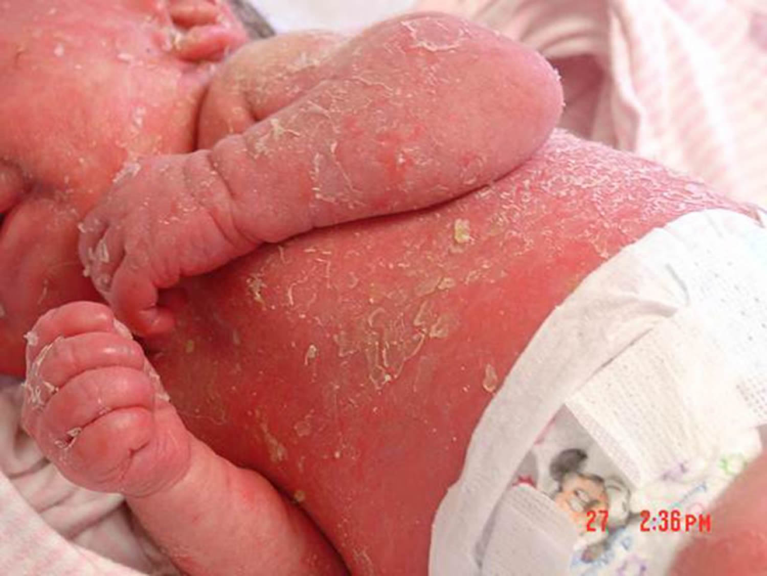
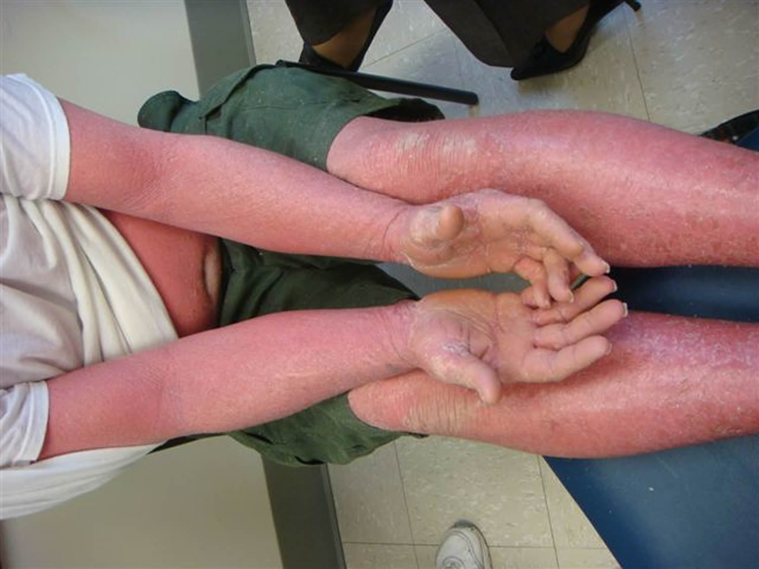
Figure 1. Generalized erythroderma
Erythroderma causes
The most common skin conditions to cause erythroderma are:
- Drug eruption — with numerous diverse drugs implicated (list of drugs)
- Dermatitis especially atopic dermatitis
- Psoriasis, especially after the withdrawal of systemic steroids or other treatment
- Pityriasis rubra pilaris
Other skin diseases that less frequently cause erythroderma may include:
- Other forms of dermatitis: contact dermatitis (allergic or irritant), stasis dermatitis (venous eczema) and in babies, seborrheic dermatitis or staphylococcal scalded skin syndrome
- Blistering diseases including pemphigus and bullous pemphigoid
- Sezary syndrome (the erythrodermic form of cutaneous T-cell lymphoma)
- Several very rare congenital ichthyotic conditions.
Erythroderma may also be a symptom or sign of systemic disease. These may include:
- Hematological malignancies, such as lymphoma and leukemia
- Internal malignancies, such as carcinoma of rectum, lung, fallopian tubes, colon, prostate (paraneoplastic erythroderma)
- Graft-versus-host disease
- HIV infection.
It is not known why some skin diseases in some people progress to erythroderma. The pathogenesis is complicated, involving keratinocytes and lymphocytes, and their interaction with adhesion molecules and cytokines. The result is a dramatic increase in turnover of epidermal cells.
Erythroderma prevention
In most cases, erythroderma cannot be prevented.
People with known drug allergy should be made aware that they should avoid the drug forever, and if their reaction was severe, wear a drug alert bracelet. All medical records should be updated if there is an adverse reaction to a medication and referred to whenever starting a new drug.
Patients with severe skin diseases should be informed if they are at known risk of erythroderma. They should be educated about the risks of recurrence should they discontinue their medication.
Erythroderma signs and symptoms
Erythroderma is often preceded by a morbilliform (measles-like) eruption, dermatitis, or plaque psoriasis. Generalized erythema can develop quite rapidly in acute erythroderma, or more gradually over weeks to months in chronic erythroderma.
By definition, generalized erythema and edema or papulation affect 90% or more of the skin surface.
- The skin feels warm to the touch.
- Itch is usually troublesome and is sometimes intolerable. Rubbing and scratching leads to lichenification.
- Eyelid swelling may result in ectropion.
- Scaling begins 2-6 days after the onset of erythema, as fine flakes or large sheets.
- Thick scaling may develop on the scalp with varying degrees of hair loss including complete baldness.
- Palms and soles may develop yellowish, diffuse keratoderma.
- Nails become dull, ridged, and thickened or develop onycholysis and may shed (onychomadesis).
- Lymph nodes become swollen (generalised dermatopathic lymphadenopathy).
Clues may be present as to the underlying cause.
- Serous ooze, resulting in clothes and dressings sticking to the skin and an unpleasant smell, is characteristic of atopic erythroderma.
- Persistence of circumscribed scaly plaques in certain sites such as elbows and knees suggests psoriasis.
- Islands of sparing, follicular prominence, orange-hue to keratoderma are typical of pityriasis rubra pilaris.
- Subungual hyperkeratosis, crusting on palms and soles, and burrows are indicative of crusted scabies.
- Sparing of abdominal creases (deck chair sign) is typical of papuloerythroderma of Ofuji.
Systemic symptoms may be due to the erythroderma or to its cause.
- Lymphadenopathy, hepatosplenomegaly, abnormal liver dysfunction and fever may suggest a drug hypersensitivity syndrome or malignancy.
- Leg edema may be due to inflamed skin, high output cardiac failure and/or hypoalbuminemia.
Erythroderma complications
Erythroderma often results in acute and chronic local and systemic complications. The patient is unwell with fever, temperature dysregulation and losing a great deal of fluid by transpiration through the skin.
- Heat loss leads to hypothermia.
- Fluid loss leads to electrolyte abnormalities and dehydration.
- Red skin leads to high-output heart failure.
- A secondary skin infection may occur (impetigo, cellulitis).
- General unwellness can lead to pneumonia.
- Hypoalbuminemia from protein loss and increased metabolic rate causes edema.
- Longstanding erythroderma may result in pigmentary changes (brown and/or white skin patches).
Erythroderma diagnosis
The blood count may show anemia, white cell count abnormalities, and eosinophilia. Marked eosinophilia should raise suspicions for lymphoma.
Investigations should be performed to identify underlying causes and complications. They may include:
- Skin swabs for bacterial culture
- Hematology and biochemistry
- Skin biopsy (results depend on underlying cause)
- > 20% circulating Sézary cells suggests Sézary syndrome
- C-reactive protein (CRP) may or may not be elevated.
- Proteins may reveal hypoalbuminemia and abnormal liver function.
- Polyclonal gamma globulins are common, and raised immunoglobulin E (IgE) is typical of idiopathic erythroderma.
Skin biopsies from several sites may be taken if the cause is unknown. They tend to show nonspecific inflammation on histopathology. Diagnostic features may also be present.
Direct immunofluorescence is of benefit if an autoimmune blistering disease or connective tissue disease is considered.
Erythroderma treatment
The underlying cause of erythroderma should be established if possible. Erythroderma is potentially serious, even life-threatening, and the patient may require hospitalization for monitoring and to restore fluid and electrolyte balance, circulatory status and body temperature.
Most patients with acute erythroderma require hospitalization to restore fluid and electrolyte balance, circulatory status and body temperature. However, erythroderma may also be relatively asymptomatic and managed as outpatient.
The following general measures apply:
- Discontinue all unnecessary medications
- Monitor fluid balance and body temperature
- Maintain skin moisture with wet wraps, other types of wet dressings, emollients and mild topical steroids
- Prescribe antibiotics for bacterial infection
- Antihistamines may or may not be helpful for the itch.
If a cause can be identified then specific treatment should be started, such as topical and systemic steroids for atopic dermatitis; acitretin or methotrexate for psoriasis.
Erythroderma prognosis
Prognosis of erythroderma depends on the underlying disease process. If the cause can be removed or corrected, the prognosis is generally good.
If erythroderma is the result of a generalized spread of a primary skin disorder such as psoriasis or dermatitis, it usually clears with appropriate treatment of the skin disease but may recur at any time.
The course of idiopathic erythroderma is unpredictable. It may persist for a long time with periods of acute exacerbation.


