Impetigo contagiosa
Impetigo contagiosa also known as impetigo, is a common highly contagious acute superficial bacterial skin infection (pyoderma) 1. Impetigo contagiosa is characterized by pustules and honey-colored crusted erosions often called ‘school sores’. Impetigo contagiosa is most common in children aged 2-5 years especially boys, but may also affect adults if they have low immunity to the bacteria. Bullous impetigo is more common in infants 2. Children younger than two account for 90% of cases of bullous impetigo 3.
Impetigo contagiosa is prevalent worldwide. Peak onset is during summer, and it is more prevalent in developing countries.
There are two principal types of impetigo contagiosa: nonbullous impetigo (70% of cases) and bullous impetigo (30% of cases). Both types usually resolve within two to three weeks without scarring, and complications are rare, with the most serious being poststreptococcal glomerulonephritis.
- Nonbullous impetigo is caused by Staphylococcus aureus or Streptococcus pyogenes and is characterized by honey-colored crusts on the face and extremities. Impetigo primarily affects the skin or secondarily infects insect bites, eczema, or herpetic lesions.
- Bullous impetigo, which is caused exclusively by Staphylococcus aureus, results in large, flaccid bullae and is more likely to affect intertriginous areas.
The following factors predispose to impetigo contagiosa:
- Atopic eczema
- Scabies
- Skin trauma: chickenpox, insect bite, abrasion, laceration, thermal burn, dermatitis, surgical wound
The term impetiginization is used for superficial secondary infection of a wound or other skin condition. Ulcerated impetigo is called ecthyma. Ecthyma is a deep tissue form of impetigo.
Left untreated, impetigo contagiosa usually clears up on its own within a few weeks, but there’s a risk of infecting other people until it does 4. Antibiotic creams are often used in order to make the symptoms go away faster and stop the infection from spreading. Antibiotic tablets may be used if the impetigo has spread over larger areas of skin. All antibiotic medications have to be prescribed by a doctor. However, there is no generally agreed standard therapy and guidelines for treatment differ widely. Treatment options include many different oral and topical antibiotics as well as disinfectants.
To compare the effectiveness of different treatment options, researchers from the Cochrane Collaboration 5 looked for and systematically analyzed all the relevant studies in this area. These studies looked at the effectiveness of antiseptic or antibiotic solutions and creams, as well as antibiotic tablets, in the treatment of impetigo. Most of this research only looked at people who had impetigo on smaller areas of skin – so their symptoms affected a limited area. The studies showed that creams containing antibiotic medication – such as mupirocin, fusidic acid or retapamulin – relieved the symptoms better than ointments that didn’t contain any antibiotic medication 5. Mupirocin cream was the most commonly tested antibiotic. The studies on mupirocin found the following:
- Without treatment: Symptoms improved or cleared up after 7 to 12 days in about 35 out of 100 people who used a placebo (fake medication).
- With treatment: Symptoms improved or cleared up after 7 to 12 days in about 75 out of 100 people who used the mupirocin cream.
In other words, treatment with mupirocin cream helped impetigo clear up faster in 40 out of 100 people. Only about 170 people participated in the studies, though, so the figures only allow a rough estimate of what can be expected from antibiotic creams.
Mupirocin cream was also somewhat more effective than tablets containing the antibiotic erythromycin (in the treatment of impetigo that covered smaller areas of skin). Erythromycin tablets were also more likely to cause side effects such as nausea, vomiting, stomach ache and diarrhea. In the studies, about 25 out of 100 people who took the erythromycin tablets had these kinds of side effects, but only 5 out of 100 people who used mupirocin cream did.
None of the studies were suitable for answering the question of whether antibiotic tablets are more effective than antibiotic creams when treating impetigo affecting larger or several areas of the body. It is also not clear if antiseptic solutions or creams can help in the treatment of impetigo.
Children with impetigo contagiosa should maintain good personal hygiene and avoid other children during the active outbreak. It is important to wash hands, linens, clothes and affected areas that may have come into contact with infected fluids. Sores can be covered with a bandage to help prevent spread by contact. If impetigo contagiosa is recurrent, evaluation for carriage of the causative bacteria should be performed. The nose is a common reservoir and carriers can be treated with mupirocin (Bactroban Nasal) applied in the nostrils.
Figure 1. Impetigo contagiosa baby

Impetigo contagiosa causes
Impetigo contagiosa is most often caused by Staphylococcus aureus, which is responsible for 80% of cases. Non-bullous impetigo can also be caused by group A beta-hemolytic streptococcus (Streptococcus pyogenes). Methicillin-resistant Staphylococcus aureus (MRSA) has become more prevalent, especially in hospitalized patients. Today, community-acquired MRSA is rapidly increasing. Methicillin-resistant Staphylococcus aureus (MRSA) is more common in populations living in close quarters, daycare centers and prisons.
Impetigo contagiosa can be classified as either primary or secondary. Primary impetigo involves previously normal skin affected by direct bacterial invasion. Secondary impetigo involves infection forming at a previous skin wound site.
Any disturbance of the skin barrier leads to access to fibronectin receptors by group A beta-hemolytic streptococcus (Streptococcus pyogenes) and Staphylococcus aureus which require fibronectin for colonization. Trauma, cuts, insect bites, surgery, atopic dermatitis, burns, and varicella are common mechanisms of skin breakdown. Once a lesion is present, self-inoculation to other sites is very common. Malnutrition, immunosuppression, daycare attendance, overcrowding, diabetes, and poor hygiene make one more susceptible to impetigo.
Triggers that breakdown skin and increase susceptibility to impetigo include:
- Varicella
- Herpes
- Scratching
- Lice
- Burns
- Trauma
- Insect bites
Bullous impetigo
- Bullous impetigo is caused almost exclusively by Staphylococcus aureus. Bullous impetigo is due to staphylococcal exfoliative toxins (exfoliatin A–D), which target desmoglein 1 (a desmosomal adhesion glycoprotein) and cleave off the superficial epidermis through the granular layer. No trauma is required, as the bacteria can infect intact skin.
Nonbullous impetigo
- In nonbullous impetigo, staphylococci and streptococci invade a site of minor trauma where exposed proteins allow the bacteria to adhere.
Ecthyma
- Ecthyma is usually due to Streptococcus pyogenes, but co-infection with Staphylococcus aureus may occur.
Risk factors for getting impetigo contagiosa
Anyone can get impetigo contagiosa, but some factors increase someone’s risk of getting this infection.
- Age: Impetigo contagiosa is most common in children 2 through 5 years old.
- Climate: Impetigo contagiosa is more common in areas with hot, humid summers and mild winters (subtropics), or wet and dry seasons (tropics).
- Infections or injuries that break the skin: People with scabies infection are at increased risk for impetigo contagiosa. Participating in activities where cuts or scrapes are common (sports) can also increase someone’s risk of impetigo.
- Close contact or crowding: Close contact with another person with impetigo is the most common risk factor for illness. For example, if someone has impetigo, it often spreads to other people in their household. Infectious illnesses also tend to spread wherever large groups of people gather together. Crowded conditions — such as those in schools and daycare centers — can increase the spread of impetigo.
Impetigo contagiosa prevention
People can get impetigo contagiosa more than once. Having impetigo does not protect someone from getting it again in the future. While there is no vaccine to prevent impetigo contagiosa, there are things people can do to protect themselves and others.
Wound care
Keep sores caused by impetigo contagiosa covered in order to help prevent spreading group A strep to others. If you have scabies, treating that infection will also help prevent impetigo.
Common sense and good wound care are the best ways to prevent bacterial skin infections, including impetigo:
- Clean all minor cuts and injuries that break the skin (like blisters and scrapes) with soap and water.
- Clean and cover draining or open wounds with clean, dry bandages until they heal.
- See a doctor for puncture and other deep or serious wounds.
- If you have an open wound or active infection, avoid spending time in:
- Hot tubs
- Swimming pools
- Natural bodies of water (e.g., lakes, rivers, oceans)
Hygiene
You should wash the clothes, linens, and towels of anyone who has impetigo every day. These items should not be shared with anyone else. After they have been washed, these items are safe for others to use.
The best way to keep from getting or spreading group A strep is to wash your hands often. This is especially important after coughing or sneezing. To practice good hygiene you should:
- Cover your mouth and nose with a tissue when you cough or sneeze
- Put your used tissue in the waste basket
- Cough or sneeze into your upper sleeve or elbow, not your hands, if you don’t have a tissue
- Wash your hands often with soap and water for at least 20 seconds
- Use an alcohol-based hand rub if soap and water are not available
Antibiotics
Someone with impetigo contagiosa is usually not able to spread the bacteria to others after the lesions heal. People diagnosed with impetigo contagiosa can return to work, school, or daycare if they:
- Have started antibiotic treatment
- Keep all sores on exposed skin covered
Use the prescription exactly as the doctor says to.
Impetigo contagiosa symptoms
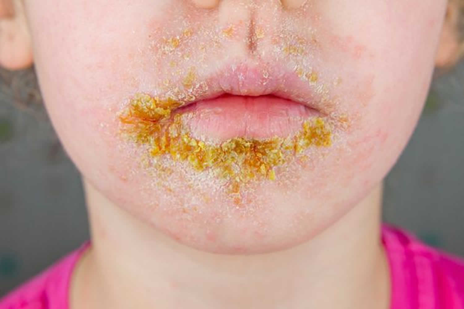
Impetigo contagiosa is an itchy and sometimes painful infection of the outer layers of skin. Primary impetigo mainly affects exposed areas such as the face and hands, but may also affect trunk, perineum and other body sites. The first signs of impetigo contagiosa can usually be seen around the mouth and nose in the form of an itchy reddish rash with small blisters. The blisters are filled with water or pus and burst easily. Once they have burst, yellowish crusts form. These fall off after some time without scarring. As well as on the face, impetigo can occur on the arms and legs. In rare cases, larger blisters develop and don’t break open as quickly. This type of impetigo (bullous impetigo) affects the neck and torso, and may be found in the armpits or in the groin area, for example.
Although many children are otherwise well, swollen lymph nodes (lymphadenopathy), mild fever and malaise may occur.
Bullous impetigo
Bullous impetigo presents with small vesicles that evolve into flaccid transparent bullae. Bullous impetigo heals without scarring. The exfoliative toxin A produced by Staphylococcus aureus causes loss of cell adhesion in the superficial epidermis. The bullae contain a clear or yellow fluid which eventually progresses to become purulent or dark. Surrounding erythema and edema are typically absent. Once the bullae rupture, an erythematous base with a rim of scale remains. Bullous impetigo does not form a honey-colored crust. Lesions most commonly form in the intertriginous regions and on the trunk and, unlike nonbullous impetigo, may occur in the buccal membranes. There are typically fewer lesions present than in non-bullous impetigo. Regional swollen lymph nodes (lymphadenopathy) is absent. Systemic symptoms, such as fever, are more common than in nonbullous impetigo.
Nonbullous impetigo
Nonbullous impetigo starts as a pink macule that evolves into a vesicle or pustule and then into crusted erosions. Multiple vesicles often coalesce and rupture after which the purulent exudate forms the characteristic honey-colored crust. An erythematous base is also present. There are often multiple lesions on the face and extremities, especially in areas in which disruption of the skin barrier has occurred. The rapid spread and satellite lesion formation follow self-inoculation, often in areas with no apparent break in the skin barrier. Mild regional swollen lymph nodes (lymphadenopathy) is a common associated finding. Systemic symptoms such as fever are typically absent in nonbullous impetigo. Untreated impetigo usually resolves within 2 to 4 weeks without scarring.
Ecthyma
Ecthyma is a deep tissue form of impetigo. Ecthyma starts as nonbullous impetigo but develops into a punched-out necrotic ulcer that heals slowly, leaving a scar. Ulcerative lesions penetrate through the epidermis and deep into the dermis. These ulcers appear as “punched out” lesions with violaceous margins. The crusts can be honey-colored or brown-black. The lesions may be purulent.
Impetigo contagiosa complications
While most patients do improve with therapy, a few patients may develop renal failure (post-streptococcal glomerulonephritis). This is more likely if the infection is due to streptococcus. The renal dysfunction appears 7-14 days after the infection. The transient hematuria and proteinuria may last a few weeks or months. Other complications include septic arthritis, scarlet fever, sepsis, and staphylococcal scalded skin syndrome.
Soft tissue infection
The bacteria causing impetigo can become invasive, leading to cellulitis and lymphangitis; subsequent bacteraemia might result in osteomyelitis, septic arthritis or pneumonia.
Staphylococcal scalded skin syndrome
In infants under six years of age or adults with renal insufficiency, localised bullous impetigo due to specific staphylococcal serotypes can lead to a sick child with generalized staphylococcal scalded skin syndrome. Superficial crusting then tender cutaneous denudation on the face, in flexures, and elsewhere is due to circulating exfoliatin or epidermolysin, rather than a direct skin infection. It does not scar.
Toxic shock syndrome
Toxic shock syndrome is rare and rarely preceded by impetigo. It causes fever, diffuse erythematous then desquamating rash, hypotension and involvement of other organs.
Post-streptococcal glomerulonephritis
Group A streptococcal infection may rarely lead to acute post-streptococcal glomerulonephritis 3–6 weeks after the skin infection. It is associated with anti-DNase B and antistreptolysin O (ASO) antibodies.
Rheumatic fever
Group A streptococcal skin infections have rarely been linked to cases of rheumatic fever and rheumatic heart disease. It is thought that this occurs because strains of group A streptococci usually found on the skin have moved to the throat (the more usual site for rheumatic fever-associated infection).
Impetigo contagiosa diagnosis
Impetigo contagiosa is usually diagnosed clinically but can be confirmed by bacterial swabs sent for microscopy (gram-positive cocci are observed), culture and sensitivity.
Bacterial cultures can be used for confirmation of diagnosis and should be obtained if methicillin-resistant staph aureus (MRSA) is suspected or if an impetigo outbreak is present. A skin biopsy may be considered if the case is refractory.
A blood count may reveal neutrophil leucocytosis when impetigo is widespread.
The anti-streptolysin O (ASO) response is weak from impetigo alone. Therefore, serologic testing for streptococcal antibodies is not indicated for the diagnosis of impetigo. However, it may be useful if post-streptococcal glomerulonephritis is suspected in a patient with a recent impetigo outbreak.
Human immunodeficiency virus (HIV) testing should be considered when a previously healthy adult develops bullous impetigo.
Skin biopsy is rarely necessary. The histological features of impetigo are characteristic.
Non-bullous impetigo
- Gram-positive cocci
- Intraepidermal neutrophilic pustules,
- Dense inflammatory infiltrate in the upper dermis
Bullous impetigo
- Split through the granular layer of the epidermis without inflammation or bacteria
- Acantholytic cells
- Minimal inflammatory infiltrate in the upper dermis
- Resembles pemphigus foliaceus
Ecthyma
- Full-thickness skin ulceration
- Gram stain shows cocci within the dermis
Impetigo contagiosa treatment
The following steps are used to treat impetigo 6.
- Cleanse the wound; use moist soaks to remove crusts gently.
- Apply antiseptic 2–3 times daily for five days (povidone-iodine, hydrogen peroxide 1% cream, chlorhexidine, superoxidised solution and others).
- Cover the affected areas.
- Oral antibiotics are recommended if:
- Symptoms are significant or severe (fever, malaise)
- There are more than three lesions
- There is a high risk of complications
- The infection is not resolving or is unlikely to resolve.
Topical antibiotics alone or in conjunction with systemic antibiotics are used to treat impetigo. Antibiotic coverage should cover both Staphylococcus aureus and Streptococcus pyogenes (group A beta-hemolytic streptococcus). While untreated impetigo is often self-limiting, antibiotics decrease the duration of illness and spread of lesions. In addition, antibiotic treatment decreases the chances of complications involving kidneys, joints, bones, and lungs, as well as acute rheumatic fever 7.
For localized, uncomplicated, non-bullous impetigo, topical therapy alone is the treatment of choice. The crust should be removed with soap and water before the application of topical antibiotic therapy. Mupirocin, retapamulin, and fusidic acid are the treatments of choice.
Systemic antibiotics should be prescribed for all cases of bullous impetigo and cases of non-bullous impetigo with more than five lesions, deep tissue involvement, systemic signs of infection, lymphadenopathy or lesions in the oral cavity. Beta-lactamase-resistant antibiotics such as cephalosporins, amoxicillin-clavulanate, dicloxacillin are the treatment of choice. Cephalexin is commonly used. If culture confirms an infection solely caused by streptococci, oral penicillin is the preferred therapy.
Suitable oral antibiotics are flucloxacillin 500 mg four times daily for 5 days (adult dose), and in case of allergy or bacterial resistance, trimethoprim + sulphamethoxazole 960 mg, twice daily, for five days (adult dose), erythromycin 800 mg twice daily for 5 days or cephalexin 1 g twice daily for 5 days 6.
In areas of high prevalence of MRSA or if cultures are positive for MRSA, clindamycin or doxycycline are the preferred treatments. Trimethoprim-sulfamethoxazole is effective against MRSA, but should only be used if group A streptococci are not the causative agent, or in addition to an anti-streptococcal antibiotic.
Table 1. Topical Antibiotics for Impetigo
| Medication | Instructions |
| Fusidic acid 2% ointment† | Apply to affected skin three times daily for seven to 12 days |
| Mupirocin 2% cream (Bactroban)‡ | Apply to affected skin three times daily for seven to 10 days; reevaluate after three to five days if no clinical response Approved for use in persons older than three months |
| Mupirocin 2% ointment‡ | Apply to affected skin three times daily for seven to 14 days Dosing in children is same as adults Approved for use in persons older than two months |
| Retapamulin 1% ointment (Altabax)§ | Apply to affected skin twice daily for five days Total treatment area should not exceed 100 cm² in adults or 2% of total body surface area in children Approved for use in persons nine months or older |
Footnotes:
*—Estimated retail price. Generic price listed first; brand listed in parentheses.
†—Coverage for Staphylococcus aureus (methicillin-susceptible) and streptococcus 8.
‡—Coverage for S. aureus (methicillin-susceptible) and streptococcus 9. Mupirocin-resistant streptococcus has now been documented 10
§—First member of the pleuromutilin class of antibiotics. Coverage for S. aureus (methicillin-susceptible) and streptococcus 11
NA = not available.
[Source 12 ]Table 2. Systemic Antibiotics for Impetigo
| Drug | Adult seven-day dose | Cost (for a typical course of treatment)* | Children seven-day dose |
| Amoxicillin/clavulanate (Augmentin)† | 875/125 mg every 12 hours | $19 ($193) | Younger than three months: 30 mg per kg per day Three months or older: 25 to 45 mg per kg per day for those weighing less than 40 kg (88 lb); 875/125 mg every 12 hours for those weighing 40 kg or more Based on mg per kg per day of the amoxicillin component in divided doses every 12 hours |
| Cephalexin (Keflex) | 250 mg every six hours or 500 mg every 12 hours | $5 ($90) | 25 to 50 mg per kg per day in divided doses every six to 12 hours |
| Clindamycin‡ | 300 to 600 mg every six to eight hours | $18 ($200) | 10 to 25 mg per kg per day in divided doses every six to eight hours |
| Dicloxacillin | 250 mg every six hours | $14 (NA) | 12.5 to 25 mg per kg per day in divided doses every six hours |
| Doxycycline§ | 50 to 100 mg every 12 hours | $15 ($95) | 2.2 to 4.4 mg per kg per day in divided doses every 12 hours Not recommend in children younger than eight years |
| Minocycline (Minocin)§ | 100 mg every 12 hours | $36 ($185) | Loading dose of 4 mg per kg for first dose (maximum dose of 200 mg), then 4 mg per kg per day in divided doses every 12 hours Maximum of 400 mg per day Not recommend in children younger than eight years |
| Trimethoprim/sulfamethoxazole§ | 160/800 mg every 12 hours | $4 (NA) | 8 to 10 mg per kg per day based on the trimethoprim component in divided doses every 12 hours |
Footnotes: Because of emerging resistance, penicillin and erythromycin are no longer recommended treatments 13.
*—Estimated retail price. Generic price listed first; brand listed in parentheses.
†—Good coverage for Staphylococcus aureus (methicillin-susceptible) and streptococcus.
‡—If methicillin-resistant Staphylococcus aureus (MRSA) is suspected or proven.
§—If methicillin-resistant Staphylococcus aureus (MRSA) is suspected or proven. There is no activity against streptococcus.
NA = not available.
[Source 12 ]To prevent recurrence of impetigo contagiosa:
- Treat carrier sites: apply antiseptic ointment to nostrils
- Wash daily with antibacterial soap or soak in a bleach bath
- Cut nails and keep hands clean
- Identify and treat the source of re-infection, usually another infected person or carrier in the household.
To reduce the chance of passing the infection to another person:
- Avoid close contact with others
- Children must stay away from school until crusts have dried out or for 24 hours after starting oral antibiotics
- Use separate towels and flannels
- Change and launder clothes and linen daily.
Natural therapies
The evidence is insufficient to recommend or dismiss popular herbal treatments for impetigo 14. Natural remedies such as tea tree oil; tea effusions; olive, garlic, and coconut oils; and Manuka honey have been anecdotally successful. The fact that impetigo is self-limited means that many “cures” could appear to be helpful without being superior to placebo. In one study 15, tea leaf ointment and oral cephalexin (Keflex) were similarly effective, with a cure rate of 81% vs. 79%. Tea tree oil (derived from Melaleuca alternifolia) appeared to be equivalent to mupirocin 2% for topical decolonization of MRSA 16.
Impetigo contagiosa prognosis
Without treatment, impetigo contagiosa heals in 14-21 days. About 20% of cases resolve spontaneously. Scarring is rare but some patients may develop pigmentation changes. Some patients may develop ecthyma. With treatment, cure occurs within 10 days. Neonates may develop meningitis. A rare complication is acute post streptococcal glomerulonephritis, whic occurs 2-3 weeks after the skin infection.
References- Koning S, van der Sande R, Verhagen AP, et al. Interventions for impetigo. Cochrane Database Syst Rev. 2012;1(1):CD003261. Published 2012 Jan 18. doi:10.1002/14651858.CD003261.pub3 https://www.ncbi.nlm.nih.gov/pmc/articles/PMC7025440
- Nardi NM, Schaefer TJ. Impetigo. [Updated 2020 Aug 8]. In: StatPearls [Internet]. Treasure Island (FL): StatPearls Publishing; 2020 Jan-. Available from: https://www.ncbi.nlm.nih.gov/books/NBK430974
- Sahu JK, Mishra AK. Ozenoxacin: A Novel Drug Discovery for the Treatment of Impetigo. Curr Drug Discov Technol. 2019;16(3):259-264. doi: 10.2174/1570163815666180502165014
- InformedHealth.org [Internet]. Cologne, Germany: Institute for Quality and Efficiency in Health Care (IQWiG); 2006-. Impetigo: What can make it go away faster? [Updated 2020 Jul 16]. Available from: https://www.ncbi.nlm.nih.gov/books/NBK279536
- Koning S, van der Sande R, Verhagen AP, van Suijlekom‐Smit LWA, Morris AD, Butler CC, Berger M, van der Wouden JC. Interventions for impetigo. Cochrane Database of Systematic Reviews 2012, Issue 1. Art. No.: CD003261. DOI: 10.1002/14651858.CD003261.pub3 https://www.cochranelibrary.com/cdsr/doi/10.1002/14651858.CD003261.pub3/full
- Koning S, Verhagen AP, van Suijlekom-Smit LW, Morris A, Butler CC, van der Wouden JC. Interventions for impetigo. Cochrane Database Syst Rev. 2004;(2):CD003261. doi: 10.1002/14651858.CD003261.pub2. Update in: Cochrane Database Syst Rev. 2012;1:CD003261 https://www.cochranelibrary.com/cdsr/doi/10.1002/14651858.CD003261.pub3/full
- Loadsman MEN, Verheij TJM, van der Velden AW. Impetigo incidence and treatment: a retrospective study of Dutch routine primary care data. Fam Pract. 2019 Jul 31;36(4):410-416. doi: 10.1093/fampra/cmy104
- Koning S, van Suijlekom-Smit LW, Nouwen JL, et al. Fusidic acid cream in the treatment of impetigo in general practice: double blind randomised placebo controlled trial. BMJ. 2002;324(7331):203–206.
- Liu C, Bayer A, Cosgrove SE, et al. Clinical practice guidelines by the Infectious Diseases Society of America for the treatment of methicillin-resistant Staphylococcus aureus infections in adults and children: executive summary. Clin Infect Dis. 2011;52(3):285–292.
- Bangert S, Levy M, Hebert AA. Bacterial resistance and impetigo treatment trends: a review. Pediatr Dermatol. 2012;29(3):243–248.
- Altabax (retapamulin) ointment [package insert]. Research Triangle Park, N.C.: GlaxoSmithKline; 2010.
- Hartman-Adams H, Banvard C, Juckett G. Impetigo: diagnosis and treatment. Am Fam Physician. 2014 Aug 15;90(4):229-35. https://www.aafp.org/afp/2014/0815/p229.html
- Silverberg N, Block S. Uncomplicated skin and skin structure infections in children: diagnosis and current treatment options in the United States. Clin Pediatr (Phila). 2008;47(3):211–219.
- Martin KW, Ernst E. Herbal medicines for treatment of bacterial infections: a review of controlled clinical trials. J Antimicrob Chemother. 2003;51(2):241–246.
- Sharquie KE, al-Turfi IA, al-Salloum SM. The antibacterial activity of tea in vitro and in vivo (in patients with impetigo contagiosa). J Dermatol. 2000;27(11):706–710.
- Caelli M, Porteous J, Carson CF, Heller R, Riley TV. Tea tree oil as an alternative topical decolonization agent for methicillin-resistant Staphylococcus aureus. J Hosp Infect. 2000;46(3):236–237.


