What is a macule
Macule is patch of skin that is altered in color but is not elevated.
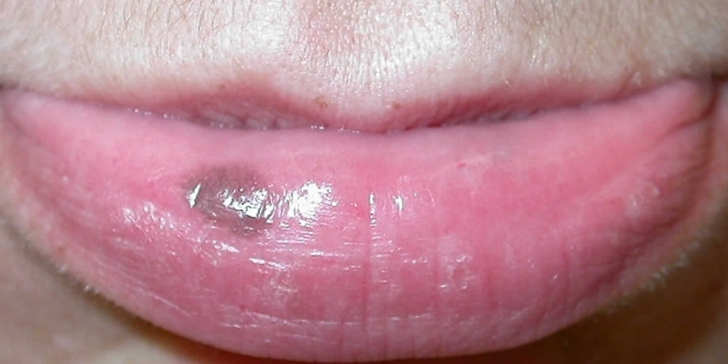
Labial melanotic macule
Labial melanotic macule is a well-defined, oval, brown to black, flat patch on the central third of the lower lip. Labial melanotic macule is the name for a freckle arising on the lip and is also sometimes called a labial lentigo of mucosal melanosis or oral melanotic macule. Traditionally, labial melanotic macule refers to oral labial lesions and represent a distinct clinicopathologic entity. Unlike (oral) labial melanotic macules, pigmented macules on genital labia are not a distinct entity and can represent a range of conditions—including melanocytic nevi, lentigines, melanocytic nevi with architectural disorder and atypia of melanocytes (dysplastic nevi), atypical melanocytic nevi of the genital type and melanomas.
Usually solitary, a (oral) labial melanotic macule is most commonly seen in adult women but it also occurs in males and in young people. Average age at time of presentation to the physician is at approximately 40 years in patients of color, though these macules may appear at any age. In patients of darker skin types, onset typically occurs in adolescence.
Occasionally labial melanotic macule can be on the upper lip. Ultraviolet light exposure is thought to be a risk factor.
Labial melanotic macules do not cause any symptoms but their appearance can be a concern to the patient.
Labial melanotic macules typically present as solitary, symmetric, asymptomatic, well-defined, brown or black macules on the medial third of the lower lip (Figure 1). Sometimes, lesions may be multiple, located on the upper lip, have variegated pigmentation, or have a history of color change—lending to confusion with other pigmented lesions. To rule out other possible entities, be sure to perform a complete skin examination, checking for lesions involving the mouth, eyes, genitals, skin, or nails. Palpation of the lip for evaluation of deeper involvement is advised.
Size ranges from 1 to 8mm. Once developed the lesions usually remain unchanged in size and color. They can occasionally have an irregular edge and there may be a history of color change which can cause confusion with other pigmented lesions, including melanoma. Luckily, melanoma is very rare on the lip (but it can occur).
Similar freckles (melanotic macule) may also occur in areas that are not exposed to the sun:
- Inside the mouth (oral melanotic macules)
- On the vulva in women (vulval labial melanotic macules, vulval melanosis)
- On the penis in men (penile melanotic macule, penile lentigo)
Patients with multiple lesions that are histologically consistent with labial melanotic macules should be evaluated for other possible conditions, like HIV or Peutz-Jeghers syndrome. Of note, skin lesions of Peutz-Jeghers syndrome usually fade after puberty while mucosal lesions may persist. In some patients with HIV, oral melanotic macules may be an early sign of HIV infection. While most of these patients have multiple macules on the buccal mucosa, some may have labial, gingival, or palatal lesions. Screening for risk factors for HIV, particularly in patients with multiple mucosal lesions, is advisable.
Figure 1. Labial melanotic macule

See your doctor for any new dark spot on the lips or inside the mouth. Similarly, any existing spot that changes size, shape, or color should also be evaluated promptly.
If the diagnosis of oral melanotic macule is not certain, your physician may wish to perform a skin biopsy in order to confirm the diagnosis. The procedure involves:
- Numbing the skin with an injectable anesthetic.
- Sampling a small piece of skin by using a flexible razor blade, a scalpel, or a tiny cookie cutter (called a “punch biopsy”). If a punch biopsy is taken, a suture or two may be placed and will need to be removed 5–10 days later.
- Having the skin sample examined under a microscope by a specially trained physician (dermatopathologist).
Your doctor is more likely to biopsy certain lesions, such as new ones, large or growing ones, or those with irregular color (pigmentation). The biopsy can help the doctor to tell whether it is a benign oral melanotic macule or a malignant melanoma, a type of skin cancer.
Most dark spots on the lips or inside the mouth are benign oral melanotic macules. Usually, your doctor will observe the lesion by measuring it, by taking a photograph of it, or both. As long as the oral melanotic macule stays stable in size, shape, and color, no treatment is needed.
Nonetheless, some people want the lesion removed for cosmetic reasons. If it is appropriate, some physicians might recommend excision or, rarely, laser treatment.
Labial melanotic macule causes
Labial melanotic macule is thought to be provoked by sun exposure, and it is more common in fair skinned people. As labial melanotic macules almost always occur on the central third of the lower lip, a site receiving maximum sun exposure, ultraviolet radiation may play a role in their development. In one study, upper lip lesions occurred following tanning bed use in 2 of 4 patients. There are also two case reports of labial melanotic macules following application of topical tacrolimus, thought possibly secondary to its activation of melanocytes or its immunosuppressive activity. However labial melanotic macule may also occur in dark skinned individuals and as described above, similar lesions can arise in sites that are never sun-exposed. Luckily, melanotic macules are harmless.
Other conditions that can cause lip pigmentation
Labial melanotic macules may be confused with other pigmented skin lesions.
- freckles (ephelides)
- lentigo simplex
- solar lentigo
- venous haemangioma (venous lake)
- amalgam tattoo
- junctional melanocytic naevus (flat mole)
- lentigo maligna
- superficial spreading melanoma
These conditions can be differentiated from labial melanotic macule by a combination of clinical and histological features.
Multiple lesions may be a sign of a widespread skin condition, such as:
- Peutz Jeghers syndrome
- Addison disease
- Laugier-Hunziker syndrome
- Multiple lentiginoses (various syndromes)
Labial melanotic macule signs and symptoms
The most common locations for an oral melanotic macule include:
- Lips, especially the lower lip
- Gums (gingiva)
- Inner cheek (buccal mucosa)
- Roof of the mouth (hard or soft palate)
An oral melanotic macule appears as a solitary, flat, tan-to-dark-brown spot usually less than 7 mm in diameter. It has a well-defined border and a uniform color.
People can have more than one oral melanotic macule.
Labial melanotic macule diagnosis
Labial melanotic macule has a characteristic pattern when examined with the magnifying glass or dermatoscope. Dermoscopy of labial melanotic macules reveals homogeneous pigmentation that may be structureless or contain partial linear or curvilinear streaks in a regular parallel pattern that gradually fades towards the periphery.
Labial melanotic macule with a typical history and appearance need not be biopsied.
If biopsy is done because the lesion is changing or looks irregular, a labial melanotic macule shows the following features on dermatopathology (under the microscope).
- Increased melanin in the melanocytes and keratinocytes of the basal layer
- Melanophages in the dermal papillae, indicating pigmentary incontinence
- Mild acanthosis without elongation of the rete ridges.
- Nuclear atypia is absent and the melanocyte count is normal.
Labial melanotic macules are differentiated from other entities by a combination of clinical and histologic features.
While ephelides and Peutz-Jeghers syndrome are histologically similar to labial melanotic macules, ephelides present as brown macules in childhood that darken on sun exposure and the macules of Peutz-Jeghers syndrome are typically multiple, more widespread (i.e., oral mucosa, lips, eyes, nose, hands, feet), and are associated with intestinal polyposis (hamartomas).
Both lentigo simplex and solar lentigines have increased basal layer pigmentation and increased melanocyte concentration. Lentigines also have elongated rete ridges and often present with irregularly shaped borders.
Labial melanotic macule treatment
Given their benign nature with apparently no reported cases of malignant transformation, labial melanotic macule can just be observed. Suspicious lesions, including lesions showing progressive change, should be biopsied. Patients should monitor lesions, seek medical attention for any lesional change, and practice good sun protection.
A paucity of published data exists regarding cosmetic treatment of labial melanotic macules, which is often debatable. Reported options for elective cosmetic augmentation include surgical excision (including by traditional, punch, or shave methods), laser therapy, and cryotherapy.
If treatment is requested the macules can be frozen (cryotherapy), or removed using a laser or intense pulsed light. Excision can also be performed but will leave a scar. However, the advantage of excisional surgery is the prospect of complete removal for histologic evaluation. This may alleviate concerns about misdiagnosis of significant pathology due to the potential biopsy sampling error or fear that other cosmetic modalities may mask detection of possible lesional change.
Cryosurgery is a relatively simple and inexpensive treatment option. However, only one published article is available regarding its use in labial melanotic macules. In this study, fifteen patients with labial melanotic macules were pretreated with topical 4% lidocaine gel, then treated with simple cryosurgery via direct cotton swab application of liquid nitrogen for 10 to 15 seconds.
Patients experienced slight erythema immediately after treatment, whitish slough within 3 to 4 days, and disappearance of melanotic macules at 1 week post-treatment. Reported patient acceptance of the procedure was good and healing was uneventful without postoperative pain, hemorrhage, infection, or scarring. Six patients had repigmentation 2 to 18 months post-cryosurgery and were retreated with the same method.
Laser therapy, although the most expensive modality, has shown favorable cosmetic results. The Q-switched lasers that target pigment (694nm, 755nm, and 1064nm) are all reasonable options for laser therapy. Multiple small studies report good response of labial melanotic macules to Q-switched lasers. In one retrospective study, 9 patients were treated by Q-switched ruby laser (694nm, 25 to 40nsec, 4mm spot, 10J/cm2) and followed for a maximum of 5.5 years. The benign nature of the lesions was assessed by dermoscopy and, in one case, by biopsy. Three patients achieved complete clearance after one treatment session, while a second treatment was needed in six patients. There were no reported recurrences, scars, or pigment alterations.
In a second study, 8 patients were treated by Q-switched ruby laser (694nm, 20 nsec, 5mm spot, 6.5J/cm2) with clearance in 5 patients after a single pulse and 3 patients requiring a second treatment session. No side effects or recurrence were reported over the 24-month follow-up period. In a third study, three patients with “labial lentigos” had dramatic clearing after one to two treatments with a Q-switched ruby laser (694nm, 10J/cm2). The use of other lasers is mentioned sparingly in the literature. In a study of six Japanese patients with labial melanosis, including “labial lentigo” and Peutz-Jeghers syndrome, rapid clearance without mucosal textural changes or recurrence was accomplished with a pulsed ruby laser (695nm, 1 to 2msec pulse).
Macule vs Papule
Papule is an elevated, solid, palpable lesion that is ≤ 1 cm in diameter. Papule may be solitary or multiple.
Papules may be:
- Acuminate (pointed)
- Dome-shaped (rounded)
- Filiform (thread-like)
- Flat-topped
- Oval or round
- Pedunculated (with a stalk)
- Sessile (without a stalk)
- Umbilicated (with a central depression)
- Verrucous (warty)


