Neuropathic arthropathy
Neuropathic arthropathy also known as Charcot joint (Charcot foot), Charcot neuroarthropathy or Charcot osteoarthropathy, refers to progressive degenerative and destructive joint disorder of a weight bearing joint, a process marked by bony destruction, bone resorption, and eventual deformity due to abnormal pain sensation and proprioception. Neuropathic arthropathy or Charcot joint is a condition that affects the bones, joints, and soft tissue in the feet and ankles. Neuropathic arthropathy can develop as a result of nerve damage in the feet due to diabetes or other nerve injuries. Patients present insidiously or are identified incidentally, or as a result of investigation for deformities. Unlike septic arthritis, Charcot joints although swollen are normal temperature without elevated inflammatory markers. Importantly, neuropathic arthropathy are painless.
Charcot joints are typically unilateral but are bilateral in ~20% (range 5.9-39.3%) of cases 1.
The pathogenesis of a Charcot neuropathic arthropathy is thought to be an inflammatory response from a minor injury that results in osteolysis. In the setting of peripheral neuropathy, both the initial insult and inflammatory response is not well appreciated, allowing ongoing inflammation and injury 1.
There are two patterns of charcot neuropathic arthropathy: atrophic and hypertrophic.
Atrophic neuropathic arthropathy
- most common form 2
- occurs earlier 3
- has an acute progression
- characterized by reabsorption of the ends of the affected bone
- joint destruction with resorption of fragments
- an absence of osteosclerosis and osteophytes
- mainly occurs in non-weight bearing joints of the upper limb 2
Hypertrophic neuropathic arthropathy
- only sensory nerves affected
- slow progression
- joint destruction with periarticular debris/bone fragmentation
- initially widened then narrowed joint space
- presence of osteosclerosis and osteophytes 2
- absence of osteoporosis (unless joint is infected) 4
Neuropathic arthropathy causes
In modern Western societies by far the most common cause of Charcot joints is diabetes mellitus, and therefore, the demographics of patients matches those of older diabetics. Prevalence differs depending on the severity of diabetes mellitus 1:
- ~0.1% in general diabetic population
- ~15% in high-risk diabetic population
- ~30% in patients with peripheral neuropathy
Sensorimotor and autonomic neuropathies of various causes are the primary predisposing factor. The most common cause vary by the involved joint 5:
- diabetes mellitus (most common cause overall and in the foot and ankle; most commonly affects the foot and ankle)
- syringomyelia (most common cause in the upper extremity and shoulder)
- neurosyphilis/tabes dorsalis (more common in the past; most commonly affects the knee)
- traumatic spinal cord injury (most common cause in the spine) 6
- alcoholism
- tumors compressing or involve the spinal cord or peripheral nerves
- amyloidosis
- pernicious anemia
- poliomyelitis
- leprosy
- multiple sclerosis
- steroid use (intraarticular or systemic)
- spina bifida/myelomeningocele
- congenital insensitivity to pain
- Charcot-Marie-Tooth disease
- familial dysautonomia (Riley-Day syndrome)
Less established causes:
Diabetes is the most common cause of this type of nerve damage. This damage is more common in people with type 1 diabetes. When blood sugar levels are high over a long time, both nerve and blood vessel damage occurs in the feet.
Nerve damage makes it harder to notice the amount of pressure on the foot or if it is being stressed. The result is ongoing small injuries to the bones and ligaments that support the foot.
- You may develop bone stress fractures in your feet, yet never know it.
- Continuing to walk on the fractured bone often leads to further bone and joint damage.
Other factors leading to foot damage include:
- Blood vessel damage from diabetes can increase or change blood flow to the feet. This can lead to bone loss. Weakened bones in the feet increase the risk of fracture.
- Injury to the foot signals the body to produce more inflammation-causing chemicals. This contributes to swelling and bone loss.
Neuropathic arthropathy symptoms
Early foot symptoms may include:
- Mild pain and discomfort
- Redness
- Swelling
- Warmth in the affected foot (noticeably warmer than the other foot)
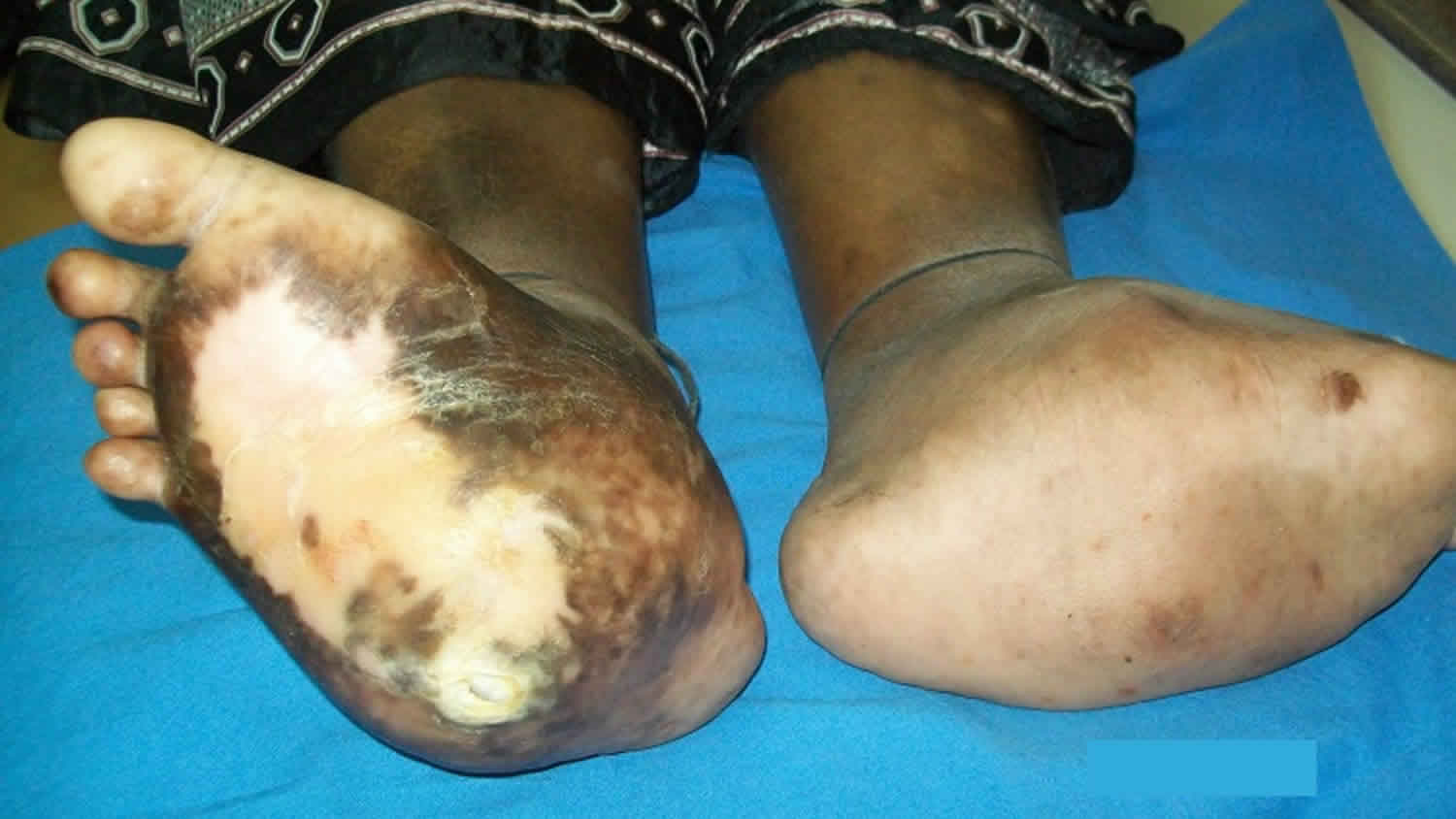
At later stages, bones in the foot break and move out of place, causing the foot or ankle to become deformed.
- A classic sign of Charcot is rocker-bottom foot. This occurs when the bones in middle of the foot collapse. This causes the arch of the foot to collapse and bow downward.
- The toes may curl downward.
Bones that stick out at odd angles can lead to pressure sores and foot ulcers.
- Because the feet are numb, these sores may grow wider or deeper before they are noticed.
- High blood sugar also makes it hard for the body to fight infection. As a result, these foot ulcers become infected.
Neuropathic arthropathy complications
Severe deformity of the foot increases the risk of foot ulcers. If ulcers become infected and hard to treat, it may require amputation.
Neuropathic arthropathy diagnosis
Charcot neuropathic arthropathy is not always easy to diagnose early on. It can be mistaken for bone infection, arthritis or joint swelling. Your health care provider will take your medical history and examine your foot and ankle.
Blood tests and other lab work may be done to help rule out other causes.
Your doctor may check for nerve damage with these tests:
- Electromyography
- Nerve conduction velocity tests
- Nerve biopsy
The following tests may be done to check for bone and joint damage:
- Foot X-rays
- MRI
- Bone scan
Foot x-rays may look normal at early stages of the condition. Diagnosis often comes down to recognizing early symptoms of Charcot foot: swelling, redness, and warmth of the affected foot.
Neuropathic arthropathy treatment
The goal of treatment is to stop bone loss, allow bones to heal, and prevent bones from moving out of place (deformity).
Immobilization
Your doctor will have you wear a total contact cast. This will help limit movement of your foot and ankle. You will likely be asked to keep your weight off your foot entirely, so you will need to use crutches, a knee-walker device, or wheelchair.
You will have new casts placed on your foot as the swelling comes down. Healing can take a couple of months or more.
Protective footwear
Once your foot has healed, your provider may suggest footwear to help support your foot and prevent re-injury. These may include:
- Splints
- Braces
- Orthotic insoles
- Charcot restraint orthotic walker, a special boot that provides even pressure to the whole foot
Activity changes
You will always be at risk for Charcot foot coming back or developing in your other foot. So your provider may recommend activity changes, such as limiting your standing or walking, to protect your feet.
Surgery
You may need surgery if you have foot ulcers that keep coming back or severe foot or ankle deformity. Surgery can help stabilize your foot and ankle joints and remove bony areas to prevent foot ulcers.
Ongoing monitoring
You will need to see your provider for checkups and take steps to protect your feet for the rest of your life.
Neuropathic arthropathy prognosis
The prognosis depends on the severity of foot deformity and how well you heal. Many people do well with braces, activity changes, and ongoing monitoring.
References- Mautone M, Naidoo P. What the radiologist needs to know about Charcot foot. J Med Imaging Radiat Oncol. 2015;59 (4): 395-402. doi:10.1111/1754-9485.12325
- Dähnert W. Radiology review manual. Lippincott Williams & Wilkins. (2007) ISBN:0781738954
- Adamand A. Diagnostic Radiology, A Textbook of Medical Imaging. Churchill Livingstone. (2001) ISBN:0443064326
- Proctor R. Final FRCR Part A Modules 1-3 Single Best Answer MCQs. Radcliffe Publishing. (2009) ISBN:184619363X
- Jones EA, Manaster BJ, May DA, Disler DG. Neuropathic osteoarthropathy: diagnostic dilemmas and differential diagnosis. (2000) Radiographics : a review publication of the Radiological Society of North America, Inc. 20 Spec No: S279-93. doi:10.1148/radiographics.20.suppl_1.g00oc22s279
- Ledbetter LN, Salzman KL, Sanders RK, Shah LM. Spinal Neuroarthropathy: Pathophysiology, Clinical and Imaging Features, and Differential Diagnosis. (2016) Radiographics : a review publication of the Radiological Society of North America, Inc. 36 (3): 783-99. doi:10.1148/rg.2016150121
- Grear BJ, Rabinovich A, Brodsky JW. Charcot arthropathy of the foot and ankle associated with rheumatoid arthritis. (2013) Foot & ankle international. 34 (11): 1541-7. doi:10.1177/1071100713500490
- Clement GB, Grizzard K, Vasey FB, Germain BF, Espinoza LR. Neuropathic arthropathy (Charcot joints) due to cervical osteolysis: a complication of progressive systemic sclerosis. (1984) The Journal of rheumatology. 11 (4): 545-8. https://www.ncbi.nlm.nih.gov/pubmed/6481729


