Pearson syndrome
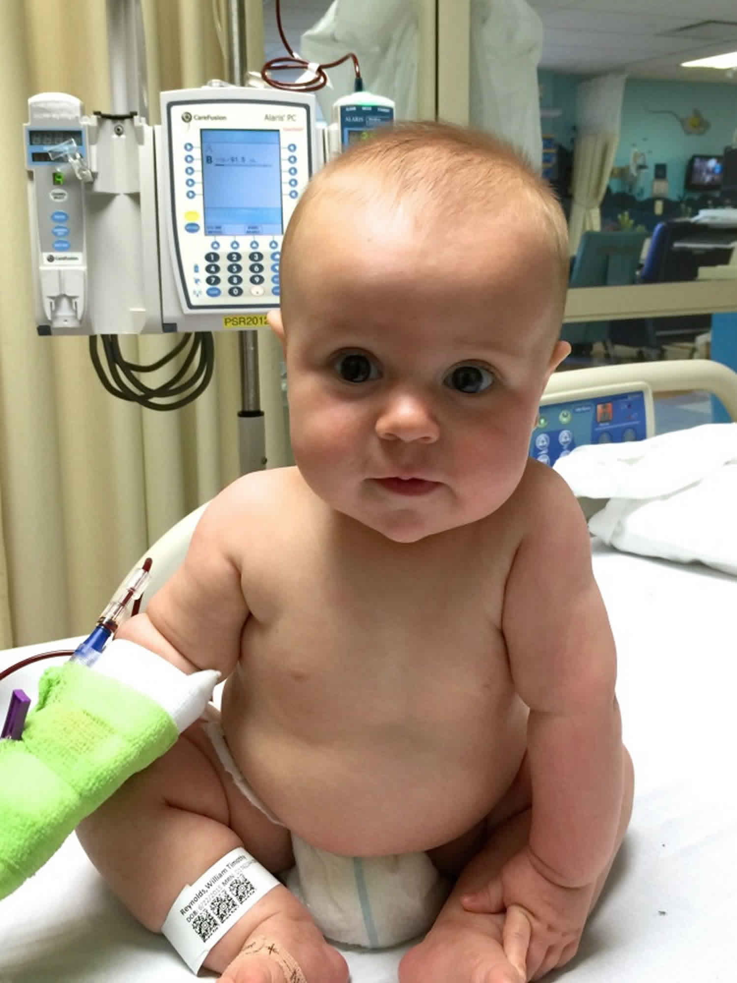
Pearson syndrome also known as Pearson marrow-pancreas syndrome, is a mitochondrial disease that affects many parts of the body but especially the bone marrow and the pancreas, that usually begins in infancy. Pearson marrow-pancreas syndrome causes problems with the development of blood-forming cells in the bone marrow (hematopoietic stem cells) that produce red blood cells, white blood cells, and platelets. For this reason, Pearson marrow-pancreas syndrome is considered a bone marrow failure disorder. Having too few red blood cells (anemia), white blood cells (neutropenia), or platelets (thrombocytopenia) can cause a child to feel weak and tired, be sick more often, bruise more easily and take a longer time to stop bleeding when cut. Pearson syndrome also affects the pancreas, which can cause frequent diarrhea and stomach pain, trouble gaining weight, and diabetes. Some children with Person syndrome may also have problems with their liver, kidneys, heart, eyes, ears, and/or brain 1.
Pearson marrow-pancreas syndrome is a rare condition. Less than 100 cases have been reported worldwide.
Pearson syndrome is caused by a change (mutation) in the mitochondrial DNA. Mitochondria make the energy for the cells in your body by combining oxygen with sugars and fats that come from the food you eat. Changes in mitochondrial DNA can make it hard for the cells of the body to make energy. Pearson syndrome is usually caused by deletions of a part of the mitochondrial DNA (pieces of the DNA are missing). Most cases of Pearson syndrome happen for the first time in a family which means it is not passed down from either parent (de novo mutation) 2. Most cases of Pearson syndrome occur by mistake, when the egg or sperm were being made (de novo mutation). This means that the disease was not passed down or inherited from either parent and no other family member has the disease.
Most affected individuals have a shortage of red blood cells (anemia), which can cause pale skin (pallor), weakness, and fatigue. Some of these individuals also have low numbers of white blood cells (neutropenia) and platelets (thrombocytopenia). Neutropenia can lead to frequent infections; thrombocytopenia sometimes causes easy bruising and bleeding. When visualized under the microscope, bone marrow cells from affected individuals may appear abnormal. Often, early blood cells (hematopoietic precursors) have multiple fluid-filled pockets called vacuoles. In addition, red blood cells in the bone marrow can have an abnormal buildup of iron that appears as a ring of blue staining in the cell after treatment with certain dyes. These abnormal cells are called ring sideroblasts.
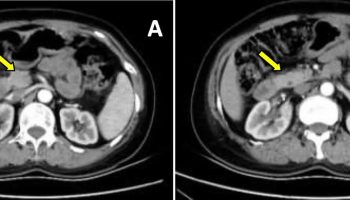
In people with Pearson marrow-pancreas syndrome, the pancreas does not work as well as usual. The pancreas produces and releases enzymes that aid in the digestion of fats and proteins. Reduced function of this organ can lead to high levels of fats in the liver (liver steatosis). The pancreas also releases insulin, which helps maintain correct blood sugar levels. A small number of individuals with Pearson marrow-pancreas syndrome develop diabetes, a condition characterized by abnormally high blood sugar levels that can be caused by a shortage of insulin. In addition, affected individuals may have scarring (fibrosis) in the pancreas.
People with Pearson marrow-pancreas syndrome have a reduced ability to absorb nutrients from the diet (malabsorption), and most affected infants have an inability to grow and gain weight at the expected rate (failure to thrive). Another common occurrence in people with this condition is buildup in the body of a chemical called lactic acid (lactic acidosis), which can be life-threatening. In addition, liver and kidney problems can develop in people with this condition.
About half of children with this severe disorder die in infancy or early childhood due to severe lactic acidosis or liver failure. Many of those who survive develop signs and symptoms later in life of a related disorder called Kearns-Sayre syndrome. This condition causes weakness of muscles around the eyes and other problems.
Diagnosis of Pearson syndrome is possible through a bone marrow biopsy, a urine test, or a special stool test. Genetic testing can be completed to confirm the diagnosis. Treatment options include frequent blood transfusions, pancreatic enzyme replacement therapy, and treatment of infections. Sadly, many children with Pearson syndrome die during infancy. Some children may survive into later childhood, but may go on to develop Kearns-Sayre syndrome 3.
Pearson syndrome causes
Pearson marrow-pancreas syndrome is caused by defects in mitochondria, which are structures within cells that use oxygen to convert the energy from food into a form cells can use. This process is called oxidative phosphorylation. Although most DNA is packaged in chromosomes within the nucleus (nuclear DNA), mitochondria also have a small amount of their own DNA, called mitochondrial DNA (mtDNA). This type of DNA contains many genes essential for normal mitochondrial function. Pearson marrow-pancreas syndrome is caused by single, large deletions of mtDNA, which can range from 1,000 to 10,000 DNA building blocks (nucleotides). The most common deletion, which occurs in about 20 percent of affected individuals, removes 4,997 nucleotides.
The mtDNA deletions involved in Pearson marrow-pancreas syndrome result in the loss of genes that provide instructions for proteins involved in oxidative phosphorylation. These deletions impair oxidative phosphorylation and decrease the energy available to cells. It is not clear how loss of mtDNA leads to the specific signs and symptoms of Pearson marrow-pancreas syndrome, although the features of the condition are likely related to a lack of cellular energy.
Inheritance pattern
Pearson marrow-pancreas syndrome is generally not inherited but arises from new (de novo) mutations that likely occur in early embryonic development.
Pearson syndrome symptoms
Pearson syndrome is a progressive disease, and its features change with age. Pearson syndrome is a form of sideroblastic anemia associated with exocrine pancreas dysfunction. With Pearson syndrome, the bone marrow fails to produce white blood cells called neutrophils. The syndrome also leads to anemia, low platelet count, and aplastic anemia. Other clinical features are failure to thrive, pancreatic fibrosis with insulin-dependent diabetes and exocrine pancreatic deficiency, muscle and neurologic impairment, and, frequently, early death. It is usually fatal in infancy. The few patients who survive into adulthood often develop symptoms of Kearns-Sayre syndrome (ophthalmoplegia with abnormal ocular movements)
Neonates may be well at birth, but some 40% of patients present in the first year with persistent hypoplastic anemia, other cytopenias, low birth weight, microcephaly, and multiple organ system involvement (gastrointestinal, neuromuscular, and metabolic) 4. Hydrops fetalis has also been reported. Anemic newborns may need transfusion.
During infancy and early childhood, failure to thrive, chronic diarrhea, and progressive hepatomegaly often occur in individuals with Pearson syndrome. These conditions are punctuated by episodic crises characterized by somnolence, vomiting, electrolytic abnormalities, lactic acidosis (elevated lactate:pyruvate ratio), and hepatic insufficiency. The lactic acidosis may become persistent with time. Typical causes of death in infants and young children with Pearson syndrome are metabolic crisis, hepatic failure, and overwhelming sepsis related to neutropenia.
Some patients survive infancy and early childhood and spontaneously recover from the hematologic dysfunction. Case reports document a shift in the phenotype of these individuals to a predominantly myopathic or encephalopathic condition. For example, some patients who survive early childhood may develop Kearns-Sayre syndrome or Leigh syndrome, whereas others may be neurologically healthy. In the Associazione Italiana Emato-Oncologia Pediatrica study 5, the investigators reported that, while all of the 11 patients in the study tested neurologically normal at birth, seven of them (64%) subsequently suffered from retardation of speech development, hypotonia, and muscle hypotrophy, with three patients eventually approaching a complete Kearns-Sayre syndrome phenotype.
History
The history is nonspecific, with the constellation of symptoms guiding the evaluation. The patient may have been pale since birth, suggesting refractory anemia.
Birth weight may have been low, and the infant may not have gained weight well. This may be confirmed with a careful growth chart.
Chronic diarrhea and fatty stools may be noted and suggest pancreatic exocrine deficiency as a cause for failure to thrive.
Dietary history is important to exclude deficiencies of copper, riboflavin, and phenylalanine, which may cause anemia with vacuolization of hematopoietic precursors, similar to that observed in Pearson syndrome.
Previous illnesses or hospitalizations may include episodes of anorexia, vomiting, fever, and lethargy in association with electrolytic abnormalities, lactic acidosis, and hepatic dysfunction.
Development may be abnormal with the presence of neuromuscular abnormalities such as tremor, abnormal tone, and lethargy. Rarely corneal edema and hemispheric dysfunction has been reported, phenomena that are more commonly associated with Kearns-Sayre syndrome and mitochondrial encephalomyopathy, lactic acidosis, and stroke-like episodes (MELAS) 6.
History of medication exposure to rule out contact with drugs that may damage the bone marrow. For example, chloramphenicol can cause sideroblastic changes and vacuolization of hematopoietic precursors in the bone marrow, similar to the changes observed in individuals with Pearson syndrome.
Family history of unexplained pancytopenia, failure to thrive, acidosis, pancreatic insufficiency, neuromuscular dysfunction, or early death are important to document.
Some constitutional anemias and inherited bone marrow failure syndromes, such as X-linked sideroblastic anemia, Shwachman-Diamond syndrome, Fanconi anemia, and Diamond-Blackfan anemia, occur in families. A careful family history is vital to guiding investigation for these disorders.
Although mitochondriopathies can be inherited maternally, Pearson syndrome appears to be sporadic.
Pearson syndrome diagnosis
Many tests may be needed to diagnose Pearson syndrome, including a bone marrow biopsy to look for signs of sideroblastic anemia or a bowel movement sample to measure the amount of fat in the stool. The doctors may also test the urine to check for certain organic acids which would be a sign of metabolic acidosis. Finally, genetic testing for changes or mutations in mitochondrial DNA would confirm the diagnosis. The results of the genetic test may be especially important. Although Pearson syndrome is usually caused by deletions of mitochondrial DNA, duplication of mitochondrial DNA can also cause symptoms of Pearson syndrome. Whether the condition is caused by a deletion or duplication of DNA may affect how the disease progresses.
Pearson syndrome treatment
Unfortunately, there is no cure for Pearson syndrome, and the goal of treatment is to decrease the seriousness of symptoms so the child can live as healthy and as long of a life as possible. Children affected by Pearson syndrome may require frequent blood transfusions to help supply the body with healthy red blood cells. Pancreatic enzyme replacement may also help to replace the missing enzymes needed to digest food, or insulin injections may be necessary to treat diabetes. It is important that children affected by Pearson syndrome avoid other people who are sick with viral or bacterial infections, as these children cannot fight off illnesses as well as other children can 7. Other treatments depend on the specific symptoms presented by each person with Pearson syndrome. It may be necessary to see specialists for the liver, kidneys, heart, and pancreas. Physical or occupational therapy may be helpful, especially in children who live past infancy 1.
Unfortunately, a stem cell transplant has not been shown to be helpful in curing a disease that affects many systems in the body like Pearson syndrome does. It is, however, important to ask your doctors about any new or promising treatments for Pearson syndrome 1.
Pearson syndrome prognosis
Unfortunately, the prognosis for Pearson syndrome is not good. Pearson syndrome usually causes a baby to die while still an infant. The usual causes of death are bacterial sepsis due to neutropenia, metabolic crisis, and liver failure. If a child lives past infancy, he or she may begin to have fewer symptoms of Pearson syndrome, and the symptoms may be less severe. Many of the children who survive past age 4 go on to have symptoms of a different disease called Kearns-Sayre syndrome. This disease is more likely to affect the brain, spinal cord, and nerves. Symptoms of Kearns-Sayre syndrome include weak eye muscles and difficulty coordinating movements. It is important for a child with Pearson syndrome to be monitored by their doctors for any changes in symptoms as they get older 1.
References- Pearson syndrome. https://emedicine.medscape.com/article/957186-overview
- Farruggia P, Di Cataldo A, Pinto RM, Palmisani E, Macaluso A, Lo Valvo L, Cantarini ME, Tornesello A, Corti P, Fioredda F, Varotto S, Martire B, Moroni I, Puccio G, Russo G, Dufour C, and Pillon M. Pearson Syndrome: A Retrospective Cohort Study from the Marrow Failure Study Group of A.I.E.O.P. (Associazione Italiana Emato-Oncologia Pediatrica). Journal of Inherited Metabolic Disease Reports. August 4, 2015; 26:37-43. https://www.ncbi.nlm.nih.gov/pmc/articles/PMC4864774
- Mancuso M, Orsucci D, Angelini C, Bertini E, Carelli V, Comi GP, Donati MA, Federico A, Minetti C, Moggio M, Mongini T, Santorelli FM, Servidei S, Tonin P, Toscano A, Bruno C, Bello L, Caldarazzo Ienco E, Cardaioli E, Catteruccia M, Da Pozzo P, Filosto M, Lamperti C, Moroni I, Musumeci O, Pegoraro E, Ronchi D, Sauchelli D, Scarpelli M, Sciacco M, Valentino ML, Vercelli L, Zeviani M, Siciliano G. Redefining phenotypes associated with mitochondrial DNA single deletion. Journal of Neurology. May 2015; 262(5):1301-1309. https://www.ncbi.nlm.nih.gov/pubmed/25808502
- Manea EM, Leverger G, Bellmann F, et al. Pearson syndrome in the neonatal period: two case reports and review of the literature. J Pediatr Hematol Oncol. 2009 Dec. 31(12):947-51.
- Farruggia P, Di Cataldo A, Pinto RM, et al. Pearson Syndrome: A Retrospective Cohort Study from the Marrow Failure Study Group of A.I.E.O.P. (Associazione Italiana Emato-Oncologia Pediatrica). JIMD Rep. 2016. 26:37-43.
- Momont AC, Trobe JD. Transient corneal edema and left hemisphere dysfunction in pearson syndrome. J Neuroophthalmol. 2009 Jun. 29(2):158-9.
- Biochemical abnormalities in Pearson syndrome. Am J Med Genet A. 2015 Mar;167A(3):621-8. doi: 10.1002/ajmg.a.36939. https://doi.org/10.1002/ajmg.a.36939


