What is pityriasis lichenoides
Pityriasis lichenoides is a rare skin disorder of unknown cause. Pityriasis lichenoides can range from a relatively mild chronic form to a more severe acute eruption. There are two types of pityriasis lichenoides: an acute form usually found in children known as pityriasis lichenoides et varioliformis acuta (PLEVA), and a more long-lasting form known as pityriasis lichenoides chronica. The mild chronic form, pityriasis lichenoides chronica is characterised by the gradual development of symptomless, small, scaling papules that spontaneously flatten and regress over weeks. At the other end of a continuous clinical spectrum of pityriasis lichenoides is the acute form, characterized by the sudden eruption of small scaling papules that develop into blisters and crusted red-brown spots. This form is also known as pityriasis lichenoides et varioliformis acuta (PLEVA).
Pityriasis lichenoides most often affects adolescents and young adults, usually appearing before the age of 30. Pityriasis lichenoides seems to be slightly more common in males. Pityriasis lichenoides is rare in infants and old age.
Often the rash will disappear after a while and no treatment is required, but if the rash is a nuisance, you may need to seek treatment from your doctor.
Reports suggest that antibiotics given for one month may help some patients. Natural sunlight may be helpful and phototherapy treatment with UVB or UVA special ultraviolet light lamps (not ordinary sun beds) can also help. A combination of tablets known as Psoralens with UVA (PUVA treatment) may also be helpful, but carries a higher risk of side effects. Severe forms of the disease may be managed by immunosuppressants.
Pityriasis lichenoides chronica
Pityriasis lichenoides chronica is a skin disease that causes the development of small, scaling, raised spots (papules) on the skin. pityriasis lichenoides chronica is the relatively mild form of the disease pityriasis lichenoides. A person with pityriasis lichenoides chronica tends to have multiple episodes of papules on the skin lasting for months or a few years, meaning the disease is chronic. The papules develop gradually. They first appear pink and scaly, and they gradually flatten and become brown in color over a period of weeks or months. Papules at various stages may be present at any one time.
Pityriasis lichenoides chronica is a chronic disease, meaning that some people experience the development of new papules after old papules have faded. This can last for several months to several years. Pityriasis lichenoides chronica most commonly affects adolescents and young adults. The disease may be slightly more common in males.
Can pityriasis lichenoides be cured?
No treatment is certain to cure pityriasis lichenoides.
Is pityriasis lichenoides hereditary?
No.
What causes pityriasis lichenoides?
The cause of pityriasis lichenoides is not yet known. The symptoms that occur in the childhood pityriasis lichenoides form suggest it is a reaction to a bacterial infection or a viral infection. It is more common in males than females. Neither type of pityriasis lichenoides is infectious, but three main theories exist:
- An inflammatory reaction triggered by infectious agents
- A relatively benign form of T-cell lymphoproliferative disorder
- An immune-complex-mediated hypersensitivity vasculitis
Infections that have been associated with both pityriasis lichenoides chronica and pityriasis lichenoides et varioliformis acuta (PLEVA) include:
- Toxoplasma gondii
- Epstein-Barr virus (EBV)
- HIV (human immunodeficiency virus)
- Cytomegalovirus (CMV)
- Parvovirus (Fifth disease)
- Staphylococcus aureus
- Group A beta-haemolytic streptococci
Lymphoproliferative disorders are conditions in which there excessive numbers of lymphocytes (T- and B-cells), including lymphoma. The idea that pityriasis lichenoides may be a lymphoproliferative disorder arises because lesions of patients with pityriasis lichenoides showed the presence of immune T-cells with specific CD30+ markers or antigens in pityriasis lichenoides et varioliformis acuta and loss of CD7 antigens on T-cells in PLC. These characteristics of T cells are indicators of lymphoproliferative disorders.
The third theory behind the cause of pityriasis lichenoides is the detection of circulating immune complexes (aggregations of antigens and antibodies) deposited in the skin in some patients with the condition.
Pityriasis lichenoides signs and symptoms
Pityriasis lichenoides main skin sign you may notice are raised small pink spots that tend to come together in groups. New spots can itch or irritate as they come up.
Pityriasis lichenoides chronica
The signs and symptoms of pityriasis lichenoides chronica include the development of papules on the skin. The papules typically begin as small, firm, pink spots, and they may have a scaly layer on the top. Over time, the papules typically turn brown in color and eventually fade. This process may occur over a few weeks 1.
The most common locations for the papules are the trunk, buttocks, arms, and legs. Rarely, the papules may also occur on the hands, feet, face, and scalp. The papules typically are located in groups, and are generally not painful or itchy 2. The papules can resemble those associated with psoriasis, chicken pox, or insect bites.
Pityriasis lichenoides chronica has a more low-grade clinical course than pityriasis lichenoides et varioliformis acuta (PLEVA). Pityriasis lichenoides chronica lesions may appear over several days, weeks or months. Lesions at various stages may be present at any one time.
- Initially, a small pink papule occurs that turns a reddish-brown color
- Usually, a fine mica-like adherent scale attached to the central spot can be peeled off to reveal a shiny, pinkish brown surface.
- Over several weeks the spot flattens out spontaneously and leaves behind a brown mark, which fades over several months.
Pityriasis lichenoides chronica most commonly occurs over the trunk, buttocks, arms and legs, but may also occur on the hands, feet, face and scalp. Unlike pityriasis lichenoides et varioliformis acuta (PLEVA), lesions are not usually painful, itchy or irritable. Often patients with pityriasis lichenoides chronica have exacerbations and relapses of the condition, which can last for months or years.
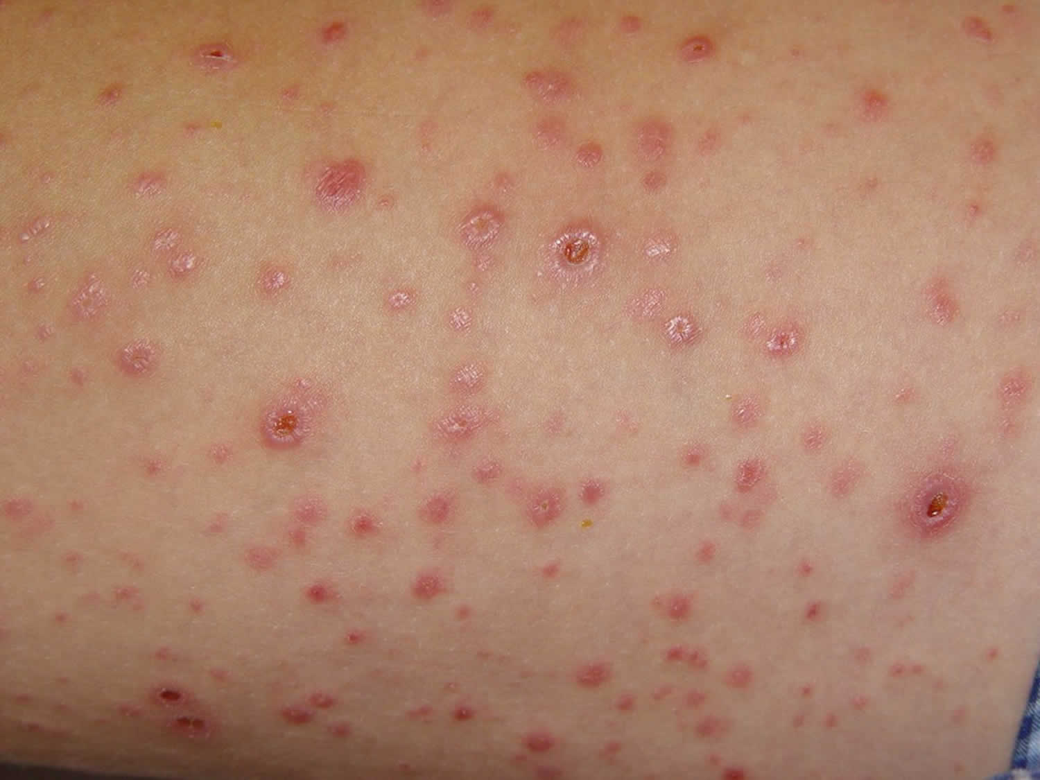
Pityriasis lichenoides et varioliformis acuta
In pityriasis lichenoides et varioliformis acuta (PLEVA) initially there may be a mild illness with a fever. The rash starts as small pink spots, which then form a little blister 5-15 mm in diameter. The center of the papules often becomes filled with pus and blood or eroded with the overlying red-brown crust. A crust (fine mica-like adherent scale) forms on the surface and drops off to leave a small scar which usually fades to some extent over several months. The spots come up in groups and so the rash consists of spots at various stages of development. The rash can often be mistaken for chickenpox but takes much longer to clear than chickenpox. It rarely affects the face, but the spots are usually scattered on the trunk, arms and legs.
Pityriasis lichenoides et varioliformis acuta (PLEVA) most often occurs on the trunk and extremities but sometimes may also be diffuse and widespread, covering any part of the body. Unlike pityriasis lichenoides chronica, which has no apparent symptoms, patients with pityriasis lichenoides et varioliformis acuta experience burning and itchiness.
A subtype of pityriasis lichenoides et varioliformis acuta (PLEVA) is febrile ulceronecrotic Mucha-Habermann disease, in which black or necrotic papules rapidly develop into large coalescent crusted ulcers, blood-filled blisters, and pustules. Mucha-Habermann lesions are usually very painful. The ulcers may become infected. Systemic symptoms may include high fever, sore throat, diarrhoea, abdominal pain, central nervous system symptoms, lung disease, enlarged spleen, arthritis, sepsis, anaemia, and conjunctival ulcers. In some cases, Mucha-Habermann disease can lead to death.
Pityriasis lichenoides diagnosis
The appearance of the rash will suggest the diagnosis; however, pityriasis lichenoides et varioliformis acuta (PLEVA) can often look like chickenpox but take much longer to clear and pityriasis lichenoides chronica can look like psoriasis (red, flaky, crusty patch of skin covered with silvery scales). The examination under the microscope of a small sample of the rash (a skin biopsy) can confirm the diagnosis.
Pityriasis lichenoides treatment
Pityriasis lichenoides may not always respond to treatment and relapses often occur when treatment is discontinued. If the rash is not causing symptoms, treatment may not be necessary. Extensive ulcerations found in febrile ulceronecrotic Muchas-Habermann disease require local wound care.
In cases where treatment is necessary, there are several different therapies available.
Current recommended first-line therapies include:
- Sun exposure may help to resolve lesions, but sunburn should be avoided.
- Topical steroids to reduce irritation. In more recent years concerns raised about their side effect profile has led to the increased use of nonsteroidal topical immunomodulators.
- Topical immunomodulators such as tacrolimus or pimecrolimus. Tacrolimus ointment applied twice daily has been used successfully to treat patients with pityriasis lichenoides chronica.
- Oral antibiotics. The most common are erythromycin and tetracyclines such as doxycycline. These antibiotics have been used to treat both pityriasis lichenoides chronica and pityriasis lichenoides et varioliformis acuta.
Second-line therapies include:
- Phototherapy – artificial ultraviolet radiation treatment with UVB or PUVA has been used with varying success both in patients with pityriasis lichenoides et varioliformis acuta and in those with pityriasis lichenoides chronica.
Third-line therapies include:
- Systemic steroids
- Methotrexate given orally or by IM injection has been used in pityriasis lichenoides chronica and pityriasis lichenoides et varioliformis acuta. It is often used to treat febrile ulceronecrotic Muchas-Habermann disease
- Acitretin
- Dapsone
- Ciclosporin
- For more resistant and severe disease a combination of the above may be used
Pityriasis lichenoides may persist for some years but is generally relatively harmless, although there have been rare reports of malignant transformation. Because of this, regular follow-up is recommended.
Pityriasis lichenoides prognosis
No clear consensus has been formed regarding duration of the disease, but most cases tend to resolve over time. Patients must be told that lesions may take time to resolve and that the duration of the disease cannot be predicted. The skin-limited form of pityriasis lichenoides is a self-limited disease.
A case series of 22 children revealed a mean duration in pityriasis lichenoides et varioliformis acuta (PLEVA) of 1.6 months to complete resolution and a mean duration in pityriasis lichenoides chronica of 7.5 months. The natural tendency of the disease is to remit spontaneously, but some cases may wax and wane over years. Disease duration may be longer in adults. A rare severe variant of pityriasis lichenoides et varioliformis acuta (PLEVA) presents with a sudden eruption of diffuse coalescent necrotic ulcerations associated with high fever 3. Ulceronecrotic pityriasis lichenoides et varioliformis acuta (PLEVA) can lead to scarring. Patients may develop complications such as interstitial pneumonitis, abdominal pain, malabsorption, central nervous system involvement, bacteremia, sepsis, and rheumatic manifestations. T-cell receptor clonal rearrangements of lymphocytic infiltrates have been detected in patients with pityriasis lichenoides et varioliformis acuta (PLEVA). Occasional cases (< 2%) have been reported to evolve into cutaneous lymphoma, although some reports may have represented misdiagnosis of lymphomatoid papulosis 4.
The febrile-ulceronecrotic variant may arise de novo or from a preexisting case of pityriasis lichenoides. Rare reports of death from the febrile-ulceronecrotic variant have been attributed to secondary pulmonary thromboembolism, pneumonia, cardiac arrest, and sepsis, among others 5.
References- Pityriasis lichenoides. https://emedicine.medscape.com/article/1099078-overview
- Pityriasis lichenoides. https://www.dermnetnz.org/topics/pityriasis-lichenoides/
- Smith JJ, Oliver GF. Febrile ulceronecrotic Mucha-Habermann disease associated with herpes simplex virus type 2. J Am Acad Dermatol. 2009 Jan. 60(1):149-52.
- Panizzon RG, Speich R, Dazzi H. Atypical manifestations of pityriasis lichenoides chronica: development into paraneoplasia and non-Hodgkin lymphomas of the skin. Dermatology. 1992. 184(1):65-9.
- Geller L, Antonov NK, Lauren CT, Morel KD, Garzon MC. Pityriasis Lichenoides in Childhood: Review of Clinical Presentation and Treatment Options. Pediatr Dermatol. 2015 Sep-Oct. 32 (5):579-92.


