What is vernix caseosa
Vernix caseosa is a white, creamy, naturally occurring biofilm covering the skin of the fetus during the last trimester of pregnancy 1. Vernix caseosa is a naturally occurring, complex, lipid-rich substance covering the skin surface of the fetus in the last trimester of pregnancy, produced in part by fetal sebaceous glands 2.
Vernix caseosa coating on the neonatal skin protects the newborn skin and facilitates extra-uterine adaptation of skin in the first postnatal week if not washed away after birth. Vernix caseosa consists of water-containing corneocytes embedded in a lipid matrix. The strategic location of the vernix on the fetal skin surface suggests participation in multiple overlapping functions required at birth, such as barrier to water loss, temperature regulation, and innate immunity 1. Vernix seems to perform various integral roles during transition of the fetus from intra-uterine to extra-uterine life. It has also found various interesting diagnostic and prognostic implications in this arena.
Vernix caseosa biology
According to present knowledge, vernix production is unique to humans. Inherent in the understanding of biology of vernix is a fundamental understanding of the epidermal barrier formation in utero. As early as 3 weeks of gestation, presumptive epidermis consisting of a single layer of cuboidal cells develops from the embryonic ectoderm4 and by the 11th week, the epidermis has three distinctive layers: basal, intermediate, and superficial (periderm) 3.
Beneath the protective cover periderm, the epidermis stratifies and differentiates, forming the four distinctive layers of the epidermis by the end of the 4th month of gestation. Periderm provides a temporary barrier suitable for aqueous environment in utero with active transport mechanism between the amniotic fluid and embryo by virtue of its microvilli at its apical surface. Durable cytoskeletal framework of keratin macro fibrils and cornified envelop is formed to provide mechanical strength analogous to ‘bricks and mortars’ 4.
Periderm cells are replaced continuously until 21 weeks when it is completely shed and replaced by the stratum corneum 5. The shed periderm cells are mixed with sebum secretions from the sebaceous glands within the epithelial walls. It is within this combination that vernix caseosa formation occurs 6.
An endocrine-based mechanism for vernix production has been proposed 7.
Hypophyseal- pituitary-adrenal axis regulates sebaceous gland activity of the fetus in utero and subsequently results in production of the superficial lipid film (sebum), first at the vicinity of pilosebaceous unit 8. This changes the transepidermal water gradient and facilitates cornification of the underlying epidermis. Enzymes required in the process, hydroxyl steroid dehydrogenases and 5 alpha-reductase are present after 16 weeks.
The glands reach a peak of activity in the third trimester and their secretion together with desquamated corneocytes into the overlying lipid matrix results in the formation of true vernix.
The development of vernix progresses in a cephalocaudal manner and is the result of an orderly progression of epithelial maturation 9. It coats the fetus until birth.
Late in the second trimester and particularly in the third trimester, fetal lung maturity parallels with sebaceous gland peak activity and increased physiological concentrations of pulmonary surfactant emulsifies surface vernix 10. There is ‘roll up’ and detachment of vernix and consequent increase in amniotic fluid turbidity 11. It is suggested that the coating spreading and detachment of vernix is facilitated by the thermal temperature in utero 11.
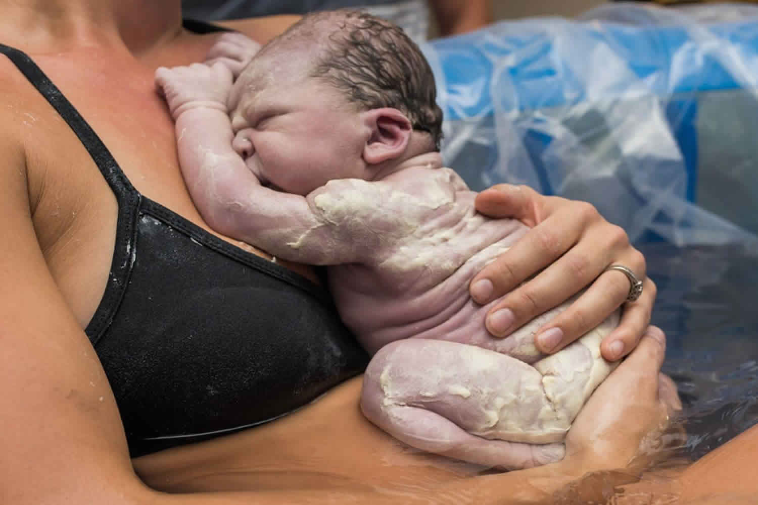
At birth, vernix may cover the entire skin surface or only confined to body folds 12. Its color may reflect intra-uterine problems such as hemolytic disease of newborn, post maturity, where it is of golden yellow color. Fetal distress in utero may stain vernix by bile pigments present in meconium.
Very low birth weight infants, i.e., <28 weeks’ gestation and < 1000g have very immature and incompetent stratum corneum 13 and also lack the protective mantle of vernix caseosa. Anecdotal reports indicate that the amount and distribution of vernix on infant at birth are highly variable. Akiba studies 14 suggest no gender or season effect on vernix coverage and the coverage to be inversely related to birth weight, with maximum for infants fewer than 2000g. Visscher et al 15, showed that the vernix coverage was higher for lower gestational age, C-section infants, females, and Caucasian infants and lower following meconium exposure. Coverage was significantly higher on the back than chest, indicating regional differences. Percentage of infants with coverage over entire body surface in these two studies has also been observed to be highly variable.
Vernix caseosa structure and composition
Vernix consists of water (81%), lipid (9%), and proteins (10%) 16.
Vernix exhibits a non-lamellar lipid matrix containing hydrated corneocytes with no intercorneal desmosomal connections, in contrast to adult stratum corneum, which contains mature corneocytes and lamellar lipid matrix 17.
Thus, the vernix structure exhibits “pasta and cheese” morphology with a “mobile” architecture.
Specific material composition reflects cholesterol esters and wax esters, ceramides derived from stratum corneum and sebaceous origin squalene, cholesterol, triglycerides, free fatty acids, phospholipids, and cellular elements 18.
Non-polar lipids such as sterol esters and triglycerides predominate among free lipids, having a chain length of up to 32 carbon atoms. The profile of fatty acids [omega]-hydroxyacids and [omega]-hydroxyceramides, representing the bound lipids of vernix shows high similarity to that of stratum corneum; however, vernix lipids show lower degree of ordering as compared to stratum corneum 19.
Though approximately 80% of vernix is water, it still has high viscosity, suggesting that its water must reside within a highly structured state which hypothetically is conferred by the abundance of water-filled fetal corneocytes in vernix. These fetal corneocytes in utero act as “cellular sponges” to facilitate and maintain cornification by interdicting water moving across the fetal skin, whereas sebaceous lipids in vernix provide a hydrophobic barrier 20.
Ultra-structural studies show hydrated corneocytes devoid of nuclei and other organelles with sparse network of keratin filaments, about 1-2 micrometers in thickness, lacking desmosomal connections, and surrounded by a thick layer of amorphous lipid without lamellae. Intercellular lipid contains unidentified inclusion bodies, presumably proteinaceous material of keratinocyte origin or sebocyte debris 20. Cells in various stages of keratinization may be seen with nuclear remnants 21.
Histochemistry shows variable acid phosphatase activity intracellularly or in amorphous lipid matrix. Immunofluorescent staining tests of frozen vernix caseosa smears show that only immunoglobulin G conjugate gives strong positive reaction at antigen sites of vernix caseosa cells 21.
Earlier studies had shown that vernix possesses antimicrobial polypeptides. Recently, Maria Tollin et al 22 conducted proteome analysis of vernix caseosa and proved the presence of potent antimicrobial polypeptides. A total of 41 proteins, of which 25 are novel to vernix, have been detected. Effectively, 39% of identified vernix proteins are components of innate immunity and 29% have direct antimicrobial properties.
Origin of vernix proteins seem to be multiple-amniotic fluid, fetal lungs, blood contamination, dermal origins, activated keratinocytes 22.
Vernix caseosa functions
The vernix within amniotic fluid when swallowed by fetus has potential effects on the developing gut. Glutamine being >20% of amino acid content of vernix is a known trophic factor for the developing gut and is generally required by rapidly proliferating cells such as intestinal epithelium and lymphocytes. Asparginase is also found in abundance, forming >30% of amino acid content 23.
Vernix performs an epidermal barrier function in utero to facilitate epidermal growth underneath it and acts as a hydrophobic barrier against amniotic fluid maceration and loss of fluids and electrolytes or trans epidermal water loss.
Vernix also acts as a protective biofilm by minimizing friction of fetal parts during delivery and as an antimicrobial cover against the bacteriologically rich environment of the mother’s genital tract along with the insulating effect on the fetus 24.
Thermal regulation at birth
Inspite of modern methods of nursing and incubation, the temperature control during the first few hours of life in very low birth weight pre-term infants remains a problematic area in the field of neonatology.
Since these infants have incompetent stratum corneum and high trans epidermal water loss, it has been suggested by some authors 25 that the hydrophobic layer of vernix is to be retained after birth and allowed to separate its natural way, which usually occurs by about the 5th day, except in folds of the body where it takes 5 more days to separate. This has shown considerable reduction in the number of cases of subnormal temperature.
However, there is considerable debate on whether vernix has an effect on body temperature regulation and on whether it is to be retained or not. Shulak speculated that vernix could provide thermal stability, but is not a primary factor 26. Vernix removal has also been linked to decreased evaporative loss 27. The latest study on this issue by Visscher et al 28 has showed no significant effect on thermal regulation by vernix retention. This aspect of function of vernix warrants further studies.
Retention of vernix in skin care of newborn infant
Traditional practice has directed nurses to wipe vernix caseosa from wet skin of the newborn as part of initial care in the birthing center. As thermoregulation and resuscitation are the priorities of care, wiping of skin was considered the preferred method for accomplishing drying and stimulation of respiratory effort. As the movement toward evidence-based practice has become a major practice effort, the practice and procedure for this nursing activity have fallen under scrutiny 29. A multiple-site national study was conducted by the National Association of Neonatal Nursing and the Association of Women’s Health Obstetrical and Neonatal Nursing in 1998 30. A consensus statement based on the results of the study directed “removal of all vernix is not necessary for hygienic reasons” and “vernix may provide antibacterial promotion and wound healing”. Interestingly, the World Health Organization (WHO) also recommends leaving vernix intact on the skin surface after birth 31.
Skin surface adaptation after birth
Newborn infants undergo a progressive adaptation immediately after birth, including a slow reduction in surface hydration, decrease in skin pH, and stratum corneum dehydration/ desquamation with formation of a dry skin surface. Moreover, there have been regional variations noted in these parameters 32.
Vernix may have a role in modulating these processes as evidenced by some studies.
Vernix loses its exogenous water slowly 20.
Vernix retention after birth results in significantly more hydrated skin surface with higher moisture accumulation rate and higher baseline hydration. This may facilitate postnatal skin hydration 28.
pH decrease following birth has been attributed to maturation of enzymes responsible for the synthesis of acidic components 32 and triglycerides in vernix could be a source of acidic fatty acids, provided conditions for hydrolysis are present 33.
Visccher et al 28 suggest that skin surface acidification appears to occur earlier in the presence of vernix retention.
An acidic stratum corneum is believed to inhibit the growth of pathogenic bacteria 34 and facilitate colonization with commensal organisms on the skin surface 35.
Antioxidant properties
Vernix is said to have antioxidant properties by virtue of the presence of antioxidants vitamin-E and melanin in it 36.
As birth marks a time of high oxidative stress, the antioxidant properties of vernix may help in coping with the pro-oxidant environment as suggested by a decrease in vitamin-E levels in vernix on exposure to ultraviolet light (pro-oxidative stressor) 37.
Anti-infective property
Earlier reports described mechanical barrier properties of vernix with respect to bacterial invasion 38. Vernix has also been shown to effectively block penetration of exogenous chymotrypsin present in the amniotic fluid from meconium contamination, while itself being devoid of alpha-chymotryptic activity, while retaining endogenous(epidermal) chymotrypsin.51 Recent studies have shown that vernix, like the epidermis, contains antimicrobial peptides and has a direct role in defense against bacteria 39.
Some identified proteins with antimicrobial properties are 22:
- [Alpha]- Defensins [human neutrophil peptide (1-3)]
- Cathelicidins (LL-37)
- Psoriasin
- Ubiquitin
- Palate lung nasal epithelial clone (PLUNC)
- Neutrophil gelatinase-associated lipocalin (NGAL)
- Ribonuclease-7
- Annexin 1
- Secretory leukocyte protease inhibitors
- Calprotectin (calgranulin A, B)
Vernix is also associated with surfactant-associated protein A and surfactant-associated protein D implicated maintenance of airway bacterial homeostasis and also against intra-uterine infection 40.
Lysozyme and lactoferrin are the other innate immune proteins present in vernix 22.
The broad-spectrum action of many of these proteins, in particular the cathelicidins and defensins, may aid in avoiding the development of resistance in bacterial pathogens 22.
Moisturizing properties
Because of its high water content, vernix acts as an agent to moisturize the stratum corneum. Comparison with various barrier creams like petrolatum, aquaphor, and eucerin, shows vernix to be having higher water content 7.
Along with providing “water-proofing” to fetus in utero, it has also been found that application of vernix to adult volar forearm results in an increased capacity to bind exogenous water 41.
Vernix contains filament aggregating protein, which when broken down forms water-binding molecules referred to as Natural Moisturizing Factor (NMF), which operates to maintain suppleness and plasticity of stratum corneum 42.
Recent studies have focused on methods to assist with epidermal barrier and evaluating the role of topical emollients in the prevention of infection in pre-term infants 43.
However, the emollients lack the active antibacterial properties and the structural barrier-enhancing properties of vernix.
It will not be very long in the future when vernix caseosa may be effectively used as a natural emollient, with all its naturally endowed properties.
Wound healing properties
Vernix has shown to increase skin metabolism in vitro by increasing glucose consumption and lactate production 44. The regulation of transepidermal water gradient is known to be important in the epidermal barrier formation and regeneration following wounding 45; and so also the effects of its trophic effects of increased glutamine content. These factors may account for its healing properties in treating adult patients with trophic ulcers of lower extremities 46 and perineal wounds following delivery 7. It may also hence be used in atopic dermatitis against bacterial skin infections 47.
Given the innate properties of vernix for the neonate-like waterproofing, barrier function, hydration, anti-infective, and antioxidant properties, has benefits for burn patients, who have analogous deficits in skin burn trauma-dehydration and hypovolemia, impaired skin integrity, increased anerobic metabolism, and oxygen-free radicals 24.
The placement of superficial layer of vernix over laboratory-cultured skin surfaces is currently under investigation, which can then be applied for grafting burn areas 24.
Skin cleansing properties
In experiments performed using human skin soiled with carbon particles, vernix had comparable efficacy to standard commercial skin cleansers 48. And unlike commercial soaps, it is capable of providing physiologically relevant lipids to the skin surface with additional moisturization, antioxidation, and infection control, all so important for skin surface integrity.
Diagnostic and Prognostic Implications of Vernix Caseosa
Identifying elements of vernix caseosa in pulmonary arterial blood using modified version of the procedure originally described by Masson et al 49 provides a rapid method of diagnosis of the amniotic fluid embolism following delivery.
A strong association between presence of vernix and mature LSR (Lecithin: Sphingomyelin Ratio) in amniotic fluid (>2) has been seen using an amnioscope. This suggests that amnioscopy may be used to assess fetal maturity before induction of labor, a less invasive procedure than amniocentesis 50.
Analysis of vernix in the amniotic fluid has been evaluated as a prognostic index of weight of the newborn 51 and prognostic of maturity status of the fetus 52.
Vernix caseosa has been used as an alternative to other biological specimens for the determination of fetal cocaine exposure due to its presence in all newborns, ease of collection and storage, and provision of historical record of drug exposure 53.
Vernix caseosa observed in urine of the parturient, vernixuria, has been reported as an additional sign of uterine rupture 54.
Vernix caseosa granuloma 55 and vernix caseosa peritonitis 56 have been reported as rare complications of cesarean section, suggesting that in cases in which there is a copious amount of vernix on infants at birth, care should be taken to meticulously irrigate and clear the peritoneal cavity of all debris.
It has been proposed that pregnancy protects against breast cancer, in part, because it results in excretion of lipophilic carcinogens by the mother through fetal fat and vernix caseosa 57.
The physical properties of vernix hypothetically contribute to the panoply of sensory cues, which attract caregivers to the skin of the newborn; and the possibility of pheromones being a part of vernix is open to investigation 58.
Vernix caseosa has been implicated in the causation of neonatal aspiration syndrome 59 and vernix caseo-granulomatous meningitis 60. Hence, pregnant women with a diffuse pattern of high-level echoes in prenatal ultrasonography, suggesting the presence of massive vernix caseosa, should be shifted to a well-equipped institution for delivery.
References- Singh G, Archana G. Unraveling the mystery of vernix caseosa. Indian J Dermatol. 2008;53(2):54–60. doi:10.4103/0019-5154.41645 https://www.ncbi.nlm.nih.gov/pmc/articles/PMC2763724
- Hoath SB, Narendran V, Visscher M. Role and biology of vernix. Neonatal Infant Nurs Rev. 2001;1:53–8
- Moore KL. Philadelphia, Penna: Saunders; 1988. The developing human: Clinically oriented embryology
- Nemes Z, Steinert PM. Bricks and mortar of the epidermal barrier. Exp Mol Med. 1999;31:5–19
- Snell RS. Clinical embryology for medical students. Boston: Little; 1983
- Pansky B. Medical embryology. New York: Macmillin; 1982
- Narendran V. Hoath SB. The Biology of Vernix Caseosa. J Neonatol. 2002;16:9–17
- Hardman MJ, Sisi P, Banbury DN, Byrne C. Patterned acquisition of skin barrier function during development. Development. 1998;125:1541–52
- Elias P. The stratum corneum revisited. J Dermatol. 1996;23:756–8
- Holbrook KA. Structural and biochemical organogenesis of skin and cutaneous appendages in the fetus and newborn. In: Polin FW, Fox RA, editors. Fetal and neonatal physiology. Philadelphia: WB Saunders Company; 1998. pp. 729–52
- Narendran V, Wickett RR, Pickens WL, Hoath SB. Interaction between pulmonary surfactant and vernix: A potential mechanism for induction of amniotic fluid turbidity. Pediatr Res. 2000;48:120–4.
- Atherton DJ, Gennery AR, Cant AJ. The neonate. In: Burns T, Breathnach S, Cox N, Griffiths C, editors. Rook’s Text Book of Dermatology. 7th ed
- Rutter N. The immature skin. Eur J Pediatr. 1996;155:S18–20.
- Akiba T. Studies on biological actions of vernix caseosa. Jpn Obstet Gynecol Soc. 1955;2:396–411
- Visscher MO, Narendran V, Pickens WL, LaRuffa AA, Meinzen-Derr J, Allen K, et al. Vernix caseosa in neonatal adaptation. J Perinatol. 2005;25:440–6.
- Hoeger PH, Schreiner V, Klaassen IA, Enzmann CC, Friedrichs K, Bleck O. Epidermal Barrier lipids in human vernix caseosa: Corresponding ceramide pattern in vernix and fetal skin. Br J Dermatol. 2002;146:194–201.
- Warner R, Lilly N. Correlation of water content with ultrastructure of stratum corneum. In: Elsner P, Berardesca E, Maibach HI, editors. Bioengineering of the skin: water and the stratum corneum. Boca Raton, Fla: CRC Press; 1994. pp. 3–12
- Sumida Y, Yakumaru M, Tokitsu Y, et al. Studies on the function of Vernix caseosa: The secrecy of Baby’s skin. Cannes, France: International Federation of the Societies of Cosmetic Chemists 20th International Conference; 1998. pp. 1–7
- Rissmann R, Groenink HW, Weerheim AM, Hoath SB, Ponec M, Bouwstra JA. New insights into ultrastructure, lipid composition and organization of Vernix Caseosa. J Invest Dermatol. 2006;126:1823–33
- Pickens W, Warner R, Boissy R, Sb H. Characterization of human vernix: Water content, morphology and elemental analysis. J Invest Dermatol. 2000;115:875–81
- Agarastos T, Hollweg G, Grussendorf EI, Papaloucas A. Features of vernix caseosa cells. Am J Perinatol. 1988;5:253–9.
- Tollin M, Jagerbrink T, Haraldsson A, Agerberth B, Jornvall H. Proteome analysis of vernix caseosa. Pediatr Res. 2006;20:430–4
- Buchman AL. Glutamine: Is it conditionally required nutrient for human gastrointestinal system? J Am Coll Nutr. 1996;15:199–205
- Haubrich RA. Role of vernix caseosa in the neonate. AACN Clin Issues. 2003;14:457–64
- Saunders C. The Vernix Caseosa and subnormal temperatures in premature infants. Br J Obstet Gynaecol. 1948;55:442–4
- Shaulak B. The antibacterial action of Vernix Caseosa. Harper Hosp Bull. 1963;21:111–7
- Riesenfeld B, Strombery B, Sedin G. The influence of Vernix Caseosa on water transport through semi permeable membranes and the skin of full- term infants. Neonatal Physiological Measurements. 1984:3–6.
- Visscher MO, Narendran V, Pickens WL, LaRuffa AA, Meinzen-Derr J, Allen K, et al. Vernix caseosa in neonatal adaptation. J Perinatol. 2005;25:440–6
- Lott J, Hoath S. Neonatal skin: The ideal nursing interface. J Pediatr Nurs. 1998;13:302–6
- Association of Women’s Health Obstetrical and Neonatal Nurses (AWHONN) and National Association of Neonatal Nursing (NANN) Evidence-based nursing practice-skin care. Washington DC: 2001
- Hoath SB, Narendran V. 50 years ago in The Journal of Pediatrics. J Pediatr. 2004;144:396
- Hoeger PH, Enzmann CC. Skin Physiology of the neonate and young infant: A prospective study of functional skin parameters during early infancy. Pediatr Dermatol. 2002;19:256–62
- Herrmann F, Behrendt H, Karp F. On the acidity of the surface of the scalp and other areas of the skin in children. J Invest Dermatol. 1946;7:215
- Fluhr JW, Kao J, Jain M, Abn SK, Feingold KR, Elias PM. Generation of free fatty acids from phospholipids regulates stratum corneum acidification and integrity. J Invest Dermatol. 2001;117:44–51
- Parra JL, Paye M. EEMCO guidance for the in vivo assessment of skin surface PH. Skin Pharmacol Appl Skin Physiol. 2003;16:188–202
- Youssef W, Hoath S. Surface free energy characterization of Vernix caseosa: Role in waterproofing the newborn infant. Skin Res Technol. 2001;7:1–17
- Thiele JJ, Traber MG, Packer L. Depletion of human stratum corneum Vitamin E: An early and sensitive in vivo marker of UV induced photooxidation. J Invest Dermatol. 1998;110:756–61
- Joglekar VM. Barrier properties of Vernix Caseosa. Arch Dis Child. 1980;55:817–9
- Tollin M, Bergsson G, Kai-Larsen Y, Lengqvist J, Sjövall J, Griffiths W, et al. Vernix Caseosa as a multi component defense system based on polypeptides, lipids and their interactions. Cell Mol Life Sci. 2005;62:2390–9
- Narendran V, Hull W, Akinbi H. Vernix Caseosa contains surfactant proteins: Potential Role in innate immune function in the fetus. Pediatr Res. 2000;47:420A
- Bautista MI, Wickett RR, Vischer MO, Pickens WL, Hoath SB. Characterization of Vernix Caseosa as a natural biofilm: Comparison to standard oil based ointments. Pediatr Dermatol. 2000;17:253–60
- Rawlings AV, Scott IR, Harding CR, Bowser PA. Stratum corneum moisturization at the molecular level. J Invest Dermatol. 1994;103:734–41
- Conner JM, Soll RF, Edward WH. Topical ointment for preventing infection in preterm infants. Cochrane Database Syst Rev. 2004. CD001150
- Barai N. College of Pharmacy. Cincinnati, OH: University of Cincinnati; 2005. Effect of Vernix Caseosa on epidermal barrier development-repair: Implications in wound healing
- Proksch E, Holleran WM, Menon GK, Elias PM, Feingold KR. Barrier function regulates epidermal lipid and DNA synthesis. Br J Dermatol. 1993;128:473–82
- Zhukov B, Neverova E, Nikitin K. A comparative evaluation of the use of Vernix Caseosa and Solcoseryl in treating patients with trophic ulcers of lower extremities. Vestnik Khirurgii Imeni I I Grekova. 1992;148:339–41
- Roos TC, Geuer S, Roos S, Brost H. Recent advances in treatment strategies for Atopic Dermatitis. Drugs. 2004;64:2639–66
- Morailli R, Pickens WL, Vischer MO, Hoath SB. A novel role for Vernix Caseosa as a skin cleanser. Biol Neonate. 2004;87:8–14
- Dolyniuk M, Orfei E, Vania H, Karlman R, Tomich P. Rapid diagnosis of amniotic fluid embolism. Obstet Gynecol. 1983;61:28S
- McLaughlan JM, Chang AM. Lecithin: Sphingomyelin ratio and presence of vernix in amniotic fluid. South Brisbane, Queensland: Department of Obstetrics and Gynaecology, Mater Misericordiae Mothers’ Hospital; 1977. pp. 1544–5
- Varela J, Pettinelli E, Molina R, Naverrete J, Zambrano N, Vera R. Analysis of the vernix in the amniotic fluid as a prognostic index of the weight of the newborn. Rev Chil Obstet Ginecol. 1974;39:49–51
- Sepulveda WH, Araneda H, Avendano A. Ultrasonic detection of fetal vernix in amniotic fluid as an index to fetal maturity. Rev Chil Obstet Ginecol. 1991;56:43–7
- Moore C, Dempsey D, Deitermann D, Lewis D, Leikin J. Fetal cocaine exposure: Analysis of vernix caseosa. J Anal Toxicol. 1996;20:509–11
- O’Grady JP, Prefontaine M, Hoffman DE. Vernixuria: Another sign of uterine rupture. J Perinatol. 2003;23:351–2
- Boothby R, Lammert N, Benrubi GI, Weiss B. Vernix caseosa granuloma: A rare complication of cesarean section. Southern Med J. 1985;78:1395–6.
- Mahmoud A, Silapaswas S, Lin K, Penney D. Vernix caseosa: An unusual cause of post-cesarean section peritonitis. Am Surg. 1997;63:382–5
- Levine RS, Dolin P. Pregnancy and breast cancer: A possible explanation for the negative association. Med Hypotheses. 1992;38:278–83
- Hoath SB, Pickens WL, Visscher MO. The biology of vernix caseosa. Int J Cosmet Sci. 2006;28:319–33
- Nishijima K, Shukunami K, Inoue S, Kotsuji F. Management for neonatal aspiration syndrome caused by vernix caseosa. Fetal Diagn Ther. 2005;20:194–6
- Midha R, Becker LE. Vernix caseo granulomatous meningitis (vernicomyelia) Can J Neurol Sci. 1991;18:63–5.


