What is Wells syndrome
Wells syndrome also called eosinophilic cellulitis, is a rare eosinophilic disorder that primarily affects the skin of unknown cause with fewer than 200 cases published in the literature 1. Affected people typically develop a skin rash that is often preceded by itching or burning skin. Wells syndrome is mainly observed in adults but also in children 2. Wells syndrome presentation usually involves a mildly pruritic or tender cellulitis-like eruption with typical histologic features characterized by edema, flame figures, and a marked infiltrate of eosinophils in the dermis 3. Papular and nodular eruptions at the clinical presentation have also been reported 4. Wells syndrome or eosinophilic cellulitis can recur and may be preceded by a pruritic papular eruption. Wells syndrome is mainly observed in adults but can occur at any age 5. Family cases have been described, but in most cases, Wells syndrome is sporadic.
One study 6 showed the successive occurrence of vasculitis, Wells syndrome, and Sweet syndrome in a patient. This finding suggests that there is an overlap between these diseases. Another report 7 describes a dominant syndrome consisting of eosinophilic cellulitis, mental retardation, and abnormal body habitus in one family.
Wells syndrome causes
The pathogenesis of Wells syndrome is unknown. However, some scientists believe that it may be an autoimmune reaction. In people affected by an autoimmune disorder, the body’s immune system mistakenly attacks it’s own healthy tissues 8. Wells syndrome may be explained as an inappropriate eosinophilic reaction to a wide variety of stimuli due to an abnormal function of eosinophil regulatory systems. Many triggering factors have been proposed: insect bites, drugs, allergic contact dermatitis, an underlying myeloproliferative disorder, and infections (e.g., dermatophytes, viruses, Toxocara canis).
Possible triggers for Wells syndrome include 9:
- Bug bites (i.e. spiders, bees, fleas, ticks, or mites)
- Viral infections
- Parasitic infections
- Leukemia
- Myeloproliferative disorders
- Atopic dermatitis
- Fungal infections
- Certain types of medications
- Churg-Strauss syndrome
Wells syndrome pathophysiology
The pathogenesis of Wells syndrome is obscure. Many triggering factors have been reported including insect bites, viral infections (parvovirus B19, herpes simplex virus, varicella zoster virus, mumps virus), parasitic infections (Ascaris, Toxocara canis, Giardia) 10, bacterial or fungal infections, drugs (antibiotics, non-steroidal anti-inflammatory drugs, thiazide diuretics, anti-TNF, biomedicines) and vaccines. Association of Wells syndrome with other diseases has also been described such as hematologic malignancies [chronic myeloid leukemia 11, chronic lymphocytic leukemia, polycythemia vera, non-Hodgkin lymphoma), malignant tumors, ulcerative colitis 12, eosinophilic granulomatosis with polyangiitis (Churg–Strauss syndrome) 13, hypereosinophilic syndrome 14]. Wells syndrome may be prior, revealing, or concomitant to these diseases. The fortuitous nature of some of these situations cannot be ruled out, but one must remain vigilant in case of prolonged evolution beyond 6 months, persistent eosinophilia and/or systemic manifestations associated with Wells syndrome 15.
The most interesting associations from a pathogenic point of view are certainly those belonging to the spectrum of eosinophilic diseases 16, such as Shulman syndrome, Churg-Strauss syndrome, and hypereosinophilic syndrome. Wells syndrome could be the first clinical manifestation of these diseases 13.
The physiopathogenic links between the triggering factors and the associations mentioned above are not clearly established. Inappropriate activation of a Th2-like T lymphocyte clone, synthesizing IL-5, and other eosinophil-stimulating cytokines in response to various, often unidentified, antigenic stimuli is the commonly accepted assumption.
Wells syndrome symptoms
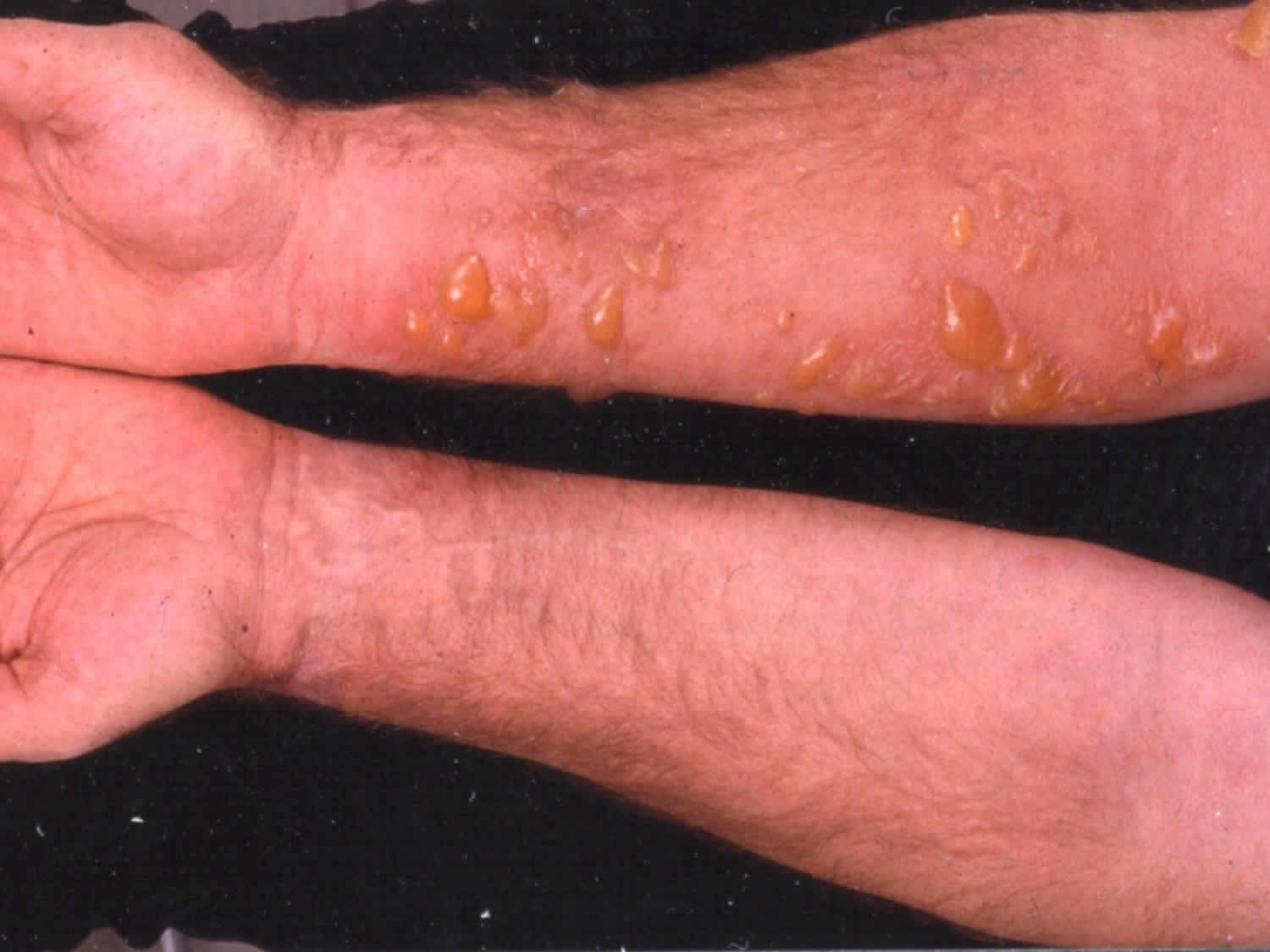
People with Wells syndrome generally develop a skin rash that is often preceded by itching or burning skin. The rash consists of raised, red, swollen areas that may be warm to the touch. Wells syndrome presents with markedly swollen nodules and plaques (lumps) with prominent borders. The patches are usually bright red at first, frequently looking like cellulitis, then fade over four to eight weeks, leaving green, grey or brown patches. They can blister. The rash most commonly occurs on the limbs, but may also affect the trunk. The limbs (arms and legs) are the most commonly affected area of the body. However, the trunk may be involved as well. Symptoms generally come on rapidly and may last for four to eight weeks. In some cases, the rash may recur (occur frequently or repeatedly) for years. Some people with Wells syndrome may experience symptoms that do not affect the skin, such as asthma, joint pain, fever, or fatigue.
The patient often feels very tired and has a fever in approximately 25% of cases.
How is Wells syndrome diagnosed?
Wells syndrome (eosinophilic cellulitis) is not often diagnosed clinically until the results of skin biopsy are available.
The diagnosis is established by the finding of typical histopathological features with many eosinophils and characteristic ‘flame figures’. However, flame figures are not diagnostic of eosinophilic cellulitis and can be seen in other conditions that have increased numbers of eosinophils.
A blood count may also reveal increased numbers of eosinophils – these are commonly associated with allergy or insect bites.
Underlying causes of eosinophilia such as parasitic disorders should be excluded (eg, a worm infestation of the bowel). Allergic contact dermatitis may be considered and excluded by patch testing.
Wells syndrome treatment
The skin symptoms associated with wells syndrome are typically treated with oral or topical corticosteroids such as Prednisone. Oral corticosteroid treatment with prednisone can lead to a dramatic improvement of eosinophilic cellulitis within days. The course is typically tapered over one month.
Other treatments include minocycline, dapsone, griseofulvin, ciclosporin and oral antihistamines.
Mild cases may respond to topical steroid therapy alone.
Therapeutic agents
- Corticosteroids: It is the reference treatment for all reactional dermatoses whether neutrophilic or eosinophilic. General corticotherapy reduces the duration and importance of relapses in 10% of cases 5. Initial doses range from 0.5 to 1 mg/kg/day, with rapid tapering. During the decrease of the treatment, it is not uncommon to observe a relapse of the lesions, which can lead, in certain cases, to a real corticosteroid dependence. Local corticosteroids represent an interesting alternative to general corticosteroids, especially in superficial forms, with inconsistent results of the order of 50% 5. However, it should not be used in deep hypodermic or extended forms.
- Dapsone: Dapsone is an interesting therapeutic alternative to corticosteroids, with doses ranging from 50 to 200 mg daily. It would reduce the duration of relapses. The optimal duration of treatment is not clearly defined but can be several months in the absence of adverse effects requiring discontinuation of treatment. It can be used alone or in combination with other treatments, including corticosteroids. It can also serve as a relay for general corticosteroids, especially in cases of corticosteroid dependence.
- Antihistamines: Antihistamines, especially hydroxyzine if pruritus, is important and may be tried as a first intention, because of their excellent tolerance even if their effectiveness does not seem to exceed 25% of the cases 5. They sometimes allow the practitioner to avoid the use of general steroid treatment. The dosage varies from 50 to 100 mg/day. Hydroxyzine can be combined with other antihistamines.
- Other treatments: These are all anecdotal, with their interest being reported in only a few clinical cases 17. Colchicine, PUVA (psoralen and ultraviolet A) therapy, interferon alpha, cyclins 18, synthetic antimalarials, ciclosporin, and anti-TNF agents can be mentioned 19.
Therapeutic strategy
In most cases, general corticosteroids (10 to 80 mg daily) allow rapid healing. Tapering the dose over one month is generally well tolerated. Continued low-dose therapy with corticosteroids allows preventing recurrences. Dapsone may be prescribed as first-line treatment in low inflammatory forms. It also seems to give good results in case of corticosteroids resistance 20. IFN-alpha and IFN-beta could represent interesting alternatives 21. For mild cases, topical corticosteroids may be sufficient. Finally, the treatment of an associated disease, when it is found, is essential. It must be prescribed as a first-line treatment and can cure Wells syndrome 22.
Wells syndrome prognosis
The prognosis for patients with Wells syndrome (eosinophilic cellulitis) is excellent. It tends to resolve in weeks or months, usually without scarring. It occasionally recurs. In these recurrent cases, it can take years to ultimately resolve.
References- Toumi A, Litaiem N. Eosinophilic (Wells syndrome) [Updated 2019 Jan 25]. In: StatPearls [Internet]. Treasure Island (FL): StatPearls Publishing; 2019 Jan-. Available from: https://www.ncbi.nlm.nih.gov/books/NBK532294
- Caputo R, Marzano AV, Vezzoli P, Lunardon L. Wells syndrome in adults and children: a report of 19 cases. Arch Dermatol. 2006 Sep;142(9):1157-61.
- Brehmer-Andersson E, Kaaman T, Skog E, Frithz A. The histopathogenesis of the flame figure in Wells’ syndrome based on five cases. Acta Derm Venereol. 1986. 66(3):213-9.
- Ghislain PD, Van Eeckhout P. Eosinophilic cellulitis of papulonodular presentation (Wells’ syndrome). J Eur Acad Dermatol Venereol. 2005 Mar. 19(2):226-7.
- Sinno H, Lacroix JP, Lee J, Izadpanah A, Borsuk R, Watters K, Gilardino M. Diagnosis and management of eosinophilic cellulitis (Wells’ syndrome): A case series and literature review. Can J Plast Surg. 2012 Summer;20(2):91-7.
- Consigny S, Courville P, Young P, et al. [Histological and clinical forms of the eosinophilic cellulitis]. Ann Dermatol Venereol. 2001 Mar. 128(3 Pt 1):213-6.
- Davis MD, Brown AC, Blackston RD, et al. Familial eosinophilic cellulitis, dysmorphic habitus, and mental retardation. J Am Acad Dermatol. 1998 Jun. 38(6 Pt 1):919-28.
- Familial Eosinophilic Cellulitis. https://rarediseases.org/rare-diseases/familial-eosinophilic-cellulitis
- Wells syndrome. https://emedicine.medscape.com/article/1124844-overview
- Canonne D, Dubost-Brama A, Segard M, Piette F, Delaporte E. Wells’ syndrome associated with recurrent giardiasis. Br. J. Dermatol. 2000 Aug;143(2):425-7.
- Nakazato S, Fujita Y, Hamade Y, Nemoto-Hasebe I, Sugita J, Nishie W, Shimizu H. Wells’ syndrome associated with chronic myeloid leukaemia. Acta Derm. Venereol. 2013 May;93(3):375-6.
- Utikal J, Peitsch WK, Kemmler N, Booken N, Hildenbrand R, Gladisch R, Goerdt S, Goebeler M. Bullous eosinophilic cellulitis associated with ulcerative colitis: effective treatment with sulfasalazine and glucocorticoids. Br. J. Dermatol. 2007 Apr;156(4):764-6.
- Ratzinger G, Zankl J, Zelger B. Wells syndrome and its relationship to Churg-Strauss syndrome. Int. J. Dermatol. 2013 Aug;52(8):949-54.
- Fujii K, Tanabe H, Kanno Y, Konishi K, Ohgou N. Eosinophilic cellulitis as a cutaneous manifestation of idiopathic hypereosinophilic syndrome. J. Am. Acad. Dermatol. 2003 Dec;49(6):1174-7.
- Powell J, Salim A, Muc R, Colloby P, Kaur MR. Persistent hypereosinophilia with Wells syndrome. Clin. Exp. Dermatol. 2013 Jan;38(1):40-3.
- Delaporte E. [From Wells syndrome to “eosinophilic disease”]. Ann Dermatol Venereol. 2001 Mar;128(3 Pt 1):207-11.
- Chung CL, Cusack CA. Wells syndrome: an enigmatic and therapeutically challenging disease. J Drugs Dermatol. 2006 Oct;5(9):908-11.
- Stam-Westerveld EB, Daenen S, Van der Meer JB, Jonkman MF. Eosinophilic cellulitis (Wells’ syndrome): treatment with minocycline. Acta Derm. Venereol. 1998 Mar;78(2):157.
- Sarin KY, Fiorentino D. Treatment of recalcitrant eosinophilic cellulitis with adalimumab. Arch Dermatol. 2012 Sep;148(9):990-2.
- Aberer W, Konrad K, Wolff K. Wells’ syndrome is a distinctive disease entity and not a histologic diagnosis. J. Am. Acad. Dermatol. 1988 Jan;18(1 Pt 1):105-14.
- Husak R, Goerdt S, Orfanos CE. Interferon alfa treatment of a patient with eosinophilic cellulitis and HIV infection. N. Engl. J. Med. 1997 Aug 28;337(9):641-2.
- Kim HS, Kang MJ, Kim HO, Park YM. Eosinophilic cellulitis in a patient with gastric cancer. Acta Derm. Venereol. 2009 Nov;89(6):644-5.


