Acroosteolysis
Acroosteolysis also called acro-osteolysis or phalangeal osteolysis, refers to the resorption of one or more of the distal phalanges of the hands or feet 1. The terminal tuft is most commonly affected. Two characteristic radiologic variants have been described, and may coexist in the same patient 2. In the transverse type, linear bands of resorption are evident in the shaft of the distal phalanx, perpendicular to its long axis. In the longitudinal type, there is resorption of the tuft of the distal phalanx. When there is linear bone resorption of the midshaft of the distal phalanx with a relatively spared terminal tuft, the condition is also referred to as band acroosteolysis and carries a more limited differential diagnosis. Band acro-osteolysis may occur in exposure to polyvinyl chloride (PVC), renal osteodystrophy (chronic kidney disease) or hyperparathyroidism, idiopathic non-familial acro-osteolysis and Hajdu-Cheney syndrome or familial acro-osteolysis 3. Whereas those with tuft resorption present a pencil-like pattern, also known as longitudinal acroosteolysis (Figure 2) 4.
Acro-osteolysis is associated with a heterogeneous group of pathological entities and, some of which can be remembered by using the mnemonic “PINCH FO“.
The causes of acro-osteolysis can be remembered using the mnemonic “PINCH FO”:
- P: Psoriasis or Pyknodysostosis. Pyknodysostosis is an extremely rare lysosomal storage disease of the bone 5. Pyknodysostosis is characterized by distinctive facial features and skeletal malformations (hypoplastic clavicles, and other craniofacial abnormalities). Affected individuals may have osteosclerosis, a condition characterized by abnormal hardening and increased density of bone. The abnormality of the bones of affected individuals cause the bones to be fragile and brittle. Affected individuals are prone to repeated fractures. Affected individuals may fail to grow and can be shorter than would otherwise be expected (short stature of less than 150 cm). Intelligence is not affected and the disorder is not believed to be life-threatening. The severity of the disorder including the frequency of fractures, final adult height, and specific symptoms can vary greatly among affected individuals. Pycnodysostosis is caused by changes (mutations) in the cathepsin K (CTSK) gene and is inherited in an autosomal recessive trait 6.
- I: Injury, e.g. thermal burn, frost bite
- N: Neuropathy, e.g. diabetes mellitus, leprosy
- C: Collagen vascular disease, e.g. scleroderma, Raynaud disease
- H: Hyperparathyroidism
- F: Familial, e.g. Hajdu-Cheney syndrome. Hajdu–Cheney syndrome is a rare genetic disease that causes acroosteolysis and generalized osteoporosis, accompanied by a series of developmental skeletal disorders and multiple clinical and radiological manifestations. Hajdu–Cheney syndrome has an autosomal dominant inheritance, although there are several sporadic non-hereditary cases. The gene that has been associated with Hajdu-Cheney syndrome is NOTCH2 7. Persons with Hajdu-Cheney syndrome typically have characteristic facies, short stature, and delayed puberty. The upper limbs tend to be affected to a greater degree in this syndrome, with acroosteolysis appearing late in childhood with characteristic transverse lytic defects across the shafts of the phalanges of the upper extremity with relative sparing of the feet. As with pyknodysostosis, there is a high incidence of fractures, but contrary to pyknodysostosis, bones tend to be osteopenic or osteoporotic on radiography 8.
- O: Other, e.g. polyvinyl chloride exposure, progeria
Figure 1. Acroosteolysis

Footnote: Radiograph of the hands showing terminal resorption of the distal phalanges (acroosteolysis) without any erosion of the articular surfaces. The patient was a 74-year-old gentleman with underlying palmoplantar psoriasis. The radiographs of his feet were normal.
[Source 9 ]Figure 2. Acroosteolysis
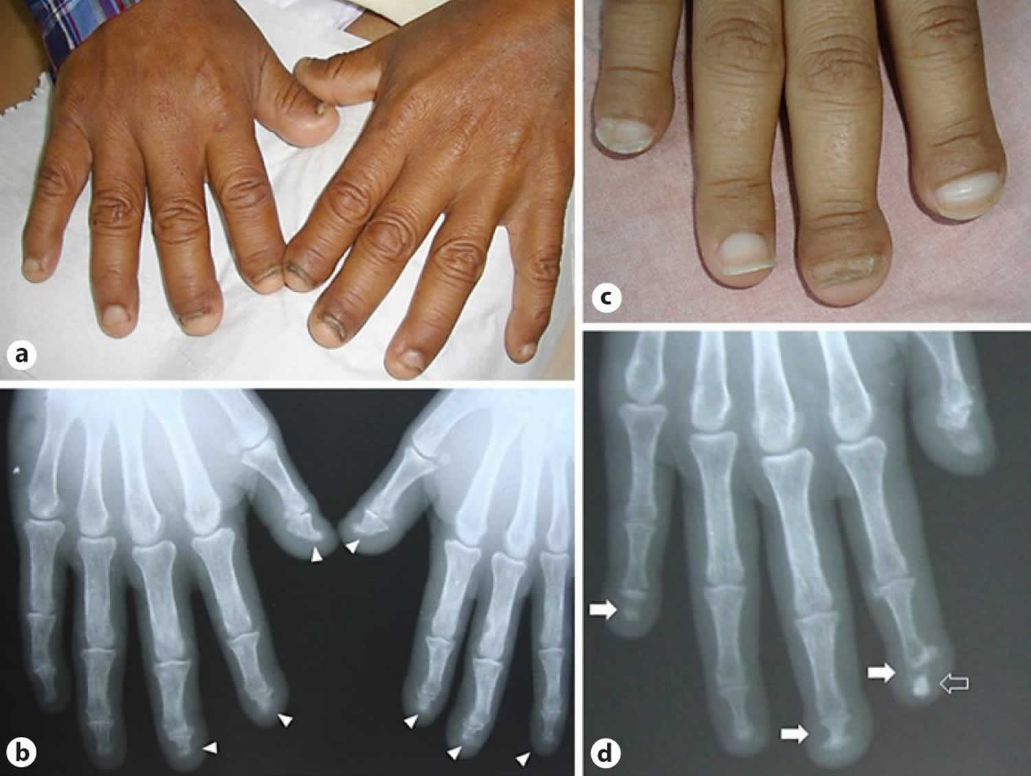
Footnote: Acroosteolysis in a patient with 16 years of chronic kidney disease and 3 years of hemodialysis. Clinical (a) and radiographic (b) images. Longitudinal acroosteolysis (white triangles) (b). Brachyonychia secondary to transverse acroosteolysis (white arrows) in a patient on hemodialysis for 8 years (c, d). Also an osteophyte is seen (white-outline arrow) (d).
[Source 10 ]Figure 3. Foot acroosteolysis

Footnote: Radiographs showing acroosteolysis of distal phalanges 2 through 5 and were without other significant bony abnormalities.
[Source 11 ]Acro-osteolysis causes
Acroosteolysis can be classified as primary or familial, idiopathic disease (unknown cause), immunological disease (systemic sclerosis and psoriasis), infections (leprosy), endocrinological disease (hyperparathyroidism and diabetes mellitus), genetic condition (Hajdu-Cheney syndrome with or without syringomyelia) and lysosomal storage disorder (Gaucher’s disease) and occupational disease (caused by exposure to polyvinyl chloride, frostbite, or trauma) or secondary as a consequence of several conditions (see the list below) 12, 10. The most common causes being inflammatory diseases, sensory neuropathy, vascular disease, metabolic diseases, exposure to vinyl chloride, and trauma (Table 1).
The causes of acro-osteolysis can be remembered using the mnemonic “PINCH FO”:
- P: Psoriasis or Pyknodysostosis. Pyknodysostosis is an extremely rare lysosomal storage disease of the bone 5. Pyknodysostosis is characterized by distinctive facial features and skeletal malformations (hypoplastic clavicles, and other craniofacial abnormalities). Affected individuals may have osteosclerosis, a condition characterized by abnormal hardening and increased density of bone. The abnormality of the bones of affected individuals cause the bones to be fragile and brittle. Affected individuals are prone to repeated fractures. Affected individuals may fail to grow and can be shorter than would otherwise be expected (short stature of less than 150 cm). Intelligence is not affected and the disorder is not believed to be life-threatening. The severity of the disorder including the frequency of fractures, final adult height, and specific symptoms can vary greatly among affected individuals. Pycnodysostosis is caused by changes (mutations) in the cathepsin K (CTSK) gene and is inherited in an autosomal recessive trait 6.
- I: Injury, e.g. thermal burn, frost bite
- N: Neuropathy, e.g. diabetes mellitus, leprosy
- C: Collagen vascular disease, e.g. scleroderma, Raynaud disease
- H: Hyperparathyroidism
- F: Familial, e.g. Hajdu-Cheney syndrome. Hajdu–Cheney syndrome is a rare genetic disease that causes acroosteolysis and generalized osteoporosis, accompanied by a series of developmental skeletal disorders and multiple clinical and radiological manifestations. Hajdu–Cheney syndrome has an autosomal dominant inheritance, although there are several sporadic non-hereditary cases. The gene that has been associated with Hajdu-Cheney syndrome is NOTCH2 7. Persons with Hajdu-Cheney syndrome typically have characteristic facies, short stature, and delayed puberty. The upper limbs tend to be affected to a greater degree in this syndrome, with acroosteolysis appearing late in childhood with characteristic transverse lytic defects across the shafts of the phalanges of the upper extremity with relative sparing of the feet. As with pyknodysostosis, there is a high incidence of fractures, but contrary to pyknodysostosis, bones tend to be osteopenic or osteoporotic on radiography 8.
- O: Other, e.g. polyvinyl chloride exposure, progeria
Primary acro-osteolysis
- Genetic disorders
- Hajdu-Cheney syndrome
- Hereditary sensory neuropathies
- Noggin mutations
- Laminopathies
- Epidermolysis bullosa
- Lysosomal storage disorders
- Gaucher disease
- Pycnodysostosis
Secondary acro-osteolysis
- Vasculopathies
- Raynaud disease/syndrome
- Scleroderma
- Thromboangiitis obliterans (Buerger)
- Inflammatory disorders
- Rheumatoid arthritis
- Psoriatic arthritis
- Dermatomyositis
- Systemic sclerosis
- Granulomatosis with polyangiitis (Wegener)
- Sarcoidosis
- Infections
- Leprosy (sensory neuropathy)
- Metabolic
- Porphyria
- Hyperparathyroidism
- Diabetes mellitus type 1
- Idiopathic
- Toxins
- Vinyl chloride
- Phenytoin
- Alcohol (peripheral neuropathy)
- Trauma
- Repetitive mechanical injury
- Frostbite
Terminal tuft resorption
- Scleroderma 13
- Raynaud disease
- Psoriatic arthritis
- Thermal injury
- extreme cold: frostbite
- extreme heat: burns, electricity
- Trauma
- Hyperparathyroidism
- Epidermolysis bullosa
- Porphyria
- Drugs
- phenytoin (occurs in infants of epileptic mothers treated with phenytoin) 14
- ergot poisoning/abuse
- Insensitivity to pain, e.g. leprosy, congenital insensitivity to pain
- Juvenile chronic arthritis
- Dermatomyositis
- Vascular occlusion 15
- Reactive arthritis 16
- Pityriasis rubra pilaris (very rare skin condition) 17
- Pachydermoperiostosis 18
- Sarcoidosis 19
Midshaft resorption (band acro-osteolysis)
- Polyvinyl chloride (PVC) exposure 20
- Primary acro-osteolysis: Hajdu-Cheney syndrome.
- Hyperparathyroidism (also causes terminal tuft resorption)
- Scleroderma (also causes terminal tuft resorption) 21
- Iiopathic non-familial acro-osteolysis 22
- Pyknodysostosis (also causes terminal tuft hypoplasia) 23
- Biomechanical in guitar players 21
Single digit resorption
Table 1. Mechanism of acquired acroosteolysis in related diseases
| Mechanism | Diseases |
| Vascular | Vascular occlusion
Frostbite |
| Nervous | Diabetic mellitus Leprosy |
| Metabolic | Hyperparathyroidism Diabetes mellitus |
| Inflammatory | Scleroderma Raynaud disease Psoriatic arthritis Juvenile chronic arthritis Epidermolysis bullosa Dermatomyositis |
| Trauma | Mechanical trauma Burns Electricity |
| Drugs | Phenytoin Ergot derivates intoxication Polyvinyl chloride exposure |
| Tumors | Epidermal inclusion cyst Glomus tumor |
There have been several physiopathologic mechanisms proposed for acroosteolysis, which vary in function with the base disease.
- Vascular:
- Occlusion and stenosis leads to bone infarction and resorption. This is secondary to elevated levels of cytokines like vascular endothelial growth factor. Osteoclast formation is also induced by hypoxia 25.
- Vasodilatation increases blood flow (hyperemia), which in turn elevates oxygen partial pressure, favoring modulation and activity of osteoclast, resulting in bone resorption. This has been proposed as the mechanism for cold induced acroosteolysis 26.
- Nervous phenomena:
- Metabolic: Mainly caused by abnormal levels in serum calcium, phosphate, parathyroid hormone (PTH) and active vitamin D 29.
Acro-osteolysis symptoms
Acroosteolysis refers to the resorption of one or more of the distal phalanges of the hands or feet 1. Symptoms associated with acro-osteolysis depends on the underlying causes.
The differential for acroosteolysis includes systemic conditions such as scleroderma, as well as inflammatory arthritidies such as psoriatic and rheumatoid arthritis 30. It can also be associated with vascular, toxic, metabolic, traumatic, and infectious etiologies 31. Acroosteolysis has been described as a component of various rare, genetic syndromes, and can be idiopathic in etiology 8.
The differential for acroosteolysis also includes acquired forms resulting from repetitive mechanical or thermal trauma 32, as well as exposure to various chemical compounds, most notably vinyl chloride 6. Reversible cases of acroosteolysis have been reported in individuals who worked in the polymerization and autoclaving of vinyl chloride 33. Among these patients, Raynaud phenomenon is the most common presenting feature, which typically correlates with vascular narrowing and vasospasm, and may precede bony changes 34.
Acroosteolyis may be a hallmark feature of rare genetic syndromes, including pyknodysostosis, an autosomal-recessive disorder also characterized by short stature of less than 150 cm, generalized diffuse osteosclerosis with a tendency for fracture after minimal trauma, hypoplastic clavicles, and other craniofacial abnormalities.10 Acroosteolysis is also seen as the primary feature in Hejdu-Cheney syndrome, an autosomal-dominant or sporadic familial form of idiopathic acroosteolysis. Persons with this syndrome typically have characteristic facies, short stature, and delayed puberty. The upper limbs tend to be affected to a greater degree in this syndrome, with acroosteolysis appearing late in childhood with characteristic transverse lytic defects across the shafts of the phalanges of the upper extremity with relative sparing of the feet. As with pyknodysostosis, there is a high incidence of fractures, but contrary to pyknodysostosis, bones tend to be osteopenic or osteoporotic on radiography 35.
Acroosteolysis has also been reported as a radiographic finding in several connective tissue disorders and inflammatory arthritidies 30. As many as one-third of patients with systemic sclerosis may show signs of moderate to severe acroosteolysis on radiography and ultrasonograpy 30. Acroosteolysis is a strong predictor of digital ischemia and disease progression in patients with systemic sclerosis 36. Acroosteolysis, and more generally osteolysis, is likewise a common radiographic finding in patients with inflammatory arthritis such as rheumatoid or psoriatic arthritis. In cases of psoriatic arthritis, acroosteolysis may be seen preceding the onset of clinical psoriasis or psoriatic arthropathy and may be the first evidence of disease 37. Findings of osteolysis differ somewhat across these disease processes, much as arthritic symptoms differ in that joint and digit involvement is typically more symmetric in rheumatoid arthritis compared with psoriatic arthritis 38.
Review of the literature has shown rare cases of acroosteolysis in the setting of repetitive mechanical stress and trauma, with 1 case being reported in an avid surfer 31. The absence of prior fractures or stress-related injuries in either dancer despite years of strenuous dance practice or any other typical phenotypic features argues against any of the aforementioned hereditary syndromes. While other systemic conditions or toxic exposures cannot be fully excluded, neither patient exhibited any symptoms characteristic of connective tissue disorders or inflammatory arthritis or reported history of exposure.
Acro-osteolysis diagnosis
A diagnosis of acroosteolysis is based upon identification of characteristic symptoms, a detailed patient, family and occupational history, a thorough clinical evaluation and x-ray studies. X-ray studies can show many of the characteristic bone changes that are associated with acroosteolysis.
Your doctor may suggest blood tests that may be done include:
- PTH blood test
- Calcium blood test
- Alkaline phosphatase
- Phosphorus
- 24-hour urine test
- Random blood sugar test
- Certain antibodies produced by the immune system
Your doctor may also remove a small sample of your affected skin so that it can be examined in the laboratory.
Bone x-rays and bone mineral density (DXA) tests can help detect bone loss, fractures, or bone softening.
X-rays, ultrasound, or CT scans of the kidneys or urinary tract may show calcium deposits or a blockage.
Ultrasound or a nuclear medicine scan of the neck (sestamibi) is used to see if a benign tumor (adenoma) in a parathyroid gland is causing hyperparathyroidism.
Molecular genetic testing can confirm a diagnosis of pycnodysostosis or Hajdu-Cheney syndrome.
Acro-osteolysis treatment
Acro osteolysis treatment involves treating the underlying cause of acroosteolysis.
References- Todd G, Saxe N. Idiopathic Phalangeal Osteolysis. Arch Dermatol. 1994;130(6):759–762. doi:10.1001/archderm.1994.01690060089011
- Allen CM, Claman L, Feldman R. The acro-osteolysis (Hadju-Cheney) syndrome. Review of the literature and report of a case. J Periodontol. 1984 Apr;55(4):224-9. doi: 10.1902/jop.1984.55.4.224. https://doi.org/10.1902/jop.1984.55.4.224
- Uchiyama T. Band acro-osteolysis in a middle-aged woman. BMJ Case Reports CP 2019;12:e229054. http://dx.doi.org/10.1136/bcr-2018-229054
- Graille J, Barry MB, Drapé JL, Doutre MS, Cogrel O. Acro-ostéolyse transversale: une cause rare d’onychopathie. Ann Dermatol Venereol. 2016;143:284–288.
- Sanjay SC, Murthy K, Shukla AK, Krishnappa N. Case Report – Pyknodysostosis. J Clin Diagn Res. 2015;9(5):TD09-TD10. doi:10.7860/JCDR/2015/11656.5894
- Ramaiah KK, George GB, Padiyath S, Sethuraman R, Cherian B. Pyknodysostosis: report of a rare case with review of literature. Imaging Sci Dent. 2011;41(4):177-181. doi:10.5624/isd.2011.41.4.177 https://www.ncbi.nlm.nih.gov/pmc/articles/PMC3251792
- Cortés-Martín J, Díaz-Rodríguez L, Piqueras-Sola B, Rodríguez-Blanque R, Bermejo-Fernández A, Sánchez-García JC. Hajdu-Cheney Syndrome: A Systematic Review of the Literature. Int J Environ Res Public Health. 2020;17(17):6174. Published 2020 Aug 25. doi:10.3390/ijerph17176174 https://www.ncbi.nlm.nih.gov/pmc/articles/PMC7504254
- Sahin A, Pepeler MS, Shimbori N. A patient with acro-osteolysis syndrome: Hajdu-Cheney. Intern Med. 2010;49(1):87-8. https://doi.org/10.2169/internalmedicine.49.2850
- Sakthiswary R, Naicker AS, Htwe O, Shahrir MS, Sazliyana SS. Severe psoriatic acroosteolysis in the absence of psoriatic arthropathy. BMJ Case Rep. 2011;2011:bcr0920114794. Published 2011 Dec 13. doi:10.1136/bcr.09.2011.4794 https://www.ncbi.nlm.nih.gov/pmc/articles/PMC3238111
- Rosales Figueroa JD, Chang P. Brachyonychia Associated with Acroosteolysis in Chronic Kidney Disease: How Phalange Shape Influences Nail Morphology. Skin Appendage Disord. 2018;4(4):264-267. doi:10.1159/000487898 https://www.ncbi.nlm.nih.gov/pmc/articles/PMC6219246
- Miller MN, Close JD. A Unique Incidental Finding in Two Young Dancers: A Case Series. Sports Health. 2015;7(5):421-423. doi:10.1177/1941738115578604 https://www.ncbi.nlm.nih.gov/pmc/articles/PMC4547113
- Botou, A., Bangeas, A., Alexiou, I. et al. Acro-osteolysis. Clin Rheumatol 36, 9–14 (2017). https://doi.org/10.1007/s10067-016-3459-7
- Avouac J, Guerini H, Wipff J, et al. Radiological hand involvement in systemic sclerosis. Ann Rheum Dis. 2006;65(8):1088-1092. doi:10.1136/ard.2005.044602 https://www.ncbi.nlm.nih.gov/pmc/articles/PMC1798258
- Davies S (editor). Chapman & Nakielny’s Aids to Radiological Differential Diagnosis: Expert Consult – Online and Print, 6e. Saunders Ltd. ISBN:0702051764
- O’Brien WT. Top 3 Differentials in Radiology, A Case Review. Thieme Medical Pub. (2009) ISBN:1604062266
- Schneiderman P, Grossman ME. A clinican’s guide to dermatologic differential diagnosis. CRC Press. ISBN:0415390516
- Liu PY, Prete PE. Arthritis associated with pityriasis rubra pilaris. BMJ Case Rep. 2010 Aug 19;2010:bcr1220092565. doi: 10.1136/bcr.12.2009.2565
- Resnick D. Acro-osteolysis in Pachydermoperiostosis. (1981) Archives of Internal Medicine. 141 (10): 1387.
- Nessrine A, Zahra AF, Taoufik H. Musculoskeletal involvement in sarcoidosis. J Bras Pneumol. 2014 Mar-Apr;40(2):175-82. doi: 10.1590/s1806-37132014000200012
- Preston BJ, Jones KL, Grainger RG. Clinical Aspects of Vinyl Chloride Disease. Proceedings of the Royal Society of Medicine. 1976;69(4):284-286. doi:10.1177/003591577606900419
- Destouet JM, Murphy WA. Acquired acroosteolysis and acronecrosis. Arthritis Rheum. 1983 Sep;26(9):1150-4. doi: 10.1002/art.1780260914
- Uchiyama T. Band acro-osteolysis in a middle-aged woman. BMJ Case Rep. 2019;12(3):e229054. Published 2019 Mar 25. doi:10.1136/bcr-2018-229054 https://www.ncbi.nlm.nih.gov/pmc/articles/PMC6453340
- Ramaiah KK, George GB, Padiyath S, Sethuraman R, Cherian B. Pyknodysostosis: report of a rare case with review of literature. Imaging Sci Dent. 2011 Dec;41(4):177-81. doi: 10.5624/isd.2011.41.4.177. Epub 2011 Dec 19.
- Miller TT. Bone tumors and tumorlike conditions: analysis with conventional radiography. Radiology. 2008 Mar;246(3):662-74. doi: 10.1148/radiol.2463061038. Epub 2008 Jan 25
- Park JK, Fava A, Carrino J, Del Grande F, Rosen A, Boin F. Association of Acroosteolysis With Enhanced Osteoclastogenesis and Higher Blood Levels of Vascular Endothelial Growth Factor in Systemic Sclerosis. Arthritis Rheumatol. 2016;68(1):201-209. doi:10.1002/art.39424 https://www.ncbi.nlm.nih.gov/pmc/articles/PMC4690758
- El-Komy MH, Baran R. Acroosteolysis presenting with brachyonychia following exposure to cold. J Eur Acad Dermatol Venereol. 2015;29:2252–2254.
- Baer A, Zahr Z, Khan S, Polydefkis M. Acroosteolysis in diabetes mellitus. J Rheumatol. 2012;39:2364–2365.
- Park J, Fava A, Carrino J, del Grande F, Rosen A, Boin F. Association of acroosteolysis with enhanced osteoclastogenesis and higher blood levels of vascular endothelial growth factor in systemic sclerosis. Arthritis Rheumatol. 2016;68:201–209. https://www.ncbi.nlm.nih.gov/pmc/articles/PMC4690758
- Lim CY, Ong KO. Various musculoskeletal manifestations of chronic renal insufficiency. Clin Radiol. 2013;68:e397–e411.
- Freire V, Bazeli R, Elhai M., et al. Hand and wrist involvement in systemic sclerosis: US features. Radiology. 2013;269:824-830.
- Lehmer LM, Ragsdale BD, Hoffman D, Clark SJ. Surfer’s toe: trauma-induced idiopathic acro-osteolysis in the toes of a 46-year-old surfer: a case report. J Am Podiatr Med Assoc. 2012;102:165-168.
- Lehmer LM, Ragsdale BD, Hoffman D, Clark SJ. Surfer’s toe: trauma-induced idiopathic acro-osteolysis in the toes of a 46-year-old surfer: a case report. J Am Podiatr Med Assoc. 2012;102:165-168
- Harris DK, Adams WG. Acro-osteolysis occurring in men engaged in the polymerization of vinyl chloride. Br Med J. 1967;3:712-714.
- Falappa P, Magnavita N, Bergamaschi A, Colavita N. Angiographic study of digital arteries in workers exposed to vinyl chloride. Br J Ind Med. 1982;39:169-172.
- O’Reilly MA, Shaw DG. Hajdu-Cheney syndrome. Ann Rheum Dis. 1994;53:276-279.
- Johnstone EM, Hutchinson CE, Vail A, Chevance A, Herrick AL. Acro-osteolysis in systemic sclerosis is associated with digital ischaemia and severe calcinosis. Rheumatology. 2012;51:2234-2238.
- Sakthiswary R, Naicker AS, Htwe O, Shahrir MS, Sazliyana SS. Severe psoriatic acroosteolysis in the absence of psoriatic arthropathy. BMJ Case Rep. 2011;2011. doi:10.1136/bcr.09.2011.479
- Ory PA, Gladman DD, Mease PJ. Psoriatic arthritis and imaging. Ann Rheum Dis. 2005;64(suppl 2):ii55-ii57.


