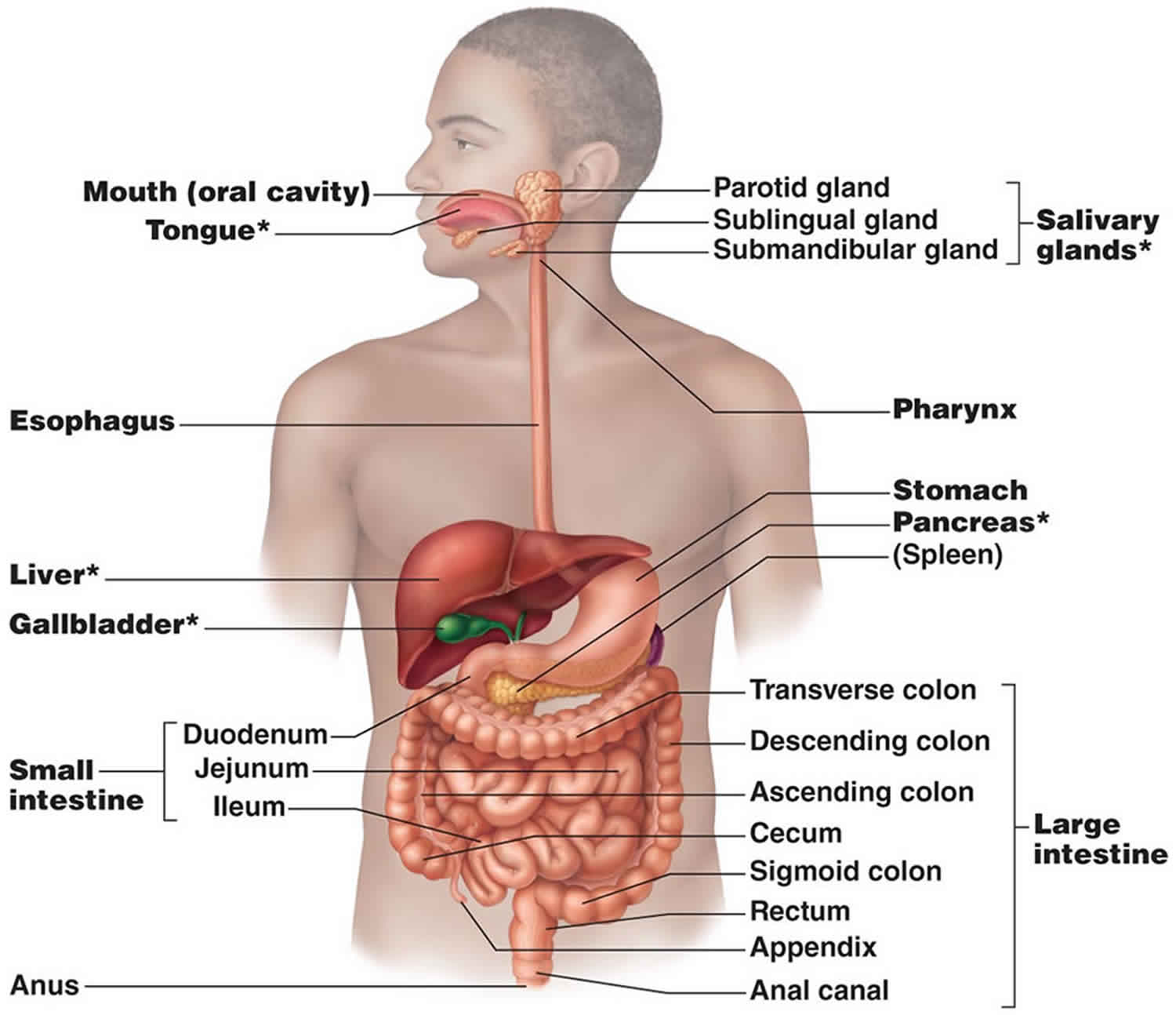
What is alimentary canal
Alimentary canal is also called gastrointestinal (GI) tract, is a continuous tube that extends from your mouth to your anus through the thoracic and abdominopelvic cavities. Organs of the alimentary canal or gastrointestinal tract include the mouth, most of the pharynx, esophagus, stomach, small intestine,
and large intestine. The length of the alimentary canal is about 5–7 meters (16.5–23 ft ) in a living person when the muscles along the wall of the GI tract organs are in a state of tonus (sustained contraction).
The alimentary canal contains food from the time it is eaten until it is digested and absorbed or eliminated. Muscular contractions in the wall of the alimentary canal physically break down the food by churning it and propel the food along the tract, from the esophagus to the anus. The contractions also help to dissolve foods by mixing them with fluids secreted into the tract. Enzymes secreted by accessory digestive organs and cells that line the alimentary canal break down the food chemically.
Figure 1. Alimentary canal
How long is the alimentary canal?
The length of the alimentary canal is about 5–7 meters (16.5–23 ft ) in a living person.
What are the main organs of the alimentary canal?
The main organs of the alimentary canal or gastrointestinal tract include the mouth, most of the pharynx, esophagus, stomach, small intestine,
and large intestine.
Layers of alimentary canal
The walls of the alimentary canal, from the esophagus to the anal canal, have the same four tissue layers (Figure 2). In fact, most such layers occur in the hollow organs of the respiratory, urinary, and reproductive systems as well. From the lumen outward, these layers are the mucosa, submucosa, muscularis externa, and serosa.
Figure 2. Layers of alimentary canal
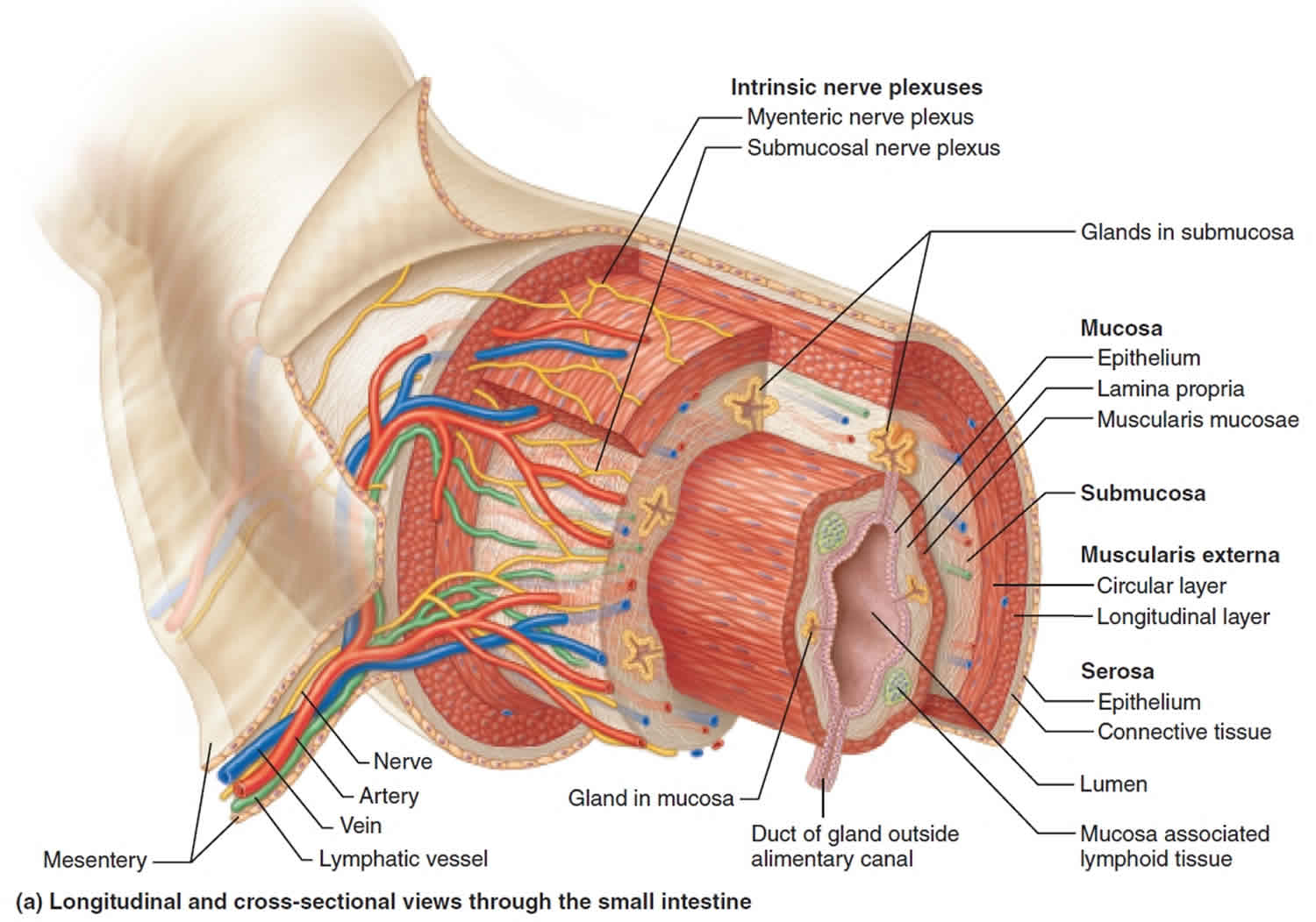
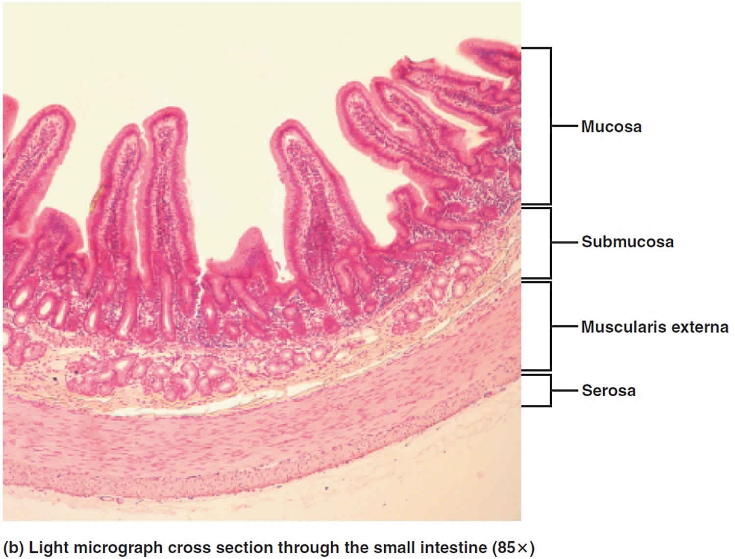
Mucosa
The innermost layer is the mucosa, or mucous membrane. More complex than other mucous membranes in the body, the typical digestive mucosa contains three sublayers: (1) a lining epithelium, (2) a lamina propria, and (3) a muscularis mucosae.
- The lining epithelium abuts the lumen of the alimentary canal and performs many functions related to digestion, such as absorbing nutrients and secreting mucus. This epithelium is continuous with the ducts and secretory cells of the various digestive glands, most of which lie fully within the wall and are called intrinsic glands. The epithelium in the mouth, pharynx, esophagus, and anal canal is mainly nonkeratinized stratified squamous epithelium that
serves a protective function. Simple columnar epithelium, which functions in secretion and absorption, lines the stomach and intestines. The tight junctions that firmly seal neighboring simple columnar epithelial cells to one another restrict leakage between the cells. The rate of renewal of alimentary canal epithelial cells is rapid: Every 5 to 7 days they slough off and are replaced by new cells. Located among the epithelial cells are exocrine cells that secrete mucus and fluid into the lumen of the tract, and several types of endocrine cells, collectively called enteroendocrine cells, which secrete hormones. - The lamina propria is a loose areolar or reticular connective tissue containing many blood and lymphatic vessels, whose capillaries nourish the lining epithelium and are the routes by which nutrients absorbed into the alimentary canal reach the other tissues of the body. The lamina propria contains most of the mucosa associated lymphoid tissue (MALT), which defends against invasion by bacteria and other microorganisms in the alimentary canal. MALT is present all along the alimentary canal, especially in the tonsils, small intestine, appendix, and large intestine. The lamina propria layer supports the epithelium and binds it to the muscularis mucosae.
- External to the lamina propria is the muscularis mucosae, a thin layer of smooth muscle that throws the mucous membrane of the stomach and small intestine into many small folds, which increase the surface area for digestion and absorption. Movements of the muscularis mucosae ensure that all absorptive cells are fully exposed to the contents of the alimentary canal. For example, the twitching of this muscle layer dislodges sharp food particles that become embedded in the mucosa. A thin layer of smooth muscle fibers called the muscularis mucosae.
Submucosa
Just external to the mucosa is the submucosa, a layer of connective tissue containing major blood and lymphatic vessels and nerve fibers. Its rich vascular network sends branches to all other layers of the wall. Its connective tissue is a type intermediate between loose areolar and dense irregular—a “moderately dense” connective tissue. The many elastic fibers in the submucosa enable the alimentary canal to return to its shape after food material passes through it.
Muscularis Externa
External to the submucosa is the muscularis externa, also simply called the muscular layer. Throughout most of the alimentary canal, this tunic consists of two layers of smooth muscle, an inner circular layer whose fibers orient around the circumference of the canal, and an outer longitudinal layer whose fibers orient along the length of the canal. Functionally, the circular layer squeezes the gut tube, and the longitudinal layer shortens it. Together, these layers are responsible for peristalsis and segmentation. In some places, the circular layer thickens to form sphincters that act as valves to prevent the backflow of food from one organ to the next.
Serosa
The serosa, which is the visceral peritoneum, is the outermost layer of the intraperitoneal organs of the alimentary canal. Like all serous membranes, it is formed of a simple squamous epithelium (mesothelium) underlain by a thin layer of areolar connective tissue.
Parts of the alimentary canal that are not associated with the peritoneal cavity lack a serosa and have an adventitia, an ordinary fibrous connective tissue, as their outer layer. For example, the esophagus in the thorax has an adventitia that binds it to surrounding structures. Secondarily retroperitoneal organs have both a serosa and an adventitia—a serosa on the anterior side facing the peritoneal cavity and an adventitia on the posterior side embedded in the posterior abdominal wall.
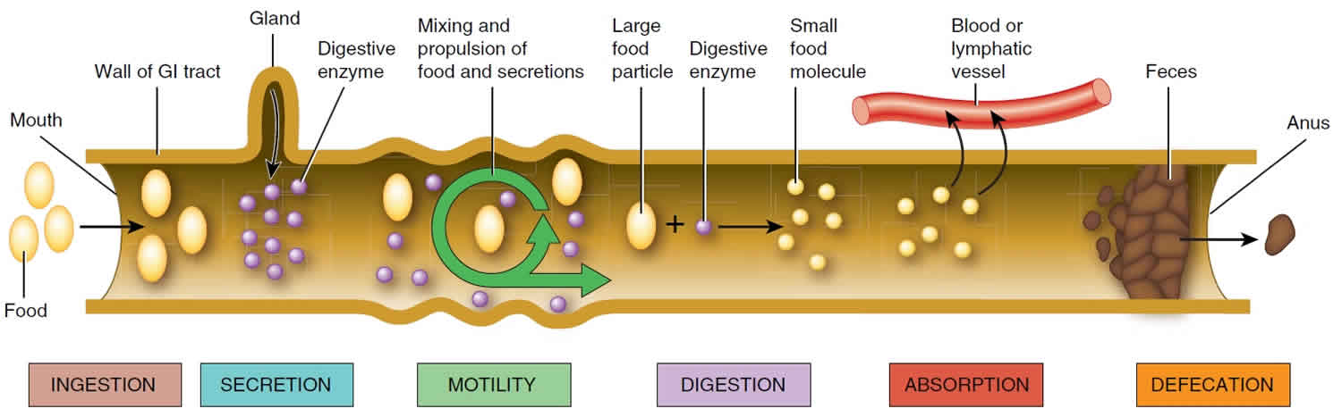
Alimentary canal functions
Overall, the alimentary canal performs six basic processes
- Ingestion. This process involves taking foods and liquids into the mouth (eating).
- Secretion. Each day, cells within the walls of the alimentary canal and accessory digestive organs secrete a total of about 7 liters of water, acid, buffers, and enzymes into the lumen (interior space) of the alimentary canal.
- Motility. Alternating contractions and relaxations of smooth muscle in the walls of the alimentary canal mix food and secretions and move them toward the anus. This capability of the alimentary canal to mix and move material along its length is called motility.
- Digestion. Digestion is the process of breaking down ingested food into small molecules that can be used by body cells. In mechanical digestion the teeth cut and grind food before it is swallowed, and then smooth muscles of the stomach and small intestine churn the food to further assist the process. As a result, food molecules become dissolved and thoroughly mixed with digestive enzymes. In chemical digestion the large carbohydrate, lipid, protein, and nucleic acid molecules in food are split into smaller molecules by hydrolysis. Digestive enzymes produced by the salivary glands, tongue, stomach, pancreas, and small intestine catalyze these catabolic reactions.
- Absorption. The movement of the products of digestion from the lumen of the alimentary canal into blood or lymph is called absorption. Once absorbed, these substances circulate to cells throughout the body. A few substances in food can be absorbed without undergoing digestion. These include vitamins, ions, cholesterol, and water.
- Defecation. Wastes, indigestible substances, bacteria, cells sloughed from the lining of the alimentary canal, and digested materials that were not absorbed in their journey through the digestive tract leave the body through the anus in a process called defecation. The eliminated material is termed feces or stool.
Figure 3. Alimentary canal functions


