close
Health topics

Epstein barr virus

epstein barr virus infection

What is Epstein Barr virus

Epstein-Barr virus, also known as human herpesvirus 4, is a gamma herpes virus that occurs only in humans 1. Epstein-Barr virus is one of the most common human viruses. Epstein Barr virus is found all over the world. According to the Centers for Disease Control and Prevention (CDC), most people in the United States are infected by Epstein Barr virus at some point in their lives 2. Epstein Barr virus is very contagious and spreads most commonly from person to person through bodily fluids, primarily saliva, which is why Epstein-Barr virus infection is sometimes called “kissing disease.” Epstein-Barr virus is present in the saliva of infected individuals and can be spread through close contact such as kissing and through sharing utensils or cups. Epstein Barr virus can cause infectious mononucleosis, also called mono, and other illnesses.

Epstein Barr virus replicates primarily in β-lymphocytes but also may replicate in the epithelial cells of the pharynx and parotid duct 3. The infection is spread primarily by saliva, and the incubation period is four to eight weeks. In an acute infection, heterophile antibodies that agglutinate sheep erythrocytes are produced. This process forms the basis for the Monospot rapid latex agglutination test. Antibodies to viral capsid antigen (i.e., VCA-IgG and VCA-IgM) are produced slightly earlier than the heterophile antibody and are more specific for Epstein Barr virus infection. The VCA-IgG antibody persists past the stage of acute infection and signals the development of immunity 3.

After initial exposure to Epstein Barr virus, there is a period of several weeks before associated symptoms may appear, called the incubation period. During the acute primary infection, the virus multiplies in number. This is followed by a decrease in viral numbers and resolution of symptoms, but Epstein-Barr virus never completely goes away. Latent Epstein Barr virus remains in the person’s body for the rest of that person’s life and may reactivate but usually causes few problems unless the person’s immune system is significantly weakened.

Most people are infected by Epstein Barr virus in childhood and experience few or no symptoms. However, when the initial infection occurs in adolescence, it can cause infectious mononucleosis, commonly called mono, a condition associated with fatigue, fever, sore throat, swollen lymph nodes, an enlarged spleen, and sometimes an enlarged liver. These symptoms occur in about 25% of infected teens and young adults and usually resolve within a month or two.

People with infectious mononucleosis (mono) are typically diagnosed by their symptoms and the findings from a complete blood count (CBC) and a mono test (which tests for a heterophile antibody). About 25% of those with infectious mononucleosis (mono) do not produce heterophile antibodies and will have a negative mono test; this is especially true with children. Tests for Epstein Barr virus antibodies can be used to determine whether or not the symptoms these people are experiencing are due to a current infection with the Epstein Barr virus virus.

Epstein Barr virus is the most common cause of infectious mononucleosis (mono). According to the CDC, examples of other causes of infectious mononucleosis (mono) include cytomegalovirus (CMV), hepatitis A, hepatitis B or hepatitis C, rubella, and toxoplasmosis. Sometimes, it can be important to distinguish Epstein Barr virus from these other illnesses. For instance, it may be important to diagnose the cause of symptoms of a viral illness in a pregnant woman. Testing can help to distinguish a primary Epstein Barr virus infection, which has not been shown to affect a developing baby, from a cytomegalovirus (CMV), herpes simplex virus, or toxoplasmosis infection, as these illnesses can cause complications during the pregnancy and may harm the fetus.

Epstein Barr virus transmission

Epstein Barr virus spreads most commonly through bodily fluids, especially saliva. However, Epstein Barr virus can also spread through blood and semen during sexual contact, blood transfusions, and organ transplantations.

Epstein Barr virus can be spread by using objects, such as a toothbrush or drinking glass, that an infected person recently used. The virus probably survives on an object at least as long as the object remains moist.

The first time you get infected with Epstein Barr virus (primary Epstein Barr virus infection) you can spread the virus for weeks and even before you have symptoms. Once the virus is in your body, it stays there in a latent (inactive) state. If the virus reactivates, you can potentially spread Epstein Barr virus to others no matter how much time has passed since the initial infection.

Health experts aren’t sure how long people with Epstein Barr virus stay contagious after symptoms are gone. They believe that people can spread the infection for many months after that — some studies show as long as 18 months. Then, the Epstein Barr virus stays dormant (inactive) in the body for the rest of a person’s life.

Sometimes the dormant Epstein Barr virus can “wake up” and find its way into a person’s saliva (spit). That person might not feel ill or show any infectious mononucleosis symptoms, but can spread the virus to other people. So there’s a very small chance that people who have had Epstein Barr virus infection in the past can pass it to others, even when they feel OK.

Epstein Barr virus infection

Infectious mononucleosis, or “mono”, is an infection usually caused by the Epstein-Barr virus. Other viruses can also cause infectious mononucleosis are cytomegalovirus (CMV), hepatitis A, hepatitis B or hepatitis C, rubella, and toxoplasmosis. Infectious mononucleosis is usually suspected in patients 10 to 30 years of age who present with sore throat and significant fatigue, palatal petechiae, posterior cervical or auricular adenopathy, marked adenopathy, or inguinal adenopathy 4. Some of the symptoms of infectious mononucleosis are similar to those of a cold or flu and serious problems are rare.

Symptoms of infectious mononucleosis include:

  • Fever
  • Sore throat
  • Swollen lymph glands in the neck and armpits
  • Extreme fatigue
  • Head and body aches
  • Swollen liver or spleen or both
  • Rash

Enlarged spleen and a swollen liver are less common symptoms. For some people, their liver or spleen or both may remain enlarged even after their fatigue ends. Jaundice and splenic rupture (spleen rupture) can occur in patients with infectious mononucleosis, but these complications are rare 5.

A ruptured spleen is rare in people who have mono. But it’s wise to be aware of the signs:

  • sharp pain in the left upper part of your abdomen (under the left chest)
  • feeling lightheaded
  • feeling confused
  • blurred vision
  • fainting.

Other complications can include anemia, nervous system problems, or hepatitis with jaundice. They could cause symptoms including:

  • breathing difficulty
  • persistent high fevers (101.5 °F or higher)
  • severe headache
  • weakness in arms or legs
  • yellow color in you eyes or skin (jaundice)
  • trouble swallowing.

Call your doctor right away if you notice any of the above symptoms.

A blood test can show if you have infectious mononucleosis (mono).

Most people get better in two to four weeks. However, you may feel tired for a few months afterward. Occasionally, the symptoms of infectious mononucleosis can last for six months or longer. Treatment focuses on helping symptoms and includes medicines for pain and fever, warm salt water gargles and plenty of rest and fluids.

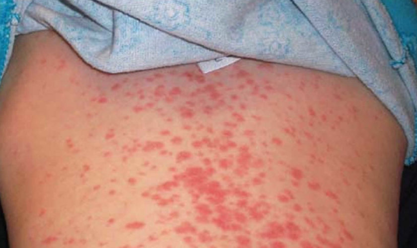
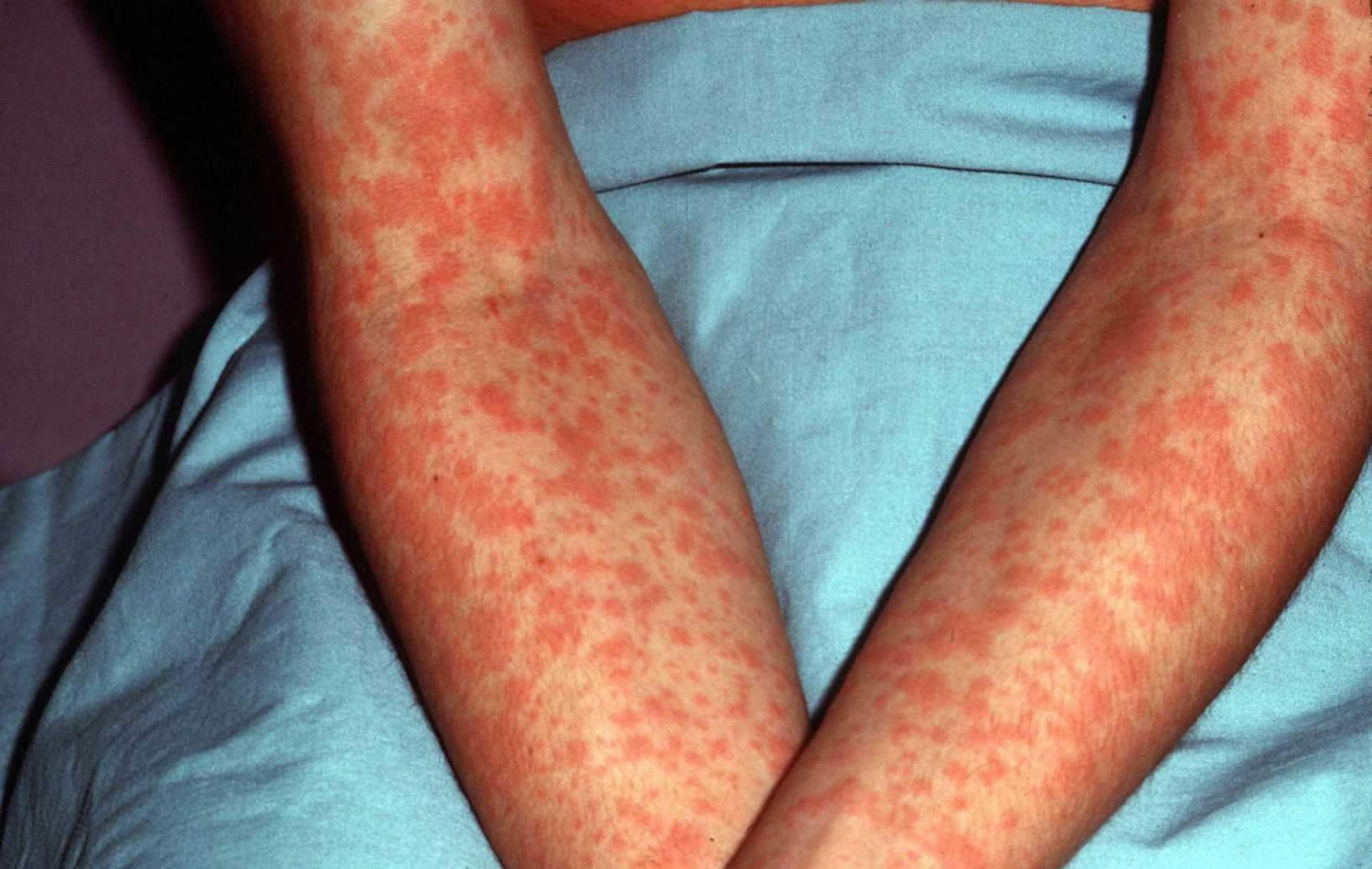
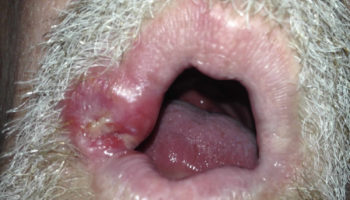
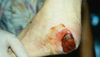
Figure 1. Epstein Barr virus rash

Epstein Barr virus rash

Can mononucleosis recur?

Most people who have infectious mononucleosis, or mono, get it only once. Rarely, however, mononucleosis symptoms may recur months or even years later.

Most cases of mononucleosis are caused by infection with the Epstein-Barr virus (Epstein Barr virus). Once you’re infected with Epstein Barr virus, you carry the virus — usually in a dormant state — for the rest of your life. Periodically, however, the virus may reactivate. When this happens, the virus can be detected in your saliva — but you’re not likely to become ill. Rarely, reactivated Epstein Barr virus may cause illness in people who have weak immune systems, such as those who have AIDS.

Mononucleosis rarely leads to a serious condition called chronic active Epstein Barr virus infection, which is characterized by persistent illness more than six months after the initial mononucleosis diagnosis.

If you’re experiencing signs or symptoms of mononucleosis — such as fatigue, weakness, fever, sore throat and swollen lymph nodes — and you’ve had mono before, consult your doctor to determine the cause of your current symptoms. Keep in mind that many other conditions, such as hepatitis and toxoplasmosis, can mimic the symptoms of mononucleosis.

Epstein Barr virus infection complications

Epstein-Barr virus can cause illnesses and complications aside from infectious mononucleosis. People with weakened immune systems may develop more severe symptoms and complications from Epstein Barr virus infection. They may also have more severe illness caused by Epstein Barr virus infection.

Complications of Epstein Barr virus infection include:

  • Peritonsillar abscesses (pus-filled tissue near the tonsils)
  • Acute bacterial sinusitis (bacterial infection of the sinus cavities)
  • Suppurative lymph nodes (swelling of lymph nodes)
  • Mastoiditis (bacterial infection of the mastoid bone of the skull)
  • Sialadenitis (swelling and injury of salivary glands)
  • Blockage of the air passages in the nose and throat
  • Acute interstitial nephritis
  • Hemolytic anemia
  • Myocarditis and cardiac conduction abnormalities
  • Neurologic abnormalities
  • Cranial nerve palsies
  • Encephalitis
  • Meningitis
  • Mononeuropathies
  • Retrobulbar neuritis
  • Thrombocytopenia
  • Upper airway obstruction

Patients with infectious mononucleosis are likely to have splenomegaly. Although most patients do not have a palpable spleen on physical examination, a study of 29 patients who were hospitalized with infectious mononucleosis (and who therefore may have had more severe disease) found that all patients had splenomegaly on ultrasound examination and that one half of them had hepatomegaly 6. Only 17 percent of the enlarged spleens and 8 percent of the enlarged livers were palpable on physical examination, a finding that is consistent with other studies 7. Because an enlarged spleen is at risk for rupture, athletes should not compete in contact or collision sports for a minimum of three to four weeks after the onset of symptoms 8.

Nervous System

Epstein Barr virus infection can affect a person’s brain, spinal cord, and nerves.

Epstein Barr virus infection can cause conditions such as:

  • Viral meningitis (swelling of the tissues that cover the brain and spinal cord)
  • Encephalitis (swelling of the brain)
  • Optic neuritis (swelling of the eye nerve)
  • Transverse myelitis (swelling of the spinal cord)
  • Facial nerve palsies (paralysis of facial muscles)
  • Guillain-Barré syndrome (an immune system disease)
  • Acute cerebellar ataxia (sudden uncoordinated muscle movement)
  • Hemiplegia (paralysis on one side of the body)
  • Sleep disorders
  • Psychoses

Hematological System

Epstein Barr virus infection can affect a person’s blood and bone marrow. The virus can cause the body to produce an excessive number of white blood cells called lymphocytes (lymphocytosis).

Epstein Barr virus can also weaken the immune system, making it more difficult for the body to fight infection.

Examples of some of these conditions include:

  • Neutropenia with secondary infections
  • Hemophagocytic syndrome (hemophagocytic lymphohistiocytosis)
  • Acquired hypogammaglobulinemia
  • X-linked lymphoproliferative disease

Other Conditions

Epstein Barr virus infection can also cause:

  • Pneumonia (injury of the lungs)
  • Interstitial lung disease (a large group of disorders, most of which cause scarring of lung tissue)
  • Pancreatitis (swelling of the pancreas)
  • Myocarditis (swelling of the heart muscle)
  • Oral cavity-oral hairy leukoplakia (raised, white patches on the tongue), which is usually seen in people infected with HIV

Cancers

Cancers associated with Epstein Barr virus infection include:

  • Burkitt’s lymphoma (cancer of the lymphatic system)
  • Nasopharyngeal carcinoma (cancer of the upper throat)
  • Hodgkin’s disease and non-Hodgkin’s lymphoma (cancers of the lymphatic system)
  • Post-transplant lymphoproliferative disorder (white blood cells are produced in excess)
  • Other tumors including leiomyosarcomas (cancer in the soft tissue) and T-cell lymphomas

Chronic Epstein Barr virus

Several cohort studies have examined the long-term outcomes of infectious mononucleosis. Between 9 and 22 percent of patients reported persistent fatigue or hypersomnia six months after clinical infectious mononucleosis, compared with zero to 6 percent of patients following uncomplicated upper respiratory infection 3. The best study of the natural history of infectious mononucleosis followed 150 patients for six months 9. In this study, sore throat, fever, headache, rash, cough, and nausea largely had resolved one month after the onset of symptoms. Fatigue resolved more slowly (77 percent initially, 28 percent at one month, 21 percent at two months, and 13 percent at six months), as did sleeping too much (45 percent initially, 18 percent at one month, 14 percent at two months, and 9 percent at six months) and sore joints (23 percent initially, 15 percent at one month, 6 percent at two months, and 9 percent at six months) 9. This study also followed functional status over the same six-month period and found that patients required about two months to achieve a stable level of recovery. The association between Epstein Barr virus infection and chronic fatigue syndrome remains uncertain, and a positive IgG test for Epstein Barr virus does not imply a causal relationship. In addition, evidence of Epstein Barr virus infection is not part of the definition of chronic fatigue syndrome or myalgic encephalomyelitis 10.

Infectious mononucleosis prevention and treatment

There is no vaccine to protect against infectious mononucleosis. You can help protect yourself by not kissing or sharing drinks, food, or personal items, like toothbrushes, with people who have infectious mononucleosis.

You can help relieve symptoms of infectious mononucleosis by:

  • drinking fluids to stay hydrated
  • getting plenty of rest
  • taking over-the-counter medications for pain and fever

If you have infectious mononucleosis, you should not take penicillin antibiotics like ampicillin or amoxicillin. Based on the severity of the symptoms, a healthcare provider may recommend treatment of specific organ systems affected by infectious mononucleosis.

Because your spleen may become enlarged as a result of infectious mononucleosis, you should avoid contact sports until you fully recover. Participating in contact sports can be strenuous and may cause the spleen to rupture.

Epstein Barr virus symptoms

Many people become infected with Epstein Barr virus in childhood. Epstein Barr virus infections in children usually do not cause symptoms, or the symptoms are not distinguishable from other mild, brief childhood illnesses. People who get symptoms from Epstein Barr virus infection, usually teenagers or adults, get better in two to four weeks. However, some people may feel fatigued for several weeks or even months.

Symptoms of Epstein Barr virus infection can include:

  • fatigue
  • fever
  • inflamed throat
  • swollen lymph nodes in the neck
  • enlarged spleen
  • swollen liver
  • rash

After you get an Epstein Barr virus infection, the virus becomes latent (inactive) in your body. In some cases, the virus may reactivate. This does not always cause symptoms, but people with weakened immune systems are more likely to develop symptoms if Epstein Barr virus reactivates.

Epstein Barr virus causes

Epstein Barr virus is a herpes virus that replicates primarily in β-lymphocytes but also may replicate in the epithelial cells of the pharynx and parotid duct.7

Epstein Barr virus is spread by saliva through:

  • kissing
  • sharing drinks and food
  • using the same cups, eating utensils, or toothbrushes
  • having contact with toys that children have drooled on

Data collected more than 30 years ago on the incidence of infectious mononucleosis show the highest rates in persons 10 to 19 years of age (six to eight cases per 1,000 persons per year) 11. The incidence in persons younger than 10 years and older than 30 years is less than one case per 1,000 persons per year 11, but mild infections in younger children often may be undiagnosed. The infection is most common in populations with many young adults, such as active-duty military personnel and college students, in whom the annual incidence for infectious mononucleosis ranges from 11 to 48 cases per 1,000 persons 12.

Infectious mononucleosis is relatively uncommon in adults, accounting for less than 2 percent of all adults presenting with sore throat 7. Family physicians should expect to diagnose one to four patients with infectious mononucleosis per year, depending on the number of adolescents in their practice 12. The incidence of infectious mononucleosis shows no consistent seasonal peak 13.

Epstein Barr virus diagnosis

Diagnosing Epstein Barr virus infection can be challenging because the symptoms are similar to other illnesses. Epstein Barr virus infection can be confirmed with a blood test that detects antibodies. About nine out of ten of adults have antibodies that show that they have a current or past Epstein Barr virus infection.

Epstein Barr virus test

Epstein-Barr virus antibody test is a blood test to detect for antibodies to Epstein Barr virus in the blood and help establish a diagnosis of Epstein Barr virus infection. Epstein-Barr virus antibody test can also be important to rule out Epstein Barr virus infection and to look for other causes of the symptoms. Those with strep throat, an infection caused by group A streptococcus, for instance, need to be identified and treated with antibiotics. A person may have strep throat instead of mono or may have both conditions at the same time.

Several tests for different types and classes of Epstein Barr virus antibodies are available. The antibodies are proteins produced by the body in an immune response to several different Epstein-Barr virus antigens. During a primary Epstein Barr virus infection, the level of each of these Epstein Barr virus antibodies rises and falls at various times as the infection progresses. Measurement of these antibodies in the blood can aid in diagnosis and typically provides the healthcare practitioner with information about the stage of infection and whether it is a current, recent, or past infection.

Healthcare practitioners can test for antibodies to the following Epstein Barr virus-associated antigens:

Viral capsid antigen (VCA)

  • Anti-VCA IgM appears early in Epstein Barr virus infection and usually disappears within four to six weeks.
  • Anti-VCA IgG appears in the acute phase of Epstein Barr virus infection, peaks at two to four weeks after onset, declines slightly then persists for the rest of a person’s life.

Early antigen (EA)

  • Anti-EA IgG appears in the acute phase of illness and generally falls to undetectable levels after three to six months. In many people, detection of antibody to EA is a sign of active infection. However, 20% of healthy people may have antibodies against EA for years.

Epstein Barr virus nuclear antigen (EBNA)

Antibody to EBNA, determined by the standard immunofluorescent test, is not seen in the acute phase of Epstein Barr virus infection but slowly appears two to four months after onset of symptoms and persists for the rest of a person’s life. Other EBNA enzyme immunoassays may report false positive results.

Monospot test

The Monospot test is not recommended for general use. The antibodies detected by Monospot can be caused by conditions other than infectious mononucleosis. Moreover, studies have shown that the Monospot produces both false positive and false negative results. For example, the heterophile antibodies detected by Monospot are often not present in children with infectious mononucleosis. At best, the Monospot test may indicate that a person has a typical case of infectious mononucleosis, but does not confirm the presence of Epstein Barr virus infection.

Interpretation of Epstein Barr virus Antibody Tests

Epstein Barr virus antibody tests are not usually needed to diagnose infectious mononucleosis. However, specific antibody tests may be needed to identify the cause of illness in people who do not have a typical case of infectious mononucleosis or have other illnesses that can be caused by Epstein Barr virus infection. Symptoms of infectious mononucleosis generally resolve within four weeks. If a person is ill for more than six months and does not have a laboratory-confirmed diagnosis of Epstein Barr virus infection, other causes of chronic illness or chronic fatigue syndrome should be considered.

The interpretation of Epstein Barr virus antibody tests requires familiarity with these tests and access to the patient’s clinical information.

Interpretation of Epstein Barr virus antibody tests and diagnosis of Epstein Barr virus infection is summarized as follows:

  • Susceptibility to infection: People are considered susceptible to Epstein Barr virus infection if they do not have antibodies to the viral capsid antigen (VCA).
  • Primary (new or recent) infection: People are considered to have a primary Epstein Barr virus infection if they have anti-VCA IgM but do not have antibody to Epstein Barr virus nuclear antigen (EBNA). Other results that strongly suggest a primary infection are a high or rising level of anti-VCA IgG and no antibody to Epstein Barr virus nuclear antigen (EBNA) after at least four weeks of illness. Resolution of the illness may occur before the diagnostic antibody levels appear. In rare cases, people with active Epstein Barr virus infections may not have detectable Epstein Barr virus-specific antibodies.
  • Past infection: The presence of antibodies to both viral capsid antigen (VCA) and Epstein Barr virus nuclear antigen (EBNA) suggests past infection (from several months to years earlier). Since over 90% of adults have been infected with Epstein Barr virus, most adults will show antibodies to Epstein Barr virus from infection years earlier. High or elevated antibody levels may be present for years and are not diagnostic of recent infection.

Testing paired acute- and convalescent-phase serum samples is not useful to distinguish between recent and past Epstein Barr virus infections. In most cases, the antibody response occurs rapidly during primary Epstein Barr virus infection. The clinical findings of infectious mononucleosis occur in conjunction with the appearance of IgG and IgM anti-VCA antibodies. However, the antibody pattern is not stable before symptoms appear.

Epstein Barr virus treatment

There isn’t a cure for Epstein Barr virus infection. The virus will go away on its own. Symptoms usually last about 4 weeks. The main goal of treatment is to relieve your symptoms. Here are some steps you can take:

  • Rest. Sleep helps your body fight infection.
  • Drink plenty of fluids. They help prevent dehydration.
  • Soothe a sore throat. Gargle with salt water or suck on throat lozenges, hard candy, or flavored frozen desserts (such as Popsicles).
  • Relieve the pain. Take acetaminophen (Tylenol) or ibuprofen (Advil, Motrin) to relieve pain and fever. Do not give aspirin to children. Aspirin has been associated with a rare disease in children called Reye’s syndrome. Reye’s syndrome is a serious illness that can lead to death.

Antibiotics are not effective against mono. Infectious mononucleosis is caused by Epstein Barr virus. Antibiotics don’t work against viruses. If you have infectious mononucleosis, you should not take penicillin antibiotics like ampicillin or amoxicillin because they may cause a morbilliform rash in patients with infectious mononucleosis. If you have a bacterial infection (such as strep throat) in addition to having infectious mononucleosis, your doctor may give you an antibiotic to treat that infection.

Based on the severity of the symptoms, a healthcare provider may recommend treatment of specific organ systems affected by infectious mononucleosis.

Because your spleen may become enlarged as a result of infectious mononucleosis, you should avoid contact sports until you fully recover. Participating in contact sports can be strenuous and may cause the spleen to rupture. The risk of spleen rupture is estimated at 0.1 percent, based on a retrospective series of 8,116 patients 14. In a review of 55 athletes with splenic rupture, almost all ruptures occurred in the first three weeks of illness 15. Interestingly, in the same series, one half of ruptures were atraumatic. These data suggest that patients should be kept out of athletics for at least three to four weeks and until they are asymptomatic. Some experts suggest a longer duration of restricted activity of five to six weeks 16 or even six months 17, although data from natural history studies do not necessarily support these recommendations. Because the physical examination is so insensitive, ultrasound imaging to assess the size of the spleen at three weeks may be a better guide for determining whether a patient should return to athletics 18. The cost to prevent one traumatic spleen rupture probably is well over $1 million when this strategy is used, given that the risk of rupture spleen overall is one in 1,000, the risk of rupture beyond four weeks is considerably less, about one half of ruptures are atraumatic, and the cost of an ultrasound scan is several hundred dollars. Although the strategy may make sense when used selectively (e.g., when an athlete would like to return to competition in less than four weeks), it cannot be endorsed as routine practice.

A meta-analysis 19 of five randomized controlled trials involving 339 patients found that patients who took acyclovir (Zovirax) had less oropharyngeal shedding at the end of therapy, but this treatment provided no significant or consistent clinical benefit and is therefore not recommended. Another trial found no significant benefit from the use of ranitidine (Zantac) in patients with infectious mononucleosis 20.

Corticosteroids have been advocated for the treatment of patients with infectious mononucleosis 21 and some early studies 22 seemed to show a benefit from these agents with regard to normalization of temperature and laboratory values. However, these studies had significant methodologic limitations. A more recent and better-designed study 23 found no benefit from a combination of acyclovir and prednisone. In a small, double-blind, randomized trial of 40 children with suspected infectious mononucleosis (33 of whom had confirmed infectious mononucleosis), those who were given oral dexamethasone (0.3 mg per kg) had less pain at 12 hours but not at 24, 48, and 72 hours 24. This finding indicates that repeated doses may be needed.

  1. Epstein-Barr Virus and Infectious Mononucleosis. Laboratory Testing. https://www.cdc.gov/epstein-barr/laboratory-testing.html[]
  2. About Epstein-Barr Virus (EBV). Epstein-Barr Virus and Infectious Mononucleosis. https://www.cdc.gov/epstein-barr/about-ebv.html[]
  3. Andersson JP. Clinical aspects of Epstein-Barr virus infection. Scand J Infect Dis Suppl. 1991;80:94–104.[][][]
  4. Epstein-Barr Virus Infectious Mononucleosis. Am Fam Physician. 2004 Oct 1;70(7):1279-1287. https://www.aafp.org/afp/2004/1001/p1279.html[]
  5. Bailey RE. Diagnosis and treatment of infectious mononucleosis. Am Fam Physician. 1994;49:879–88.[]
  6. Dommerby H, Stangerup SE, Stangerup M, Hancke S. Hepatosplenomegaly in infectious mononucleosis, assessed by ultrasonic scanning. J Laryngol Otol. 1986;100:573–9[]
  7. Aronson MD, Komaroff AL, Pass TM, Ervin CT, Branch WT. Heterophil antibody in adults with sore throat: frequency and clinical presentation. Ann Intern Med. 1982;96:505–8.[][]
  8. Burroughs KE. Athletes resuming activity after infectious mononucleosis. Arch Fam Med. 2000;9:1122–3.[]
  9. Rea TD, Russo JE, Katon W, Ashley RL, Buckwald DS. Prospective study of the natural history of infectious mononucleosis caused by Epstein-Barr virus. J Am Board Fam Pract. 2001;14:234–42.[][]
  10. Myalgic Encephalomyelitis/Chronic Fatigue Syndrome Possible Causes. https://www.cdc.gov/me-cfs/about/possible-causes.html[]
  11. Fry J. Infectious mononucleosis: some new observations from a 15-year study. J Fam Pract. 1980;10:1087–9.[][]
  12. Candy B, Chalder T, Cleare AJ, Wessley S, White PD, Hotopf M. Recovery from infectious mononucleosis: a case for more than symptomatic therapy? A systematic review. Br J Gen Pract. 2002;52:844–51.[][]
  13. Henke CE, Kurland LT, Elveback LR. Infectious mononucleosis in Rochester, Minnesota, 1950 through 1969. Am J Epidemiol. 1973;98:483–90.[]
  14. Farley DR, Zietlow SP, Bannon MP, Farnell MB. Spontaneous rupture of the spleen due to infectious mononucleosis. Mayo Clin Proc. 1992;67:846–53.[]
  15. Maki DG, Reich RM. Infectious mononucleosis in the athlete. Diagnosis, complications, and management. Am J Sports Med. 1982;10:162–73[]
  16. Eichner ER. Infectious mononucleosis. Phys Sportsmed. 1996;24:49–54[]
  17. Rutkow IM. Rupture of the spleen in infectious mononucleosis: a critical review. Arch Surg. 1978;113:718–20.[]
  18. Sevier TL. Infectious disease in athletes. Med Clin North Am. 1994;78:389–412.[]
  19. Torre D, Tambini R. Acyclovir for treatment of infectious mononucleosis: a meta-analysis. Scand J Infect Dis. 1999;31:543–7.[]
  20. Vendelbo Johansen L, Lildholdt T, Bende M, Toft A, Brahe Pedersen C, Danielsson GP. Infectious mononucleosis treated by an antihistamine: a comparison of the efficacy of ranitidine (Zantac) vs placebo in the treatment of infectious mononucleosis. Clin Otolaryngol. 1997;22:123–5.[]
  21. Hoagland RJ. Infectious mononucleosis. Prim Care. 1975;2:295–307.[]
  22. Bolden KJ. Corticosteroids in the treatment of infectious mononucleosis. An assessment using a double blind trial. J R Coll Gen Pract. 1972;22:87–95.[]
  23. Tynell E, Aurelius E, Brandell A, Julander I, Wood M, Yao QY, et al. Acyclovir and prednisolone treatment of acute infectious mononucleosis: a multicenter, double-blind, placebo-controlled study. J Infect Dis. 1996;174:324–31.[]
  24. Roy M, Bailey B, Amre DK, Girodias JB, Bussieres JF, Gaudreault P. Dexamethasone for the treatment of sore throat in children with suspected infectious mononucleosis: a randomized, double-blind, placebo-controlled, clinical trial. Arch Pediatr Adolesc Med. 2004;158:250–4.[]
read more
Health topics

Syphilis

syphilis
Syphilis Syphilis is a complex sexually transmitted disease (STD) or sexually transmitted infection (STI) caused by the bacteria Treponema pallidum. Syphilis can cause serious health
read more
Health topics

Chorea

chorea

What is chorea

Chorea is a random-appearing, continuous (while awake), abnormal involuntary movement disorder, which can affect the entire body. Chorea often includes the face and tongue. Symptoms in arms and legs are often worse on one side of the body. Chorea is classed as one of a group of neurological disorders called dyskinesias, which are caused by overactivity of the neurotransmitter dopamine in the areas of the brain that control movement 1. Chorea is characterized by brief, irregular contractions that are not repetitive or rhythmic, but appear to flow from one muscle to the next. Chorea often occurs with athetosis, which adds twisting and writhing movements. Chorea is a primary feature of Huntington’s disease, a progressive, hereditary movement disorder that appears in adults, but it may also occur in a variety of other conditions. Sydenham’s chorea occurs in a small percentage (20 percent) of children and adolescents as a complication of rheumatic fever 2. Chorea can also be induced by drugs (levodopa, anti-convulsants, and anti-psychotics) metabolic and endocrine disorders, and vascular incidents.

Chorea can be due to a multitude of disorders with a wide variety of pathophysiologies. In addition to metabolic and structural causes, the list of potential investigations continues to grow as more potentially causative genes are discovered. Although treatments for genetic causes are not yet possible, correct diagnosis is essential for appropriate genetic counseling.

The prognosis for individuals with chorea varies depending on the type of chorea and the associated disease. Huntington’s disease is a progressive, and ultimately, fatal disease. Sydenham’s chorea is treatable and curable.

Types of chorea

Sydenham’s chorea

Sydenham chorea also called St. Vitus dance, is a rare neurological disorder of childhood characterized by sudden onset chorea, resulting from infection via Group A beta-hemolytic streptococcus, the bacterium that causes rheumatic fever 3. Sydenham chorea is characterized by rapid, irregular, and aimless involuntary movements of the arms and legs, trunk, and facial muscles. Additional symptoms of Sydenham chorea may include slurring of speech and difficulty maintaining steady hand grip. Anxiety, sadness, inattention, and obsessive compulsive thoughts and behaviors may also occur.

Sydenham chorea most often affects children over the age of 5 and 15 years of age. According to most studies, Sydenham chorea affects girls more often than boys. One distinct finding of Sydenham chorea is that it is almost never seen in children below 5 years of age. Rarely, the disorder has been reported in adults. Sydenham chorea affects individuals of all races and ethnicities.

Some children will have a sore throat several weeks before the symptoms begin, but the disorder can also strike up to 6 months after the fever or infection has cleared. Symptoms can appear gradually or all at once, and also may include uncoordinated movements, muscular weakness, stumbling and falling, slurred speech, difficulty concentrating and writing, and emotional instability. The symptoms of Sydenham chorea can vary from a halting gait and slight grimacing to involuntary movements that are frequent and severe enough to be incapacitating.

Sydenham chorea usually develops within weeks to months following group A beta-hemolytic streptococcal infection and may occur as an isolated finding or as a major complication of acute rheumatic fever. Rheumatic fever is considered an autoimmune disorder, meaning it occurs when the body’s immune system (which normally responds to foreign substances) mistakenly targets part of the body, disrupting normal function. The random, writhing movements of chorea are caused by an auto-immune reaction to the Group A beta-hemolytic streptococcus bacterium that interferes with the normal function of a part of the brain (the basal ganglia) that controls motor movements. Due to better sanitary conditions and the use of antibiotics to treat streptococcal infections, rheumatic fever, and consequently Sydenham chorea, are rare in North America and Europe. Rheumatic fever can still be found in developing countries.

Approximately, 25 percent of individuals with rheumatic fever develop Sydenham chorea. The incidence of rheumatic fever in North America declined steadily in the past 50 years, although there have been occasional outbreaks. Sydenham chorea is the most common cause of acute chorea during childhood in the United States. Sydenham chorea remains a major public health problem in many developing countries due primarily to cases where there is damage to heart valves.

Sydenham chorea symptoms

The severity of chorea and the presence of non-chorea symptoms of Sydenham chorea may vary greatly from one person to another. Most cases follow a streptococcal infection. Streptococcus is a group of bacteria that can cause several different infections, most commonly “strep throat” – often presenting with a sore throat (pharyngitis) or fever. Symptoms of Sydenham chorea may appear anywhere from 1 week to 6 months following streptococcal infection.

The onset of the abnormal movements (chorea) that characterize Sydenham chorea are most often sudden – appearing over hours and peaking within a few hours or days. Pediatricians and emergency physicians seldom see chorea and may not recognize it. Initially, doctors may misattribute the restless movements and involuntary facial expressions of Sydenham chorea to a child being extremely fidgety, hyperactive, clumsy and/or purposely uncooperative. Parents (and children) generally recognize however that these movements are a clear change from the child’s usual status.

The abnormal movements in Sydenham chorea range from mild symptoms, affecting coordination and tasks such as writing, to severe symptoms, disrupting walking, talking, and performing basic tasks such as dressing, eating, or simply holding objects. Choreic movements may fluctuate through the day. In most cases, chorea disappears during sleep.

In addition to choreic movements, individuals with Sydenham chorea may develop muscle weakness, slurred speech (dysarthria), diminished muscle tone (hypotonia), tics, obsessions, compulsions, inattention, anxiety, labile mood, and decreased verbal output. In some extremely rare cases (less than 2 percent), severe muscle weakness, irritability, or confusion may be profound and affected children may become bedridden, a condition sometimes referred to as paralytic chorea.

Because Sydenham chorea is a complication of rheumatic fever, some individuals will have additional symptoms of joint arthritis or arthralgia, inflammation of the heart valves causing permanent damage to the valves, and ongoing fever.

Sydenham chorea symptoms usually resolve within three weeks to six months. However, symptoms may last longer than one year. Occasionally, the symptoms of Sydenham chorea have recurred later during adult life, particularly in young women during the first trimester of pregnancy (so called chorea gravidarum, which may represent a recurrence of Sydenham chorea in some cases).

Sydenham chorea causes

Sydenham chorea is believed to be an autoimmune disorder. Most cases develop following a streptococcal infection or more severe rheumatic fever. An autoimmune disorder occurs when the body’s immune system mistakenly reacts against healthy tissue. In Sydenham chorea, streptococcal infection induces the body’s immune system to produce antibodies to combat the infection. For unknown reasons, these antibodies persist and subsequently target certain cells in the joints, kidneys, heart, and, in the brain, specifically cells of the basal ganglia (a key part of the brain for controlling motor movements). Researchers believe this ultimately leads to the characteristic symptoms of Sydenham chorea.

The exact underlying mechanisms that cause Sydenham chorea are not fully understood. Researchers believe that antigens (substances that are capable of stimulating an immune system response) on streptococcal bacterial cells are similar to antigens found on cells of the basal ganglia. When the immune system creates antibodies to combat the streptococcal infection, the antibodies also, in genetically predisposed individuals, mistakenly bind to healthy brain cells and disrupt their function.

Sydenham chorea diagnosis

A diagnosis of Sydenham chorea is made based upon identification of new onset choreic movements, a detailed patient history, and a thorough clinical evaluation.

If a streptococcus infection is suspected, tests will be done to confirm the infection. These include:

  • Throat swab
  • Anti-DNAse B blood test
  • Antistreptolysin O (ASO) blood test

Further testing may include:

  • Blood tests such as ESR, CBC
  • MRI or CT scan of the brain

In the presence of new onset chorea, which is uncommon in childhood, the documentation of a prior streptococcal infection through throat swabs and/or the current presence of high blood titers of streptococcal antibodies (ASO, anti DNAseB) is useful, as are identification of co-occurring arthritis or cardiac valve abnormalities. In some cases, certain imaging techniques such as magnetic resonance imaging (MRI) may be recommended to exclude other causes. Usually, brain imaging is normal in Sydenham chorea. Of note, because the onset of Sydenham chorea usually occurs weeks after the infection, the characteristic signs of rheumatic fever or streptococcal infection are usually no longer present.

Individuals who are diagnosed with Sydenham chorea should receive an evaluation for inflammation of the heart (carditis).

Sydenham chorea treatment

A confirmed diagnosis of Sydenham chorea is nearly always an indication for long-term antibiotic treatment until adulthood. Antibiotics are used to kill the streptococcus bacteria. Your doctor may also prescribe antibiotics to prevent future rheumatic fever infections. This is called preventive antibiotics, or antibiotic prophylaxis. The purpose if this treatment is to prevent permanent heart valve damage which could result if the child experiences recurrent streptococcal infections. Most often penicillin is used. Physicians should consult current rheumatic fever guidelines.

Chorea-suppressing treatment

Chorea suppressing medications should be considered. Some mild cases may not require intervention because the disorder usually resolves on its own within weeks. When chorea symptoms are disabling, low doses of potent dopamine receptor blocking agents such as haloperidol, dopamine depleting agents such as tetrabenazine, anti-seizure medications such as valproic acid, or benzodiazepines may help. Because in most cases the treatment will only be needed for weeks to months and at low doses, side effects such as tardive dyskinesia are extremely unlikely. As is the case for any neurological medications, however, a careful discussion of potential benefits as well as risks is advised.

Immune system treatment

Additional short-term immune therapies have been used to treat individuals with impairing Sydenham chorea during the first weeks of symptoms, based on the idea that ongoing acute inflammation is contributing to symptoms. There is some scientific support for using oral steroids and intravenous immunoglobulins from small but rigorous clinical trials.

Huntington’s disease

Huntington disease is a genetic, progressive, neurodegenerative disorder that causes uncontrolled movements, emotional problems, and loss of thinking ability (cognition) 4. Huntington’s disease is characterized by the gradual development of involuntary muscle movements affecting the hands, feet, face, and trunk and progressive deterioration of cognitive processes and memory (dementia). Neurologic movement abnormalities may include uncontrolled, irregular, rapid, jerky movements (chorea) and athetosis, a condition characterized by relatively slow, writhing involuntary movements. Dementia is typically associated with progressive disorientation and confusion, personality disintegration, impairment of memory control, restlessness, agitation, and other symptoms and findings. In individuals with the disorder, disease duration may range from approximately 10 years up to 25 years or more. Life-threatening complications may result from pneumonia or other infections, injuries related to falls, or other associated developments.

Adult-onset Huntington disease, the most common form of this disorder, usually appears in a person’s thirties or forties. Early signs and symptoms can include irritability, depression, small involuntary movements, poor coordination, and trouble learning new information or making decisions. Many people with Huntington disease develop involuntary jerking or twitching movements known as chorea. As the disease progresses, these movements become more pronounced. Affected individuals may have trouble walking, speaking, and swallowing. People with this disorder also experience changes in personality and a decline in thinking and reasoning abilities. Individuals with the adult-onset form of Huntington disease usually live about 15 to 20 years after signs and symptoms begin.

A less common form of Huntington disease known as the juvenile form begins in childhood or adolescence. It also involves movement problems and mental and emotional changes. Additional signs of the juvenile form include slow movements, clumsiness, frequent falling, rigidity, slurred speech, and drooling. School performance declines as thinking and reasoning abilities become impaired. Seizures occur in 30 percent to 50 percent of children with this condition. Juvenile Huntington disease tends to progress more quickly than the adult-onset form; affected individuals usually live 10 to 15 years after signs and symptoms appear.

Huntington disease affects an estimated 3 to 7 per 100,000 people of European ancestry. Huntington’s disease appears to be less common in some other populations, including people of Japanese, Chinese, and African descent. About 30,000 people in the United States have Huntington’s disease and another 200,000 are at risk of developing the condition. Symptoms commonly develop between ages 30 and 50. Huntington’s disease progresses slowly and a person may live for another 15-20 years after the onset of symptoms.

Huntington disease signs and symptoms

Huntington’s disease is characterized by rapid uncontrollable muscle movements such as tics or muscle jerks (choreiform movements or chorea). This disorder causes a loss of coordination and personality changes. As the disease progresses, the ability to speak may be impaired, memory may fade, and the involuntary jerky muscle movements (chorea) become more severe.

Huntington’s disease runs a ten to 25 year progressive course. As the disorder progresses, the chorea may subside and there may be an absence of movement (akinesia). Dementia gradually develops. Patients with Huntington’s disease are at high risk of developing pneumonia as a result of being bedridden and undernourished.

Huntington disease causes

Huntington’s disease is inherited as an autosomal dominant trait. In dominant disorders, a single copy of the disease gene (received from either the mother or father) will be expressed “dominating” the other normal gene and resulting in the appearance of the disease. The risk of transmitting the disorder from affected parent to offspring is 50 percent for each pregnancy regardless of the sex of the resulting child.

Huntington’s disease is caused by changes (mutations) of a HTT gene (huntingtin gene) that is located on the short arm (p) of chromosome 4 (4p16.3). The HTT gene provides instructions for making a protein called huntingtin. Although the function of this protein is unknown, it appears to play an important role in nerve cells (neurons) in the brain. Chromosomes are found in the nucleus of all body cells. They carry the genetic characteristics of each individual. Pairs of human chromosomes are numbered from 1 through 22, with an unequal 23rd pair of X and Y chromosomes for males and two X chromosomes for females. Each chromosome has a short arm designated as “p” and a long arm identified by the letter “q.” Chromosomes are further subdivided into bands that are numbered.

Known as huntingtin, the gene controls the production of a protein found in nerve cells (neurons) throughout the brain. However, the specific function of the huntingtin protein is not known. In individuals with the disorder, the huntingtin gene contains errors in the coded “building blocks” that make up its specific genetic instructions. The instructions within every gene consist of different arrangements of four basic chemicals (nucleotide bases) called adenine (A), cytosine (C), guanine (G), and thymine (T). In those with Huntington’s Disease, the huntingtin gene contains abnormally long repeats of coded instructions consisting of cytosine, adenine, and guanine (CAG trinucleotide repeat expansion). For example, individuals with the disease have over 35 CAG repeats within the huntingtin gene, with most having more than 39. However, individuals without the disorder tend to have about 20 repeats in the gene. Expanded CAG repeats are unstable and may expand further over time and with successive generations. This is thought to be responsible for genetic anticipation, a phenomenon in which an individual with Huntington’s disease may have symptom onset at a significantly earlier age than his or her affected parent. In addition, some researchers suggest that expanded CAG repeats of the huntingtin gene may become more unstable when the gene is transmitted from the father.

The specific symptoms associated with Huntington’s disease are caused by degenerative changes of nerve cells (neurons) within certain regions of the brain, including the basal ganglia and cerebral cortex. The basal ganglia are specialized nerve cells deep within the brain that play a role in regulating movements. The cerebral cortex, the outer region of the brain, is responsible for conscious thought and movement.

Huntington disease diagnosis

The diagnosis of Huntington’s disease may be confirmed by a thorough clinical evaluation, detailed patient history, and a variety of specialized tests. Specialized x-ray studies such as computerized tomography (CT) scanning, magnetic resonance imaging (MRI), or electroencephalography (EEG) may help confirm the diagnosis of Huntington’s Disease. During CT scanning, a computer and x-rays are used to create a file showing cross-sectional images of the brain. During MRI, a magnetic field and radio waves are used to create cross-sectional images of the brain. During an EEG, an instrument records electrical activity of the brain. Neuropsychological and/or genetic tests are also used to aid the diagnosis of Huntington’s disease.

Huntington disease treatment

In August 2008, the Food and Drug Administration (FDA) approved tetrabenazine (Xenazine) for the treatment of the repetitive, involuntary movements (chorea. associated with Huntington’s disease. This is the first and only FDA-approved treatment specifically for any symptom of Huntington’s disease. The drug has been available in Europe for several years.

Other treatment for Huntington’s disease is symptomatic and supportive. There are some treatments that may alleviate various symptoms temporarily. Neuroleptic medication such as haloperidon can partially suppress the involuntary movement, especially in the early stages. Other medication can often help depression and other emotional symptoms. Special high calorie food preparations may help an affected individual maintain weight and avoid choking during the later stages of Huntington’s disease.

Genetic counseling will be of benefit for affected individuals and their families. Family members of affected individuals should also receive clinical evaluations to detect any symptoms and physical characteristics that may be potentially associated with Huntington’s disease.

Chorea symptoms

Chorea is characterized by repetitive, brief, irregular, somewhat rapid, involuntary movements that typically involve the face, mouth, trunk and limbs. However, the appearance of chorea is not typically helpful in elucidating the diagnosis. The exceptions to this are when there is prominent orofacial involvement, or markedly asymmetric involvement of the limbs.

In neuroleptic-induced tardive dyskinesia facial muscles alone are most commonly affected. Prominent and severe orofacial and lingual involvement can be seen in chorea-acanthocytosis and pantothenate kinase-associated neurodegeneration. In chorea-acanthocytosis the tongue movements are dystonic, and specifically induced by eating.

Unilateral chorea in adults is often due to structural lesions. These are most commonly due to focal lesions of the basal ganglia, but may be seen with lesions in other locations, for reasons which are unclear.

Peripheral neuropathy

Peripheral neuropathy, as demonstrated by hyporeflexia and peripheral sensory changes is characteristic of the two core neuroacanthocytosis syndromes, chorea-acanthocytosis and McLeod syndrome, and of some of the inherited ataxias, both autosomal recessive (Friedreich’s ataxia, ataxias with oculomotor apraxia1and ataxias with oculomotor apraxia 2) and autosomal dominant (spinocerebellar ataxia 1, 2, 3).

Hyperreflexia

Upper motor neuron signs can be seen with disorders in which there is involvement of the cortex, e.g. Huntington’s disease, Huntington’s disease-like 2, and the spinocerebellar ataxias. Asymmetric hyperreflexia may indicate a focal lesion.

Ataxia

The inherited ataxias, both autosomal dominant and autosomal recessive, typically cause ataxia and eye movement abnormalities, however, occasionally these may be absent.

Cognitive and psychiatric symptoms

Changes in personality, frontal or subcortical dementia, and psychiatric disease can be seen in a number of the neurodegenerative causes of chorea, and are most likely due to involvement of the frontal cortex and caudate nucleus. (see accompanying article by Walterfang) Subtle changes in cognition can be detected by neuropsychological testing, for example, in pre-symptomatic carriers of the Huntington’s disease mutation.

Ophthalmologic findings

Analogous to the fragments of movements intruding into voluntary limb movements, intrusions of square-wave jerks can interfere with fixation of gaze. These are best documented in Huntington’s disease, but can be seen also in chorea of other etiologies. Impaired smooth pursuit and saccadic gaze, or ophthalmoplegia, may suggest a cerebellar disorder.

Pigmentary retinopathy is indicative of pantothenate kinase-associated neurodegeneration and is an invariable feature of typical (young-onset) disease.

The characteristic telangiectactic blood vessels seen in the conjunctivae of the eye may be helpful in making the diagnosis of ataxia-telangiectasia but may be absent in atypical, late-onset disease.

Other local ophthalmological findings may indicate autoimmune disease.

Seizures

Seizures are seen in approximately 50% of patients with a neuroacanthocytosis syndrome. These can predate the appearance of the movement disorder by several years. Seizures can also be present in young-onset Huntington’s disease, although these patients tend to have an akinetic-rigid phenotype rather than chorea. Younger onset patients with dentatorubropallidoluysian atrophy tend to present with seizures, although in these cases chorea is less prominent than ataxia. Older onset patients with chorea may develop seizures later in the disease course.

Myoclonus

Myoclonus is a frequent finding in prion diseases, and in younger patients with dentatorubropallidoluysian atrophy.

Chorea causes

A large number of neurological disorders can cause chorea and diagnosis can be challenging (see the flowchart in Figure 1). While Sydenham chorea should be suspected as the most likely cause of acute chorea in children, there are other conditions doctors should consider as well.

Chorea can be present in a number of well-characterized genetic disorders, but also may occur as a rarer phenotypic variation in others. Despite extensive work-up some patients inevitably remain undiagnosed. Clues to the diagnosis of chorea may be found in features of the family and medical history and with careful clinical examination.

Many different disorders and conditions can be associated with acute chorea. In many developed countries, chorea is observed when psychiatric medications are started, increased, or stopped too abruptly. Specifically, abrupt withdrawal of dopamine receptor blocking medications (for example, haloperidol, risperidone, or aripiprazole) can cause chorea. These medications, also known as anti-psychotics or neuroleptics, are prescribed for conditions such as bipolar disorder, autism with irritability, schizophrenia, and Tourette syndrome. Other medications such as stimulants used to treat ADHD, or levodopa or anticholinergic medications can induce chorea when first started or if the dose is increased to a high level. Other acute choreas can be autoimmune, due to lupus or antiphospholipid antibody syndrome, or metabolic, due mitochondrial or other genetic metabolic diseases.

There are also many neurological diseases which are chronic or degenerative which may include chorea as one symptom. Typically, in these conditions neurologists recognize that the time course, other non-chorea symptoms, and neurological examination are different than Sydenham chorea. Such diseases include hereditary (genetic) disorders such as Huntington’s disease, neuroacanthocytosis, ataxia-telangiectasia, Wilson disease and dentatorubral-pallidoluysian atrophy.

Figure 1. Chorea disease causes

chorea disease causes

[Source 5]

Age of onset

Infancy/childhood onset chorea

The commonest cause of acute chorea in childhood is Sydenham’s chorea, following beta-hemolytic streptococcal group A infection 3. A more insidious onset, especially where there are other neurological abnormalities, suggests a pediatric metabolic disorder. Dystonia appears to be more common than chorea for the disorders with early onset, probably related to the degree of long-tract myelination.

The commonest etiology of chronic movement disorders in this population is cerebral palsy. Spasticity may evolve to a hyperkinetic disorder with age. The history of the disorder should be diagnostic. Dystonia is more commonly seen, but chorea can also occur, and in some cases can be quite violent, being precipitated, for example by metabolic stress such as infection. Severe cases may require general anesthesia or neurosurgical intervention.

Adult onset chorea

Adult onset, often following the reproductive years, is typical for the autosomal dominant disorders. For the trinucleotide repeat expansion, the age of onset is inversely related to the size of the trinucleotide repeat disorders. However, young-onset forms are not uncommon, particularly with paternal inheritance.

Atypical forms of autosomal recessive disorders can present during adulthood, for example, when the causative mutations result in only partial enzyme deficiency. This can be seen with pantothenate kinase-associated neurodegeneration and ataxia-telangiectasia 6. Other autosomal recessive disorders, such as chorea-acanthocytosis 7, present later, for reasons which are unclear. In chorea-acanthocytosis subtle psychiatric signs of basal ganglia dysfunction may be present during adolescence. In other disorders, such as aceruloplasminemia, it is likely that symptoms emerge once a critical amount of substrate, e.g. iron in the case of aceruloplasminemia, has accumulated in the target organs 8.

The X-linked McLeod neuroacanthocytosis syndrome may be detected prior to neuropsychiatric manifestations if blood-typing is performed, or if serological tests demonstrate elevated levels of creatine kinase or liver enzymes; however, symptoms typically emerge in middle-age 9.

Late adult onset

The entity of “senile chorea” is obsolete as these cases are now recognized as most probably being due to late onset of genetic disorders, including Huntington’s disease. Elderly patients who develop chorea should be thoroughly evaluated for a paraneoplastic syndrome or for an autoimmune disorder, especially if female. Late onset of Huntington’s disease may be seen with CAG repeats in the borderline range. As the trinucleotide repeat will most likely expand with inheritance, genetic counseling issues in this age group may potentially involve several younger generations.

Disease progression

As with many other neurological and medical disorders, the characteristics of disease onset and progression can narrow the differential diagnosis. Sudden onset of chorea which does not progress indicates a stroke, either ischemic or hemorrhagic. Acute onset of chorea in childhood is most often due to Sydenham’s chorea.

Acute or subacute onset of chorea related to a medical illness suggests a common underlying metabolic, post-infectious, or autoimmune process. Examples of these include non-ketotic hyperglycemia in a non-insulin-dependent diabetic patient, and other metabolic disorders such as hyper-and hyponatremia, hyper- and hypocalcemia, hypomagnesemia, hyperthyroidism, hypo- and hyperparathyroidism. A variety of autoimmune diseases have been associated with movement disorders, including chorea; these include systemic lupus erythematosus (SLE), Sjögren’s syndrome, and anti-phospholipid antibody syndrome.

A sub-acute presentation may be seen with prion diseases, specifically new variant Creutzfeldt-Jakob disease 10 or with slowly growing mass lesions. Paraneoplastic syndromes can present similarly, and have been reported to occur with a variety of tumors, including renal, small cell lung, breast, Hodgkins and non-Hodgkins lymphoma. The causative auto-antibodies include anti-CRMP-5/CV2, anti-Hu, anti-Yo, and others. Bizarre movements can be seen in anti-NMDA-receptor antibody syndrome, which may be choreiform in nature, although are typically more complex 11.

A chronic, progressive disease course is likely to indicate a neurodegenerative process, for example due to Huntington’s disease, the autosomal dominant ataxias, a neuroacanthocytosis syndrome, or a neurodegeneration with iron accumulation syndrome. The appearance and progression of neurological, cognitive or psychiatric findings due to more widespread neurodegeneration can be informative.

Stable, chronic chorea which develops gradually, but does not progress, may be attributed to a medication, either as a direct side-effect, or as a tardive syndrome. A detailed medication history should reveal this diagnosis. Benign hereditary chorea should be considered in the setting of childhood onset and an autosomal dominant family history.

Episodic chorea may be due to one of the paroxysmal hyperkinetic disorders, although these are more typically dystonic and are classified with the genetic dystonias. The diagnosis of these disorders was previously based upon their phenomenology and precipitating factors, but this is being replaced by molecular identification 12. These include paroxysmal non-kinesigenic dyskinesia (paroxysmal dystonic choreoathetosis; DYT8), which is precipitated by alcohol, caffeine, stress, extremes of temperature, fatigue, or fasting; paroxysmal choreoathetosis with spasticity (DYT9), which is brought on by exercise, stress, alcohol consumption and sleep deprivation; paroxysmal kinesigenic dyskinesia (paroxysmal kinesigenic dystonia; DYT10), characterized by very frequent, brief, episodes of limb dystonia following movement; paroxysmal exertional dyskinesia (DYT18) develops following more prolonged exertion than paroxysmal kinesigenic dystonia, and is due to mutations of the glucose transporter GLUT1. Many of these disorders appear to be due to mutations of ion channels and respond to anticonvulsants. Rarely, episodic chorea may be psychogenic in etiology.

Medication and drug use

Chorea can be caused by a variety of medications, and can be either as a direct, and often predictable, effect, as with dopaminergic agents in Parkinson’s disease.

The movements of tardive dyskinesia (tardive dyskinesia) are more accurately described as chorea, although dystonia can also be seen, and is more disabling. Although much less common than with typical neuroleptics, tardive dyskinesia has been reported with many of the newer atypical antipsychotics. Other dopaminergic antagonists, such as those used for nausea, specifically compazine and metoclopromide, may also cause tardive dyskinesia.

It may be hard to confirm or refute the association of other medications with tardive dyskinesia, as the movements may get worse, rather than resolving, when the offending agent is stopped. Other classes of medications have also been implicated including selective serotonin re-uptake inhibitors (SSRIs), lithium, and anticonvulsant medications.

Chorea may be seen as a direct side-effect of other medications, including dilantin, gabapentin, lamotrigine, and lithium. This may be seen particularly when there is an underlying structural brain lesion, as in cerebral palsy. Stimulants which release catecholamines can be causative, whether used therapeutically, or recreationally, such as amphetamine and cocaine. The timing and the resolution of the movements with discontinuation of the offending agent makes the diagnosis and treatment straightforward.

Practitioners should be alert to the appearance of atypical neurologic and cognitive features in patients who have been diagnosed with tardive dyskinesia following neuroleptic use for psychiatric disease, as these may be indicative of a neurodegenerative basal ganglia disorder.

Chorea disease diagnosis

Diagnostic tests

Neuroimaging

Neuroimaging should be performed in all patients with undiagnosed chorea. Ideally this should be brain MRI with contrast to exclude a space-occupying lesion, and with sequences sensitive to iron deposition. In addition to tumors, benign or malignant, causative lesions may be vascular, most commonly stroke, but also vasculitis, moya-moya disease, cavernous angioma, or arteriovenous malformation. Multiple sclerosis is a rare cause of chorea.

In the neurodegenerative disorders the head of the caudate nucleus is typically atrophic, and may be interpreted as being consistent with a diagnosis of Huntington’s disease.

Mineral deposition can be seen particularly in the basal ganglia, especially the globus pallidus, probably due to high metabolic demands. CT scan may be necessary to distinguish calcium from iron deposition. “Fahr’s disease” refers to the non-specific neuroradiological finding of intra-cranial deposition of calcium, which may be associated with a variety of neurologic features, including chorea, dystonia, parkinsonism. The structural lesion due to calcium deposition in the putamen may be the direct cause of chorea, but this could also be attributable to effects upon neurotransmission. Intra-cranial calcium deposition may be due to a number of different disorders, including those involving calcium and mitochondrial metabolism.

Neurodegeneration with brain iron accumulation refers to several distinct genetic disorders. These include neuroferritinopathy, aceruloplasminemia, and mutations of PLA2G6. The classical MRI finding is the “eye-of-the-tiger”, due to basal ganglia iron deposition and central edema, although there may be some variation in the different disorders.

Cases of reversible chorea associated with herniated cervical discs have been reported, although the pathophysiology of chorea originating outside the basal ganglia is obscure.

Serological tests

Sydenham’s chorea can be diagnosed by the presence of anti-streptolysin O and anti-DNAse B antibodies 3. Testing for appropriate autoantibodies should be performed to evaluate for autoimmune diseases including SLE, antiphospholipid antibody syndrome and Sjögren’s syndrome.

If indicated, serum and cerebrospinal fluid can be tested for the presence of autoantibodies due to a paraneoplastic syndrome. If these are found, or if there is a high degree of suspicion for a neoplasm, patients should undergo neuroimaging of the thorax, abdomen and pelvis. Sometimes, for example with ovarian teratomas, neoplasms are only detected following exploratory surgery.

The finding of elevated levels of serum liver enzymes raises the suspicion of Wilson’s disease 13 or a neuroacanthocytosis syndrome 14. Elevated creatine kinase is typical of either of the NA syndromes due to myopathy, especially in McLeod syndrome.

In addition to Wilson’s disease, markedly reduced ceruloplasmin may be found in aceruloplasminemia. Low ferritin can be indicative of neuroferritinopathy.

Serum alpha-fetoprotein (AFP) may be elevated in ataxia-telangiectasia and help distinguish this disorder from other childhood-onset ataxias. However, atypical forms have recently been reported, thus this may be a useful screening test for adults with unusual movement disorders.

Hypoalbuminemia and elevated cholesterol can be seen in ataxias with oculomotor apraxiaI. Findings of elevated cholesterol, creatine kinase, and alpha-fetoprotein support the diagnosis, although alpha-fetoprotein levels are typically lower than those seen in ataxia-telangiectasia 15.

Peripheral blood smear

Acanthocytosis refers to the finding of contracted thorny erythrocytes on peripheral blood smear. The presence of acanthocytes suggests, but is not necessary for the diagnosis of, one of the core neuroacanthocytosis syndromes. Acanthocytes can be found in approximately 10% of cases of pantothenate kinase-associated neurodegeneration and Huntington’s disease-like 2. The presence of acanthocytosis may vary over time for reasons which are not understood, and may not be detected even with repeated testing, despite utilization of a sensitive protocol 16. In this circumstance the serum enzymes mentioned above may be more informative.

Erythrocyte phenotyping

Erythrocyte antigen phenotyping should be performed if the diagnosis of McLeod syndrome is suspected 17. Testing for these antigens requires a panel of anti-Kx and anti-Kell antibodies, and is usually performed at regional blood banks, which should be asked explicitly to exclude McLeod phenotype.

Electroencephalography

EEG may be informative in identifying disorders in which myoclonus and seizures co-occur with chorea. These include prion diseases, dentatorubropallidoluysian atrophy, and the core neuroacanthocytosis syndromes.

Chorea treatment

There is no standard course of treatment for chorea. Treatment depends on the type of chorea and the associated disease. Treatment for Huntington’s disease is supportive, while treatment for Sydenham’s chorea usually involves antibiotic drugs to treat the infection, followed by drug therapy to prevent recurrence. Adjusting medication dosages can treat drug-induced chorea. Metabolic and endocrine-related choreas are treated according to the cause(s) of symptoms.

  1. Chorea Information Page. https://www.ninds.nih.gov/Disorders/All-Disorders/Chorea-Information-Page[]
  2. Sydenham Chorea Information Page. https://www.ninds.nih.gov/Disorders/All-Disorders/Sydenham-Chorea-Information-Page[]
  3. Gilbert DL. (2009). Acute and chronic chorea in childhood. Semin.Pediatr.Neurol. 16:71-76.[][][]
  4. Huntington disease. https://ghr.nlm.nih.gov/condition/huntington-disease[]
  5. An approach to the patient with chorea. https://www.movementdisorders.org/MDS-Files1/News/Archived-Content/choreaposter12.pdf[]
  6. Verhagen MMM, Abdo WF, Willemsen MAAP et al. (2009). Clinical spectrum of ataxia-telangiectasia in adulthood. Neurology 73:430-437.[]
  7. Danek A, Walker RH. (2005). Neuroacanthocytosis. Curr.Opin.Neurol. 18:386-392.[]
  8. McNeill A, Pandolfo M, Kuhn J et al. (2008). The neurological presentation of ceruloplasmin gene mutations. Eur.Neurol. 60:200-205.[]
  9. Danek A, Rubio JP, Rampoldi L et al. (2001). McLeod neuroacanthocytosis: genotype and phenotype. Ann Neurol 50:755-764.[]
  10. Bowen J, Mitchell T, Pearce R et al. (2000). Chorea in new variant Creutzfeldt-Jacob disease. Mov Disord. 15:1284-1285.[]
  11. Dalmau J, Tuzun E, Wu HY et al. (2007). Paraneoplastic anti-N-methyl-D-aspartate receptor encephalitis associated with ovarian teratoma. Ann.Neurol. 61:25-36.[]
  12. Mink JW. (2007). Paroxysmal dyskinesias. Curr.Opin.Pediatr. 19:652-656.[]
  13. Machado A, Fen CH, Mitiko DM et al. (2006). Neurological manifestations in Wilson’s disease: Report of 119 cases. Mov Disord. 21:2192-2196.[]
  14. Rampoldi L, Danek A, Monaco AP. (2002). Clinical features and molecular bases of neuroacanthocytosis. J.Mol.Med. 80:475-491.[]
  15. Le Ber I, Bouslam N, Rivaud-Pechoux S et al. (2004). Frequency and phenotypic spectrum of ataxia with oculomotor apraxia 2: a clinical and genetic study in 18 patients. Brain. 127:759-767.[]
  16. Storch A, Kornhass M, Schwarz J. (2005). Testing for acanthocytosis – A prospective reader-blinded study in movement disorder patients. J.Neurol. 252:84-90.[]
  17. Russo D, Redman C, Lee S. (1998). Association of XK and Kell blood group proteins. J.Biol.Chem. 273:13950-13956.[]
read more
1 375 376 377 378 379 544
Page 377 of 544
Health Jade