What is a barium swallow
Barium swallow also called barium esophagram, is a test that uses x-ray and barium, a thick white chalky substance as contrast agent to diagnose problems of the pharynx, esophagus, and proximal stomach. Barium swallow may be performed as a single or double contrast study. The barium liquid coats the esophagus or stomach and a series of x-rays are taken to track its path through the digestive system. Barium swallow test shows where foods and liquids go when a person swallows. Barium swallow study is often “modified” to suit the history and symptoms of the individual patient, but it is often useful to evaluate the entire pathway from the lips to the gastric fundus.
Barium swallow or barium esophagram, may be ordered for patients with difficult or painful swallowing, coughing, choking, a sensation of something stuck in the throat, or chest pain. Barium swallow test is performed when a patient drinks the barium and X-ray images or a video is made that shows the food/liquid in the mouth, moving over the tongue, through the pharynx or throat and into the esophagus. Sometimes you are also asked to swallow a barium tablet if you have trouble swallowing. This enables the doctor to detect a subtle narrowing in the esophagus called a stricture.
However, nowadays upper gastrointestinal endoscopy has largely replaced the barium swallow for the assessment of peptic ulcer disease and the evaluation of hematemesis.
The following list of esophageal pathologies (categorized roughly by type) that may benefit from barium swallow study in workup is not fully inclusive, but it serves to highlight the diversity of clinical situations where it may play some role 1.
Structural
- Esophageal diverticula (Zenker’s, mid-esophageal, and epiphrenic)
- Strictures
- Ulcerations
- Hiatal hernia
Neoplastic, Benign
- Fibrovascular polyps
- Lipomas
- Leiomyomas
Neoplastic, Malignant
- Adenocarcinoma
- Small cell carcinoma
- Gastrointestinal stromal tumor (potentially)
- Leiomyosarcoma
Mobility
- Achalasia
- Hypertensive lower esophageal sphincter
- Diffuse esophageal spasm
- Ineffective esophageal motility/hypotensive peristalsis
Traumatic
- Iatrogenic injury – endoscopy, laryngoscopy
- Perforation – blunt/penetrating trauma
- Perforation – effort (Boerhaave’s Syndrome)
- Post-caustic injury stricture
Pediatric
- Esophageal atresia/stricture
- Tracheoesophageal fistula
It is important to note that, regardless of the appearance of a lesion discovered on barium swallow study, all masses, strictures, and complaints of dysphagia require consideration of endoscopy for a complete workup. Additionally, small perforations or fistula may sometimes be detected on endoscopy even with a negative barium swallow study.
What does barium taste like?
Barium comes in several different consistencies and is artificially sweetened and flavored.
How long will the barium swallow test take?
Barium swallow test will take about 30 minutes. Some of the time is taken preparing for the study.
Modified barium swallow vs Barium swallow
Modified barium swallow test also called video fluoroscopic swallowing exam, uses a form of real-time x-ray called fluoroscopy to evaluate the mouth and the throat while swallowing. Modified barium swallow test shows where foods and liquids go when a person swallows. Modified barium swallow study helps clinicians to identify the reasons for swallowing problems and to determine if there are ways to keep swallowing safe. A videotape is made that shows the food/liquid in the mouth, moving over the tongue, through the pharynx or throat and into the esophagus.
The modified barium swallow study only evaluates the area from the back of the mouth through the pharynx (throat) to the top of the chest. In some cases, the patient’s symptoms may be due to abnormalities in the esophagus, which is lower in the chest. A barium swallow or barium esophagram, may be performed if the problem is thought to be lower in the esophagus.
Fluoroscopy uses a continuous or pulsed x-ray beam to create a sequence of images that are projected onto a fluorescent screen, or television-like monitor. When used with a contrast material, which clearly defines the area being examined by making it appear dark (or by electronically reversing the image contrast to white), this special x-ray technique makes it possible for the physician to view joints or internal organs in motion. Still images or movies are also captured and stored electronically on a computer.
Modified barium swallow study is typically well tolerated, noninvasive, and can help identify the consistencies of liquid and food that a patient can most safely consume.
Tell your doctor if there’s a possibility you are pregnant and discuss any recent illnesses, medical conditions, medications you’re taking and allergies, especially to contrast materials. This procedure requires little to no special preparation, although you may be instructed to not smoke, chew gum, eat or drink several hours prior to your exam. Leave jewelry at home and wear loose, comfortable clothing. You may be asked to wear a gown.
The modified barium swallow study is performed on patients of all ages with dysphagia, the technical term for difficulty swallowing. It is used primarily for evaluating the swallowing function and any evidence of aspiration, which is liquid or food going into the airway (the trachea and bronchi) instead of staying in the pharynx and esophagus.
In order to help a patient swallow more safely and efficiently, speech-language pathologists may suggest maneuvers, such as tucking or tilting the chin or turning the head while swallowing. The modified barium swallow study can also be used to evaluate and observe the effectiveness of these swallowing strategies. The speech-language pathologist may also suggest thickening liquids to help prevent aspiration.
The modified barium swallow study may be performed because of a known or suspected swallowing problem or because of the presence of conditions that are strongly associated with swallowing difficulty, such as:
- coughing and/or choking while eating or drinking
- coughing, choking or drooling with swallowing
- wet-sounding voice
- changes in breathing when eating or drinking
- frequent respiratory infections
- known or suspected aspiration pneumonia
- masses on the tongue, pharynx or larynx
- muscle weakness, or myopathy, involving the pharynx
- neurologic disorders likely to affect swallowing.
What are the benefits vs. risks of modified barium swallow study?
Benefits of modified barium swallow study
- The modified barium swallow study is a noninvasive procedure.
- Allergic reactions to barium are extremely rare.
- The modified barium swallow study can help determine the consistencies of food that a patient can most safely eat, which can limit the risk of aspiration (liquids and/or food entering the airway and lungs).
- No radiation remains in a patient’s body after an x-ray examination.
- X-rays usually have no side effects in the typical diagnostic range for this exam.
Risks of modified barium swallow study
- There is always a slight chance of cancer from excessive exposure to radiation. However, the benefit of an accurate diagnosis far outweighs the risk.
- The effective radiation dose from this procedure varies.
- Occasionally, patients may be allergic to the flavoring added to some brands of barium. If you have experienced allergic reactions after eating chocolate, certain berries or citrus fruit, be sure to tell your physician or the technologist before the procedure.
- If barium accidentally gets into your lungs because you aspirate during the exam, it does not cause any lasting harm. Barium may be visible on future images, however.
- There is a slight chance that barium could be retained in the gastrointestinal (GI) system, leading to a blockage. Therefore, patients who have a known obstruction in the gastrointestinal tract should not undergo this examination.
- Women should always inform their physician or x-ray technologist if there is any possibility that they are pregnant. See the Safety page for more information about pregnancy and x-rays.
Modified barium swallow study prep
You should inform your physician of any medications being taken and if there are any allergies, especially to iodinated contrast materials. Also inform your doctor about recent illnesses or other medical conditions.
Other than medications, you may be instructed to not eat or drink anything for several hours before your procedure.
You may also be asked to refrain from smoking or chewing gum prior to the exam.
You will be asked to remove some of your clothes and to wear a gown during the exam. You may also be asked to remove jewelry, removable dental appliances, eye-glasses and any metal objects or clothing that might interfere with the x-ray images.
Women should always inform their physician and x-ray technologist if there is any possibility that they are pregnant. Many imaging tests are not performed during pregnancy so as not to expose the fetus to radiation. If an x-ray is necessary, precautions will be taken to minimize radiation exposure to the baby. See the Safety page for more information about pregnancy and x-rays.
Video fluoroscopic swallowing studies are also commonly performed on infants and children. Your doctor will give you detailed instructions to prepare your child for the examination. You may be asked to bring small amounts of the foods and liquids your child is able to eat and drink as well as things he or she has difficulty swallowing. You may also be asked to bring the things your child normally uses when eating or drinking, such as the bottles and nipples you use at home, sipper (“sippy”) cups, and/or eating utensils such as spoons.
The foods you bring to the exam will be mixed with a material called barium that will show up on the x-ray. You may want to explain to your child that the barium may change the way the food looks and tastes.
How is the procedure modified barium swallow study performed?
Your physician and/or speech-language pathologist will take your medical history, including complaints of difficulty swallowing.
A radiologist or radiologic technologist and a speech-language pathologist will guide you through the swallow exam.
You will be positioned upright on a chair or stool or be standing on a platform. If necessary, you may remain in a wheelchair. Infants and children are positioned in secure seats.
You will be directed to eat and drink controlled amounts of foods and liquids in a variety of consistencies to which barium, a contrast material, has been added. The speech pathologist may try to help you swallow better by using different cups or utensils or changing your body position.
As you eat and drink, the x-ray camera will be moved near your throat. The speech-language pathologist and radiologist will watch you swallow in real-time through a fluoroscope, a device that projects x-ray images in a movie-like sequence onto a monitor. The images are usually captured digitally, so they can be reviewed later.
The imaging portion of this procedure is usually completed within 15 minutes.
After the examination, you can resume your usual diet and take orally administered medications unless told otherwise by your doctor. You may also resume your normal activities.
For a day or two following your exam, your bowel movements may look white because of the barium. White bowel movements are normal. Sometimes the barium can cause temporary constipation, which is usually treated by an over-the-counter laxative.
Drinking increased quantities of fluids for several days following the test can also help. If you are unable to have a bowel movement, or if your bowel habits undergo any significant changes following the exam, you should contact your physician.
Barium swallow indications
Barium swallow test helps clinicians to identify the reasons for swallowing problems and to determine if there are ways to keep swallowing safe.
Barium swallow test may be used in the diagnosis of a wide range of pathologies including esophageal motility disorders, strictures, and perforations. Barium swallow test may also be used to characterize more distal issues such as hiatal hernias or gastric volvulus 1. Barium swallow test may also be used in some capacity to evaluate swallowing at the pharyngeal level, but that is a function that is often served by a Videofluoroscopic Swallow Study 2.
Barium swallow indications include:
- high or low dysphagia
- gastro-esophageal reflux disease (GORD/GERD)
- assessment of a hiatus hernia
- generalized epigastric pain
- globus hystericus
- persistent vomiting
- assessment of fistula
- inability to pass the endoscope during upper gastrointestinal endoscopy
Barium swallow studies are used to define the structure and to a lesser extent, function. Pathologies typically seen with barium swallow include esophageal perforations, neoplasms, hiatal hernias, and diverticula 1. Some motility disorders are also readily diagnosed with barium swallow due to the secondary effects they have on esophageal morphology. This includes achalasia, which is characterized famously by a “bird beak” appearance and a dilated, tortuous, proximal esophagus. Diffuse esophageal spasm is identifiable by a “corkscrew” sign 3.
Another disease that also may benefit from a barium swallow study is that of chemical esophageal injury. In the long term after injury, contrast may be used to identify strictures or sometimes more subtle findings such as ulceration in conjunction with endoscopy. It is important not to obtain contrast studies in the acute setting after chemical esophageal injury.
Further consideration is involved regarding the choice of swallowed contrast agent in the setting of trauma. Trauma will be defined as injury resultant from non-iatrogenic causes such as gunshot wounds to the chest. Guidelines for nontraumatic esophageal perforation investigations often involve the use of Gastrografin/diatrizoate (water-soluble) contrast swallow studies initially, followed by a thinned barium swallow study if the study is negative in the face of significant clinical suspicion. This is because barium extravasation into the mediastinum carries a risk of resultant inflammation and is more difficult to wash out than Gastrografin/diatrizoate during surgical exploration. However, a barium contrast study has better sensitivity than that of Gastrografin/diatrizoate in the detection of perforation. In such clinical situations, timeliness of diagnosis is important, and surgical exploration may be likely on a positive swallow study or even already warranted due to other criteria. This would allow for a washout of extravasated contrast, thus minimizing the risks associated with barium while maximizing sensitivity during an urgent workup. Additionally, aspiration of Gastrografin/diatrizoate is known to cause severe pneumonitis. This is of concern in the setting of concomitant tracheal injury that would allow for Gastrografin/diatrizoate to extravasate into the airway. Barium sulfate is not innocuous when aspirated but causes a less severe pneumonitis.
Barium swallow contraindications
Barium swallow studies should be used in cases of suspected esophageal perforation only after considering the risk of inflammatory mediastinitis. This is not an absolute contraindication and should be weighed against the benefit of obtaining a timely diagnosis. Patients must be capable of swallowing relatively large amounts of contrast without assistance and be able to protect their airway.
Do not administer barium in cases of suspected, acute, chemical esophageal injury. There is little useful information to be gained from a contrast swallow study in those cases.
Water-soluble contrast agents (Gastrografin or diatrizoate) should be used instead of barium in the following cases:
- suspected perforation
- post-operative assessment for leak
Caution should be exercised when using water-soluble contrast agents in patients with a risk for aspiration. Aspiration of high-osmolarity water-soluble contrast agents has been associated with massive pulmonary edema and subsequent death. A low osmolarity agent such as Omnipaque may be used in this setting.
Barium swallow prep
Adults – No solid foods 4 hours prior to procedure. Adult patients may be on a clear liquid diet 2 hours prior to the procedure. Take medications at the regular times with a small sip of water.
Children – No solid foods 6 hours prior to procedure. A clear liquid diet anytime prior to the procedure is fine. Give medications if necessary at the regular times with small sip of water.
Infants – Do not eat or drink 4 hours prior to procedure. Your child should be thirsty and ready to drink upon arrival for the barium swallow study. Bring a special cup or sippy cup your child likes to use. If your child drinks best from a bottle, bring a bottle. Give medications at the regular times with a small amount of water.
Wear clothing that you can remove from the waist up. You will be given a gown to wear.
Barium swallow uses x-rays and anyone who is pregnant cannot be in the room during the x-ray.
What happens during a barium swallow study?
Before the barium swallow study you/your child will need to remove all clothing and jewelry from the waist up. You will be given a hospital gown to wear. Barium swallow study is completed in a radiology (x-ray/fluoroscopy) room. If a child is having the barium swallow, a family member or caregiver will be encouraged to stay in the x- ray room to make it easier for the child. The radiologist or radiology practitioner assistant operates the fluoroscopy equipment and takes
images of the esophagus. A report will be sent to your physician within 24 hours.
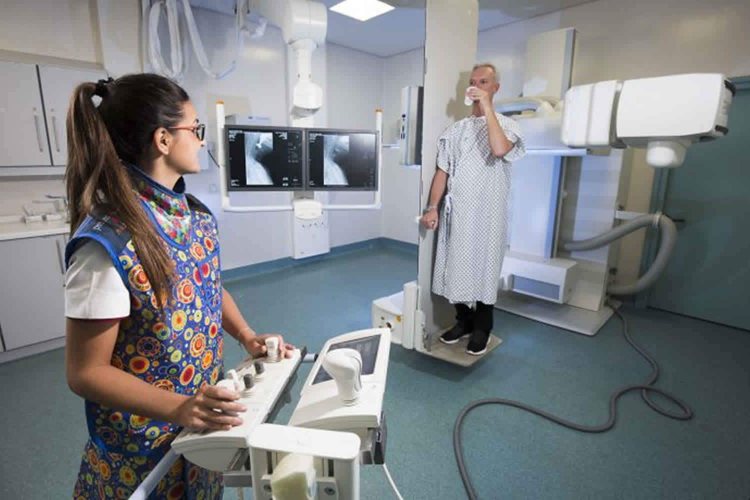
You/your child will be asked to drink 1 to 2 cups of barium. The barium is a contrast material that makes liquids show up on the image screen as gray or black. You/your child will drink the barium while standing up and while lying down.
You/your child will be placed in a prone (lying on your back) or supine (face down) position with some tilt, essentially a partial lateral decubitus position, to displace the esophagus away from the spine for imaging. One common position is “right anterior oblique” (RAO), where the patient’s right side is on the table. Their left arm and knee will be flexed and the head rotated left, allowing for some elevation of the left side. The patient is then asked to rapidly swallow contrast, roughly 100 to 200 cc. The goal, typically, is to distend the esophagus for best resolution. The contrast media may be thinned with water if needed to reveal more subtle lesions. The exact thickness of contrast and amount will depend on the local radiology protocol and also the reason for the exam. Equipment for a still-image X-ray is sufficient in the majority of cases. For certain uses, such as for oropharyngeal dysphagia, fluoroscopy is utilized. Standard posteroanterior and lateral radiographs also may be taken as needed.
If an air-contrast study is desired to further distend the esophagus, effervescent sodium bicarbonate granules may be administered immediately prior to the contrast media.
If you have any questions or don’t understand the instructions please ask.
What should I do after the barium swallow test?
Barium rarely causes any problems. It passes through your digestive system. Drink extra fluids for 12 to 24 hours after the study. If your/your child becomes constipated after the study, drink more liquids. If the constipation continues, talk with your doctor.
Barium swallow side effects
Oral barium contrast has relatively few adverse effects in standard practice. Most commonly, patients complain of nausea and vomiting within 30 minutes of ingestion. Hypersensitivity reactions have been reported but are uncommon. Most adverse effects are related to extravasation of contrast into the mediastinum or from aspiration 4.
References- Chen A, Tuma F. Barium Swallow. [Updated 2019 Feb 16]. In: StatPearls [Internet]. Treasure Island (FL): StatPearls Publishing; 2019 Jan-. Available from: https://www.ncbi.nlm.nih.gov/books/NBK493176
- Yadlapati R, Furuta GT, Menard-Katcher P. New Developments in Esophageal Motility Testing. Curr Treat Options Gastroenterol. 2019 Feb 02
- Ishii T, Akaishi T, Abe M, Takayama S, Koseki K, Kamei T, Nakano T. Importance of Barium Swallow Test and Chest CT Scan for Correct Diagnosis of Achalasia in the Primary Care Setting. Tohoku J. Exp. Med. 2019 Jan;247(1):41-49.
- Hamid M, Ullah W, Ur Rashid M, Amjad W, Mukhtar M, Hurairah A. An Esophagogram or Tracheobronchogram? A Review of Barium Sulfate Aspiration. J Investig Med High Impact Case Rep. 2018 Jan-Dec;6:2324709618802872


