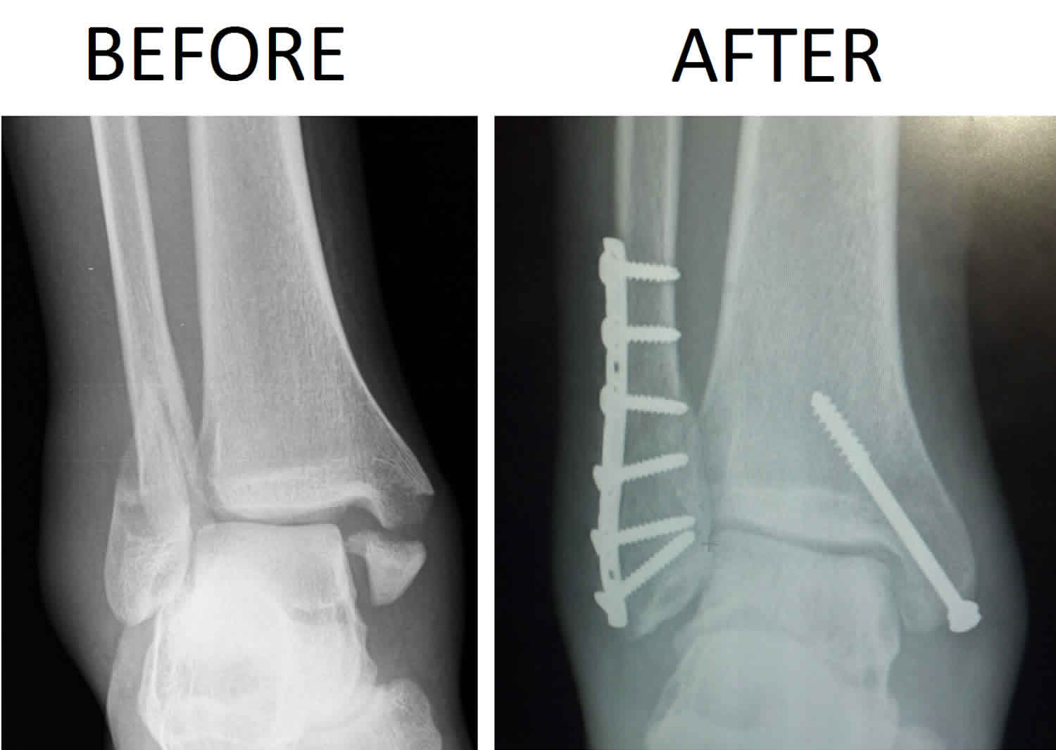
Bimalleolar fracture
A bimalleolar fracture is a type of broken ankle that happens when parts of both the tibia and fibula called the malleoli are fractured. “Bi” means two. “Bimalleolar” means that two of the three parts or malleoli of the ankle are broken. Malleoli is plural for malleolus. In most cases of bimalleolar fracture, the lateral malleolus and the medial malleolus are broken and the ankle is not stable. A “bimalleolar equivalent” fracture means that in addition to one of the malleoli being fractured, the ligaments on the inside (medial) side of the ankle are injured. Usually, this means that the fibula is broken along with injury to the medial ligaments, making the ankle unstable.
Bimalleolar ankle fracture often happens as a result of the foot and ankle rolling inward, but it can also be caused by a trip or fall, or by a direct blow to the ankle.
A stress test x-ray may be done to see whether the medial ligaments are injured.
Bimalleolar fractures or bimalleolar equivalent fractures are unstable fractures and can be associated with a dislocation.
Bimalleolar fractures can cause severe pain, swelling, and bruising in the injured ankle. Bimalleolar fracture also can be tender to the touch and make walking or putting any weight on the affected foot very difficult and painful.
Bimalleolar fracture makes the ankle unstable and typically require surgery to implant metal plates, screws, wires and rods to keep the bones aligned. Following surgery, the ankle is usually put in a short leg cast. In general, it takes at least 6 weeks for the broken malleoli to heal.
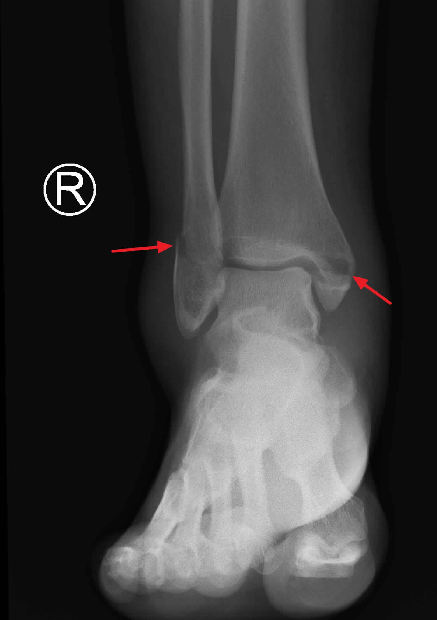
Figure 1. Bimalleolar ankle fracture
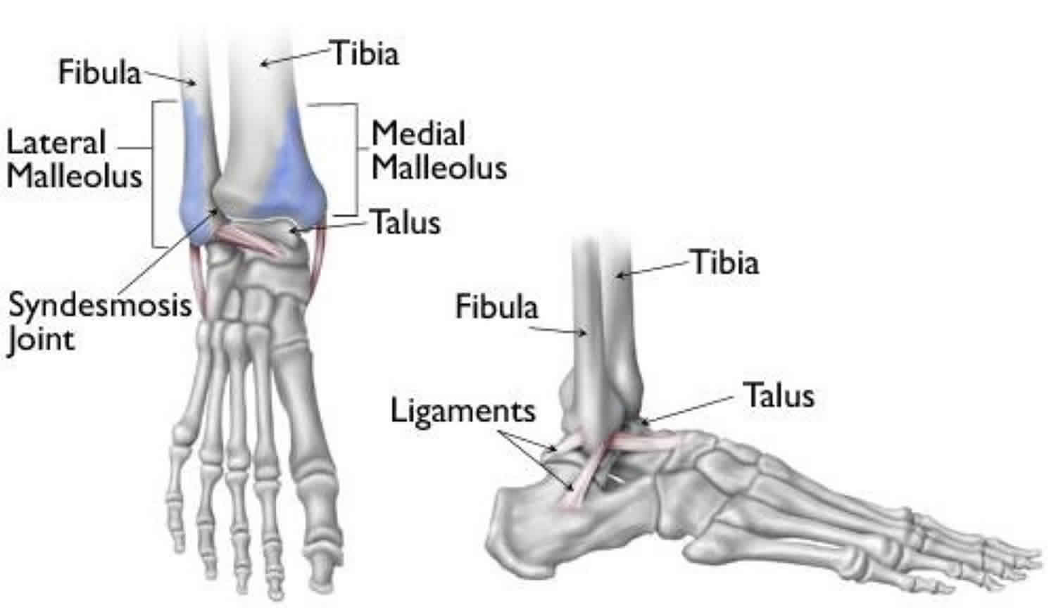
Ankle anatomy
Three bones make up the ankle joint:
- Tibia – shinbone
- Fibula – smaller bone of the lower leg
- Talus – a small bone that sits between the heel bone (calcaneus) and the tibia and fibula
The tibia and fibula have specific parts that make up the ankle:
- Medial malleolus – inside part of the tibia
- Posterior malleolus – back part of the tibia
- Lateral malleolus – end of the fibula
Figure 2. Anatomy of the ankle
Bimalleolar fracture causes
Bimalleolar fracture can be caused by:
- Twisting or rotating your ankle
- Rolling your ankle
- Tripping or falling
- Impact during a car accident
Bimalleolar fracture symptoms
Because a severe ankle sprain can feel the same as a broken ankle, every ankle injury should be evaluated by a physician.
Bimalleolar fractures can cause severe pain, swelling, and bruising in the injured ankle. Bimalleolar fracture also can be tender to the touch and make walking or putting any weight on the affected foot very difficult and painful.
Common symptoms for a bimalleolar ankle fracture include:
- Immediate and severe pain
- Swelling
- Bruising
- Tender to touch
- Cannot put any weight on the injured foot
- Deformity (“out of place”), particularly if the ankle joint is dislocated as well
Bimalleolar fracture complications
People who smoke, have diabetes, or are elderly are at a higher risk for complications after surgery, including problems with wound healing. This is because it may take longer for their bones to heal.
Nonsurgical treatment
Without surgery, there is a risk that the bimalleolar fracture will move out of place before it heals. This is why it is important to follow up with your physician as scheduled.
If the bimalleolar fracture fragments do move out of place and the bones heal in that position, it is called a “malunion.” Treatment for this is determined by how far out of place the bones are and how the stability of the ankle joint is affected.
If a malunion does occur or if your ankle becomes unstable after it heals, this can eventually lead to arthritis in your ankle.
Surgical treatment
General surgical risks include:
- Infection
- Bleeding
- Pain
- Blood clots in your leg
- Damage to blood vessels, tendons, or nerves
Risks from the surgical treatment of ankle fractures include
- Difficulty with bone healing
- Arthritis
- Pain from the plates and screws that are used to fix fracture. Some patients choose to have them removed several months after their fracture heals
Bimalleolar fracture diagnosis
After discussing your medical history, symptoms, and how the injury occurred, your doctor will do a careful examination of your ankle, foot, and lower leg.
Imaging tests
If your doctor suspects an ankle fracture, he or she will order additional tests to provide more information about your injury.
- X-rays. X-rays are the most common and widely available diagnostic imaging technique. X-rays can show if the bone is broken and whether there is displacement (the gap between broken bones). They can also show how many pieces of broken bone there are. X-rays may be taken of the leg, ankle, and foot to make sure nothing else is injured.
- Stress test. Depending on the type of ankle fracture, the doctor may put pressure on the ankle and take a special x-ray, called a stress test. This x-ray is done to see if certain ankle fractures require surgery.
- Computed tomography (CT) scan. This type of scan can create a cross-section image of the ankle and is sometimes done to further evaluate the ankle injury. It is especially useful when the fracture extends into the ankle joint.
- Magnetic resonance imaging (MRI) scan. These tests provide high resolution images of both bones and soft tissues, like ligaments. For some ankle fractures, an MRI scan may be done to evaluate the ankle ligaments.
Bimalleolar fracture treatment
Surgical treatment is often recommended because bimalleolar fractures make the ankle unstable. Surgical treatment typically require to implant screws, a plate and screws, or different wiring techniques to keep the bones aligned. During this type of procedure, the bone fragments are first repositioned (reduced) into their normal alignment. They are held together with special screws and metal plates attached to the outer surface of the bone. In some cases, a screw or rod inside the bone may be used to keep the bone fragments together while they heal. Following surgery, the ankle is usually put in a short leg cast. In general, it takes at least 6 weeks for the broken malleoli to heal.
Nonsurgical treatment
Bimalleolar fractures are considered unstable and surgery is usually recommended.
Nonsurgical treatment might be considered if you have significant health problems, where the risk of surgery may be too great, or if you usually do not walk.
Immediate treatment typically includes a splint to immobilize the ankle until the swelling goes down. A short leg cast is then applied. Casts may be changed frequently as the swelling subsides in the ankle.
You will need to see your physician regularly to repeat your x-rays to make sure your ankle remains stable.
In most cases, weightbearing is not be allowed for 6 weeks. After 6 weeks, the ankle may be protected by a removable brace as it continues to heal.
Bimalleolar fracture recovery
A bimalleolar fracture usually requires someone to keep weight off the affected foot for at least 6 weeks, but in most cases, people return to normal daily activities within 3 to 4 months. It takes at least 6 weeks for the broken bones to heal. It may take longer for the involved ligaments and tendons to heal. Stretching and strengthening exercises supervised by a doctor or physical therapist can help improve ankle function and mobility during the healing process.
Your doctor will most likely monitor the bone healing with repeated x-rays. This is typically done more often during the first 6 weeks if surgery is not chosen.
Rehabilitation
Rehabilitation is very important regardless of how an ankle fracture is treated.
When your physician allows you to start moving your ankle, physical therapy and home exercise programs are very important. Doing your exercises regularly is key.
Eventually, you will also start doing strengthening exercises. It may take several months for the muscles around your ankle to get strong enough for you to walk without a limp and to return to your regular activities.
Again, exercises only make a difference if you actually do them.
Weightbearing
It is very important to not put weight on your ankle until your physician says you can. If you put weight on the injured ankle too early, the fracture fragments may move or your surgery may fail and you may have to start over.
Supports
It is very common to have several different kinds of things to wear on the injured ankle, depending on the injury.
Initially, most ankle fractures are placed in a splint to protect your ankle and allow for the swelling to go down. After that, you may be put into a cast or removable brace.
Even after the fracture has healed, your physician may recommend wearing an ankle brace for several months while you are doing sporting activities.
Bimalleolar fracture prognosis
Although most people return to normal daily activities, except for sports, within 3 to 4 months, studies have shown that people can still be recovering up to 2 years after their ankle fractures. It may take several months for you to stop limping while you walk, and before you can return to sports at your previous competitive level. Most people return to driving within 9 to 12 weeks from the time they were injured.


