close

Cardiovascular System

BloodConditions & DiseasesImmune and Hematology System

Anemia of prematurity

anemia of prematurity

Anemia of prematurity

Anemia of prematurity means that a baby born early (prematurely) does not have enough red blood cells. Red blood cells carry oxygen to the body. Preterm infants with birth weight <1.0 kg (commonly designated as extremely low birth weight or ELBW, infants) have completed ≤29 weeks of gestation, and nearly all will need red blood cell (RBC) transfusions during the first weeks of life. Every week in the United States, approximately 10,000 infants are born prematurely (ie, <37 weeks of gestation), with 600 (6%) of these preterm infants being extremely low birth weight 1. Approximately 90% of extremely low birth weight neonates will receive at least one red blood cell transfusion 2.

All babies have some anemia (decrease in hemoglobin concentration) when they are born. In healthy term infants, the nadir hemoglobin value rarely falls below 10 g/dL at an age of 10 to 12 weeks 3. This is normal and is called “physiological anemia of infancy”. For the term infant, a physiologic and usually asymptomatic anemia is observed 8-12 weeks after birth. But in premature babies, the number of red blood cells may decrease faster and go lower than in full-term babies. This may happen because:

  • A premature baby may not make enough red blood cells.
  • A premature baby may need tests that require blood samples. It may be hard for the baby to produce enough red blood cells to make up for the blood that’s taken out and used in the tests.
  • A baby’s red blood cells don’t live as long as an older child’s red blood cells.

Anemia of prematurity is an exaggerated, pathologic response of the preterm infant to this transition. Anemia of prematurity is a normocytic, normochromic, hyporegenerative anemia characterized by a low serum erythropoietin (EPO) level, often despite a remarkably reduced hemoglobin concentration 4. Nutritional deficiencies of iron, vitamin E, vitamin B-12, and folate may exaggerate the degree of anemia, as may blood loss and/or a reduced red cell life span.

The anemia of prematurity is caused by untimely birth occurring before placental iron transport and fetal erythropoiesis are complete, by phlebotomy blood losses taken for laboratory testing, by low plasma levels of erythropoietin due to both diminished production and accelerated catabolism, by rapid body growth and need for commensurate increase in red cell volume/mass, and by disorders causing red blood cell losses due to bleeding and/or hemolysis 3.

The risk of anemia of prematurity is inversely related to gestational maturity and birthweight 4. As many as half of infants of less than 32 weeks gestation develop anemia of prematurity. Anemia of prematurity is not typically a significant issue for infants born beyond 32 weeks’ gestation.

Race and sex have no influence on the incidence of anemia of prematurity.

Testosterone is believed to be at least partially responsible for a slightly higher hemoglobin level in male infants at birth, but this effect is of no significance with regard to risk of anemia of prematurity. The nadir of the hemoglobin level is typically observed 4-10 weeks after birth in the tiniest infants, with hemoglobin levels of 8-10 g/dL if birthweight was 1200-1400 grams, or 6-9 g/dL at birth weights of less than 1200 grams and to approximately 7 g/dL in infants with birth weights <1 kg 4.

Anemia of prematurity is usually not serious. Anemia of prematurity spontaneously resolves in many premature infants within 3-6 months of birth 4. In others, however, medical intervention is required, because the low oxygen levels in a premature infant can make other problems worse, such as heart and lung problems. Most infants with birth weight <1.0 kg are given multiple red blood cell (RBC) transfusions within the first few weeks of life 3.

Red blood cell transfusions are the mainstay of therapy for anemia of prematurity with recombinant human erythropoietin (EPO) largely unused because it fails to substantially diminish red blood cell transfusion needs despite exerting substantial erythropoietic effects on neonatal marrow.

Figure 1. Blood composition

blood composition

blood composition

Footnote: Blood consists of a liquid portion called plasma and a solid portion (the formed elements) that includes red blood cells, white blood cells, and platelets. When blood components are separated by centrifugation, the white blood cells and platelets form a thin layer, called the “buffy coat,” between the plasma and the red blood cells, which accounts for about 1% of the total blood volume. Blood cells and platelets can be seen under a light microscope when a blood sample is smeared onto a glass slide.

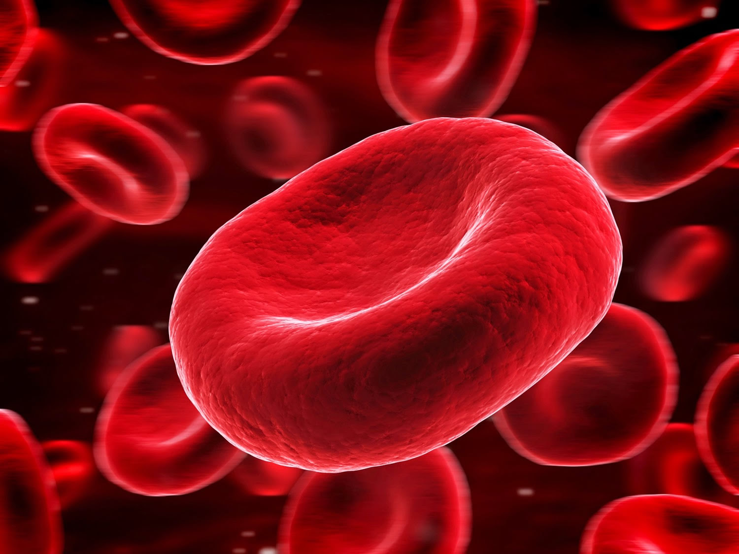
Figure 2. Red blood cells (normal red blood cells)

red blood cells

Anemia of prematurity causes

The three basic mechanisms for the development of anemia of prematurity include:

  1. Inadequate red blood cell production,
  2. Shortened red blood cell life span,
  3. Blood loss.

Taken together, the premature infant is at risk for the development of anemia of prematurity because of limited red blood cell synthesis during rapid growth, a diminished red blood cell life span, and an increased loss of red blood cells.

Inadequate red blood cell production

The first mechanism of anemia is inadequate red blood cell production for the growing premature infant. The location of erythropoietin (EPO) and red blood cell production changes during gestation. Erythropoietin (EPO) synthesis initially occurs in the fetal liver but gradually shifts toward the kidney as gestation advances. By the end of gestation, however, the liver remains the major source of erythropoietin (EPO).

Fetal erythrocytes are produced in the yolk sac during the first few weeks of embryogenesis. The fetal liver becomes more important as gestation advances and, by the end of the first trimester, has become the primary site of erythropoiesis. Bone marrow then begins to take on a more active role in producing erythrocytes. By about 32 weeks’ gestation, the burden of erythrocyte production in the fetus is shared evenly by liver and bone marrow. By 40 weeks’ gestation, the marrow is the sole erythroid organ. Premature delivery does not accelerate the ontogeny of these processes.

Although erythropoietin (EPO) is not the only erythropoietic growth factor in the fetus, it is the most important. Erythropoietin (EPO) is synthesized in response to anemia and consequent relative tissue hypoxia. The degree of anemia and hypoxia required to stimulate erythropoietin (EPO) production is far greater for the fetal liver than for the fetal kidney. Erythropoietin (EPO) production may not be stimulated until a hemoglobin concentration of 6-7 g/dL is reached. As a result, new red blood cell production in the extremely premature infant, whose liver remains the major site of erythropoietin (EPO) production, is blunted despite what may be marked anemia. In addition, erythropoietin (EPO), whether endogenously produced or exogenously administered, has a larger volume of distribution and is more rapidly eliminated by neonates, resulting in a curtailed time for bone marrow stimulation.

Erythroid progenitors in premature infants are quite responsive to erythropoietin (EPO), but the response may be blunted if iron or other substrate or co-factor stores are insufficient. Another potential problem is that while the infant may respond appropriately to increased erythropoietin (EPO) concentrations with increased reticulocyte counts, rapid growth may prevent the appropriate increase in hemoglobin concentration.

Shortened red blood cell life span or hemolysis

Also important in the development of anemia of prematurity is that the average life span of a neonatal red blood cell is only one half to two thirds that of an adult red blood cell. Cells of the most immature infants may survive only 35-50 days. The shortened red blood cell life span of the neonate is a result of multiple factors, including diminished levels of intracellular adenosine triphosphate (ATP), carnitine, and enzyme activity; increased susceptibility to lipid peroxidation; and increased susceptibility of the cell membrane to fragmentation.

Blood loss

Finally, blood loss may contribute to the development of anemia of prematurity. If the neonate is held above the placenta for a time after delivery, fetal-placental transfer of blood may occur. Conversely, delayed cord clamping may lessen the degree of anemia of prematurity 5, although a study by Elimian et al 6 did not find this to be true. More commonly, because of the need to closely monitor the tiny infant, frequent samples of blood are removed for various tests. These losses are often 5-10% of the total blood volume.

Anemia of prematurity differential diagnoses

Conditions to consider in the differential diagnosis of anemia of prematurity are those which diminish red cell production, increase red cell destruction, or cause blood loss.

  • Acute Anemia
  • Birth Trauma
  • Chronic Anemia
  • Head Trauma
  • Hemolytic Disease of the Newborn
  • Parvovirus B19 Infection
  • Intraventricular Hemorrhage in the Preterm Infant

Conditions that diminish red blood cell synthesis are as follows:

  • Bone marrow infiltration
  • Bone marrow depression (eg, pancytopenia, drugs)
  • Diamond-Blackfan anemia
  • Substrate deficiencies (eg, iron, vitamin E, folic acid)
  • Congenital fetal infections (eg, cytomegalovirus, parvovirus, syphilis)

Conditions that cause hemolysis are as follows:

  • Congenital fetal infections (eg, cytomegalovirus, parvovirus, syphilis)
  • Acute systemic infections (leading to disseminated intravascular coagulation)
  • Abnormal red blood cells (spherocytosis, elliptocytosis)
  • Nonspherocytic hemolytic anemias (eg, G6PD deficiency, kinase and isomerase deficiencies)
  • Hemolytic disease of the newborn (Rh, ABO, other major blood-group incompatibilities between mother and fetus)

Conditions that reduce blood volume are as follows:

  • Twin-to-twin transfusion syndrome (donor twin)
  • Iatrogenic (eg, excessive blood sampling)
  • Hemorrhage (eg, gastrointestinal, central nervous system, subcutaneous tissues)

Anemia of prematurity symptoms

Many clinical findings have been attributed to anemia of prematurity, but they are neither specific nor diagnostic. These symptoms may include the following:

  • Poor weight gain despite adequate caloric intake
  • Cardiorespiratory symptoms such as tachycardia, tachypnea, and flow murmurs
  • Decreased activity, lethargy, and difficulty with oral feeding
  • Pallor
  • Increase in apneic and bradycardic episodes, and worsened periodic breathing
  • Metabolic acidemia – Increased lactic acid secondary to increased cellular anaerobic metabolism in relatively hypoxic tissues

Anemia of prematurity diagnosis

The following are useful laboratory studies:

  • Complete blood count (CBC) – White blood cell (WBC) and platelet values are normal in anemia of prematurity. Low hemoglobin values, below 10 g/dL, are found. They may descend to a nadir of 6-7 g/dL. Lowest levels are generally observed in the smallest infants. Red blood cell indices are normal (eg, normochromic, normocytic) for age.
  • Reticulocyte count – The reticulocyte count is low when the degree of anemia is considered, as a result of the low levels of erythropoietin (EPO). Conversely, an elevated reticulocyte count is not consistent with the diagnosis of anemia of prematurity.
  • Peripheral blood smear – Red blood cell morphology should be normal. Red blood cell precursors may appear to be more prominent.
  • Maternal and infant blood typing; direct antibody test (Coombs) – The direct Coombs test result may be coincidentally positive. Despite this, it is important to ensure an immune-mediated hemolytic process related to maternal-fetal blood group incompatibility (hemolytic disease of the newborn) is not present.
  • Serum bilirubin – An elevated serum bilirubin level should suggest other possible explanations for the anemia. These would include hemolytic entities, such as G-6-PD deficiency or other kinase/isomerase/enzyme deficiencies, or more common causes such as infection or hemolytic disease of the newborn.
  • Lactic acid – Elevated lactic acid levels have been suggested by some to be useful as an aid to determine the need for transfusion.

Anemia of prematurity treatment

Medical treatment options are blood transfusion(s), recombinant erythropoietin (EPO) treatment, and observation.

Observation may be the best course of action for infants who are asymptomatic, not acutely ill, and are receiving adequate nutrition. Adequate amounts of vitamin E, vitamin B-12, folate, and iron are important to blunt the expected decline in hemoglobin levels in the premature infant. Periodic measurements of the hematocrit level in infants with anemia of prematurity are necessary after hospital discharge. Once a steady increase in the hematocrit level has been established, only routine checks are required.

Packed red blood cell transfusions

Packed red blood cell transfusions are the mainstay of therapy for anemia of prematurity. The frequency of blood transfusion varies with gestational age, degree of illness, and, interestingly, the hospital evaluated. Unfortunately, there is considerable disagreement about the indication, timing, and efficacy of packed red blood cell transfusion.

Guidelines for transfusing red blood cells to preterm neonates are controversial, and practices vary greatly 7. This lack of a universal approach stems from limited knowledge of the cellular and molecular biology of erythropoiesis during the perinatal period, an incomplete understanding of infant physiological/adaptive responses to anemia, and contrary/controversial transfusion practice guidelines as based on results of randomized clinical trials and expert opinions. Generally, red blood cell transfusions are given to maintain a level of blood hemoglobin or hematocrit believed to be optimal for each neonate’s clinical condition. Guidelines for red blood cell transfusions, judged to be reasonable by most neonatologists to treat the anaemia of prematurity, are listed by Table 1. These guidelines are very general, and it is important that terms such as “severe” and “symptomatic” be defined to fit local transfusion practices/policies. Importantly, guidelines are not mandates for red blood cell transfusions that must be followed; they simply suggest situations when an red blood cell transfusion would be judged to be reasonable/acceptable.

The decision to give a transfusion should not be made lightly, because significant infectious, hematologic, immunologic, and metabolic complications are possible. Late-onset necrotizing enterocolitis has been reported in stable-growing premature infants electively transfused for anemia of prematurity 8. Transfusions also transiently decrease erythropoiesis and EPO levels. There is also agreement that the number of transfusions, as well as the number of donor exposures, should be reduced as much as possible.

Clinical trials designed to determine the efficacy of blood transfusions in relieving symptoms ascribed to anemia of prematurity have produced conflicting results 9. Improved growth has been an inconsistent finding. While some studies have demonstrated a decrease in apneic episodes after blood transfusion, others have found similar results with simple crystalloid volume expansion.

Subjective improvement in activity, decreased lethargy, and improved feeding have been described in some studies. Blood transfusions have been documented to decrease lactic acid levels in otherwise healthy preterm infants who are anemic. Blood transfusions have reduced tachycardia in anemic infants who are transfused.

Some medical professionals have suggested using lactate levels as an aid in determining the need for transfusion.

Table 1. Allogeneic red blood cell transfusions for the anemia of prematurity

Transfuse to maintain the blood hematocrit per each clinical situation:

  • > 40% (35 to 45% *) for severe cardiopulmonary disease
  • > 30% for moderate cardiopulmonary disease
  • > 30% for major surgery
  • >25% (20 to 25% *) for symptomatic anemia
  • > 20% for asymptomatic anemia

*Reflects practices that vary among neonatologists. Thus, any value within range is acceptable for local practices.

Reducing the number of transfusions

Studies from individual centers have documented a marked decrease in the administration of packed red blood cell transfusions in the past decades, even before the use of EPO became more frequent. This decrease in transfusions is almost certainly multifactorial in origin. Adoption of standardized transfusion protocols that take various factors into account, including hemoglobin levels, degree of cardiorespiratory disease, and traditional signs and symptoms of pathologic anemia, are acknowledged as an important factor in this reduction. A restricted transfusion protocol may decrease the number of transfusions while also decreasing the hematocrit at discharge 10.

A 2011 study 11 evaluated 41 preterm infants with birth weights of 500-1300 g who were enrolled in a clinical trial that compared high and low hematocrit thresholds for transfusion. A rise in systemic oxygen transport and a decrease in lactic acid after transfusion was noted in both groups; however, oxygen consumption did not change significantly in either group. In the low hematocrit group only, cardiac output and fractional oxygen extraction fell after transfusion, which shows that these infants had increased their cardiac output to maintain adequate tissue oxygen delivery in response to anemia. The results demonstrate that infants with low hematocrit thresholds may benefit from transfusion, while transfusion in those with high hematocrit thresholds may provide no acute physiological benefit 11.

The Premature Infant in Need of Transfusion study 12 showed that transfusing infants to maintain higher hemoglobin level (8.5-13.5 g/dL) conferred no benefit in terms of mortality, severe morbidity, or apnea intervention compared with infants transfused to maintain a low hemoglobin levels (7.5-11.5 g/dL).

These findings differ from the Iowa study, which found less parenchymal brain hemorrhage, periventricular leukomalacia, and apnea in infants whose transfusion criteria was not restricted and whose hemoglobin level was higher. Clearly, no universally accepted guidelines for transfusion in infants with anemia of prematurity are available at this time, and clinicians must determine their individual standardized transfusion practices.

Anemia of prematurity guidelines

No universally accepted guidelines for transfusion in infants with anemia of prematurity are available at this time, and clinicians must determine their individual standardized transfusion practices.

As an example, note the Children’s Hospital of Wisconsin Transfusion Committee guidelines for consideration:

  • An infant with a hemoglobin (Hb) level of less than 8 g/dL may be transfused at the discretion of the attending physician
  • A stable infant with a hemoglobin level of 8-10 g/dL without clinical symptoms or other exceptions listed below may be transfused
  • An infant with a hemoglobin level of 11-13 g/dL without a supplemental oxygen or continuous positive airway pressure (CPAP) requirement, apnea/bradycardia, significant tachycardia or tachypnea, or other exceptions listed below should not be transfused
  • An infant with a hemoglobin level of more than 13 g/dL without an oxygen requirement of more than 40% by hood, CPAP, or ventilator; hypotension that requires pressor medication; major surgery; or other exceptions listed below should not be transfused
  • An infant with a hemoglobin level of more than 15 g/dL without cyanotic heart disease, extracorporeal membrane oxygenation (ECMO) therapy, regional oxygen saturations less than 50%, or hypotension that requires pressor medications should not be transfused
  • An infant with a history of massive blood loss may be transfused at the discretion of the attending physician

It is of obvious importance to discuss with the family their child’s need for transfusion and to obtain consent before the transfusion. It is also important to discuss with parents the normal course of anemia, the criteria for and risks associated with transfusions, and the advantages and disadvantages of erythropoietin (EPO) administration. Also necessary is consideration of the family’s religious beliefs regarding transfusions.

Reducing the number of donor exposures

Reducing the number of donor exposures is also important. Preservatives and additive systems allow blood to be stored safely for as long as 35-42 days. This can be accomplished by using packed red blood cells stored in preservatives (eg, citrate-phosphate-dextrose-adenine [CPDA-1]) and additive systems (eg, Adsol). Infants may be assigned a specific unit of blood, which may suffice for treatment during their entire hospitalization and thus limit exposure to the single donor of that unit. Concerns that stored blood might increase serum potassium levels are unfounded if the transfused volume is low.

Complications

Potential complications of transfusion include the following:

  • Infection (eg, hepatitis, cytomegalovirus [CMV], human immunodeficiency virus [HIV], syphilis)
  • Fluid overload and electrolyte imbalances
  • Exposure to plasticizers
  • Hemolysis
  • Posttransfusion graft versus host disease

An important tool in reducing at least one of these transfusion risks is to use all available screening techniques for infection. The risk of cytomegalovirus (CMV) transmission can be dramatically reduced by use of CMV-safe blood. This can be accomplished by using CMV serology–negative cells, along with blood processed through leukocyte-reduction filters or inverted spin technique. These methods also reduce other WBC-associated infectious agents (eg, Epstein-Barr virus, retroviruses, Yersinia enterocolitica) by yielding a leukocyte-poor suspension of packed red blood cells. The American Red Cross now provides exclusively leukocyte-reduced blood to hospitals in the United States.

Recombinant Erythropoietin treatment

Multiple investigations have established that premature infants respond to exogenously administered recombinant human EPO and supplemental iron with a brisk reticulocytosis. Subcutaneous administration of EPO may be preferred, as intravenous administration has increased urinary losses. Although EPO cannot prevent early transfusions, modest decreases in the frequency of late packed red blood cell transfusions have been documented. Additional iron supplementation is necessary during exogenous EPO treatment.

Trials have evaluated the impact of EPO treatment in populations of the most immature neonates. These studies likewise have demonstrated that infants with very low birth weight (VLBW) are capable of responding to EPO with a reticulocytosis.

Studies and a Cochrane Neonatal Systemic review suggest an association between exogenous EPO administration and retinopathy of prematurity 13.

Yasmeen et al 14 studied 60 preterm low birth weight infants and concluded that short-term recombinant human erythropoietin with iron and folic acid was effective in preventing anemia of prematurity.

EPO with iron does not adversely affect growth or developmental outcomes, but the impact on the number of transfusions a premature infant receives ranges from nonexistent to small.

At this time, no agreement regarding the safety, timing, dosing, route, or duration of therapy has been established. In short, the cost-benefit ratio for EPO has yet to be clearly established, and this medication is not universally accepted as a standard therapy for an infant with anemia of prematurity.

Anemia of prematurity prognosis

Spontaneous recovery of mild anemia of prematurity may occur 3-6 months after birth. In more severe, symptomatic cases, medical intervention may be required.

References
  1. Martin JA, Hamilton BE, Sutton PD, et al. Births: final data for 2008 national Vital Statistics Reports. Centers for Disease Control and Prevention. 2009;57:7.
  2. Maier RJ, Sonntag J, Walka MM, et al. Changing practices of red blood cell transfusions in infants with birth weights less than 1000 g. J Pediatr. 2000;136:220–224.
  3. Strauss RG. Anaemia of prematurity: pathophysiology and treatment. Blood Rev. 2010;24(6):221-225. doi:10.1016/j.blre.2010.08.001 https://www.ncbi.nlm.nih.gov/pmc/articles/PMC2981681
  4. Anemia of Prematurity. https://emedicine.medscape.com/article/978238-overview
  5. Ultee CA, van der Deure J, Swart J, Lasham C, van Baar AL. Delayed cord clamping in preterm infants delivered at 34 36 weeks’ gestation: a randomised controlled trial. Arch Dis Child Fetal Neonatal Ed. 2008 Jan. 93(1):F20-3.
  6. Elimian A, Goodman J, Escobedo M, Nightingale L, Knudtson E, Williams M. Immediate compared with delayed cord clamping in the preterm neonate: a randomized controlled trial. Obstet Gynecol. 2014 Dec. 124 (6):1075-9.
  7. dos Santos AMN, Guinsburg R, Procianoy RS, dos SR, Sadeck L, Netto AA, Rugolo LM, et al. Variability on red blood cell transfusion practices among Brazilian neonatal intensive care units. Transfusion. 2010;50:150–159.
  8. Singh R, Shah BL, Frantz ID 3rd. Necrotizing enterocolitis and the role of anemia of prematurity. Semin Perinatol. 2012 Aug. 36(4):277-82.
  9. Bell EF, Nahmias C, Sinclair JC, Zipursky A. Changes in circulating red cell volume during the first 6 weeks of life in very-low-birth-weight infants. Pediatr Res. 2014 Jan. 75 (1-1):81-4.
  10. Bell EF, Strauss RG, Widness JA, et al. Randomized trial of liberal versus restrictive guidelines for red blood cell transfusion in preterm infants. Pediatrics. 2005 Jun. 115(6):1685-91.
  11. Fredrickson LK, Bell EF, Cress GA, et al. Acute physiological effects of packed red blood cell transfusion in preterm infants with different degrees of anaemia. Arch Dis Child Fetal Neonatal Ed. 2011 Jul. 96(4):F249-53.
  12. Kirpalani H, Whyte RK, Andersen C, et al. The Premature Infants in Need of Transfusion (PINT) study: a randomized, controlled trial of a restrictive (low) versus liberal (high) transfusion threshold for extremely low birth weight infants. J Pediatr. 2006 Sep. 149(3):301-307.
  13. Suk KK, Dunbar JA, Liu A, et al. Human recombinant erythropoietin and the incidence of retinopathy of prematurity: a multiple regression model. J AAPOS. 2008 Jun. 12(3):233-8.
  14. Yasmeen BH, Chowdhury MA, Hoque MM, Hossain MM, Jahan R, Akhtar S. Effect of short-term recombinant human erythropoietin therapy in the prevention of anemia of prematurity in very low birth weight neonates. Bangladesh Med Res Counc Bull. 2012 Dec. 38(3):119-23.
read more
Veins

Portal vein

Portal vein

Portal vein

Portal vein sometimes referred to as the hepatic portal vein, is the main vessel in the portal venous system and drains blood from the gastrointestinal tract, spleen, pancreas, and gallbladder to the liver. The superior mesenteric and splenic veins unite to form the hepatic portal vein (see Figure 1). This special flow of venous blood, called the hepatic portal circulation. After passing through the liver for processing, blood drains into the hepatic veins, which empty into the inferior vena cava.

Portal vein anatomy

The portal vein usually measures approximately 8 cm in length in adults with a maximum diameter of 13 mm. The portal vein originates behind the neck of the pancreas where it is classically formed by the confluence of the superior mesenteric and splenic veins (the portovenous confluence), and also receives blood from the inferior mesenteric, gastric, and cystic veins.

Immediately before reaching the liver, the portal vein divides in the porta hepatis into left and right portal veins. The right portal vein divides into anterior (supplying segments V and VIII) and posterior (supplying segments VI and VII) branches. The left portal vein may be divided into transverse and umbilical portions, as delineated by the ligamentum venosum, and is mostly extrahepatic in course. The main branches of the left portal vein originate from the umbilical portion, and supply liver segments II, III, IV 1.

It ramifies further, forming smaller venous branches and ultimately portal venules. Each portal venule courses alongside a hepatic arteriole and the two vessels form the vascular components of the portal triad. These vessels ultimately empty into the hepatic sinusoids to supply blood to the liver.

75% of the blood supplied to the liver comes from the portal vein, but it only supplies 50% of the oxygen supply to the liver.

Portal vein variant anatomy

The overall incidence of portal vein variation is reported to be ~25% (range 20-30%), which should be recognized prior to procedures such as liver transplantation, complex hepatectomy and portal vein embolization 2:

  • portal vein trifurcation (most common)
    • portal vein divides into three branches: left portal vein, right anterior portal vein, and right posterior portal vein
  • absent right portal vein (rare) 3
    • right sectional portal veins originate independently from the common portal vein
    • if the right anterior section portal vein branches higher from the common portal vein vs posterior sectional portal vein, the surgeon may mistake the posterior sectional portal vein for the right portal vein
  • portal vein duplication (rare)
  • absent left extrahepatic portal vein (rare) 3
    • a single right portal vein originates from the porta hepatis, supplying the right hemiliver, then following an intrahepatic course with distalmost branches supplying the left liver.

There is an increased risk of bile duct hilar anatomical variation in the presence of portal vein variants.

Figure 1. Portal vein

Hepatic portal vein function

Like all portal systems, the hepatic portal vein delivers the digested nutrient-rich blood from your gastrointestinal tract into your liver for processing. The liver cells also break down toxins that enter the blood through the digestive tract. After passing through the liver sinusoids, the blood enters the hepatic veins and inferior vena cava, thereby reentering the general systemic circulation.

Portal vein embolization

Portal vein embolization is a technique used to selectively occlude the blood supply to one of the liver lobes, diverting portal blood flow to the other lobe, the future liver remnant.

This diversion will increase the size of the post-hepatectomy future liver remnant which improves surgical outcomes by preventing liver insufficiency. The minimum limit of the future liver remnant is 20–40% of total liver volume dependent on the presence of background liver disease 4.

Portal vein embolization is a procedure performed by interventional radiology.

Portal vein embolization indications

  • Future liver remnant that would be too small for the patient’s body mass, post-hepatectomy (typically <20%)
  • Elevated ICG-R15 serum values 15 minutes after injection
    ICG (indocyanine green) binds to albumin and is excreted by the biliary system
    elevated values imply decreased hepatic reserve
  • Patients who underwent hepatotoxic chemotherapy, if future liver remnant <30%
  • Cirrhosis, Child-Pugh class A, ICG-R15 <10%, if future liver remnant <40%
  • Patients with hepatic steatosis
  • Concomitant pancreas resection and patients with diabetes due to poor post-hepatectomy hypertrophy rates

Portal vein embolization contraindications

  • ipsilateral portal tumor thrombus precluding catheter placement
  • clinically overt portal hypertension (procedure exacerbates portal hypertension)

Portal vein embolization procedure

Can be performed on an outpatient basis. The future liver remnant (on CT or MRI) should be obtained prior to undertaking this procedure.

Portal vein embolization technique

The right lobe is almost always targeted. The approach is usually through the right lobe, as well.

Different embolic agents have been used, including:

  • n-butyl cyanoacrylate (NBCA)
  • ethiodized oil
  • fibrin glue
  • ethanol
  • microparticles (such as polyvinyl alcohol, PVA)
  • microspheres followed by coils are used by some.

The portal vein can be approached surgically through a transileocolic approach, but interventional radiology usually approaches the portal vein transhepatically. Portal vein pressures are checked pre-procedure, to ensure that there is no portal hypertension.

Postprocedural care

  • minor fluctuations in postprocedure liver function tests (50%)
  • liver synthetic functions usually not affected
  • nausea, fever, and pain are rare

Portal vein embolization complications

  • reported 0% procedure-related mortality 5
  • reported overall morbidity of 2.2% 5
  • nontarget embolization
  • complete portal vein thrombus
  • risks similar to other transhepatic procedures
    • hemobilia
    • hemoperitoneum
    • cholangitis
    • subcapsular hematoma
    • pneumothorax

Portal vein embolization prognosis

Patients with otherwise normal livers regenerate two weeks postprocedure at 12-21 cm³/ day (9 cm³/day for cirrhotic patients) 6. 2-4 weeks is usually enough for most patients with normal liver function (>4 weeks for patients with cirrhosis).

There is some evidence from volumetric analyzes to indicate that right portal vein embolization + segment 4 embolization results in a greater degree of hypertrophy of segments 2/3 than right portal vein embolization alone in patients with colorectal liver metastases and this may become the recommended strategy in those with a relatively low future liver remnant (<20%) and are planned to undergo an extended right hemihepatectomy 4.

Portal vein obstruction

Portal vein obstruction is a common complication of several metabolic and autoimmune diseases. It is most commonly the result of thrombosis of the portal vasculature, but it can also result from malignancies. Due to the vast range of diseases that result in portal vein obstruction, understanding the common causes, pathophysiologies, and relevant management is key to treating patients suffering from this disease.

The prevalence of portal vein obstruction varies in different populations. In patients with cirrhosis or portal hypertension, it is estimated to be anywhere between 1.6% and 15.8% 7. The incidence is higher where the cirrhosis results from alcohol use disorder or Hepatitis B infection. The prevalence is as low as 1% in patients with compensated liver cirrhosis 8, while as high as 25% in patients awaiting a liver transplant 9. Possible causes for the higher incidence in liver transplant patients include advanced underlying disease, immobility due to possibly worse ascites, and a higher degree of imbalance of clotting factors.

Portal vein obstruction causes

The causes of portal vein thrombosis can be divided into two categories; inherited and acquired.

Inherited portal vein thrombosis:

  • Factor V Leiden mutation
  • Prothrombin gene mutation
  • Anti-thrombin III deficiency
  • Protein C deficiency
  • Protein S deficiency

Acquired portal vein thrombosis:

  • Lupus anticoagulant syndrome
  • Liver disease
  • Iatrogenic
  • Disseminated intravascular coagulation
  • Burns
  • Sepsis
  • Malignancy
  • Myeloproliferative disorders
  • Peripartum
  • Oral contraceptives
  • Inflammatory states

Rare iatrogenic causes include bariatric surgery, radiofrequency ablation for hepatocellular carcinoma (liver cancer), or fine needle aspiration of pancreatic cancer 7.

Portal vein obstruction pathophysiology

The pathophysiology of portal vein obstruction depends upon the cause. In liver cirrhosis patients, endothelial dysfunction is implicated along with an imbalance of coagulation factors leading to a net hypercoagulable state. Blood samples of cirrhotic patients have been found to have high quantities of thrombin 10.

Similarly, stasis or low portal velocity has also been found to have an association with portal vein thrombosis 7. There could be an associated link with the use of beta blockers, but the results of a study demonstrating this link are yet to be replicated.

In cancer patients, the obstruction can occur due to thrombosis (from stasis or hypercoagulability caused by cancer) or direct invasion from a growing tumor.

Portal vein obstruction symptoms

Patients typically present with signs of portal hypertension. Although individual presentations vary depending on the cause, patients commonly demonstrate 11:

  • Abdominal pain (91%)
  • Fever (53%)
  • Ascites (38%)

Depending upon the severity of the disease, splenomegaly will present in about 75 to 100% of patients. In patients with liver cirrhosis as the primary cause, signs such as spider angiomata and palmar erythema may be evident. If there has been longstanding portal hypertension, collaterals might be clinically evident with caput-medusae (umbilical veins), hemorrhoids (rectal veins), or in patients presenting with upper gastrointestinal bleeding from enlarged esophageal veins (varices).

In cases where malignancies are the primary cause, either from thrombus formation or direct invasion, clinical manifestations of the neoplasm could be prominent. In cases of pancreatic carcinoma, fatigue and jaundice are usually present. Similarly, jaundice is also present in hepatocellular carcinoma and cholangiocarcinoma. In patients with jaundice, associated pruritis is a common finding as well.

Portal vein obstruction complications

Complications of portal vein obstruction include:

Portal hypertension

  • This can present in multiple forms, including ascites, variceal hemorrhage, or hypersplenism.

Mesenteric infarction

  • Usually seen in acute portal vein thrombosis, leading to blockage of blood flow from the mesenteries.

Worsening hepatic function

  • In patients with cirrhosis, portal vein obstruction can lead to worsening liver function.

Acute pylephlebitis

  • Septic portal vein thrombosis can occur if there is a concurrent abdominal focus of infection (appendicitis, diverticulitis, etc.)

Portal vein obstruction diagnosis

When portal vein obstruction is suspected, several modalities can help confirm or exclude the diagnosis. The first line of investigation is Doppler ultrasound. Contrast-enhanced ultrasound seems to be superior to Doppler ultrasound for the characterization and further evaluation of portal vein obstruction 12.

Liver function tests are expected to be normal unless there is underlying liver disease. Other recommended blood tests should encompass extensive procoagulant factors workup, including antiphospholipid syndrome, protein C, S, antithrombin III levels, factor V, and Leiden mutation.

CT and MRI provide additional information such as the extension of thrombus, evidence of bowel infarction, and the status of adjacent organs. The sensitivity and specificity for MRI in detecting a primary portal vein thrombosis are 100% and 98%, respectively. MRI is valuable in determining the resectability of neoplasm involving the portal venous system and follow-up after therapeutic procedures 13.

Endoscopy is essential in patients with overt upper gastrointestinal bleeding and can be helpful in patients presenting with symptoms of gastritis. Esophageal varices are a common finding in patients with chronic portal vein obstruction. If identified early, esophageal varices can be cauterized or clipped to prevent potentially life-threatening hemorrhage.

Portal vein obstruction treatment

Treatment of thrombosis in cirrhosis patients can present a significant challenge as balancing anticoagulation with the risk of bleeding can be problematic. The Anticoagulation Forum recommends that cirrhotic patients with portal vein thrombosis should undergo endoscopic screening of esophageal varices and, if indicated, banding treatment should precede low molecular weight heparin (LMWH) treatment 14.

Choosing the right anticoagulant for a patient is difficult as well, as each agent has its benefits and risks. As cirrhotic patients have a raised international normalized ratio (INR) at baseline, monitoring warfarin treatment can be challenging. Despite some disadvantages, low molecular weight heparin and vitamin K antagonists have been successfully used to treat thrombosis in cirrhotic patients. According to the guidelines of the American Association for the Study of Liver Diseases, acute portal vein thrombosis should be treated for at least 3 months with low molecular weight heparin and switched to oral anticoagulant agents after patient stabilization. One study demonstrated partial or complete recanalization rates of up to 60% in cirrhotic patients treated early with low molecular weight heparin or vitamin K antagonists 15.

The use of vitamin K antagonists has been the object of study, but no target INR has been defined. The study quoted above used a target INR of 2.5 for the patient’s using warfarin. However, no data yet suggests what the goal INR should be for portal vein thrombosis patients treated with warfarin. With low molecular weight heparin being well-studied and not requiring monitoring, it might be the best option for some 16.

Despite recanalization, the possibility of recurrent DVT remains. One trial noted a recurrence rate of 38% after complete recanalization while another showed 27% 15.

Despite the interest in direct oral anticoagulants, there is insufficient data to recommend their use. There have been some in vitro and theoretical literature supporting their use, but studies establishing their safety and efficacy are lacking at the moment.

A transvenous intrahepatic portosystemic shunt (TIPS) is considered a highly effective and relatively safe treatment modality. In a recent study of 70 cirrhosis patients who received TIPS, partial and complete recanalization was found in 57% and 30% respectively 17. However, TIPS is associated with worse outcomes in liver transplant recipients. It is associated with increased post-transplant morbidity, graft loss, and mortality 18.

Other surgical modalities used in the treatment of portal vein occlusion associated with variceal bleeding include shunt surgery (such as splenorenal and mesogonadal) and the controversial Sugiura procedure. However, the Sugiura procedure is rarely an option 19.

One should keep in mind the possibility of liver nodules forming in patients undergoing shunt procedures. Such nodules are known to present in patients with congenital portosystemic shunts without liver disease 20.

For obstruction caused by local invasion, treatment of the underlying malignancy might be helpful. In patients with an obstruction due to pancreatic cancer, chemotherapy has led to recanalization and improvement in survival 21.

Portal vein obstruction prognosis

The overall prognosis is excellent, with 10-year mortality of 25% and an overall mortality rate of approximately 10%. In the presence of cirrhosis and malignancy, the prognosis is worse and is dependent upon the underlying condition.

Portal vein thrombosis

Portal vein thrombosis is a narrowing or blockage of the portal vein by a blood clot. Thrombosis can develop in the main body of the portal vein or its intrahepatic branches and may even extend to the splenic or superior mesenteric veins 22.

Portal vein thrombosis frequently occurs with cirrhosis of the liver 22. Portal vein thrombosis may also occur without an associated liver disease like malignancy, abdominal sepsis, pancreatitis, etc. The terminology of extra hepatic portal venous obstruction should be considered as a separate entity which refers to the development of portal cavernoma or collaterals around chronic portal vein thrombosis 22.

The prevalence of portal vein thrombosis in cirrhosis has been reported to be 0.6% to 16%, and more commonly reported in patients awaiting liver transplantation. portal vein thrombosis is seen in up to 35% of cirrhotic patients with hepatocellular carcinoma. The lifetime risk of portal vein thrombosis in the general population is reported to be 1% 7.

Portal vein thrombosis causes

Portal vein thrombosis, like thrombosis elsewhere, can occur due to disturbance of any one of the Virchow triad, and causes can be thought of in these terms 23:

Reduced flow / portal hypertension

  • cirrhosis: most common
  • hepatobiliary malignancies
    • hepatocellular carcinoma (liver cancer)
    • pancreatic ductal carcinoma, or other pancreatic neoplasms
    • cholangiocarcinoma
  • gastric carcinoma
  • extrinsic compression by an adjacent tumor (bland thrombus) 24

Hypercoagulable state

  • inherited prothrombotic conditions:
    • protein S deficiency
    • protein C deficiency
    • factor V Leiden mutation
    • antiphospholipid syndrome
  • malignancy
    • myeloproliferative disorders
    • inflammatory bowel disease
    • dehydration
    • oral contraceptive pills
    • pregnancy
    • trauma

Endothelial disturbance

  • local inflammation/infection (most common in some series) 25
    • acute pancreatitis
    • ascending cholangitis
    • abdominal surgery
    • perinatal omphalitis 26

Also, hepatocellular carcinoma (liver cancer) has a predilection for invading the portal vein, with tumor thrombus occluding the lumen 27.

The most common cause of portal vein thrombosis is liver cirrhosis. In a non-cirrhotic liver, portal vein thrombosis is mainly due to inherited or acquired pro-thrombotic states. Primary myeloproliferative disorders are the most common procoagulant state found. Other pro-thrombotic conditions that cause portal vein thrombosis include paroxysmal nocturnal hemoglobinuria, antiphospholipid syndrome, hyperhomocysteinemia, inherited pro-thrombotic disorders such as protein C, S and antithrombin 3 deficiencies, and less frequently, factor 5 Leiden mutation, factor 2 mutation, and methylenetetrahydrofolate reductase (MTHFR) gene mutation 28. Rare conditions that are associated with portal vein thrombosis are pregnancy, chronic inflammatory diseases, oral contraceptives, and malignancies with or without the above prothrombotic causes 29. Malignancy is responsible for portal vein thrombosis in around 25% of cases.

The intra-abdominal inflammatory conditions leading vascular endothelial injury can cause portal vein thrombosis. These include pancreatitis, cholangitis, appendicitis, and liver abscess. Local injury to portal venous axis following splenectomy, laparoscopic colectomy, or abdominal trauma with the above acquired or inherited pro-thrombotic conditions can lead to portal vein thrombosis.

Cause for extra hepatic portal venous obstruction in children is phlebosclerosis with thrombosis as a secondary event. Omphalitis, neonatal umbilical sepsis, umbilical vein cannulation, repeated abdominal infections, sepsis, abdominal surgery, and trauma later progress to extra hepatic portal venous obstruction 30.

Portal vein thrombosis pathophysiology

The pathophysiology of portal vein thrombosis encompasses one or more features of Virchow’s triad, which includes reduced portal blood flow, a hypercoagulable state, or vascular endothelial injury.

A confluence of splenic and superior mesenteric veins forms a portal vein, which carries blood from the spleen and small intestine to the liver. Patients with cirrhosis usually have slow blood flow through the severely scarred liver. Theses altered portal hemodynamics more likely to produce clot and can cause portal vein thrombosis.

Malignant portal vein obstruction is seen by direct vascular invasion by hepatocellular carcinoma, and cholangiocarcinoma or compression by tumor mass or lymph node are the other mechanisms involved.

Portal vein thrombosis symptoms

Portal vein thrombosis is asymptomatic in a majority of patients. Clinical presentation is often vague and non-specific. If extensive acute portal vein thrombosis is present, especially if the superior mesenteric venous system is also involved, then the presentation is likely to be with acute ischemic bowel, mimicking superior mesenteric artery occlusion.

Clinically portal vein thrombosis may be acute or chronic, although no time frame exists to distinguish acute from chronic portal vein thrombosis. Portal hypertension develops as a result of chronic obstruction to flow within the portal venous system. Portal hypertension can present with left upper quadrant abdominal fullness due to splenomegaly or upper gastrointestinal (GI) bleeding from esophageal or gastric varices.

Non-cirrhotic non-malignant acute portal vein thrombosis usually presents with abdominal pain (91%), fever (53%) and ascites (38%) 11. Extension of portal vein thrombus into a superior mesenteric vein may lead to intestinal ischemia, bowel infarction, ileus presenting as hematochezia, fever, and sepsis and is responsible for high mortality in this subset of patients.

If new portal vein thrombosis develops in people with cirrhosis, they present with hepatic decompensation in the form of ascites, jaundice or variceal bleeding. In patients with underlying cirrhosis, ascites usually develop when large amounts of fluids are given intravenously to treat massive bleeding from ruptured esophageal or gastric varices.

Patients with extra hepatic portal venous obstruction present with only portal hypertension-related complications like a well-tolerated upper GI bleed, splenomegaly, anemia, and thrombocytopenia or may be asymptomatic with incidental detection following an imaging procedure.

Portal vein thrombosis complications

Portal hypertension

Portal hypertension is responsible for the majority of the complications seen in patients with chronic portal vein thrombosis. It presents with splenomegaly, varices or ascites. Portal vein thrombosis commonly forms varices in sites other than the esophagus and stomach (ectopic varices).

Intestinal ischemia

Intestinal ischemia It is typically seen when acute portal vein thrombosis progresses to obstruction of mesenteric venous outflow with reflex arterial constriction and occlusion.

Septic Portal Vein Thrombosis

Septic portal vein thrombosis (acute pylephlebitis) occurs when portal vein thrombosis develops in a patient with an abdominal focus of an infection like appendicitis, diverticulitis, among others.

Portal Cholangiopathy

Portal cholangiopathy is a complication that may develop with longstanding portal vein thrombosis due to extrinsic compression of large bile ducts from venous collaterals around portal vein. It may progress to ischemic strictures of bile ducts presenting with obstructive jaundice and cholangitis 31.

Portal vein thrombosis diagnosis

Liver Function Tests

The liver functions are normal or near normal except if portal vein thrombosis occurs in a patient with cirrhosis. Portal hypertension due to chronic portal vein thrombosis may cause thrombocytopenia due to splenomegaly. Patients with portal biliopathy may show a rise in alkaline phosphatase and bilirubin.

Doppler Ultrasound

Doppler ultrasound is an investigation of choice with sensitivity and specificity ranging from 80% to 100% with an accuracy of 88% to 98%. It will show solid isoechoic or hypoechoic material within portal vein either filling the lumen partially or completely with the absence or reduced portal venous flow. Portal cavernoma will be seen as multiple tortuous small vessels replacing the portal vein suggestive of chronic portal vein thrombosis. Ultrasound will also pick up associated with splenomegaly. Contrast-enhanced ultrasound and endoscopic ultrasound are other modalities that have been found to be superior to ultrasound in demonstrating the presence or absence of flow in portal vein when it is very small.

Computed Tomography and Magnetic Resonance Imaging

CT and MRI provide additional information such as the extension of thrombus, evidence of bowel infarction and status of adjacent organs. CT scan with contrast also helps to distinguish bland thrombus from the malignant one. Bland thrombus is typically seen as a low density, non-enhancing defect within portal veins, while a tumor thrombus enhances following contrast administration with distension of vessel wall or intra-thrombus contrast enhancement due to neovascularization. The sensitivity and specificity of MRI for detecting the main portal vein thrombosis are 100% and 98%, respectively. It is valuable in determining the resectability of neoplasm involving the portal venous system and follow-up after therapeutic procedures.[6]. PET CT also has been shown to be helpful in differentiating benign and malignant portal vein obstruction.

Splenoportovenography

This is invasive, but now an obsolete procedure is done in the past which involves injecting dye in the splenic pulp and visualizing the splenoportal venous axis. It helps not only in diagnosing portal vein thrombosis but also identifying the patency of splenoportal axis for future splenorenal or mesocaval shunt surgery. In the pre-ultrasound/CT/MRI era, it was proved to be a safe procedure which also helped in measuring portal pressure.

Endoscopy

It is important to have an endoscopy in patients with portal vein thrombosis as portal hypertensive gastropathy is often present in the acute portal vein thrombosis with cancer or cirrhosis, while large ectopic/esophageal/gastric varices are present more often in patients with chronic portal vein thrombosis.

Procoagulant Workup

Once the diagnosis of portal vein thrombosis is made, the extensive investigation of prothrombotic disorders and local factors is recommended including antiphospholipid syndrome, protein C, S, antithrombin III levels, factor V, Leiden mutation, among others.

Portal vein thrombosis treatment

Anticoagulation

The aim of the treatment is to reverse or prevent advancement of thrombosis in the portal venous system and to treat complications of established portal vein thrombosis. There is a clear recommendation for the use of anticoagulation in non-cirrhotic acute portal vein thrombosis with good safety and efficacy data for both low molecular weight heparin new oral anticoagulants. However, the data in the setting of cirrhosis is limited. Anticoagulation is indicated with impending intestinal ischemia, decompensated liver disease awaiting liver transplantation, a compensated liver disease with a new diagnosis of acute portal vein thrombosis or portal vein thrombosis with asymptomatic mesenteric venous occlusion, while anticoagulation in non-transplant candidates with advanced liver disease and patients with portal cavernoma formation in the absence of thrombotic risk factors may not benefit survival. Enoxaparin was safe with no significant side effects or hemorrhagic events in cirrhosis. There is still not enough data with newer oral anticoagulants in cirrhosis as the majority are metabolized in the liver.

Thrombolysis

Thrombolytic therapy in very recent non-cirrhotic portal vein thrombosis can be done via indirect intraarterial infusion of tissue plasminogen activator, urokinase or streptokinase into the superior mesenteric artery (SMA) or directly via the catheter introduced into a portal vein either transhepatically or through transjugular approach 32. Prolonged catheterization of superior mesenteric artery may itself pose a risk of embolizing superior mesenteric artery and its arterial branches. Hence, direct access to portal vein via transjugular or percutaneous intrahepatic route is preferred mode as being less time-consuming and a more efficient technique with a requirement of a reduced dose of thrombolytics, thereby reducing the thrombolysis-related complications.

Thrombectomy

Surgical thrombectomy or mechanical thrombectomy by percutaneous transhepatic route is associated with recurrence of thrombosis from intimal or vascular trauma to the portal vein 33. Percutaneous transhepatic thrombo-aspiration within 72 hours has been done successfully in some patients.

Transvenous Intrahepatic Portosystemic Shunt

Transvenous intrahepatic portosystemic shunt (TIPS) placement in the setting of portal vein thrombosis is technically challenging for radiologists. However, when placed successfully, there is a possibility of achieving recanalization by disrupting the thrombus and mechanical thrombectomy.

Portal vein thrombosis prognosis

In acute non-cirrhotic portal vein thrombosis with an early diagnosis with improved diagnostic techniques and use of early anticoagulation, the 5-year survival rate has now improved to 85%. The outcome of portal vein thrombosis is good, and mortality primarily is due to an underlying cause or as consequences of portal hypertension. Acute portal vein thrombosis usually has a good prognosis if it does not progress to intestinal infarction. In chronic extrahepatic portal vein thrombosis, bleeding-related mortality is much lower due to preserved liver function compared to cirrhosis. In contrast, portal vein thrombosis in a patient with cirrhosis, 2-year survival is reduced by 55% secondary to hepatic dysfunction.

Portal vein hypertension

Portal hypertension is increased pressure within the portal venous system 34. Normal portal venous pressure is 5 to 10 millimeters of mercury. The portal venous pressure should never exceed the pressure within the inferior vena cava or the hepatic vein by 5 millimeters of mercury or more. A pressure gradient of 6 millimeters of mercury or more between the portal and hepatic veins (or inferior vena cava) suggests the presence of portal hypertension in most cases. This gradient is measured by determination of the hepatic venous pressure gradient. Portal hypertension develops when resistance to portal blood flow increases. This resistance often occurs within the liver, as in cirrhosis. It can also be outside of the liver, such as prehepatic in portal vein thrombosis or posthepatic in the case of constrictive pericarditis or Budd-Chiari syndrome. Identification of the level of resistance to portal blood flow allows determination of the cause of portal hypertension. Portal hypertension is the most frequent cause of hospitalization, variceal bleed, liver transplantation, and death in patients with cirrhosis.

Cirrhosis of the liver is the most prevalent cause of portal hypertension in the Western world. However, schistosomiasis is the most frequent cause in the African continent where schistosomiasis is endemic.

Portal vein hypertension causes

Numerous causes of portal hypertension exist. The cause can be classified as prehepatic, intrahepatic, or posthepatic reasons.

The common causes of pre-hepatic portal hypertension are either due to increased blood flow or obstruction within the portal vein or splenic vein. Instances of increased blood flow include idiopathic tropical splenomegaly, arterio-venous malformations, or fistula. A blockage within the portal or splenic vein may be due to thrombosis or to invasion or compression of these veins by the tumor.

Intrahepatic portal hypertension causes are classified into pre-sinusoidal, sinusoidal, or post-sinusoidal. Pre-sinusoidal intrahepatic causes can be produced by schistosomiasis, congenital hepatic fibrosis, early primary biliary cholangitis, sarcoidosis, chronic active hepatitis, and toxins such as vinyl chloride, arsenic, and copper. Sinusoidal causes arise from cirrhosis, alcoholic hepatitis, vitamin A intoxication, or cytotoxic drugs. Post-sinusoidal causes result from sinusoidal obstruction syndrome or veno-occlusive disease.

Finally, posthepatic portal hypertension causes can be at the level of the heart, hepatic vein, as in Budd-Chiari syndrome, or inferior vena cava. Posthepatic causes at the level of the heart are due to a rise in atrial pressure, as in constrictive pericarditis. If these causes occur the level of the inferior vena cava, it is due to stenosis, thrombosis, webs or tumor invasion.

Portal vein hypertension pathophysiology

The superior mesenteric vein and splenic vein join to form the portal vein. It drains into the liver before dividing into right and left portal veins into both lobes respectively. It supplies two-thirds of the blood to the liver. The portal vein pressure is typically between 1 to 4 millimeters of mercury more than hepatic vein pressure. This pressure differential enables blood to flow through the liver into the systemic circulation. The veins do not have valves. If there is resistance to the flow of blood in the portal venous tract, it leads to elevated portal venous pressure as seen in portal hypertension. The resistance occurs more commonly within the liver as seen in cirrhosis, but it can also be pre-hepatic or post-hepatic.

The increased resistance within the organ can be due to structural or dynamic changes. Structural changes are due to the alteration of the hepatic microcirculation. Such an alteration is caused by hepatic stellate cell activation and the resultant fibrosis, regenerative nodules, vascular occlusion, and angiogenesis. The increased production of endothelial vasoconstrictors and decreased release of vasodilators within the liver leads to sinusoidal constriction. Portal hypertension stemming from this is augmented and perpetuated by the increased blood flow within the splanchnic circulation. This increased blood flow is due to the increased release of splanchnic vasodilators because of increased shear stress and reduced effective arterial volume. Thus portal hypertension is a result of both increased resistance to portal venous flow and increased portal blood flow due to splanchnic vasodilation. When the portal pressure remains elevated, developing collaterals attempt to reduce it.

Portal vein hypertension symptoms

Patients usually have no symptoms until complications arise. Hematemesis from bleeding varices is the most common presentation. Melena without hematemesis can also be present. As cirrhosis is the most common cause of portal hypertension, patients may present with stigmata of cirrhosis. These include jaundice, gynecomastia, palmar erythema, spider nevi, testicular atrophy, ascites, pedal edema, or asterixis due to hepatic encephalopathy. Prominent abdominal wall veins may be visible, which is an attempt to divert the portal blood flow via the paraumbilical veins into the caval system. In caput medusae, the blood flow is away from the umbilicus. However, in inferior venacaval obstruction, the blood flow is toward the umbilicus to reach the superior venacaval system. A venous hum may be heard near the xiphoid process or umbilicus. Cruveilhier-Baumgarten syndrome is characterized by dilated abdominal wall veins and a low venous murmur at the umbilicus. An arterial systolic murmur is often due to hepatocellular carcinoma or alcoholic hepatitis. Splenomegaly is a reliable sign in the diagnosis of portal hypertension. If the spleen is not enlarged on physical examination or imaging studies, the diagnosis of portal hypertension should be questioned. The pancytopenia seen with hypersplenism is due to reticuloendothelial hyperplasia. Therefore, it cannot be reversed by the reduction of portal hypertension via a portocaval shunt. While a firm liver supports a diagnosis of cirrhosis, hepatomegaly does not correlate with the severity of portal hypertension.

Portal hypertension complications

Complications of portal hypertension include:

  • Thrombocytopenia due to congestive hepatopathy
  • Abdominal wall collaterals
  • Variceal bleeding secondary to hemorrhage from gastroesophageal, anorectal, retroperitoneal, stomal, and other varices
  • Acute bleeding or iron deficiency anemia due to chronic blood loss from portal hypertensive gastropathy, enteropathy, or coagulopathy
  • Ascites
  • Spontaneous bacterial peritonitis
  • Hepatic hydrothorax
  • Hepatorenal syndrome
  • Hepatic encephalopathy
  • Hepatopulmonary syndrome
  • Portopulmonary hypertension
  • Cirrhotic cardiomyopathy

Portal vein hypertension diagnosis

The diagnosis requires obtaining a good history and utilizing the relevant lab data. A complete blood count helps to distinguish the presence of thrombocytopenia which is secondary to hypersplenism and anemia from gastrointestinal blood loss. A complete metabolic panel identifies renal failure and liver enzyme elevation present in liver disease, viral hepatitis, and also hypoalbuminemia. A coagulation profile helps to identify the synthetic function of the liver. A prolonged prothrombin time, together with a low serum albumin level, reliably predicts hepatic synthetic function. Dopplers of portal vein can detect the presence of stenosis or thrombosis. An abdominal ultrasound can find evidence of cirrhosis of the liver, ascites, and splenomegaly. An endoscopy helps to look for the presence of varices. Finally, patients who present with ascites need paracentesis to determine their etiology, and to rule out spontaneous bacterial peritonitis.

Measurement of portal pressure is often not needed to make a diagnosis of portal hypertension in cases where clinical signs and symptoms are readily manifest. The patency of the portal and hepatic veins may be assessed by duplex Doppler ultrasound, magnetic resonance, or computed tomography angiography. Direct measurement of portal pressure is invasive, expensive, and complicated. The indirect method of portal pressure determination is thus the preferred method. It is often achieved by cannulation of the hepatic vein and measurement of free hepatic vein pressure, followed by balloon occlusion of the hepatic vein and measurement of the wedged hepatic vein pressure. These measurements are used to calculate the hepatic venous pressure gradient.

Portal vein hypertension treatment

Management of portal hypertension depends on its cause. If there are reversible causes, they should be attempted to be corrected. For example, if there is thrombosis in the portal vein or the inferior vena cava due to a hypercoagulable state, it needs anticoagulation.

Other treatment options are based on concurrent complications. Patients who have cirrhosis of the liver should undergo endoscopy to screen for varices. If large varices or varices with high-risk stigmata are present, the patient should start therapy with non-selective beta-blockers and/or endoscopic variceal ligation. Patients with an acute variceal bleed should receive endoscopic therapy or the placement of a transjugular intrahepatic portosystemic shunt. They should also start taking empiric antibiotics for prophylaxis against spontaneous bacterial peritonitis. Ascites treatments depend on the severity of the underlying liver disease and the patient’s response to therapy. These treatments include dietary sodium restriction, diuretics such as spironolactone in combination with furosemide, large volume paracentesis, transjugular intrahepatic portosystemic shunt placement, and liver transplantation. The definitive treatment for portal hypertension caused by cirrhosis is liver transplantation.

Portal vein hypertension prognosis

The prognosis depends on the underlying cause of portal hypertension.

References
  1. Strasberg, Steven. Hepatic, biliary and pancreatic anatomy. In: Garden JO, Parks RW, ed. Hepatobiliary and Pancreatic Surgery. : Elsevier; 2013: 17-38.
  2. Sureka B, Patidar Y, Bansal K et-al. Portal vein variations in 1000 patients: surgical and radiological importance. Br J Radiol. 2015;88 (1055): 20150326. doi:10.1259/bjr.20150326
  3. Strasberg, Steven. Hepatic, biliary and pancreatic anatomy. In: Garden JO, Parks RW, ed. Hepatobiliary and Pancreatic Surgery. : Elsevier; 2013: 17-38
  4. Christopher J. Hammond, Saadat Ali, Hafizul Haq, Lorna Luo, Judith I. Wyatt, Giles J. Toogood, J. Peter A. Lodge, Jai V. Patel. Segment 2/3 Hypertrophy is Greater When Right Portal Vein Embolisation is Extended to Segment 4 in Patients with Colorectal Liver Metastases: A Retrospective Cohort Study. (2019) CardioVascular and Interventional Radiology. 42 (4): 552
  5. Abulkhir A, Limongelli P, Healey AJ et-al. Preoperative portal vein embolization for major liver resection: a meta-analysis. Ann. Surg. 2008;247 (1): 49-57. doi:10.1097/SLA.0b013e31815f6e5b
  6. Madoff DC, Hicks ME, Vauthey JN et-al. Transhepatic portal vein embolization: anatomy, indications, and technical considerations. Radiographics. 2002;22 (5): 1063-76. doi:10.1148/radiographics.22.5.g02se161063
  7. Mantaka A, Augoustaki A, Kouroumalis EA, Samonakis DN. Portal vein thrombosis in cirrhosis: diagnosis, natural history, and therapeutic challenges. Ann Gastroenterol. 2018 May-Jun;31(3):315-329
  8. Okuda K, Ohnishi K, Kimura K, Matsutani S, Sumida M, Goto N, Musha H, Takashi M, Suzuki N, Shinagawa T. Incidence of portal vein thrombosis in liver cirrhosis. An angiographic study in 708 patients. Gastroenterology. 1985 Aug;89(2):279-86
  9. Nonami T, Yokoyama I, Iwatsuki S, Starzl TE. The incidence of portal vein thrombosis at liver transplantation. Hepatology. 1992 Nov;16(5):1195-8.
  10. Delahousse B, Labat-Debelleix V, Decalonne L, d’Alteroche L, Perarnau JM, Gruel Y. Comparative study of coagulation and thrombin generation in the portal and jugular plasma of patients with cirrhosis. Thromb. Haemost. 2010 Oct;104(4):741-9
  11. Plessier A, Darwish-Murad S, Hernandez-Guerra M, Consigny Y, Fabris F, Trebicka J, Heller J, Morard I, Lasser L, Langlet P, Denninger MH, Vidaud D, Condat B, Hadengue A, Primignani M, Garcia-Pagan JC, Janssen HL, Valla D., European Network for Vascular Disorders of the Liver (EN-Vie). Acute portal vein thrombosis unrelated to cirrhosis: a prospective multicenter follow-up study. Hepatology. 2010 Jan;51(1):210-8
  12. Danila M, Sporea I, Popescu A, Șirli R. Portal vein thrombosis in liver cirrhosis – the added value of contrast enhanced ultrasonography. Med Ultrason. 2016 Jun;18(2):218-33.
  13. Lin J, Zhou KR, Chen ZW, Wang JH, Wu ZQ, Fan J. Three-dimensional contrast-enhanced MR angiography in diagnosis of portal vein involvement by hepatic tumors. World J. Gastroenterol. 2003 May;9(5):1114-8
  14. Ageno W, Beyer-Westendorf J, Garcia DA, Lazo-Langner A, McBane RD, Paciaroni M. Guidance for the management of venous thrombosis in unusual sites. J. Thromb. Thrombolysis. 2016 Jan;41(1):129-43
  15. Delgado MG, Seijo S, Yepes I, Achécar L, Catalina MV, García-Criado A, Abraldes JG, de la Peña J, Bañares R, Albillos A, Bosch J, García-Pagán JC. Efficacy and safety of anticoagulation on patients with cirrhosis and portal vein thrombosis. Clin. Gastroenterol. Hepatol. 2012 Jul;10(7):776-83
  16. Amitrano L, Guardascione MA, Menchise A, Martino R, Scaglione M, Giovine S, Romano L, Balzano A. Safety and efficacy of anticoagulation therapy with low molecular weight heparin for portal vein thrombosis in patients with liver cirrhosis. J. Clin. Gastroenterol. 2010 Jul;44(6):448-51
  17. Luca A, Miraglia R, Caruso S, Milazzo M, Sapere C, Maruzzelli L, Vizzini G, Tuzzolino F, Gridelli B, Bosch J. Short- and long-term effects of the transjugular intrahepatic portosystemic shunt on portal vein thrombosis in patients with cirrhosis. Gut. 2011 Jun;60(6):846-52
  18. Englesbe MJ, Schaubel DE, Cai S, Guidinger MK, Merion RM. Portal vein thrombosis and liver transplant survival benefit. Liver Transpl. 2010 Aug;16(8):999-1005
  19. Selzner M, Tuttle-Newhall JE, Dahm F, Suhocki P, Clavien PA. Current indication of a modified Sugiura procedure in the management of variceal bleeding. J. Am. Coll. Surg. 2001 Aug;193(2):166-73
  20. Pupulim LF, Vullierme MP, Paradis V, Valla D, Terraz S, Vilgrain V. Congenital portosystemic shunts associated with liver tumours. Clin Radiol. 2013 Jul;68(7):e362-9.
  21. Jaseanchiun W, Kato H, Hayasaki A, Fujii T, Iizawa Y, Tanemura A, Murata Y, Azumi Y, Kuriyama N, Kishiwada M, Mizuno S, Usui M, Sakurai H, Isaji S. The clinical impact of portal venous patency ratio on prognosis of patients with pancreatic ductal adenocarcinoma undergoing pancreatectomy with combined resection of portal vein following preoperative chemoradiotherapy. Pancreatology. 2019 Mar;19(2):307-315
  22. Samant H, Garfield K. Portal Vein Thrombosis. [Updated 2019 Mar 18]. In: StatPearls [Internet]. Treasure Island (FL): StatPearls Publishing; 2019 Jan-. Available from: https://www.ncbi.nlm.nih.gov/books/NBK534157
  23. Janssen HL, Wijnhoud A, Haagsma EB et-al. Extrahepatic portal vein thrombosis: aetiology and determinants of survival. Gut. 2001;49 (5): 720-4. doi:10.1136/gut.49.5.720
  24. Wolfgang Dähnert. Radiology Review Manual. (2011) ISBN: 9781609139438
  25. Sheen CL, Lamparelli H, Milne A et-al. Clinical features, diagnosis and outcome of acute portal vein thrombosis. QJM. 2000;93 (8): 531-4. doi:10.1093/qjmed/93.8.531
  26. Chawla YK, Bodh V. Portal vein thrombosis. (2015) Journal of clinical and experimental hepatology. 5 (1): 22-40. doi:10.1016/j.jceh.2014.12.008
  27. Lee JK. Computed body tomography with MRI correlation. Lippincott Williams & Wilkins. (2006) ISBN:0781745268
  28. Salembier A, Verhamme M, Verhamme P, Van Moerkercke W. Acute non-cirrhotic portal vein thrombosis : review. Acta Gastroenterol. Belg. 2018 Apr-Jun;81(2):318-322
  29. Denninger MH, Chaït Y, Casadevall N, Hillaire S, Guillin MC, Bezeaud A, Erlinger S, Briere J, Valla D. Cause of portal or hepatic venous thrombosis in adults: the role of multiple concurrent factors. Hepatology. 2000 Mar;31(3):587-91
  30. Chawla YK, Bodh V. Portal vein thrombosis. J Clin Exp Hepatol. 2015 Mar;5(1):22-40
  31. Gebreselassie A, Bukhari M, Awan A, Khashab M. Recurrent Biliary Obstruction Secondary to Portal Biliopathy and the Role of Cholagioscopy: A Case Report. Cureus. 2018 Jan 09;10(1):e2046
  32. Wolter K, Decker G, Kuetting D, Trebicka J, Manekeller S, Meyer C, Schild H, Thomas D. Interventional Treatment of Acute Portal Vein Thrombosis. Rofo. 2018 Aug;190(8):740-746
  33. Rosenqvist K, Ebeling Barbier C, Rorsman F, Sangfelt P, Nyman R. Treatment of acute portomesenteric venous thrombosis with thrombectomy through a transjugular intrahepatic portosystemic shunt: a single-center experience. Acta Radiol. 2018 Aug;59(8):953-958
  34. Oliver TI, John S. Hypertension, Portal. [Updated 2018 Oct 27]. In: StatPearls [Internet]. Treasure Island (FL): StatPearls Publishing; 2019 Jan-. Available from: https://www.ncbi.nlm.nih.gov/books/NBK507718
read more
12 Body SystemsCardiovascular System

Blood vessels

Blood vessels

What are blood vessels

Blood vessels are not rigid tubes that simply direct the flow of blood but rather are dynamic structures that pulsate, constrict and relax, and even proliferate, according to the changing needs of the body.

When the heart contracts, it forces blood into the large arteries that leave the ventricles. The blood then moves into successively smaller branches of arteries, finally reaching the smallest branches, the arterioles, which feed into the capillaries of the organs. Blood leaving the capillaries is collected by venules, small veins that merge to form larger veins that ultimately empty into the heart. This pattern of vessels applies to both the pulmonary and systemic circuits.

Altogether, the blood vessels in an adult human body stretch for 100,000 km or 60,000 miles, a distance equivalent to almost 2.5 times around the Earth. Notice that arteries are said to “branch,” “diverge,” or “fork” as they carry blood away from the heart. Veins, by contrast, are said to “join,” “merge,” “converge,” or “serve as tributaries” as they carry blood toward the heart.

Figure 1. Blood vessels

types of blood vessels

Major Systemic Arteries

The total blood volume is unevenly distributed among arteries, veins, and capillaries. The heart, arteries, and capillaries contain approximately 30–35 percent of the blood volume (about 1.5 Liter of whole blood), and the venous system contains the rest (65–70 percent, or about 3.5 Liters). Because the walls of veins are thinner and contain more elastic tissue and less smooth muscle than those of arteries, veins are much more distensible (stretchable) than arteries. For a given rise in blood pressure, a typical vein stretches about eight times as much as a corresponding artery.

The capacitance of a blood vessel is the relationship between the volume of blood it contains and the blood pressure, and veins are called capacitance vessels. Because veins have high capacitance, they act as blood reservoirs, which can accommodate large changes in blood volume. If the blood volume rises or falls, the elastic walls stretch or recoil, respectively, changing the volume of blood in the venous system. If serious hemorrhaging (blood loss) occurs, sympathetic nerves stimulate smooth muscle cells in the walls of medium-sized veins in the systemic system to constrict. This process, called venoconstriction reduces the volume of blood within the venous system, increasing the volume within the arterial system and capillaries. Reducing the amount of blood in the venous system maintains the blood volume within the arterial system at near-normal levels despite a significant blood loss. As a result, blood flow to active skeletal muscles and delicate organs, such as the brain, can be increased or maintained.

Layers of blood vessels

The wall of a blood vessel consists of three layers, or tunics, of different tissues:

  • an epithelial inner lining,
  • a middle layer consisting of smooth muscle and elastic connective tissue, and
  • a connective tissue outer covering.

The three structural layers of a generalized blood vessel from innermost to outermost are the tunica interna (intima), tunica media, and tunica externa (adventitia) (Figure 2). Modifications of this basic design account for the five types of blood vessels and the structural and functional differences among the various vessel types. Always remember that structural variations correlate to the differences in function that occur throughout the cardiovascular system.

Figure 2. Layers of blood vessels

Layers of blood vessels
Tunica Interna

The tunica interna (intima) forms the inner lining of a blood vessel and is in direct contact with the blood as it flows through the lumen or interior opening, of the vessel. Although this layer has multiple parts, these tissue components contribute minimally to the thickness of the vessel wall. Its innermost layer is called endothelium, which is continuous with the endocardial lining of the heart. The endothelium is a thin layer of flattened cells that lines the inner surface of the entire cardiovascular system (heart and blood vessels). It is now known that endothelial cells are active participants in a variety of vessel-related activities, including physical influences on blood flow, secretion of locally acting chemical mediators that influence the contractile state of the vessel’s overlying smooth muscle, and assistance with capillary permeability. In addition, their smooth luminal surface facilitates eff icient blood flow by reducing surface friction.

The second component of the tunica interna is a basement membrane deep to the endothelium. It provides a physical support base for the epithelial layer. Its framework of collagen fibers affords the basement membrane significant tensile strength, yet its properties also provide resilience for stretching and recoil. The basement membrane anchors the endothelium to the underlying connective tissue while also regulating molecular movement. It appears to play an important role in guiding cell movements during tissue repair of blood vessel walls.

The outermost part of the tunica interna, which forms the boundary between the tunica interna and tunica media, is the internal elastic lamina. The internal elastic lamina is a thin sheet of elastic fibers with a variable number of window like openings that give it the look of Swiss cheese. These openings facilitate diffusion of materials through the tunica interna to the thicker tunica media.

Tunica Media

The tunica media (media = middle) is a muscular and connective tissue layer that displays the greatest variation among the different vessel types. In most vessels, it is a relatively thick layer comprising mainly smooth muscle cells and substantial amounts of elastic fibers. The primary role of the smooth muscle cells, which extend circularly around the lumen like a ring encircles your finger, is to regulate the diameter of the lumen. An increase in sympathetic stimulation typically stimulates the smooth muscle to contract, squeezing the vessel wall and narrowing the lumen. Such a decrease in the diameter of the lumen of a blood vessel is called vasoconstriction. In contrast, when sympathetic stimulation decreases, or in the presence of certain chemicals (such as nitric oxide, H+, and lactic acid) or in response to blood pressure, smooth muscle fibers relax. The resulting increase in lumen diameter is called vasodilation.The rate of blood flow through different parts of the body is regulated by the extent of smooth muscle contraction in the walls of particular vessels. Furthermore, the extent of smooth muscle contraction in particular vessel types is crucial in the regulation of blood pressure.

In addition to regulating blood flow and blood pressure, smooth muscle contracts when a small artery or arteriole is damaged (vascular spasm) to help limit loss of blood through the injured vessel. Smooth muscle cells also help produce the elastic fibers within the tunica media that allow the vessels to stretch and recoil under the applied pressure of the blood.

The tunica media is the most variable of the tunics. Separating the tunica media from the tunica externa is a network of elastic fibers, the external elastic lamina, which is part of the tunica media.

Tunica Externa

The outer covering of a blood vessel, the tunica externa (externa = outermost), consists of elastic and collagen fibers. The tunica externa contains numerous nerves and especially in larger vessels, tiny blood vessels that supply the tissue of the vessel wall. These small vessels that supply blood to the tissues of the vessel are called vasa vasorum, or vessels to the vessels. They are easily seen on large vessels such as the aorta. In addition to the important role of supplying the vessel wall with nerves and self-vessels, the tunica externa helps anchor the vessels to surrounding tissues.

Types of blood vessels

The five main types of blood vessels are:

  1. Arteries,
  2. Arterioles,
  3. Capillaries,
  4. Venules,
  5. Veins.

Arteries carry blood away from the heart to other organs. Large, elastic arteries leave the heart and divide into medium-sized, muscular arteries that branch out into the various regions of the body.

Medium-sized arteries then divide into small arteries, which in turn divide into still smaller arteries called arterioles. As the arterioles enter a tissue, they branch into numerous tiny vessels called blood capillaries or simply capillaries.

The thin walls of capillaries allow the exchange of substances between the blood and body tissues. Groups of capillaries within a tissue re -unite to form small veins called venules (little veins). These in turn merge to form progressively larger blood vessels called veins.

Veins are the blood vessels that convey blood from the tissues back to the heart.

Circulatory Routes

The simplest and most common route of blood flow is heart → arteries → capillaries → veins → heart. Blood usually passes through only one network of capillaries from the time it leaves the heart until the time it returns (Figure 3), but there are exceptions, notably portal systems and anastomoses.

Figure 3. Circulatory Routes

Circulatory Routes

Arteries

Arteries are vessels that carry blood away from the heart. It is a common misconception that all arteries carry oxygen-rich blood, whereas all veins carry oxygen-poor blood. This statement is correct for the systemic circuit, but it is not correct for the pulmonary circuit, whose arteries carry oxygen-poor blood to the lungs for oxygenation.

The passage of blood through the arteries proceeds from elastic arteries, to muscular arteries, to arterioles.

Elastic Arteries

Elastic arteries are the largest arteries near the heart—the aorta and its major branches—with diameters ranging from 2.5 cm (about the width of the thumb) to 1 cm (slightly less than the width of the little finger). Their large lumen allows them to serve as low-resistance conduits for conducting blood between the heart and the medium-sized muscular arteries. For this reason, elastic arteries are sometimes called conducting arteries. More elastin occurs in the walls of these arteries than in any other type of vessel, and the sheets of elastin in the tunica media are remarkably thick. The high elastin content of conducting arteries dampens the surges of blood pressure resulting from the rhythmic contractions of the heart. When the heart forces blood into the arteries, the elastic elements in these vessels expand in response to increased blood pressure, in effect storing some of the energy of the flowing fluid; then, when the heart relaxes, the elastic elements recoil, propelling the blood onward. As the blood proceeds through smaller arteries, there is a marked decline both in its absolute pressure (due to resistance imparted by the arterial walls) and in the size of the pressure vacillations (due to the elastic recoil of the arteries just described). By the time the blood reaches the thin-walled capillaries, which are too fragile to withstand strong surges in blood pressure, the pressure of the blood is considerably lower and completely steady.

Muscular Arteries

Muscular (distributing) arteries lie distal to the elastic arteries and supply groups of organs, individual organs, and parts of organs. These “middle-sized” arteries constitute most of the named arteries seen in the anatomy laboratory. They range in diameter from about 1 cm to 0.3 mm.

The following features distinguish muscular arteries:

  • The tunica media of muscular arteries is thicker relative to the size of the lumen than that of any other type of vessel. By actively changing the diameter of the artery, this muscular layer regulates the amount of blood flowing to an organ according to the specific needs of that organ.
  • The smooth muscle of the tunica media of muscular arteries is sandwiched between two thick sheets of elastin: A wavy internal elastic membrane forms the outer layer of the tunica intima, and an external elastic membrane forms the outer layer of the tunica media. These elastic membranes, in addition to the thin sheets of elastin found within the tunica media, help to dampen the pulsatile pressure produced by the heartbeat.

Arterioles

Arterioles are the smallest arteries, with diameters ranging from about 0.3 mm to 10 μm. Their tunica media contains only one or two layers of smooth muscle cells. Larger arterioles have all three tunics plus an internal elastic network in the tunica intima. Smaller arterioles, which lead into the capillary beds, are little more than a single layer of smooth muscle cells spiraling around an underlying endothelium.

The diameter of each arteriole is regulated in two ways:

  1. Local factors in the tissues signal the smooth muscle cells to contract or relax, thus regulating the amount of blood sent downstream to each capillary bed; and
  2. The sympathetic nervous system adjusts the diameter of arterioles throughout the body to regulate systemic blood pressure. For example, a widespread sympathetic vasoconstriction raises blood pressure during fight-or-flight responses.

Capillaries

Capillaries are the smallest blood vessels, with a diameter of 8–10 μm, just large enough to enable erythrocytes to pass through in single file. They are composed of only a single layer of endothelial cells surrounded by a basement membrane. They are the body’s most important blood vessels because they renew and refresh the surrounding tissue fluid (interstitial fluid) with which all body cells are in contact. Capillaries deliver to this fluid the oxygen and nutrients cells need, and they remove the carbon dioxide and nitrogenous wastes that cells deposit into the fluid. Along with these universal functions, some capillaries also perform site-specific functions. In the lungs, oxygen enters the blood (and carbon dioxide leaves it) through capillaries. Capillaries in the small intestine receive digested nutrients; those in the endocrine glands pick up hormones; and those in the kidneys remove nitrogenous wastes from the body. There are three types of capillaries: continuous, fenestrated, and sinusoids.

Types of Capillaries

There are three types of capillaries, distinguished by the ease with which they allow substances to pass through their walls and by structural differences that account for their greater or lesser permeability.

1)Continuous Capillaries

Continuous capillaries are the most common type of capillary, occurring in most organs of the body such as skeletal muscles, skin, and the central nervous system.

Tight junctions and occasional desmosomes hold the capillary endothelial cells together. These tight junctions block the passage of small molecules, but they do not surround the whole perimeter of the endothelial cells. There are gaps of unjoined membrane called intercellular clefts that allow small molecules to pass into and out of the capillary. External to the endothelial cells, the delicate capillary is strengthened and stabilized by scattered pericytes, spider-shaped contractile stem cells whose thin processes form a widely spaced network around the capillary. Pericytes help control capillary permeability and can give rise to new vessels.

2) Fenestrated Capillaries

Like continuous capillaries, the endothelial cells of fenestrated capillaries are joined by tight junctions and contain intercellular clefts. In addition, fenestrated capillaries have pores (fenestrations, or “windows”) spanning the endothelial cells. Fenestrated capillaries occur only where there are exceptionally high rates of exchange of small molecules between the blood and the surrounding tissue fluid. For example, capillaries in the small intestine, which receive the digested nutrients from food; capillaries in the glomeruli of the kidneys, which filter blood; capillaries in the endocrine glands, which pick up secreted hormones; and capillaries in the synovial membranes of joints, where many water molecules exit the blood to contribute to the synovial fluid.

3) Sinusoid Capillaries (Sinusoids)

Some organs contain wide, leaky capillaries called sinusoids or sinusoid capillaries. Each sinusoid follows a twisted path and has both expanded and narrowed regions. Sinusoids are usually fenestrated, and their endothelial cells have fewer cell junctions than do other capillaries. In some sinusoids, in fact, the intercellular clefts are wide open. Sinusoids occur wherever there is an extensive exchange of large materials, such as proteins or cells, between the blood and surrounding tissue. For example, they occur in the bone marrow and spleen, where many blood cells move through their walls. The large diameter and twisted course of sinusoids ensure that blood slows when flowing through these vessels, allowing time for the many exchanges that occur across their walls.

Capillary Permeability

Molecules pass into and out of capillaries through four routes.

  1. Direct diffusion through the endothelial cell membranes. Carbon dioxide and oxygen seem to be the only important molecules that diffuse directly through endothelial cells, because these uncharged molecules easily diffuse through the lipid-containing membranes of cells.
  2. Intercellular clefts. Most small molecules are exchanged through the intercellular clefts. In sinusoids, larger molecules and cells are exchanged through the wide intercellular clefts.
  3. Fenestrations. In fenestrated capillaries, the pores in the endothelial cells allow passage of many small molecules.
  4. Pinocytotic vesicles. Pinocytotic vesicles invaginate from the plasma membrane and migrate across the endothelial cells, transporting dissolved gases, nutrients, and waste products into the capillary.

Low-Permeability Capillaries: The Blood Brain Barrier

The blood brain barrier, which prevents all but the most vital molecules (and normally even leukocytes) from leaving the blood and entering brain tissue, derives from the structure of capillaries in the brain. These capillaries lack the structural features that account for capillary permeability: Brain capillaries are continuous capillaries with complete tight junctions; intercellular clefts are absent. The vital molecules, such as glucose, that must cross brain capillaries are “ushered through” by highly selective transport mechanisms in the plasma membranes of the endothelial cells. The blood brain barrier is not a barrier against uncharged and lipid-soluble molecules such as oxygen, carbon dioxide, and some anesthetics, which diffuse unhindered through the endothelial cells and freely enter brain tissue.

During prolonged emotional stress, the tight junctions between the endothelial cells of brain capillaries are opened, so that the blood brain barrier fails and toxic substances in the blood can enter brain tissue. Further study of the disruption of the blood brain barrier could one day help medical researchers who are seeking ways to deliver beneficial drugs—antibiotics and chemicals to kill brain tumors—into the brain.

Capillary Beds

Capillaries supply body tissues through structures called capillary beds. A capillary bed is a network of the body’s smallest vessels. Capillary beds run throughout almost all tissues, especially the loose connective tissues. A terminal arteriole leads to a metarteriole—a vessel that is structurally intermediate between an arteriole and a capillary—from which branch true capillaries. The metarteriole continues into a thoroughfare channel, a vessel structurally intermediate between a capillary and a venule. True capillaries merge into the thoroughfare channel, which then joins a venule. Smooth muscle cells called precapillary sphincters wrap around the root of each true capillary where it leaves the metarteriole.

The precapillary sphincters regulate the flow of blood to the tissue according to that tissue’s needs for oxygen and nutrients. When the tissue is functionally active, the sphincters are relaxed, enabling blood to flow through the wide-open capillaries and supply the surrounding tissue cells. When the tissue has lower demands (such as when nearby tissue cells already have adequate oxygen), the precapillary sphincters contract, closing off the true capillaries and forcing blood to flow straight from the metarteriole into the thoroughfare channel and venule—thereby bypassing the true capillaries. In this way, capillary beds precisely control the amount of blood supplying a tissue at any time.

Most tissues and organs have a rich capillary supply, but not all do. Tendons and ligaments are poorly vascularized. Epithelia and cartilage contain no capillaries; instead, they receive nutrients indirectly via diffusion from nearby vascularized connective tissues. The cornea and the lens have no capillary supply at all and are nourished by the aqueous humor and other sources.

Venous Vessels

Veins are the blood vessels that conduct blood from the capillaries toward the heart. Veins in the systemic circuit carry blood that is relatively oxygen-poor, but the pulmonary veins carry oxygen-rich blood returning from the lungs.

Venules

The smallest veins are called venules and are 8–100 μm in diameter. The smallest venules, called postcapillary venules, consist of an endothelium on which lie pericytes. These venules function very much like capillaries. In fact, during inflammatory responses, more fluid and leukocytes leave the circulation through postcapillary venules than through capillaries. Larger venules have a tunica media that consists of one or two layers of smooth muscle cells and a thin tunica externa.

Veins

Venules join to form veins. Structurally, veins differs from arteries in the following ways:

  • The lumen of a vein is larger than that of an artery of comparable size. At any given time, veins hold fully 65% of the body’s blood.
  • In a vein, the tunica externa is thicker than the tunica media. In an artery, the tunica media is the thicker layer. In the body’s largest veins—the venae cavae, which return systemic blood to the heart—longitudinal bands of smooth muscle further thicken the tunica externa.
  • Veins have less elastin in their walls than do arteries because veins do not need to dampen any pulsations (all of which are smoothed out by arteries before the blood reaches the veins).
  • The wall of a vein is thinner than that of a comparable artery. Blood pressure declines substantially while blood passes through the high-resistance arterioles and capillary beds; thus, blood pressure in the veins is much lower than in the arteries.

Several mechanisms counteract the low venous blood pressure and help move the blood back to the heart. One structural feature of some veins is valves that prevent the backflow of blood away from the heart. Each of these valves has several cusps formed from the tunica intima. The flow of blood toward the heart pushes the cusps apart, opening the valve, and any backflow pushes the cusps together, closing the valve. Valves are most abundant in veins of the limbs, where the superior direction of venous flow is most directly opposed by gravity. A few valves occur in the veins of the head and neck, but none are located in veins of the thoracic and abdominal cavities.

One functional mechanism that aids the return of venous blood to the heart is the normal movement of the body and limbs, for instance, during walking.  Swinging a limb moves the blood in the limb, and the venous valves ensure that this blood moves only in the proper direction. Another mechanism aiding venous return is the skeletal muscular pump, in which contracting skeletal muscles press against the thin-walled veins, forcing valves proximal to the area of contraction to open and propelling blood toward the heart. Valves distal to the contracting muscles are closed by backflowing blood.

The effectiveness of venous valves in preventing the backflow of blood is easily demonstrated. Hang one hand by your side until the veins on its dorsal surface become distended with blood. Next, place two fingertips against one of the distended veins, and, pressing firmly, move the superior finger proximally along the vein, and then release that finger. The vein stays flat and collapsed despite the pull of gravity. Finally, remove the distal fingertip, and watch the vein refill rapidly with blood.

Anastomoses

Most tissues of the body receive blood from more than one artery. The union of the branches of two or more arteries supplying the same body region is called an anastomosis. Anastomoses between arteries provide alternative routes for blood to reach a tissue or organ. If blood flow stops for a short time when normal movements compress a vessel, or if a vessel is blocked by disease, injury, or surgery, then circulation to a part of the body is not necessarily stopped. The alternative route of blood flow to a body part through an anastomosis is known as collateral circulation. Anastomoses may also occur between veins and between arterioles and venules. Arteries that do not anastomose are known as end arteries. Obstruction of an end artery interrupts the blood supply to a whole segment of an organ, producing necrosis (death) of that segment. Alternative blood routes may also be provided by nonanastomosing vessels that supply the same region of the body.

Arterial Sense Organs

Certain major arteries above the heart have sensory structures in their walls that monitor blood pressure and composition. These receptors transmit information to the brainstem that serves to regulate the heartbeat, blood vessel diameters, and respiration.

They are of three kinds:

  1. Carotid sinuses. These are baroreceptors—sensors that monitor blood pressure. Ascending the neck on each side is a common carotid artery, which branches near the angle of the mandible, forming the internal carotid artery to the brain and external carotid artery to the face. The carotid sinuses are located in the wall of the internal carotid artery just above the branch point. The carotid sinus has a relatively thin tunica media and an abundance of  glossopharyngeal nerve fibers in the tunica externa. The role of the baroreceptors in adjusting blood pressure, called the baroreflex.
  2. Carotid bodies. Also located near the branch of the common carotid arteries, these are oval receptors about 3 × 5 mm in size, innervated by sensory fibers of the glossopharyngeal nerves. They are chemoreceptors—sensors that monitor changes in blood composition. They primarily transmit signals to the brainstem respiratory centers, which adjust breathing to stabilize the blood pH and its CO2 and O2 levels.
  3. Aortic bodies. These are one to three chemoreceptors located in the aortic arch near the arteries to the head and arms. They are structurally similar to the carotid bodies and have the same function, but transmit their signals to the brainstem via the vagus nerves.

Blood vessels function

Your heart and blood vessels make up your overall blood circulatory system. Your blood circulatory system is made up of four subsystems.

Arterial Circulation

Arterial circulation is the part of your circulatory system that involves arteries, like the aorta and pulmonary arteries. Arteries are blood vessels that carry blood away from your heart. (The exception is the coronary arteries, which supply your heart muscle with oxygen-rich blood.) The arteries carry oxygenated blood from the heart to the capillaries of organs throughout the body.

Healthy arteries are strong and elastic (stretchy). They become narrow between heartbeats, and they help keep your blood pressure consistent. This helps blood move through your body.

Arteries branch into smaller blood vessels called arterioles. Arteries and arterioles have strong, flexible walls that allow them to adjust the amount and rate of blood flowing to parts of your body.

Venous Circulation

Venous circulation is the part of your circulatory system that involves veins, like the vena cavae and pulmonary veins. Veins are blood vessels that carry blood to your heart.

Veins have thinner walls than arteries. Veins can widen as the amount of blood passing through them increases.

Capillary Circulation

Capillary circulation is the part of your circulatory system where oxygen, nutrients, and waste pass between your blood and parts of your body.

Capillaries are very small blood vessels. They connect the arterial and venous circulatory subsystems.

The importance of capillaries lies in their very thin walls. Oxygen and nutrients in your blood can pass through the walls of the capillaries to the parts of your body that need them to work normally.

Capillaries’ thin walls also allow waste products like carbon dioxide to pass from your body’s organs and tissues into the blood, where it’s taken away to your lungs.

Pulmonary Circulation

Blood entering the right atrium is returning from capillary beds in the peripheral tissues and myocardium where oxygen was released and carbon dioxide absorbed. After traveling through the right atrium and right ventricle, blood passes through the pulmonary valve and enters the pulmonary trunk, the start of the pulmonary circuit. This circuit, which contains approximately 9 percent of the total blood volume at any given moment, begins at the pulmonary valve and ends at the entrance to the left atrium. In the pulmonary circuit, oxygen is replenished and carbon dioxide is released. Compared with the systemic circuit, the pulmonary circuit is short: The base of the pulmonary trunk and the lungs are only about 15 cm (6 in.) apart.

The arteries of the pulmonary circuit carry deoxygenated (oxygen-poor) blood, which is different from the arteries of the systemic circuit. (For this reason, color-coded diagrams usually show the pulmonary arteries in blue, the same color as systemic veins.) The pulmonary trunk curves over the superior border of the heart, where it divides into the left and right pulmonary arteries. These large arteries enter the lungs before branching repeatedly, giving rise to smaller and  smaller arteries. The smallest branches, the pulmonary arterioles, provide blood to capillary networks surrounding small air pockets, or alveoli, in the lungs. The  walls of the alveoli are thin enough to allow gas exchange between the capillary blood and inspired air. As oxygenated (oxygen-rich) blood leaves the alveolar capillaries, it enters venules that merge to form larger and larger vessels carrying blood toward the pulmonary veins. These four veins, typically two from each lung, empty into the left atrium, to complete the pulmonary circuit.

Dilated blood vessels

Aneurysm

An aneurysm is a bulge or “ballooning” in the wall of an artery 1. Arteries have thick walls to withstand normal blood pressure. However, certain medical problems, genetic conditions, and trauma can damage or injure artery walls. The force of blood pushing against the weakened or injured walls can cause an aneurysm. If an aneurysm grows large, it can burst and cause dangerous bleeding or even death 2. About 13,000 Americans die each year from aortic aneurysms. Most of the deaths result from rupture or dissection.

An aneurysm can grow large and rupture (burst) or dissect. A rupture causes dangerous bleeding inside the body. A dissection is a split in one or more layers of the artery wall. The split causes bleeding into and along the layers of the artery wall.

Both rupture and dissection often are fatal.

Most aneurysms occur in the aorta, the main artery that runs from the heart through the chest and abdomen. An aneurysm that occurs in the chest portion of the aorta is called a thoracic aortic aneurysm. An aneurysm that occurs in the abdominal portion of the aorta is called an abdominal aortic aneurysm.

Aneurysms also can happen in arteries in the brain, heart and other parts of the body. If an aneurysm in the brain bursts, it causes a stroke.

Early diagnosis and treatment can help prevent rupture and dissection. However, aneurysms can develop and become large before causing any symptoms. Often doctors can stop aneurysms from bursting if they find and treat them early. They use imaging tests to find aneurysms. Often aneurysms are found by chance during tests done for other reasons. Medicines and surgery are the two main treatments for aneurysms.

Figure 5. Aortic Aneurysm

aneurysm

Note: (A) shows a normal aorta. Figure B shows a thoracic aortic aneurysm, which is located behind the heart. Figure C shows an abdominal aortic aneurysm, which is located below the arteries that supply blood to the kidneys.

Types of Aneurysms

Aortic Aneurysms

The two types of aortic aneurysm are abdominal aortic aneurysm and thoracic aortic aneurysm. Some people have both types.

Abdominal Aortic Aneurysms

An aneurysm that occurs in the abdominal portion of the aorta is called an abdominal aortic aneurysm. Most aortic aneurysms are abdominal aortic aneurysms.

These aneurysms are found more often now than in the past because of computed tomography scans, or CT scans, done for other medical problems.

Small abdominal aortic aneurysms rarely rupture. However, abdominal aortic aneurysms can grow very large without causing symptoms. Routine checkups and treatment for an abdominal aortic aneurysm can help prevent growth and rupture.

Thoracic Aortic Aneurysms

An aneurysm that occurs in the chest portion of the aorta (above the diaphragm, a muscle that helps you breathe) is called a thoracic aortic aneurysm.

Thoracic aortic aneurysms don’t always cause symptoms, even when they’re large. Only half of all people who have thoracic aortic aneurysms notice any symptoms. Thoracic aortic aneurysms are found more often now than in the past because of chest CT scans done for other medical problems.

With a common type of thoracic aortic aneurysm, the walls of the aorta weaken and a section close to the heart enlarges. As a result, the valve between the heart and the aorta can’t close properly. This allows blood to leak back into the heart.

A less common type of thoracic aortic aneurysm can develop in the upper back, away from the heart. A thoracic aortic aneurysm in this location may result from an injury to the chest, such as from a car crash.

Other Types of Aneurysms

Brain Aneurysms

Aneurysms in the arteries of the brain are called cerebral aneurysms or brain aneurysms. Brain aneurysms also are called berry aneurysms because they’re often the size of a small berry.

Most brain aneurysms cause no symptoms until they become large, begin to leak blood, or rupture (burst). A ruptured brain aneurysm can cause a stroke.

Figure 6. Brain aneurysm

brain aneurysm

Peripheral Aneurysms

Aneurysms that occur in arteries other than the aorta and the brain arteries are called peripheral aneurysms. Common locations for peripheral aneurysms include the popliteal, femoral and carotid arteries.

The popliteal arteries run down the back of the thighs, behind the knees. The femoral arteries are the main arteries in the groin. The carotid arteries are the two main arteries on each side of your neck.

Peripheral aneurysms aren’t as likely to rupture or dissect as aortic aneurysms. However, blood clots can form in peripheral aneurysms. If a blood clot breaks away from the aneurysm, it can block blood flow through the artery.

If a peripheral aneurysm is large, it can press on a nearby nerve or vein and cause pain, numbness, or swelling.

What Causes an Aneurysm ?

The force of blood pushing against the walls of an artery combined with damage or injury to the artery’s walls can cause an aneurysm.

Many conditions and factors can damage and weaken the walls of the aorta and cause aortic aneurysms. Examples include aging, smoking, high blood pressure, and atherosclerosis. Atherosclerosis is the hardening and narrowing of the arteries due to the buildup of a waxy substance called plaque.

Rarely, infections—such as untreated syphilis (a sexually transmitted infection)—can cause aortic aneurysms. Aortic aneurysms also can occur as a result of diseases that inflame the blood vessels, such as vasculitis.

A family history of aneurysms also may play a role in causing aortic aneurysms.

In addition to the factors above, certain genetic conditions may cause thoracic aortic aneurysms. Examples of these conditions include Marfan syndrome, Loeys-Dietz syndrome, Ehlers-Danlos syndrome (the vascular type), and Turner syndrome.

These genetic conditions can weaken the body’s connective tissues and damage the aorta. People who have these conditions tend to develop aneurysms at a younger age than other people. They’re also at higher risk for rupture and dissection.

Trauma, such as a car accident, also can damage the walls of the aorta and lead to thoracic aortic aneurysms.

Researchers continue to look for other causes of aortic aneurysms. For example, they’re looking for genetic mutations (changes in the genes) that may contribute to or cause aneurysms.

Who Is at Risk for an Aneurysm ?

Certain factors put you at higher risk for an aortic aneurysm. These factors include:

  • Male gender. Men are more likely than women to have aortic aneurysms.
  • Age. The risk for abdominal aortic aneurysms increases as you get older. These aneurysms are more likely to occur in people who are aged 65 or older.
  • Smoking. Smoking can damage and weaken the walls of the aorta.
  • A family history of aortic aneurysms. People who have family histories of aortic aneurysms are at higher risk for the condition, and they may have aneurysms before the age of 65.
  • A history of aneurysms in the arteries of the legs.
  • Certain diseases and conditions that weaken the walls of the aorta. Examples include high blood pressure and atherosclerosis.

Having a bicuspid aortic valve can raise the risk of having a thoracic aortic aneurysm. A bicuspid aortic valve has two leaflets instead of the typical three.

Car accidents or trauma also can injure the arteries and increase the risk for aneurysms.

If you have any of these risk factors, talk with your doctor about whether you need screening for aneurysms.

What Are the Signs and Symptoms of an Aneurysm ?

The signs and symptoms of an aortic aneurysm depend on the type and location of the aneurysm. Signs and symptoms also depend on whether the aneurysm has ruptured (burst) or is affecting other parts of the body.

Aneurysms can develop and grow for years without causing any signs or symptoms. They often don’t cause signs or symptoms until they rupture, grow large enough to press on nearby body parts, or block blood flow.

Abdominal Aortic Aneurysms

Most abdominal aortic aneurysms develop slowly over years. They often don’t cause signs or symptoms unless they rupture. If you have an abdominal aortic aneurysm, your doctor may feel a throbbing mass while checking your abdomen.

When symptoms are present, they can include:

  • A throbbing feeling in the abdomen
  • Deep pain in your back or the side of your abdomen
  • Steady, gnawing pain in your abdomen that lasts for hours or days

If an abdominal aortic aneurysm ruptures, symptoms may include sudden, severe pain in your lower abdomen and back; nausea (feeling sick to your stomach) and vomiting; constipation and problems with urination; clammy, sweaty skin; light-headedness; and a rapid heart rate when standing up.

Internal bleeding from a ruptured abdominal aortic aneurysm can send you into shock. Shock is a life-threatening condition in which blood pressure drops so low that the brain, kidneys, and other vital organs can’t get enough blood to work well. Shock can be fatal if it’s not treated right away.

Thoracic Aortic Aneurysms

A thoracic aortic aneurysm may not cause symptoms until it dissects or grows large. If you have symptoms, they may include:

  • Pain in your jaw, neck, back, or chest
  • Coughing and/or hoarseness
  • Shortness of breath and/or trouble breathing or swallowing

A dissection is a split in one or more layers of the artery wall. The split causes bleeding into and along the layers of the artery wall.

If a thoracic aortic aneurysm ruptures or dissects, you may feel sudden, severe, sharp or stabbing pain starting in your upper back and moving down into your abdomen. You may have pain in your chest and arms, and you can quickly go into shock.

If you have any symptoms of thoracic aortic aneurysm or aortic dissection, call your local emergency number. If left untreated, these conditions may lead to organ damage or death.

How Is an Aneurysm Diagnosed ?

If you have an aortic aneurysm but no symptoms, your doctor may find it by chance during a routine physical exam. More often, doctors find aneurysms during tests done for other reasons, such as chest or abdominal pain.

If you have an abdominal aortic aneurysm, your doctor may feel a throbbing mass in your abdomen. A rapidly growing aneurysm about to rupture (burst) can be tender and very painful when pressed. If you’re overweight or obese, it may be hard for your doctor to feel even a large abdominal aortic aneurysm.

If you have an abdominal aortic aneurysm, your doctor may hear rushing blood flow instead of the normal whooshing sound when listening to your abdomen with a stethoscope.

Specialists Involved

Your primary care doctor may refer you to a cardiothoracic or vascular surgeon for diagnosis and treatment of an aortic aneurysm.

A cardiothoracic surgeon does surgery on the heart, lungs, and other organs and structures in the chest, including the aorta. A vascular surgeon does surgery on the aorta and other blood vessels, except those of the heart and brain.

Diagnostic Tests and Procedures

To diagnose and study an aneurysm, your doctor may recommend one or more of the following tests.

  • Ultrasound and Echocardiography

Ultrasound and echocardiography (echo) are simple, painless tests that use sound waves to create pictures of the structures inside your body. These tests can show the size of an aortic aneurysm, if one is found.

  • Computed Tomography Scan

A computed tomography scan, or CT scan, is a painless test that uses x rays to take clear, detailed pictures of your organs.

During the test, your doctor will inject dye into a vein in your arm. The dye makes your arteries, including your aorta, visible on the CT scan pictures.

Your doctor may recommend this test if he or she thinks you have an abdominal aortic aneurysm or a thoracic aortic aneurysm. A CT scan can show the size and shape of an aneurysm. This test provides more detailed pictures than an ultrasound or echo.

  • Magnetic Resonance Imaging

Magnetic resonance imaging (MRI) uses magnets and radio waves to create pictures of the organs and structures in your body. This test works well for detecting aneurysms and pinpointing their size and exact location.

  • Angiography

Angiography is a test that uses dye and special x rays to show the insides of your arteries. This test shows the amount of damage and blockage in blood vessels.

Aortic angiography shows the inside of your aorta. The test may show the location and size of an aortic aneurysm.

How Is an Aneurysm Treated ?

Aortic aneurysms are treated with medicines and surgery. Small aneurysms that are found early and aren’t causing symptoms may not need treatment. Other aneurysms need to be treated.

The goals of treatment may include:

  • Preventing the aneurysm from growing
  • Preventing or reversing damage to other body structures
  • Preventing or treating a rupture or dissection
  • Allowing you to continue doing your normal daily activities

Treatment for an aortic aneurysm is based on its size. Your doctor may recommend routine testing to make sure an aneurysm isn’t getting bigger. This method usually is used for aneurysms that are smaller than 5 centimeters (about 2 inches) across.

How often you need testing (for example, every few months or every year) is based on the size of the aneurysm and how fast it’s growing. The larger it is and the faster it’s growing, the more often you may need to be checked.

Medicines

If you have an aortic aneurysm, your doctor may prescribe medicines before surgery or instead of surgery. Medicines are used to lower blood pressure, relax blood vessels, and lower the risk that the aneurysm will rupture (burst). Beta blockers and calcium channel blockers are the medicines most commonly used.

Surgery

Your doctor may recommend surgery if your aneurysm is growing quickly or is at risk of rupture or dissection.

The two main types of surgery to repair aortic aneurysms are open abdominal or open chest repair and endovascular repair.

Open Abdominal or Open Chest Repair

The standard and most common type of surgery for aortic aneurysms is open abdominal or open chest repair. This surgery involves a major incision (cut) in the abdomen or chest.

General anesthesia is used during this procedure. The term “anesthesia” refers to a loss of feeling and awareness. General anesthesia temporarily puts you to sleep.

During the surgery, the aneurysm is removed. Then, the section of aorta is replaced with a graft made of material such as Dacron® or Teflon.® The surgery takes 3 to 6 hours; you’ll remain in the hospital for 5 to 8 days.

If needed, repair of the aortic heart valve also may be done during open abdominal or open chest surgery.

It often takes a month to recover from open abdominal or open chest surgery and return to full activity. Most patients make a full recovery.

Endovascular Repair

In endovascular repair, the aneurysm isn’t removed. Instead, a graft is inserted into the aorta to strengthen it. Surgeons do this type of surgery using catheters (tubes) inserted into the arteries; it doesn’t require surgically opening the chest or abdomen. General anesthesia is used during this procedure.

The surgeon first inserts a catheter into an artery in the groin (upper thigh) and threads it to the aneurysm. Then, using an x ray to see the artery, the surgeon threads the graft (also called a stent graft) into the aorta to the aneurysm.

The graft is then expanded inside the aorta and fastened in place to form a stable channel for blood flow. The graft reinforces the weakened section of the aorta. This helps prevent the aneurysm from rupturing.

The recovery time for endovascular repair is less than the recovery time for open abdominal or open chest repair. However, doctors can’t repair all aortic aneurysms with endovascular repair. The location or size of an aneurysm may prevent the use of a stent graft.

Figure 7. Endovascular repair

Endovascular Repair

Note: The illustration shows the placement of a stent graft in an aortic aneurysm. In figure A, a catheter is inserted into an artery in the groin (upper thigh). The catheter is threaded to the abdominal aorta, and the stent graft is released from the catheter. In figure B, the stent graft allows blood to flow through the aneurysm.

How Can an Aneurysm Be Prevented ?

The best way to prevent an aortic aneurysm is to avoid the factors that put you at higher risk for one. You can’t control all aortic aneurysm risk factors, but lifestyle changes can help you lower some risks.

For example, if you smoke, try to quit. Talk with your doctor about programs and products that can help you quit smoking. Also, try to avoid secondhand smoke. For more information about how to quit smoking, go to the Diseases and Conditions Index (DCI) Smoking and Your Heart article.

Another important lifestyle change is following a healthy diet. A healthy diet includes a variety of fruits, vegetables, and whole grains. It also includes lean meats, poultry, fish, beans, and fat-free or low-fat milk or milk products. A healthy diet is low in saturated fat, trans fat, cholesterol, sodium (salt), and added sugar.

Be as physically active as you can. Talk with your doctor about the amounts and types of physical activity that are safe for you.

Work with your doctor to control medical conditions such as high blood pressure and high blood cholesterol. Follow your treatment plans and take all of your medicines as your doctor prescribes.

Screening for Aneurysms

Although you may not be able to prevent an aneurysm, early diagnosis and treatment can help prevent rupture and dissection.

Aneurysms can develop and grow large before causing any signs or symptoms. Thus, people who are at high risk for aneurysms may benefit from early, routine screening.

Your doctor may recommend routine screening if you’re:

  • A man between the ages of 65 and 75 who has ever smoked
  • A man or woman between the ages of 65 and 75 who has a family history of aneurysms

If you’re at risk, but not in one of these high-risk groups, ask your doctor whether screening will benefit you.


Varicose Veins

Varicose veins are swollen, twisted veins that you can see just under the surface of the skin 3. These veins usually occur in the legs, but they also can form in other parts of the body.

Veins have one-way valves that help keep blood flowing toward your heart. If the valves are weak or damaged, blood can back up and pool in your veins. This causes the veins to swell, which can lead to varicose veins.

Many factors can raise your risk for varicose veins. Examples of these factors include family history, older age, gender, pregnancy, overweight or obesity, lack of movement, and leg trauma.

Varicose veins are a common condition. They usually cause few signs and symptoms. Sometimes varicose veins cause mild to moderate pain, blood clots, skin ulcers (sores), or other problems.

Varicose veins are treated with lifestyle changes and medical procedures. The goals of treatment are to relieve symptoms, prevent complications, and improve appearance.

What Causes Varicose Veins ?

Weak or damaged valves in the veins can cause varicose veins. After your arteries and capillaries deliver oxygen-rich blood to your body, your veins return the blood to your heart. The veins in your legs must work against gravity to do this.

One-way valves inside the veins open to let blood flow through, and then they shut to keep blood from flowing backward. If the valves are weak or damaged, blood can back up and pool in your veins. This causes the veins to swell.

Weak vein walls may cause weak valves. Normally, the walls of the veins are elastic (stretchy). If these walls become weak, they lose their normal elasticity. They become like an overstretched rubber band. This makes the walls of the veins longer and wider, and it causes the flaps of the valves to separate.

When the valve flaps separate, blood can flow backward through the valves. The backflow of blood fills the veins and stretches the walls even more. As a result, the veins get bigger, swell, and often twist as they try to squeeze into their normal space. These are varicose veins.

Older age or a family history of varicose veins may raise your risk for weak vein walls. You also may be at higher risk if you have increased pressure in your veins due to overweight or obesity or pregnancy.

Figure 8. Varicose veins

Varicose veins

Varicose veins

Vein Problems Related to Varicose Veins

Many vein problems are related to varicose veins, such as telangiectasias, spider veins, varicoceles and other vein problems.

Telangiectasias

Telangiectasias are small clusters of blood vessels. They’re usually found on the upper body, including the face.

These blood vessels appear red. They may form during pregnancy, and often they develop in people who have certain genetic disorders, viral infections, or other conditions, such as liver disease.

Because telangiectasias can be a sign of a more serious condition, see your doctor if you think you have them.

Figure 9. Telangiectasias

Telangiectasias

Spider Veins

Spider veins are a smaller version of varicose veins and a less serious type of telangiectasias. Spider veins involve the capillaries, the smallest blood vessels in the body.

Spider veins often appear on the legs and face. They’re red or blue and usually look like a spider web or tree branch. These veins usually aren’t a medical concern.

Figure 10. Spider veins

Spider veins

Varicoceles

Varicoceles are varicose veins in the scrotum (the skin over the testicles). Varicoceles may be linked to male infertility. If you think you have varicoceles, see your doctor.

Other Related Vein Problems

Other types of varicose veins include venous lakes, reticular veins, and hemorrhoids. Venous lakes are varicose veins that appear on the face and neck. Reticular veins are flat blue veins often seen behind the knees. Hemorrhoids are varicose veins in and around the anus.

How Are Varicose Veins Treated ?

Varicose veins are treated with lifestyle changes and medical procedures. The goals of treatment are to relieve symptoms, prevent complications, and improve appearance.

If varicose veins cause few symptoms, your doctor may simply suggest making lifestyle changes. If your symptoms are more severe, your doctor may recommend one or more medical procedures. For example, you may need a medical procedure if you have a lot of pain, blood clots, or skin disorders caused by your varicose veins.

Some people who have varicose veins choose to have procedures to improve how their veins look.

Although treatment can help existing varicose veins, it can’t keep new varicose veins from forming.

Lifestyle Changes

Lifestyle changes often are the first treatment for varicose veins. These changes can prevent varicose veins from getting worse, reduce pain, and delay other varicose veins from forming. Lifestyle changes include the following:

  • Avoid standing or sitting for long periods without taking a break. When sitting, avoid crossing your legs. Keep your legs raised when sitting, resting, or sleeping. When you can, raise your legs above the level of your heart.
  • Do physical activities to get your legs moving and improve muscle tone. This helps blood move through your veins.
  • If you’re overweight or obese, try to lose weight. This will improve blood flow and ease the pressure on your veins.
  • Avoid wearing tight clothes, especially those that are tight around your waist, groin (upper thighs), and legs. Tight clothes can make varicose veins worse.
  • Avoid wearing high heels for long periods. Lower heeled shoes can help tone your calf muscles. Toned muscles help blood move through the veins.

Your doctor may recommend compression stockings. These stockings create gentle pressure up the leg. This pressure keeps blood from pooling and decreases swelling in the legs.

There are three types of compression stockings. One type is support pantyhose. These offer the least amount of pressure. A second type is over-the-counter compression hose. These stockings give a little more pressure than support pantyhose. Over-the-counter compression hose are sold in medical supply stores and pharmacies.

Prescription-strength compression hose are the third type of compression stockings. These stockings offer the greatest amount of pressure. They also are sold in medical supply stores and pharmacies. However, you need to be fitted for them in the store by a specially trained person.

Medical Procedures

Medical procedures are done either to remove varicose veins or to close them. Removing or closing varicose veins usually doesn’t cause problems with blood flow because the blood starts moving through other veins.

You may be treated with one or more of the procedures described below. Common side effects right after most of these procedures include bruising, swelling, skin discoloration, and slight pain.

The side effects are most severe with vein stripping and ligation. Rarely, this procedure can cause severe pain, infections, blood clots, and scarring.

Sclerotherapy

Sclerotherapy uses a liquid chemical to close off a varicose vein. The chemical is injected into the vein to cause irritation and scarring inside the vein. The irritation and scarring cause the vein to close off, and it fades away.

This procedure often is used to treat smaller varicose veins and spider veins. It can be done in your doctor’s office, while you stand. You may need several treatments to completely close off a vein.

Treatments typically are done every 4 to 6 weeks. Following treatments, your legs will be wrapped in elastic bandaging to help with healing and decrease swelling.

Microsclerotherapy

Microsclerotherapy is used to treat spider veins and other very small varicose veins.

A small amount of liquid chemical is injected into a vein using a very fine needle. The chemical scars the inner lining of the vein, causing it to close off.

Laser Surgery

This procedure applies light energy from a laser onto a varicose vein. The laser light makes the vein fade away.

Laser surgery mostly is used to treat smaller varicose veins. No cutting or injection of chemicals is involved.

Endovenous Ablation Therapy

Endovenous ablation therapy uses lasers or radiowaves to create heat to close off a varicose vein.

Your doctor makes a tiny cut in your skin near the varicose vein. He or she then inserts a small tube called a catheter into the vein. A device at the tip of the tube heats up the inside of the vein and closes it off.

You’ll be awake during this procedure, but your doctor will numb the area around the vein. You usually can go home the same day as the procedure.

Endoscopic Vein Surgery

For endoscopic vein surgery, your doctor will make a small cut in your skin near a varicose vein. He or she then uses a tiny camera at the end of a thin tube to move through the vein. A surgical device at the end of the camera is used to close the vein.

Endoscopic vein surgery usually is used only in severe cases when varicose veins are causing skin ulcers (sores). After the procedure, you usually can return to your normal activities within a few weeks.

Ambulatory Phlebectomy

For ambulatory phlebectomy, your doctor will make small cuts in your skin to remove small varicose veins. This procedure usually is done to remove the varicose veins closest to the surface of your skin.

You’ll be awake during the procedure, but your doctor will numb the area around the vein. Usually, you can go home the same day that the procedure is done.

Vein Stripping and Ligation

Vein stripping and ligation typically is done only for severe cases of varicose veins. The procedure involves tying shut and removing the veins through small cuts in your skin.

You’ll be given medicine to temporarily put you to sleep so you don’t feel any pain during the procedure.

Vein stripping and ligation usually is done as an outpatient procedure. The recovery time from the procedure is about 1 to 4 weeks.

How Can Varicose Veins Be Prevented ?

You can’t prevent varicose veins from forming. However, you can prevent the ones you have from getting worse. You also can take steps to delay other varicose veins from forming.

Avoid standing or sitting for long periods without taking a break. When sitting, avoid crossing your legs. Keep your legs raised when sitting, resting, or sleeping. When you can, raise your legs above the level of your heart.

Do physical activities to get your legs moving and improve muscle tone. This helps blood move through your veins.

If you’re overweight or obese, try to lose weight. This will improve blood flow and ease the pressure on your veins.

Avoid wearing tight clothes, especially those that are tight around your waist, groin (upper thighs), and legs. Tight clothes can make varicose veins worse.

Avoid wearing high heels for long periods. Lower heeled shoes can help tone your calf muscles. Toned muscles help blood move through the veins.

Wear compression stockings if your doctor recommends them. These stockings create gentle pressure up the leg. This pressure keeps blood from pooling in the veins and decreases swelling in the legs.

Blood Vessels to the Brain

Although the brain is only 2% of the adult body weight, it receives 15% of the blood (about 750 mL/min.) and consumes 20% of its oxygen and glucose. Because neurons have such a high demand for ATP (adenosine triphosphate is a high-energy molecule found in every cell, its job is to store and supply the cell with needed energy) and therefore glucose and oxygen, the constancy of blood supply is especially critical to the nervous system. A mere 10-second interruption in blood flow can cause loss of consciousness; an interruption of 1 to 2 minutes can significantly impair neural function; and 4 minutes without blood usually causes
irreversible brain damage.

The brain receives its arterial supply from two pairs of vessels, the vertebral and internal carotid arteries, which are interconnected in the cranial cavity to produce a cerebral arterial circle (of Willis). The two vertebral arteries enter the cranial cavity through the foramen magnum and just inferior to the pons fuse to form the basilar artery. The two internal carotid arteries enter the cranial cavity through the carotid canals on either side.

Figure 11. Brain blood supply

brain blood supply

References
  1. Aneurysms. Medline Plus. https://medlineplus.gov/aneurysms.html
  2. Aneurysm. National Heart, Lung and Blood Institute. https://www.nhlbi.nih.gov/health/health-topics/topics/arm/
  3. Varicose Veins. Medline Plus. https://medlineplus.gov/varicoseveins.html
read more
ArteriesCardiovascular System

Vertebral artery

Vertebral artery segments

Vertebral artery

The brain receives its arterial supply from two pairs of vessels, the vertebral and internal carotid arteries (Figure 1), which are interconnected in the cranial cavity to produce a cerebral arterial circle (of Willis). The vertebral artery, a component of the vertebrobasilar artery system, supplies 20% of the blood to the brain (primarily the posterior cranial fossa), with the remaining 80% being supplied by the carotid system. The vertebral artery supply blood to the brainstem, spinal cord, and to the vertebrae and their associated ligaments and muscles.

In the cranial cavity, the vertebral arteries unite to form a single basilar artery. This vessel passes along the ventral brainstem and gives rise to branches leading to the pons, midbrain, and cerebellum. The basilar artery ends by dividing into two posterior cerebral arteries that supply parts of the occipital and temporal lobes of the cerebrum. The posterior cerebral arteries also help form the cerebral arterial circle (circle of Willis) at the base of the brain, which connects the vertebral artery and internal carotid artery systems (Figure 1). The union of these systems provides alternate pathways for blood to circumvent blockages and reach brain tissues. It also equalizes blood pressure in the brain’s blood supply.

The vertebral artery usually originates from the posterior surface of the subclavian artery as the first branch of the subclavian artery, but it can also originate from the aortic arch and common carotid artery. Each vertebral artery arises from the first part of each subclavian artery in the lower part of the neck, and passes superiorly through the transverse foramina of the upper six cervical vertebrae (Figure 3 and 4). In approximately 88% of individuals, the artery enters the transverse foramen of C VI (cervical spine C6), but it has been shown to enter as far superior as the transverse foramen of C IV (cervical spine C4). Continuing to pass superiorly, the vertebral artery passes through the foramina of vertebrae C V (cervical spine C5) to C 1 (cervical spine C1 also known as the Atlas).

At the superior border of vertebra C I (cervical spine C1 or the Atlas), the artery turns medially and crosses the posterior arch of vertebra CI. From here it passes through the foramen magnum to enter the posterior cranial fossa.

On entering the cranial cavity through the foramen magnum each vertebral artery gives off a small meningeal branch. Continuing forward, the vertebral artery gives rise to three additional branches before joining with its companion vessel to form the basilar artery (Figure 1).

One branch joins with its companion from the other side to form the single anterior spinal artery, which then descends in the anterior median fissure of the spinal cord.

A second branch is the posterior spinal artery, which passes posteriorly around the medulla and then descends on the posterior surface of the spinal cord in the area of the attachment of the posterior roots, there are two posterior spinal arteries, one on each side (although the posterior spinal arteries can originate directly from the vertebral arteries, they more commonly branch from the posterior inferior cerebellar arteries).

Just before the two vertebral arteries join, each gives off a posterior inferior cerebellar artery.

The basilar artery travels in a rostral direction along the anterior aspect of the pons. Its branches in a caudal to rostral direction include the anterior inferior cerebellar arteries, several small pontine arteries, and the superior cerebellar arteries. The basilar artery ends as a bifurcation, giving rise to two posterior cerebral arteries.

Figure 1. Brain blood supply

brain blood supply

Figure 2. Vertebral artery origin

subclavian artery branches to the brain

Figure 3. Vertebral artery in the neck

Vertebral artery segments

Vertebral artery segments

The vertebral artery usually arises from the subclavian artery and is angiographically divided into five segments 1.

  • The first segment (V1) begins at the origin of the subclavian artery and extends to the point where the artery enters the transverse foramen of the sixth cervical vertebra.
  • The second segment (V2) begins from the level of the fifth or sixth cervical vertebra to the second cervical vertebra travelling through the transverse foramina at each vertebral level, with an alternating intra and inter-osseous course. On account of having such a unique anatomic environment, the V2 segment may be exposed to the extrinsic compression from spondylotic exostosis of the spine 2. This segment can be extremely tortuous, which can make the placement of a stent in the mid or distal extracranial vertebral artery difficult 3.
  • The third segment (V3) traverses the transverse foramen of C2 and terminates as the artery pierces the posterior atlanto-occipital membrane.
  • The fourth segment (V4) is demarcated by the atlanto-occipital membrane and where the artery finally enters the foramen magnum at the base of the skull.
  • The fifth segment (V5) traverses the foramen magnum and courses along the anterior lateral surface of the medulla oblongata, before it finally unites with the opposite vertebral artery at the inferior border of the pons to form the basilar artery. V5 commonly gives rise to the ipsilateral posterior inferior cerebellar artery (PICA).

The branches of the vertebral artery can be classified as cervical and cranial. The cervical vertebral artery produces spinal and muscular branches. The muscular branches typically originate from the second (V2) and third (V3) segments of the vertebral artery. These branches typically supply the dorsal cervical musculature and are best visualized in the presence of the common carotid artery or vertebral artery occlusion. Small branches from the second segment (V2) may anastomose with spinal arteries. Branches of the V3 segment typically anastomose with a branch of the occipital artery. The cranial branches of the vertebral artery are meningeal, posterior spinal, anterior spinal, posterior inferior cerebellar artery (PICA) and medullary arteries. The posterior inferior cerebellar artery (PICA) is the largest branch of the vertebral artery, coursing backward to the inferior surface of the cerebellum. It is divided into medial and lateral branches and may anastomose with the anterior cerebellar artery and superior cerebellar artery of the basilar artery.

Figure 4. Vertebral artery segments

Vertebral artery segments

Figure 5. Vertebral artery segments (angiogram)

Vertebral artery segments angiogram

Note: The right subclavian artery with visualization of the vertebral artery origin and the V1 and V2 segments. The right internal mammary artery (RIMA) can also be seen, as can the thyrocervical trunk with an ascending cervical branch.

[Source 1]

Figure 6. Vertebral artery intracranial segments (angiogram)

Vertebral artery segments - intracranial

Note: Intracranial run-off of an injection of the vertebral artery in a cross-table lateral projection using digital subtraction. This shows a solitary posterior inferior cerebellar artery; two superimposed anterior inferior cerebellar arteries; two superimposed superior cerebellar arteries; and a solitary posterior cerebral artery with obvious branching. The blush of the cerebellum is faintly visualized.

[Source 1]

Variants of the vertebral artery anatomy

Anatomic variants of vertebral artery are much more common than those of the carotid artery and most often involve the origin of vertebral artery from the aortic arch in V1 segment or the distal branches in V3 and V4 segments 2. The vertebral artery generally arises from the superior-posterior aspect of the first part of the subclavian artery. However, in approximately 5%-6% of cases, the left vertebral artery arises directly from the aortic arch between the left common carotid artery and the left subclavian artery 4. On rare occasions, the right vertebral artery may arise distal to the left subclavian artery or from the right common carotid artery (0.18%) 5. In 50% of individuals, the diameter of the left vertebral artery is larger than the right vertebral artery diameter 2. In 25% of individuals, the vertebral artery diameters are equal to each other. On the other hand, in approximately 10% of individuals one vertebral artery is prominently smaller in diameter than the other 2. In these kinds of cases, the smaller vertebral artery may terminate in the posterior inferior cerebellar artery (PICA) or have a hypoplastic segment between the posterior inferior cerebellar artery (PICA) and basilar artery that contributes little to basilar artery blood flow 2.

The posterior inferior cerebellar artery (PICA) generally originates from the intradural segment of the vertebral artery, but may alternatively originate from the extracranial segment. The posterior inferior cerebellar artery (PICA) may be absent unilaterally or bilaterally, in approximately 20% and 2% of individuals, respectively. In 1% of cases, the vertebral artery terminates in the posterior inferior cerebellar artery (PICA) 6.

In the literature, the incidence of normal entrance to the transverse foramina ranges from 90% to 93% 3. Additionally, it may enter into the transverse foramen at other levels than C6 7. The prevalence of vertebral artery entry at C4 level ranges from 0.5% to 1.3% and at C5 level ranges from 5% to 6.6%, which often makes the V1 segment longer 8. However, it may enter at the C7 level (0.8%-5.4%), which makes the V1 segment shorter.

Tortuosity of the vertebral artery from the origin to the transverse foramen is another variation. Matula et al. 9 reported 47.2% tortuosity of the V1 segment. It is important in endovascular procedures, because severe tortuosity of the proximal V1 segment combined with a stenosis can preclude safe stent placement. Moreover, it was also reported as an independent and significant predictor of in-stent restenosis owing to unnatural straightening of the tortuous segment 10.

These anatomic variations must be considered in clinical assessment and treatment.

Vertebral artery stenosis

Vertebral artery stenosis may occur either extra- or intra-cranially, but it is often localized at the origin of the vessel as it arises from the subclavian artery 11. In a large series which included 4748 patients with ischemic stroke, some degree of proximal extracranial vertebral artery stenosis was seen in 18% of cases on the right and 22.5% on the left 12. This is the second most common location of stenosis after internal carotid artery stenosis at the carotid bifurcation 13. About 25% of ischemic strokes occur in the vertebrobasilar territory 14. Around one fifth of posterior circulation strokes occur in the setting of extracranial vertebral artery stenosis 15, 16, 17, 18.

Extracranial vertebral artery stenosis

Atherosclerosis is the most common cause of extracranial vertebral artery stenosis 19. However, an atherosclerotic plaque situated at the vertebral artery origin is considered to be less prone to ulceration and smoother than that seen at the carotid system 20. The less common causes of extracranial vertebral artery stenosis are arterial dissection, extrinsic compression due to trauma, osteophytes, fibrous bands and vasculitis (most commonly in giant cell arteritis) 13.

The most common mechanism of stroke in patients with vertebral artery stenosis is intra-arterial embolism, rather than hemodynamic failure 21. Hemodynamic stroke, however, is less commonly caused by vertebral artery stenosis, because both vertebral arteries feed into one basilar artery 11. Also, in contrast to the internal carotid artery, the vertebral artery gives off numerous branches at the neck region, therefore facilitating a considerable collateral blood supply, which often reconstitutes the distal artery after occlusion at the origin 11.

Individuals with occlusive disease of proximal segments of the vertebral artery are at relatively high risk for posterior or vertebrobasilar circulation ischemia 21. Indeed, a systematic review suggested that patients with symptomatic vertebral artery stenosis may have a greater recurrent stroke risk in the first 7 d after symptoms onset than patients with recently symptomatic carotid stenosis 22. Nevertheless, the best medical therapy for these patients is unclear, and the precise role of invasive treatment remains uncertain 23.

The role of imaging in diagnosis of vertebral artery stenosis

The American Heart Association/American Stroke Association guideline recommends an evidence-based diagnostic approach for diagnosing vertebral artery disease 2. In this American Heart Association/American Stroke Association guideline, 11 studies about comparing noninvasive methods and digital subtraction angiography (DSA) for the detection of vertebral artery stenosis have been systematically reviewed. According to these studies, computed tomography angiography (CTA) and contrast-enhanced magnetic resonance angiography (MRA) were associated with higher sensitivity (94%) and specificity (95%) than Doppler ultrasonography (US) (sensitivity 70%) 2. Also, of these noninvasive imaging methods, computed tomography angiography (CTA) had slightly superior accuracy 2. Doppler US is relatively less suitable and technically difficult for detection of vertebral artery stenosis in related anatomic regions. Digital subtraction angiography (DSA) as a noninvasive method is typically required before revascularization for patients with symptomatic posterior cerebral ischemia, because of the fact that neither contrast-enhanced magnetic resonance angiography (MRA) nor computed tomography angiography (CTA) reliably delineates the origins of the vertebral arteries 24. Thus the gold standard for diagnosing vertebral artery stenosis remains digital subtraction angiography (DSA), although it has a small morbidity and associated mortality. The complications of digital subtraction angiography (DSA) associated with morbidity and mortality can be divided into two major groups: clinical and technical. The former includes groin hematoma, contrast medium reaction, transient neurological event and permanent neurological deficit; the latter includes carotid and vertebral artery dissection and femoral or iliac artery dissection 25. Although the overall neurological complication rate related to cerebral angiography is 1.5% 26, recent reports using diffusion-weighted imaging suggest that there is a much higher rate of subclinical neurological events 27, 28, 29.

How is vertebral artery stenosis treated ?

Although optimum management of patients with vertebral artery stenosis is not well established in the literature, treatment options fundamentally include medical, surgical and endovascular therapies. For all patients with vertebral artery stenosis, optimal medical therapy should include risk factor modifications, antiplatelet and statin therapies 2. In patients with ongoing symptoms despite optimal medical treatment, endovascular and surgical options should be considered.

Medical therapy

Aspirin (81-325 mg daily), clopidogrel (75 mg daily) or the combination of aspirin and extended-release dipyridamole (25 and 200 mg twice daily, respectively) are acceptable options. Combination antiplatelet regimens (e.g., aspirin and clopidogrel) are emerging as the mainstay of medical therapy for patients with vertebrobasilar insufficiency. A combination of aspirin and dipyridamole was shown to significantly reduce the rate of stroke in patients with vertebrobasilar insufficiency compared to placebo 30. Selection of an antiplatelet regimen should be individualized on the basis of patient risk factor profiles, cost, tolerance, resistance and other clinical characteristics 31.

Surgery

Surgery to this region of the vertebral artery is technically difficult due to poor access to the vessel origin, hence surgery is not considered in most centers. It may be the only viable treatment option in those patients who fail medical therapy but have lesions or anatomy that are unfavorable for angioplasty and/or stent therapy. In the study by Buerger et al. 32 that includes 369 consecutive extracranial vertebral artery reconstructions, stroke and death rates of the procedure were found to be low (5.1% in the 215 patients treated before 1991 and 1.9% in the 154 patients treated since 1991). The combined morbidity and mortality rates of surgical therapy for vertebral artery stenosis range from 10% to 20% and have dampened enthusiasm for this option 33, 34, 35. Horner’s syndrome and lymphocele are considerable postoperative complications of the surgery.

Prognosis (Outlook) of vertebral artery stenosis

In a review of 300 interventions for proximal vertebral artery stenosis, the risk of death was 0.3%, the risk of periprocedural neurological complications was 5.5%, and the risk of posterior system stroke was 0.7% at a mean follow-up of 14.2 mo. Restenosis occurred in 26% of cases (range: 0%-43%) after a mean of 12 mo (range: 3-25 mo), although restenosis was not consistently correlated with recurrent symptoms 36. In a Cochrane review of 313 cases of vertebral artery intervention, 173 cases which underwent a vertebral artery stenting procedure were identified from 20 studies 37. Analysis of these studies revealed a 30-d major stroke and death rate of 3.2% and a 30-d transient ischemic attack and non-disabling stroke rate of 3.2% 37. These analyses suggest that vertebral artery stenting is safe and effective.

In summary, although angioplasty and stenting of the vertebral vessels are technically feasible, there is insufficient evidence from randomized trials to demonstrate that endovascular management is superior to best medical management 19.

Vertebral artery dissection

The term dissection implies a tear in the wall of a major artery leading to the intrusion of blood within the layers of an arterial wall (intramural hematoma). An arterial wall consists of three layers-intima (the innermost layer), media (the middle muscular layer), and adventitia (the outermost layer). It is generally agreed that a tear in the wall of an artery leads to a collection of blood between the layers of the artery, leading to formation of an intramural hematoma. This causes stenosis of the lumen when blood collects between the intima and media or an aneurysmal dilatation of the artery when the hematoma predominantly involves the media and adventitia 38.

Figure 7. Vertebral artery dissection

Vertebral artery dissection

The overall incidence of vertebral artery dissection is approximately 1-1.5 per 100,000 39. Spontaneous dissections of the carotid and vertebral artery account for only about 2 percent of all ischemic strokes 40, but they are an important cause of ischemic stroke in patients under the age of 45 years and account for 10 to 25 percent of such cases.

Extracranial vertebral dissections account for about 15% of all cervicocerebral arterial dissections 38.

Spontaneous dissections of the vertebral arteries affect all age groups, including children, but there is a distinct peak in the fifth decade of life 41. Although there is no overall sex-based predilection, women are on average about five years younger than men at the time of the dissection 42.

Aetiopathogenesis of cervicocerebral arterial dissections is incompletely understood, though trauma, genetic factors, respiratory infections, and underlying arteriopathy are considered important. A typical picture of local pain, headache, and ipsilateral Horner’s syndrome followed after several hours by cerebral or retinal ischaemia is rare. A history of a minor precipitating event is frequently elicited in patients with a spontaneous dissection of the vertebral artery 43. Some precipitating events associated with hyperextension or rotation of the neck include practicing yoga, painting a ceiling, coughing, vomiting, sneezing, the receipt of anesthesia, and the act of resuscitation 44. Such neck movements, particularly when they are sudden, may injure the artery as a result of mechanical stretching.

Chiropractic manipulation of the neck has been associated with vertebral artery dissection 45. It has been estimated that as many as 1 in 20,000 spinal manipulations causes a stroke.

A recent history of a respiratory tract infection is a risk factor for spontaneous dissections of the vertebral artery 46. The possibility of an infectious trigger is supported by the finding of a seasonal variation in the incidence of spontaneous dissections of the vertebral arteries, with a peak incidence in the fall.

A potential link with common risk factors for vascular disease, such as tobacco use, hypertension, and the use of oral contraceptives, has not been systematically evaluated. One case-control study suggested migraine as a risk factor for dissection 47.

Patients with a spontaneous dissection of the vertebral artery are thought to have an underlying structural defect of the arterial wall, although the exact type of arteriopathy remains elusive in most cases 43. Foremost among the heritable connective tissue disorders that are associated with an increased risk of spontaneous dissections of the vertebral arteries is Ehlers-Danlos syndrome type IV 48. Others include Marfan’s syndrome, autosomal dominant polycystic kidney disease, and osteogenesis imperfecta type I 49. Although these well-characterized heritable connective tissue disorders have been identified in only 1 to 5 percent of patients with spontaneous dissection of the vertebral artery, one-fifth of patients have a clinically apparent but as yet unnamed connective tissue disorder 50.

Spontaneous vertebral artery dissection is mainly divided into two types 51:

  1. Ischemic type, which is manifest by ischemic symptoms and/or infarction of the vertebrobasilar circulation due to arterial narrowing and thromboembolism; and
  2. Hemorrhagic type, which presents as a subarachnoid hemorrhage caused by rupture of an intradural vertebral artery dissecting aneurysm.

More than a quarter of patients with stroke caused by cervical artery dissection develop relevant disability, while almost half report a decreased quality of life 52. The socio-economic consequences are significant, because patients with cervical artery dissection are on average 45 years of age and play an important role in private, business and social life 53. Brain imaging studies and detection of micro-embolic signals by transcranial ultrasound in patients with cervical artery dissection suggest that arterial embolism is the main mechanism of stroke 54, 55.

Vertebral artery dissection symptoms

The cervical arteries are innervated with pain-sensitive nerve fibers that may generate neck pain and headache when provoked. Several studies have shown that pain is typically the first symptom associated with cervical artery dissection 56 and a recent descriptive study involving 245 cervical artery dissection patients reported that 8% of them presented with head or neck pain as their only symptom 57. In this study, pain-only cervical artery dissection patients were mostly composed of vertebral artery or multiple artery dissection cases, with only 3 cases of internal carotid artery dissection. Pain related to cervical artery dissection frequently occurs suddenly and is of severe intensity, often described by patients as being different from any previous pain. Accordingly, the clinical manifestations of cervical artery dissection typically include severe head and neck pain that involves mostly the ipsilateral occipitocervical area when the vertebral artery is affected 58 or the periorbital, frontal, and upper cervical region when the internal carotid artery is involved 59. These symptoms may or may not be followed by ischemic involvement in the brain, cerebellum, or brain stem. The interval of time between the initial pain of cervical artery dissection and ischemic symptoms is quite variable, however, with reports ranging from almost immediately to several weeks.

Prognosis of vertebral artery dissection

Extracranial vertebral artery dissections generally carry a good prognosis. Most dissections of the vertebral arteries heal spontaneously and especially, extracranial vertebral artery dissections generally carry a good prognosis. However, intracranial vertebral artery dissections are usually associated with severe neurological deficits or subarachnoid hemorrhage and carry a poor prognosis, so an urgent surgical intervention may be required in patients presenting with hemorrhage. A literature review reports 50% of cases having no neurological deficit, 21% mild deficits only, and 25% moderate to severe deficits, the remaining 4% having died 38. In a recent study looking at the relation between recanalization rate and neurological outcome, no relation was found between these two variables 60, the neurological outcome was dependent on the lesion localization and the presence of good collaterals.

Intracranial dissections are usually associated with severe neurological deficits or subarachnoid hemorrhage and carry a poor prognosis. The risk of a recurrent dissection in an initially unaffected artery is about 2 percent during the first month but then decreases to a rate of only about 1 percent per year 42. However, the increased risk persists for at least a decade and possibly longer 50. The risk of a recurrence is higher in young patients with a heritable arteriopathy 61. Only rarely do dissections recur in the same artery 42.

Doppler ultrasound, MRI/MRA, and CT angiography are useful non-invasive diagnostic tests.

How is vertebral artery dissection treated ?

Medical Treatment

The treatment of extracranial cervicocerebral arterial dissection is mainly medical using anticoagulants or antiplatelet agents although controlled studies to show their effectiveness are lacking. In a 2013 meta-analysis 62 comparing antiplatelets versus anticoagulants for the treatment of cervical artery dissection found antiplatelets should be given precedence over anticoagulants as a first line treatment in patients with cervical artery dissection unless results of an adequately powered randomised trial suggest the opposite. The prognosis of extracranial cervicocerebral arterial dissection is generally much better than that of the intracranial cervicocerebral arterial dissection. Recurrences are rare in cervicocerebral arterial dissection.

One approach is to obtain a magnetic resonance angiogram (MRA) after three months, continue anticoagulant therapy for three more months if luminal irregularities are found, and then repeat the magnetic resonance study and change to antiplatelet therapy if the luminal irregularities are still present. The rationale behind this approach is the high rate of recanalization within the first three months after the dissection and the observation that, after the discontinuation of anticoagulation, symptoms occasionally recur within three to six months after the onset of dissection but rarely after six months. The fear that anticoagulant therapy or intravenous thrombolysis with tissue-plasminogen activator will extend the dissection appears to be unfounded 43. Nevertheless, anticoagulation is not innocuous, and some patients are treated with antiplatelet therapy alone, particularly those who have no symptoms of ischemia.

Surgical Interventions

Most vertebral artery dissections heal spontaneously 43. However, an urgent surgical intervention may be required in patients presenting with subarachnoid hemorrhage. Symptomatic aneurysmal dilatation of the artery may also warrant surgery. Chronic vertebral artery dissections have also been treated by surgical reconstruction to prevent further ischemic or thromboembolic complications, if medical treatment with six month anticoagulation fails or if the dissecting aneurysms and/or high grade stenosis persist 38. Surgical interventions include endovascular treatment and the arterial repair.

Endovascular Therapy

Endovascular treatment has largely supplanted surgery as the initial therapy of choice once medical therapy fails or is contraindicated 63.

Endovascular occlusion of the parent artery

A saccular aneurysm will persist unless treated by surgical clipping or endovascular embolization. In contrast, reflecting the intrinsic mechanism of healing, a dissection can resolve spontaneously. The aims of treatment are, first, to reduce the “hemodynamic stress” on the vessel wall that could produce rebleeding and, second, to provide a suitable environment for healing. Both goals can be achieved by eliminating or reversing the flow within the dissection through sacrificing the parent artery close to or even far proximal to the dissection 64. If the dissection is not completely excluded from the antegrade arterial circulation following proximal occlusion, the potential for rebleeding still exists 65. Rebleeding can be anticipated when the dissection cavity increases in size after proximal occlusion 66. Rebleeding from a “caecum-like” dissection usually occurs within several hours after proximal occlusion, presumably as result of hemodynamic changes in its lumen.

Endovascular trapping

Because histopathology shows that the rupture point is in close proximity to the entrance into the dissection 67, an option in treatment is double catheterization and simultaneous embolization of the proximal and distal portions of the dissection 68. Endovascular trapping does not cross the dissected segment (which should be avoided). However, ischemia may occur in the territory of brainstem perforators arising from the healthy vessel distal to the dissected portion that will be occluded. Therefore, it is not likely that trapping is superior to occlusion of the proximal parent vessel 64. This is because the theoretical benefits (exclusion of the diseased vessel segment) are outweighed by the risk of occluding brain stem perforators in the occluded distal healthy vessel.

Intracranial stenting

Stent implantation has the advantage of preserving the patency of the parent vessel and remodeling blood flow. Nevertheless, there are unnecessary difficulties and hazards. These are that the stent has to be navigated through the dissection, inducing the risk of further dissecting the wall, that uncovered stents that do not fully exclude the dissection from the circulation may lead to early rebleeding, and that stent-assisted coiling of the dissection (as well as coiling of the dissection itself) might cause rupture of the dissection or lead to recanalization if the coil migrates through the dissected vessel wall, so not completely securing the situation 69. Finally, the use of stent grafts in the intracranial circulation is experimental, and likely to occlude perforators to the brain stem and even may induce a secondarily symptomatic excessive neointimal proliferation.

Surgery

Surgical treatment should be considered for patients with persistent ischemic symptoms refractory to optimal medical care who are not candidates for endovascular treatment 70. Arterial ligation can be a very safe and simple treatment, assuming the presence of adequate collateral blood flow has been established. However, delayed ischemia because of propagation of thrombus or embolization is a potential threat in these patients in the early postoperative period. Trapping of the dissected segment of vertebral artery with or without extracranial-intracranial arterial bypass also can be a curative treatment option.

Rebleeding after surgical proximal occlusion has been reported 71, whereas trapping excludes the dissection from the circulation thus eliminating the risk of rebleeding which has not been reported after surgical trapping for vertebral artery dissections. However, the location of dissection makes the surgical approach technically demanding with a high risk of cranial nerve damage. Rigorous comparison between surgical and endovascular treatment is difficult, because most reports of surgical treatment include less than 10 patients and their features are not comparable.

References
  1. Bajzer CT. Vertebral artery. In: Bhatt DL, editor. Guide to Peripheral and Cerebrovascular Intervention. London: Remedica; 2004. Available from: https://www.ncbi.nlm.nih.gov/books/NBK27430/
  2. Brott TG, Halperin JL, Abbara S, Bacharach JM, Barr JD, Bush RL, Cates CU, Creager MA, Fowler SB, Friday G, et al. 2011 ASA/ACCF/AHA/AANN/AANS/ACR/ASNR/CNS/SAIP/SCAI/SIR/SNIS/SVM/SVS guideline on the management of patients with extracranial carotid and vertebral artery disease. Stroke. 2011;42:e464–e540. http://stroke.ahajournals.org/content/42/8/e464.long
  3. Wehman JC, Hanel RA, Guidot CA, Guterman LR, Hopkins LN. Atherosclerotic occlusive extracranial vertebral artery disease: indications for intervention, endovascular techniques, short-term and long-term results. J Interv Cardiol. 2004;17:219–232. https://www.ncbi.nlm.nih.gov/pubmed/15318894
  4. Lemke AJ, Benndorf G, Liebig T, Felix R. Anomalous origin of the right vertebral artery: review of the literature and case report of right vertebral artery origin distal to the left subclavian artery. AJNR Am J Neuroradiol. 1999;20:1318–1321. http://www.ajnr.org/content/20/7/1318.long
  5. Gluncic V, Ivkic G, Marin D, Percac S. Anomalous origin of both vertebral arteries. Clin Anat. 1999;12:281–284. https://www.ncbi.nlm.nih.gov/pubmed/10398389
  6. Wollschlaeger G, Wollschlaeger PB, Lucas FV, Lopez VF. Experience and result with postmortem cerebral angiography performed as routine procedure of the autopsy. Am J Roentgenol Radium Ther Nucl Med. 1967;101:68–87. https://www.ncbi.nlm.nih.gov/pubmed/6037344
  7. Lin YH, Hung CS, Tseng WY, Lee RK, Wang YC, Lin MS, Yeh MH, Chao CL, Ho YL, Jeng JS, et al. Safety and feasibility of drug-eluting stent implantation at vertebral artery origin: the first case series in Asians. J Formos Med Assoc. 2008;107:253–258. https://www.ncbi.nlm.nih.gov/pubmed/18400611
  8. Voetsch B, DeWitt LD, Pessin MS, Caplan LR. Basilar artery occlusive disease in the New England Medical Center Posterior Circulation Registry. Arch Neurol. 2004;61:496–504. https://www.ncbi.nlm.nih.gov/pubmed/15096396
  9. Matula C, Trattnig S, Tschabitscher M, Day JD, Koos WT. The course of the prevertebral segment of the vertebral artery: anatomy and clinical significance. Surg Neurol. 1997;48:125–131. https://www.ncbi.nlm.nih.gov/pubmed/9242236
  10. Zhou Z, Yin Q, Xu G, Yue X, Zhang R, Zhu W, Fan X, Ma M, Liu X. Influence of vessel size and tortuosity on in-stent restenosis after stent implantation in the vertebral artery ostium. Cardiovasc Intervent Radiol. 2011;34:481–487. https://www.ncbi.nlm.nih.gov/pubmed/20683721
  11. Caplan L. Posterior circulation ischemia: then, now, and tomorrow. The Thomas Willis Lecture-2000. Stroke. 2000;31:2011–2023. http://stroke.ahajournals.org/content/31/8/2011.long
  12. Hass WK, Fields WS, North RR, Kircheff II, Chase NE, Bauer RB. Joint study of extracranial arterial occlusion. II. Arteriography, techniques, sites, and complications. JAMA. 1968;203:961–968. https://www.ncbi.nlm.nih.gov/pubmed/5694318
  13. Cloud GC, Markus HS. Diagnosis and management of vertebral artery stenosis. QJM. 2003;96:27–54. https://www.ncbi.nlm.nih.gov/pubmed/12509646
  14. Bamford J, Sandercock P, Dennis M, Burn J, Warlow C. Classification and natural history of clinically identifiable subtypes of cerebral infarction. Lancet. 1991;337:1521–1526. https://www.ncbi.nlm.nih.gov/pubmed/1675378
  15. Caplan LR, Amarenco P, Rosengart A, Lafranchise EF, Teal PA, Belkin M, DeWitt LD, Pessin MS. Embolism from vertebral artery origin occlusive disease. Neurology. 1992;42:1505–1512. https://www.ncbi.nlm.nih.gov/pubmed/1641144
  16. Koroshetz WJ, Ropper AH. Artery-to-artery embolism causing stroke in the posterior circulation. Neurology. 1987;37:292–295. https://www.ncbi.nlm.nih.gov/pubmed/3808311
  17. Pessin MS, Daneault N, Kwan ES, Eisengart MA, Caplan LR. Local embolism from vertebral artery occlusion. Stroke. 1988;19:112–115. http://stroke.ahajournals.org/content/19/1/112.long
  18. Wityk RJ, Chang HM, Rosengart A, Han W, DeWitt LD, Pessin MS, Caplan LR. Proximal Extracranial Vertebral Artery Disease in the New England Medical Center Posterior Circulation Registry. Arch Neurol. 1998;55(4):470–478. doi:10.1001/archneur.55.4.470 http://jamanetwork.com/journals/jamaneurology/fullarticle/773717
  19. Kocak B, Korkmazer B, Islak C, Kocer N, Kizilkilic O. Endovascular treatment of extracranial vertebral artery stenosis. World Journal of Radiology. 2012;4(9):391-400. doi:10.4329/wjr.v4.i9.391. https://www.ncbi.nlm.nih.gov/pmc/articles/PMC3460226/
  20. Caplan L. Stroke: A Clinical Approach. 3rd ed. Stoneham, MA: Butterworth-Heinemann; 2000.
  21. Wityk RJ, Chang HM, Rosengart A, Han WC, DeWitt LD, Pessin MS, Caplan LR. Proximal extracranial vertebral artery disease in the New England Medical Center Posterior Circulation Registry. Arch Neurol. 1998;55:470–478. https://www.ncbi.nlm.nih.gov/pubmed/9561974
  22. Flossmann E, Rothwell PM. Prognosis of vertebrobasilar transient ischaemic attack and minor stroke. Brain. 2003;126:1940–1954. https://www.ncbi.nlm.nih.gov/pubmed/12847074
  23. Sacco RL, Adams R, Albers G, Alberts MJ, Benavente O, Furie K, Goldstein LB, Gorelick P, Halperin J, Harbaugh R, et al. Guidelines for prevention of stroke in patients with ischemic stroke or transient ischemic attack: a statement for healthcare professionals from the American Heart Association/American Stroke Association Council on Stroke: co-sponsored by the Council on Cardiovascular Radiology and Intervention: the American Academy of Neurology affirms the value of this guideline. Stroke. 2006;37:577–617. http://stroke.ahajournals.org/content/37/2/577.long
  24. Long A, Lepoutre A, Corbillon E, Branchereau A. Critical review of non- or minimally invasive methods (duplex ultrasonography, MR- and CT-angiography) for evaluating stenosis of the proximal internal carotid artery. Eur J Vasc Endovasc Surg. 2002;24:43–52. https://www.ncbi.nlm.nih.gov/pubmed/12127847
  25. Dawkins AA, Evans AL, Wattam J, Romanowski CA, Connolly DJ, Hodgson TJ, Coley SC. Complications of cerebral angiography: a prospective analysis of 2,924 consecutive procedures. Neuroradiology. 2007;49:753–759. https://www.ncbi.nlm.nih.gov/pubmed/17594083
  26. Willinsky RA, Taylor SM, TerBrugge K, Farb RI, Tomlinson G, Montanera W. Neurologic complications of cerebral angiography: prospective analysis of 2,899 procedures and review of the literature. Radiology. 2003;227:522–528. https://www.ncbi.nlm.nih.gov/pubmed/12637677
  27. Chuah KC, Stuckey SL, Berman IG. Silent embolism in diagnostic cerebral angiography: detection with diffusion-weighted imaging. Australas Radiol. 2004;48:133–138. https://www.ncbi.nlm.nih.gov/pubmed/15230745
  28. Kato K, Tomura N, Takahashi S, Sakuma I, Watarai J. Ischemic lesions related to cerebral angiography: Evaluation by diffusion weighted MR imaging. Neuroradiology. 2003;45:39–43. https://www.ncbi.nlm.nih.gov/pubmed/12525953
  29. Bendszus M, Koltzenburg M, Bartsch AJ, Goldbrunner R, Günthner-Lengsfeld T, Weilbach FX, Roosen K, Toyka KV, Solymosi L. Heparin and air filters reduce embolic events caused by intra-arterial cerebral angiography: a prospective, randomized trial. Circulation. 2004;110:2210–2215. http://circ.ahajournals.org/content/110/15/2210.long
  30. Sivenius J, Riekkinen PJ, Smets P, Laakso M, Lowenthal A. The European Stroke Prevention Study (ESPS): results by arterial distribution. Ann Neurol. 1991;29:596–600. https://www.ncbi.nlm.nih.gov/pubmed/1892362
  31. Sacco RL, Diener H-C, Yusuf S, et al. Aspirin and Extended-Release Dipyridamole versus Clopidogrel for Recurrent Stroke. The New England journal of medicine. 2008;359(12):1238-1251. doi:10.1056/NEJMoa0805002. https://www.ncbi.nlm.nih.gov/pmc/articles/PMC2714259/
  32. Berguer R, Flynn LM, Kline RA, Caplan L. Surgical reconstruction of the extracranial vertebral artery: management and outcome. J Vasc Surg. 2000;31:9–18. https://www.ncbi.nlm.nih.gov/pubmed/10642704
  33. Bhatt DL, Kapadia SR, Bajzer CT, Chew DP, Ziada KM, Mukherjee D, Roffi M, Topol EJ, Yadav JS. Dual antiplatelet therapy with clopidogrel and aspirin after carotid artery stenting. J Invasive Cardiol. 2001;13:767–771. https://www.ncbi.nlm.nih.gov/pubmed/11731685
  34. Koskas F, Kieffer E, Rancurel G, Bahnini A, Ruotolo C, Illuminati G. Direct transposition of the distal cervical vertebral artery into the internal carotid artery. Ann Vasc Surg. 1995;9:515–524. https://www.ncbi.nlm.nih.gov/pubmed/8746828
  35. Berguer R. Long-term results of vertebral artery reconstruction. In: Yao JST, Pearce WH, et al., editors. Long-term results in vascular surgery. Norwalk, CT: Appleton and Lange; 1993. p. 69.
  36. Eberhardt O, Naegele T, Raygrotzki S, Weller M, Ernemann U. Stenting of vertebrobasilar arteries in symptomatic atherosclerotic disease and acute occlusion: case series and review of the literature. J Vasc Surg. 2006;43:1145–1154. https://www.ncbi.nlm.nih.gov/pubmed/16765230
  37. Coward L, Featherstone R, Brown MM. Percutaneous transluminal angioplasty and stenting for vertebral artery stenosis. Cochrane Database of Systematic Reviews 2005, Issue 2. Art. No.: CD000516. DOI: 10.1002/14651858.CD000516.pub2. http://onlinelibrary.wiley.com/doi/10.1002/14651858.CD000516.pub2/full
  38. Thanvi B, Munshi S, Dawson S, Robinson T. Carotid and vertebral artery dissection syndromes. Postgraduate Medical Journal. 2005;81(956):383-388. doi:10.1136/pgmj.2003.016774.
  39. Bogousslavsky J, Regli F. Ischemic stroke in adults younger than 30 years of age. Cause and prognosis. Arch Neurol. 1987;44:479–482. https://www.ncbi.nlm.nih.gov/pubmed/3579657
  40. Bassetti C, Carruzzo A, Sturzenegger M, Tuncdogan E. Recurrence of cervical artery dissection. A prospective study of 81 patients. Stroke. 1996;27:1804–1807. http://stroke.ahajournals.org/content/27/10/1804.long
  41. Schievink WI, Mokri B, Piepgras DG. Spontaneous dissections of cervicocephalic arteries in childhood and adolescence. Neurology. 1994;44:1607–1612. https://www.ncbi.nlm.nih.gov/pubmed/7936283
  42. Schievink WI, Mokri B, O’Fallon WM. Recurrent spontaneous cervical-artery dissection. N Engl J Med. 1994;330:393–397. http://www.nejm.org/doi/full/10.1056/NEJM199402103300604
  43. Schievink WI. Spontaneous dissection of the carotid and vertebral arteries. N Engl J Med. 2001;344:898–906. http://www.nejm.org/doi/full/10.1056/NEJM200103223441206
  44. Fisher CM, Ojemann RG, Roberson GH. Spontaneous dissection of cervico-cerebral arteries. Can J Neurol Sci. 1978;5:9–19. https://www.ncbi.nlm.nih.gov/pubmed/647502
  45. Hufnagel A, Hammers A, Schonle PW, Bohm KD, Leonhardt G. Stroke following chiropractic manipulation of the cervical spine. J Neurol. 1999;246:683–688. https://www.ncbi.nlm.nih.gov/pubmed/10460445
  46. Grau AJ, Brandt T, Buggle F, Orberk E, Mytilineos J, Werle E, et al. Association of cervical artery dissection with recent infection. Arch Neurol. 1999;56:851–856. http://jamanetwork.com/journals/jamaneurology/fullarticle/775214
  47. D’Anglejan-Chatillon J, Ribeiro V, Mas JL, Youl BD, Bousser MG. Migraine–a risk factor for dissection of cervical arteries. Headache. 1989;29:560–561. https://www.ncbi.nlm.nih.gov/pubmed/2583992
  48. Schievink WI, Michels VV, Piepgras DG. Neurovascular manifestations of heritable connective tissue disorders. A review. Stroke. 1994;25:889–903. https://www.ncbi.nlm.nih.gov/pubmed/8160237
  49. Schievink WI, Bjornsson J, Piepgras DG. Coexistence of fibromuscular dysplasia and cystic medial necrosis in a patient with Marfan’s syndrome and bilateral carotid artery dissections. Stroke. 1994;25:2492–2496. https://www.ncbi.nlm.nih.gov/pubmed/7974595
  50. Schievink WI, Wijdicks EF, Michels VV, Vockley J, Godfrey M. Heritable connective tissue disorders in cervical artery dissections : a prospective study. Neurology. 1998;50:1166–1169. https://www.ncbi.nlm.nih.gov/pubmed/9566419
  51. Park K-W, Park J-S, Hwang S-C, Im S-B, Shin W-H, Kim B-T. Vertebral Artery Dissection: Natural History, Clinical Features and Therapeutic Considerations. Journal of Korean Neurosurgical Society. 2008;44(3):109-115. doi:10.3340/jkns.2008.44.3.109. https://www.ncbi.nlm.nih.gov/pmc/articles/PMC2588305/
  52. Fischer U, Ledermann I, Nedeltchev K, Meier N, Gralla J, et al. (2009) Quality of life in survivors after cervical artery dissection. J Neurol 256: 443–449. https://www.ncbi.nlm.nih.gov/pubmed/19319463
  53. Arnold M, Kappeler L, Georgiadis D, Berthet K, Keserue B, et al. (2006) Gender differences in spontaneous cervical artery dissection. Neurology 67: 1050–1052. https://www.ncbi.nlm.nih.gov/pubmed/17000975
  54. Benninger DH, Georgiadis D, Kremer C, Studer A, Nedeltchev K, et al. (2004) Mechanism of ischemic infarct in spontaneous carotid dissection. Stroke 35: 482–485. http://stroke.ahajournals.org/content/35/2/482.long
  55. Srinivasan J, Newell DW, Sturzenegger M, Mayberg MR, Winn HR (1996) Transcranial Doppler in the evaluation of internal carotid artery dissection. Stroke 27: 1226–1230. http://stroke.ahajournals.org/content/27/7/1226.long
  56. De Bray JM, Penisson-Besnier I, Dubas F, Emile J. Extracranial and intracranial vertebrobasilar dissections: diagnosis and prognosis. Journal of Neurology, Neurosurgery, and Psychiatry. 1997;63(1):46-51. https://www.ncbi.nlm.nih.gov/pmc/articles/PMC2169649/
  57. Arnold M, Cumurciuc R, Stapf C, Favrole P, Berthet K, Bousser M. Pain as the only symptom of cervical artery dissection. Journal of Neurology, Neurosurgery, and Psychiatry. 2006;77(9):1021-1024. doi:10.1136/jnnp.2006.094359. https://www.ncbi.nlm.nih.gov/pmc/articles/PMC2077740/
  58. Triano J., Kawchuk G.N., editors. Current concepts in spinal manipulation and cervical arterial incidents. NCMIC Group Inc; West Des Moines (Iowa): 2005.
  59. Haneline MT, Lewkovich G. Identification of internal carotid artery dissection in chiropractic practice. The Journal of the Canadian Chiropractic Association. 2004;48(3):206-210. https://www.ncbi.nlm.nih.gov/pmc/articles/PMC1769453/
  60. Caso V, Paciaroni M, Corea F, Hamam M, Milia P, Pelliccioli GP, et al. Recanalization of cervical artery dissection : influencing factors and role in neurological outcome. Cerebrovasc Dis. 2004;17:93–97. https://www.karger.com/Article/Abstract/75775
  61. Schievink WI, Mokri B, Piepgras DG, Kuiper JD. Recurrent spontaneous arterial dissections : risk in familial versus nonfamilial disease. Stroke. 1996;27:622–624. http://stroke.ahajournals.org/content/27/4/622.long
  62. Sarikaya H, da Costa BR, Baumgartner RW, et al. Antiplatelets versus Anticoagulants for the Treatment of Cervical Artery Dissection: Bayesian Meta-Analysis. Schlachetzki F, ed. PLoS ONE. 2013;8(9):e72697. doi:10.1371/journal.pone.0072697. https://www.ncbi.nlm.nih.gov/pmc/articles/PMC3764185/
  63. Schievink WI. The treatment of spontaneous carotid and vertebral artery dissections. Curr Opin Cardiol. 2000;15:316–321. https://www.ncbi.nlm.nih.gov/pubmed/11128183
  64. Zhao WY, Krings T, Alvarez H, Ozanne A, Holmin S, Lasjaunias P. Management of spontaneous haemorrhagic intracranial vertebrobasilar dissection: review of 21 consecutive cases. Acta Neurochir(Wien) 2007;149:585–596. discussion 596. https://www.ncbi.nlm.nih.gov/pubmed/17514349
  65. Yasui T, Komiyama M, Nishikawa M, Nakajima H. Subarachnoid hemorrhage from vertebral artery dissecting aneurysms involving the origin of the posteroinferior cerebellar artery : report of two cases and review of the literature. Neurosurgery. 2000;46:196–200. discussion 200-201. https://www.ncbi.nlm.nih.gov/pubmed/10626950
  66. Aoki N, Sakai T. Rebleeding from intracranial dissecting aneurysm in the vertebral artery. Stroke. 1990;21:1628–1631. http://stroke.ahajournals.org/content/21/11/1628.long
  67. Mizutani T, Kojima H, Asamoto S, Miki Y. Pathological mechanism and three-dimensional structure of cerebral dissecting aneurysms. J Neurosurg. 2001;94:712–717. https://www.ncbi.nlm.nih.gov/pubmed/11354401
  68. Kai Y, Hamada JI, Morioka M, Todaka T, Mizuno T, Ushio Y. Endovascular coil trapping for ruptured vertebral artery dissecting aneurysms by using double microcatheters technique in the acute stage. Acta Neurochir(Wien) 2003;145:447–451. discussion 451. https://www.ncbi.nlm.nih.gov/pubmed/12836068
  69. MacKay CI, Han PP, Albuquerque FC, McDougall CG. Recurrence of a vertebral artery dissecting pseudoaneurysm after successful stentsupported coil embolization : case report. Neurosurgery. 2003;53:754–759. discussion 760-761. https://www.ncbi.nlm.nih.gov/pubmed/12943592
  70. Schievink WI, Piepgras DG, McCaffrey TV, Mokri B. Surgical treatment of extracranial internal carotid artery dissecting aneurysms. Neurosurgery. 1994;35:809–815. discussion 815-816. https://www.ncbi.nlm.nih.gov/pubmed/7838327
  71. Kawamata T, Tanikawa T, Takeshita M, Onda H, Takakura K, Toyoda C. Rebleeding of intracranial dissecting aneurysm in the vertebral artery following proximal clipping. Neurol Res. 1994;16:141–144. https://www.ncbi.nlm.nih.gov/pubmed/7914000
read more
ArteriesCardiovascular System

Subclavian artery

subclavian artery branches

Subclavian artery

Three large arteries arise from the aortic arch to collectively supply the head, neck, shoulder, and upper limbs: the brachiocephalic trunk, the left common carotid artery, and the left subclavian artery.

The brachiocephalic trunk (brachiocephalic artery) gives rise to the right subclavian artery and the right common carotid artery.

Each subclavian artery arches over the respective lung, rising as high as the base of the neck slightly superior to the clavicle. It then passes posterior to the clavicle, downward over the first rib, and ends in name only at the first rib’s lateral margin. In the shoulder, it gives off several small branches to the thoracic wall and viscera.

As the  subclavian artery continues past the first rib, it is named the axillary artery. It continues through the axillary region, gives off small thoracic branches and ends, again in name only, at the neck of the humerus. Here, it gives off a pair of circumflex humeral arteries, which encircle the humerus, anastomose with each other laterally, and supply blood to the shoulder joint and deltoid muscle. Beyond this loop, the vessel is called the brachial artery.

The brachial artery continues down the medial and anterior sides of the humerus and ends just distal to the elbow, supplying the anterior flexor muscles of the brachium along the way. This artery is the most common site of blood pressure measurement with the sphygmomanometer. The deep brachial artery arises from the proximal end of the brachial and supplies the humerus and triceps brachii muscle.

About midway down the arm, the deep brachial artery continues as the radial collateral artery. The radial collateral artery descends in the lateral side of the arm and empties into the radial artery slightly distal to the elbow.

The superior ulnar collateral artery arises about midway along the brachial artery and descends in the medial side of the arm. It empties into the ulnar artery slightly distal to the elbow.

Just distal to the elbow, the brachial artery forks into the radial and ulnar arteries. The radial artery descends the forearm laterally, alongside the radius, nourishing the lateral forearm muscles. The most common place to take a pulse is at the radial artery just proximal to the thumb.

The ulnar artery descends medially through the forearm, alongside the ulna, nourishing the medial forearm muscles.

The interosseous arteries of the forearm lie between the radius and ulna. They begin with a short common interosseous artery branching from the upper end of the ulnar artery. The common interosseous quickly divides into anterior and posterior branches. The anterior interosseous artery travels down the anterior side of the interosseous membrane, nourishing the radius, ulna, and deep flexor muscles. It ends distally by passing through the interosseous membrane to join the posterior interosseous artery. The posterior interosseous artery descends along the posterior side of the interosseous membrane and nourishes mainly the superficial extensor muscles.

Two U-shaped palmar arches arise by anastomosis of the radial and ulnar arteries at the wrist. The deep palmar arch is fed mainly by the radial artery and the superficial palmar arch mainly by the ulnar artery. The arches issue arteries to the palmar region and fingers.

Figure 1. Origins of Left and Right subclavian arteries

subclavian-artery-from-brachiocephalic-artery

Figure 2. Left Subclavian artery directly coming off the Aortic arch

brachiocephalic artery

Figure 3. Branches of subclavian artery to the shoulder and upper limb

subclavian artery branches

Branches of the Subclavian artery in the head and neck

All branches from the right and left subclavian arteries in the head and neck arise from the first part of the artery, except in the case of one branch (the costocervical trunk) on the right side (Figure 4). The 4 majors branches include:

  1. the Vertebral artery,
  2. the Thyrocervical trunk,
  3. the Internal thoracic artery, and
  4. the Costocervical trunk.

Figure 4. Branches of subclavian artery in the neck and head

subclavian artery branches in the neck and head

Vertebral Artery

The brain receives its arterial supply from two pairs of vessels, the vertebral and internal carotid arteries (Figure 5), which are interconnected in the cranial cavity to produce a cerebral arterial circle (of Willis). The cerebral arterial circle (of Willis) is formed at the base of the brain by the interconnecting vertebrobasilar and internal carotid systems of vessels (Figure 5). This anastomotic interconnection is accomplished by an anterior communicating artery connecting the left and right anterior cerebral arteries to each other, and two posterior communicating arteries, one on each side, connecting the internal carotid artery with the posterior cerebral artery.

Each vertebral artery arises as the first branch of each subclavian artery (Figure 5) in the lower part of the neck, and passes superiorly through the transverse foramina of the upper six cervical vertebrae. On entering the cranial cavity through the foramen magnum each vertebral artery gives off a small meningeal branch. Continuing forward, the vertebral artery gives rise to three additional branches before joining with its companion vessel to form the basilar artery (Figure 5).

One branch joins with its companion from the other side to form the single anterior spinal artery, which then descends in the anterior median fissure of the spinal cord.

A second branch is the posterior spinal artery, which passes posteriorly around the medulla and then descends on the posterior surface of the spinal cord in the area of the attachment of the posterior roots, there are two posterior spinal arteries, one on each side (although the posterior spinal arteries can originate directly from the vertebral arteries, they more commonly branch from the posterior inferior cerebellar arteries).

Just before the two vertebral arteries join, each gives off a posterior inferior cerebellar artery.

The basilar artery travels in a rostral direction along the anterior aspect of the pons. Its branches in a caudal to rostral direction include the anterior inferior cerebellar arteries, several small pontine arteries, and the superior cerebellar arteries. The basilar artery ends as a bifurcation, giving rise to two posterior cerebral arteries.

Figure 5. Branches of subclavian artery to the brain

subclavian artery branches to the brain

Figure 6. Brain blood supply

brain blood supply

Thyrocervical trunk

The second branch of the subclavian artery is the thyrocervical trunk (Figure 4). It arises from the first part of the subclavian artery medial to the anterior scalene muscle, and divides into three branches- the inferior thyroid, the transverse cervical. and the suprascapular arteries.

Inferior thyroid artery. The inferior thyroid artery is the superior continuation of the thyrocervical trunk. It ascends, anterior to the anterior scalene muscle, and eventually turns medially, crossing posterior to the carotid sheath and its contents and anterior to the vertebral artery. Reaching the posterior surface of the thyroid gland it supplies the thyroid gland. When the inferior thyroid artery turns medially, it gives off an important branch (the ascending cervical artery), which continues to ascend on the anterior surface of the prevertebral muscles, supplying these muscles and sending branches to the spinal cord.

Transverse cervical artery. The middle branch of the thyrocervical trunk is the transverse cervical artery. This branch passes laterally, across the anterior surface of the anterior scalene muscle and the phrenic nerve, and enters and crosses the base of the posterior triangle of the neck. It continues to the deep surface of the trapezius muscle, where it divides into superficial and deep branches :

  • The superficial branch continues on the deep surface of the trapezius muscle.
  • The deep branch continues on the deep surface of the rhomboid muscles near the medial border of the scapula.

Suprascapular artery. The lowest branch of the thyrocervical trunk is the suprascapular artery. This branch passes laterally, crossing anterior to the anterior scalene muscle, the phrenic nerve, the third part of the subclavian artery, and the trunks of the brachial plexus. At the superior border of the scapula, it crosses  over the superior transverse scapular ligament and enters the supraspinatus fossa.

Internal thoracic artery

The third branch of the subclavian artery is the internal thoracic artery (Figure 4). This artery branches from the inferior edge of the subclavian artery and descends. It passes posterior to the clavicle and the large veins in the region and anterior to the pleural cavity. It enters the thoracic cavity posterior to the ribs and anterior to the transversus thoracis muscle and continues to descend giving off numerous branches .

Costocervical trunk

The final branch of the subclavian artery in the root o f the neck is the costocervical trunk (Figure 4). It arises in a slightly different position, depending on the side:

  • On the left, it arises from the first part of the subclavian artery, just medial to the anterior scalene muscle.
  • On the right, it arises from the second part of the subclavian artery.

On both sides, the costocervical trunk ascends and passes posteriorly over the dome of the pleural cavity and continues in a posterior direction behind the anterior scalene muscle. Eventually it divides into two branches the deep cervical and the supreme intercostal arteries.

  • The deep cervical artery ascends in the back of the neck and anastomoses with the descending branch of the occipital artery.
  • The supreme intercostal artery descends anterior to first rib and divides to form the posterior intercostal arteries for the first two intercostal spaces.


Aberrant subclavian artery

Aberrant subclavian artery is a rare vascular anomaly that is present from birth 1. It usually causes no symptoms and is often discovered as an incidental finding (such as through a barium swallow or echocardiogram). Occasionally the anomaly causes swallowing difficulty (dysphagia) 2, 3. Swallowing symptoms in children may present as feeding difficulty and/or recurrent respiratory tract infection 3. When aberrant subclavian artery causes no symptoms, treatment is not needed. If the anomaly is causing significant symptoms, treatment may involve surgery 2, 3. Swallowing symptoms in children may present as feeding difficulty and/or recurrent respiratory tract infection 3. Children with symptomatic aberrant subclavian artery should be carefully evaluated for additional vascular and heart anomalies 3.

Aberrant right subclavian artery

Aberrant right subclavian artery or Lusoria artery is a relatively common congenital anomaly. It has a prevalence of up to 1.8% 4. About a third of people with this anatomic variant experience symptoms. Dysphagia (difficulty swallowing) is experienced in 90% of such cases, whereas dyspnea (difficult or laboured breathing), is less common 5. Dysphagia secondary to extrinsic esophageal compression by an aberrant right subclavian artery is known as dysphagia lusoria 4. The term, Lusoria artery, dates back to the first description of the condition in 1794 by David Bayford, who called it “lusus naturae,” meaning “freak or jest of nature” 6.

Essentially, right subclavian artery originates from the brachiocephalic artery, but in 0.4-1.8% of the general population it may arise directly from the aortic arch distal to the left subclavian artery 4, 7, 8. Aberrant right subclavian artery on its way to the right arm crosses the midline posterior to esophagus.

The anomaly may be associated with some clinical manifestations such as dyspnea, stridor (a harsh vibrating noise when breathing), dysphagia (which is called dysphagia lusoria), chest pain or fever 9, 10, 11, but majority of cases with aberrant right subclavian artery are asymptomatic.

In the symptomatic cases, particularly in the cases with high-risk dilated aneurysm, the general census is advocating surgical treatment 7, 12, 13. In asymptomatic cases, the anomaly has low importance, and there is no need for further intervention.

Even if it is asymptomatic, this anomaly should be taken into consideration during surgical procedures around esophagus, such as esophagectomy. Any unintentional injury of this artery during surgical procedures could be extremely life threatening.

Figure 7. Aberrant right subclavian artery

aberrant right subclavian artery

Note:

(A) A barium esophagogram (lateral projection) showing an oblique indentation of the posterior esophageal wall (arrow) that suggests extrinsic compression of the esophagus by an aberrant right subclavian artery.

(B) Volume-rendered, 3-dimensional computed tomography scan (cranial view) of the aortic arch. The right subclavian artery arises as the last branch from the aortic arch distal to the origin of the left subclavian artery. Notice the aneurysmal origin of the aberrant right subclavian artery.

RCA = right common carotid artery, LCA = left common carotid artery, LSA = left subclavian artery, aRSA = aberrant right subclavian artery.

[Source 4]

Subclavian artery stenosis

Subclavian artery stenosis or occlusion has a low prevalence (1.9%) of the free-living population and is asymptomatic in most cases 14:618–623. https://www.ncbi.nlm.nih.gov/pubmed/15358030)). Additionally, subclavian artery stenosis is correlated with current and past smoking histories, systolic blood pressure (hypertension), HDL levels (low HDL “good” cholesterol levels), and the presence of peripheral artery disease. These findings suggest that bilateral brachial blood pressure measurements should routinely be performed in patients with an elevated risk profile, both to screen for subclavian artery stenosis and to avoid missing a hypertension or peripheral artery disease diagnosis because of unilateral pressure measurement in an obstructed arm.

Patients with atherosclerotic occlusive plaques in the subclavian artery are usually asymptomatic. Intervention is warranted in the symptomatic patient 15, 16. Hemodynamically significant stenosis of the subclavian artery usually presents with symptoms of upper limb ischemia on the ipsilateral side as the lesion. It may also present as subclavian steal syndrome with symptoms of vertebro-basilar insufficiency as a result of retrograde flow in the ipsilateral vertebral artery 17. The most common cause of subclavian artery stenosis is atherosclerosis but other causes include congenital abnormalities such as arteria lusoria (aberrant subclavian artery) or right sided aortic arch that can cause compression of the right subclavian artery leading to congenital subclavian steal syndrome 18.

There are several methods of treating symptomatic occlusive lesions of the proximal subclavian artery. An endovascular approach is attempted before proceeding to open subclavian artery revascularization as it is a less invasive procedure 19. Surgical revascularization is attempted either via transposition of subclavian to carotid artery, carotid – subclavian bypass using a synthetic graft or by subclavian – axillary bypass if the carotid is not feasible 20.

Angioplasty and stenting are first line interventions for symptomatic subclavian occlusive disease. A retrospective study on the long-term outcome of this endovascular intervention concluded that there was high primary success with satisfactory outcomes beyond 10 years 21. Angioplasty can however, can cause intraluminal hyperplasia and the re-stenosis rates are higher than for extrathoracic surgical revascularisation 22. Extrathoracic surgical revascularisation becomes necessary when endoluminal measures fail, or when anatomical variations make it more technically difficult.

The study conducted by Stone et al. 23 compared endovascular treatment and open surgery in subclavian occlusive disease. The study concluded that endoluminal therapy for subclavian disease is effective and safe; however, open surgery still carries a better long-term durability and should be the preferred approach in low-risk patients. Symptomatic patients who failed endovascular treatment or subsequent loss of patency by stent occlusion should be considered for surgical revascularization.

The carotid subclavian bypass is a safe and commonly used surgical procedure when endovascular intervention has failed 24. A retrospective study analysing outcomes of common carotid-subclavian artery bypass versus transposition of subclavian artery on the common carotid artery concluded that the latter should be considered the first line surgical management of proximal subclavian artery lesions. The study showed the six-year patency rate was one hundred percent for transposition of subclavian artery on the common carotid artery and sixty-six percent for carotid subclavian bypass 25.

Transposition of the subclavian artery onto the carotid artery is suitable if there is proximal stenosis of the subclavian artery, allowing for mobilisation of the subclavian artery distal to the stenosis. In the management of proximal subclavian artery stenosis it is safe and re-occlusion rates are low 26. Re-occlusion rates are lower than those for carotid-subclavian artery bypass using a graft 27:1275–1282. https://www.ncbi.nlm.nih.gov/pubmed/23384492)). This technique has been used to treat subclavian steal due to a congenital aberrant right subclavian artery 28. Another common reason for doing this operation is in preparation for stenting of thoracic aortic aneurysms, to reduce the risk of cerebral ischemia 29.

Carotid-subclavian artery bypass using a prosthetic graft is considered as a safe surgical intervention with mortality rates of 0–3% and stroke risk of 0–5% 30. These rates are comparable with those for transposition of the subclavian artery onto the carotid artery 31. A wide range of grafts may be used, including Dacron, autologous vein and PTFE. Takach et al. 32 have demonstrated the safety and effectiveness of carotid-subclavian bypass in their analysis of 287 patients treated for subclavian artery stenosis in this way. Carotid-subclavian bypass has also been used in repair of aberrant right subclavian artery aneurysm following endoluminal aortic stent graft exclusion 33.

Carotid-axillary bypass is less widely used than the other techniques. It has the advantage of avoiding the area of stenosis in cases where the subclavian artery has extensive disease and damage from previous angioplasty and stenting. It can be performed even with more distal disease in the subclavian artery 34.

It is important to consider the options available when considering surgical management of subclavian artery stenosis. In addition to atherosclerosis and damage from previous endovascular treatments, anatomical variations such arteria lusoria may complicate surgical decisions.

References
  1. Aberrant subclavian artery. Genetic and Rare Diseases Information Center. https://rarediseases.info.nih.gov/diseases/5706/aberrant-subclavian-artery
  2. Pramesh CS, Saklani AP, Parmar V, Acharya S, Badwe RA. Aberrant subclavian artery causing difficulty in transhiatal esophageal dissection. Diseases of the Esophagus. 2003;16:173-176.
  3. Woods RK, Sharp RJ, Holcomb GW, Snyder CL, Laofland GK, Ashcraft KW, Holder TM. Vascular anomalies and tracheoesophageal compression: A single institution’s 25-year experience. Ann Thorac Surg. 2001;72:434-9.
  4. Dandelooy J, Coveliers JPM, Van Schil PEY, Anguille S. Dysphagia lusoria. CMAJ : Canadian Medical Association Journal. 2009;181(8):498. doi:10.1503/cmaj.081651. https://www.ncbi.nlm.nih.gov/pmc/articles/PMC2761442/
  5. Levitt B, Richter J. Dysphagia lusoria: a comprehensive review. Dis Esophagus. 2007;20:455–60. https://www.ncbi.nlm.nih.gov/pubmed/17958718
  6. Bayford D. An account of a singular case of obstructed deglutition. Memoirs Med Soc London. 1794;2:275–86.
  7. Hart PA, Kamath PS. Dysphagia Lusoria. Mayo Clinic Proceedings. 2012;87(3):e17. doi:10.1016/j.mayocp.2011.11.015. https://www.ncbi.nlm.nih.gov/pmc/articles/PMC3497995/
  8. Pramesh CS, Saklani AP, Parmar V, Acharya S, Badwe RA. Aberrant subclavian artery causing difficulty in transhiatal esophageal dissection. Dis Esophagus. 2003;16:173–6. https://www.ncbi.nlm.nih.gov/pubmed/12823224
  9. Abraham V, Mathew A, Cherian V, Chandran S, Mathew G. Aberrant subclavian artery: Anatomical curiosity or clinical entity. Int J Surg. 2009;7:106–9. https://www.ncbi.nlm.nih.gov/pubmed/19246267
  10. Saeed G, Ganster G, Friedel N. Arteria lusoria aneurysm with truncus bicaroticus: Surgical resection without restoring blood supply to the right arm. Tex Heart Inst J. 2010;37:602–7. https://www.ncbi.nlm.nih.gov/pmc/articles/PMC2953214/
  11. Saito T, Tamatsukuri Y, Hitosugi T, Miyakawa K, Shimizu T, Oi Y, et al. Three cases of retroesophageal right subclavian artery. J Nippon Med Sch. 2005;72:375–82. https://www.ncbi.nlm.nih.gov/pubmed/16415518
  12. Saeed G, Ganster G, Friedel N. Arteria Lusoria Aneurysm with Truncus Bicaroticus: Surgical Resection without Restoring Blood Supply to the Right Arm. Texas Heart Institute Journal. 2010;37(5):602-607. https://www.ncbi.nlm.nih.gov/pmc/articles/PMC2953214/
  13. Myers PO, Fasel JH, Kalangos A, Gailloud P. Arteria lusoria: Developmental anatomy, clinical, radiological and surgical aspects. Ann Cardiol Angeiol (Paris) 2010;59:147–54. https://www.ncbi.nlm.nih.gov/pubmed/19962688
  14. Shadman R., Criqui M.H., Bundens W.P., Fronek A., Denenberg J.O., Gamst A.C. Subclavian artery stenosis: prevalence, risk factors, and association with cardiovascular diseases. J Am Coll Cardiol. 2004;44(August (3
  15. Iared W., Mourão J.E., Puchnick A., Soma F., Shigueoka D.C. Angioplasty versus stenting for subclavian artery stenosis. Cochrane Database Syst. Rev. 2014;16(5). https://www.ncbi.nlm.nih.gov/pubmed/24833157
  16. Labropoulos N., Nandivada P., Bekelis K. Prevalence and impact of the subclavian steal syndrome. Ann. Surg. 2010;252(1):166–170. https://www.ncbi.nlm.nih.gov/pubmed/20531004
  17. Potter B.J., Pinto D.S. Subclavian steal syndrome. Circulation. 2014;129(22):2320–2323. http://circ.ahajournals.org/content/129/22/2320.long
  18. Stefańczyk L., Szymczyk K., Stefańczyk K., Polguj M. The presence of a right asortic arch associated with severe stenosis of the right common carotid artery and steal phenomenon. Ann. Vasc. Surg. 2015;29(8):1656. https://www.ncbi.nlm.nih.gov/pubmed/26184367
  19. Eisenhauer A.C. Subclavian and innominate revascularization: surgical therapy versus catheter-based intervention. Curr. Interv. Cardiol. Rep. 2000;2:101–110. https://www.ncbi.nlm.nih.gov/pubmed/11096657
  20. van der Vliet J.A., Palamba H.W., Scharn D.M., van Roye S.F., Buskens F.G. Arterial reconstruction for subclavian obstructive disease: a comparison of extrathoracic procedures. Eur. J. Vasc. Endovasc. Surg. 1995;4:454–458. https://www.ncbi.nlm.nih.gov/pubmed/7633992
  21. Mousa A.Y., AbuRahma A.F., Bozzay J., Broce M., Barsoum E., Bates M. Anatomic and clinical predictors of reintervention after subclavian artery stenting. J. Vasc. Surg. 2015;(15):S0741–S5214. https://www.ncbi.nlm.nih.gov/pubmed/25864043
  22. AbuRahma A.F., Bates M.C., Stone P.A., Dyer B., Armistead L., Scott Dean L., Scott Lavigne P. Angioplasty and stenting versus carotid-subclavian bypass for the treatment of isolated subclavian artery disease. J. Endovasc. Ther. 2007;14(5):698–704. https://www.ncbi.nlm.nih.gov/pubmed/17924737
  23. Stone P.A., Srivastiva M., Campbell J.E., Mousa A.Y. Diagnosis and treatment of subclavian artery occlusive disease. Expert Rev. Cardiovasc. Ther. 2010;8(9):1275–1282. https://www.ncbi.nlm.nih.gov/pubmed/20828350
  24. AbuRahma A.F., Robinson P.A., Jennings T.G. Carotid-subclavian bypass grafting with polytetrafluoroethylene grafts for symptomatic subclavian artery stenosis or occlusion: a 20-year experience. J. Vasc. Surg. 2000;32(3):411–419. https://www.ncbi.nlm.nih.gov/pubmed/10957647
  25. Deriu G.P., Milite D., Verlato F., Cognolato D., Frigatti P., Zaramella M., Mellone G., Greco F. Surgical treatment of atherosclerotic lesions of subclavian artery: carotid-subclavian bypass versus subclavian-carotid transposition. J. Cardiovasc. Surg. (Torino) 1998;39(6):729–734. https://www.ncbi.nlm.nih.gov/pubmed/9972889
  26. Duran M., Grotemeyer D., Danch M.A., Grabitz K., Schelzig H., Sagban T.A. Subclavian carotid transposition: immediate and long-term outcomes of 126 surgical reconstructions. Ann. Vasc. Surg. 2015;29(3):397–403. https://www.ncbi.nlm.nih.gov/pubmed/25463327
  27. Madenci A.L., Ozaki C.K., Belkin M., McPhee J.T. Carotid-subclavian bypass and subclavian-carotid transposition in the thoracic endovascular aortic repair era. J. Vasc. Surg. 2013;57(May (5
  28. De Vleeschauwer P., Horsch S. Subclavian steal syndrome in a congenitaly anomalous subclavian artery: a case report. Ann. Vasc. Surg. 1986;1(3):389–391. https://www.ncbi.nlm.nih.gov/pubmed/3504353
  29. Xydas S., Wei B., Takayama H., Russo M., Bacchetta M., Smith C.R., Stewart A. Use of carotid-subclavian arterial bypass and thoracic endovascular aortic repair to minimize cerebral ischemia in total aortic arch reconstruction. J. Thorac. Cardiovasc. Surg. 2010;139(3):717–722. https://www.ncbi.nlm.nih.gov/pubmed/20080269
  30. Law M.M., Colburn M.D., Moore W.S., Quinones-Baldrich W.J., Machleder H.I., Gelabert H.A. Carotid-subclavian bypass for brachiocephalic occlusive disease. Stroke. 1995;26:1565–1571. http://stroke.ahajournals.org/content/26/9/1565.long
  31. Madenci A.L., Ozaki C.K., Belkin M., McPhee J.T. Carotid-subclavian bypass and subclavian-carotid transposition in the thoracic endovascular aortic repair era. J. Vasc. Surg. 2013;57(5):1275–1282. https://www.ncbi.nlm.nih.gov/pubmed/23384492
  32. Takach T.J., Duncan J.M., Livesay J.J., Ott D.A., Cervera R.D., Cooley D.A. Contemporary relevancy of carotid-subclavian bypass defined by an experience spanning five decades. Ann. Vasc. Surg. 2011;25(7):895–901. https://www.ncbi.nlm.nih.gov/pubmed/21831586
  33. Daniels L., Coveliers H.M., Hoksbergen A.W., Nederhoed J.H., Wisselink W. Hybrid treatment of aberrant right subclavian artery and its aneurysms. Acta. Chir. Belg. 2010;110(3):346–349. https://www.ncbi.nlm.nih.gov/pubmed/20690521
  34. Salman R, Hornsby J, Wright LJ, et al. Treatment of subclavian artery stenosis: A case series. International Journal of Surgery Case Reports. 2016;19:69-74. doi:10.1016/j.ijscr.2015.12.011. https://www.ncbi.nlm.nih.gov/pmc/articles/PMC4756098/
read more
ArteriesCardiovascular System

Renal artery

renal artery

Renal artery

Twenty to twenty-five percent of the total cardiac output, or about 1200 mL of blood, flows through the kidneys each minute. Each kidney receives blood from a renal artery. Every day, the two kidneys filter about 120 to 150 quarts (115 to 142 liters) of blood to produce about 1 to 2 quarts (950 mL to 1.9 liter) of urine, composed of wastes and extra fluid.

Right and left renal arteries usually arise from lateral aspects of abdominal aorta at superior border of second lumbar (L2) vertebra, about 1 cm inferior to superior mesenteric artery (see Figure 1 and 2). Right renal artery, which is longer than left, arises slightly lower than left and passes posterior to right renal vein and inferior vena cava. Left renal artery is posterior to left renal vein and is crossed by inferior mesenteric vein.

As each renal artery approaches the renal hilum, it divides into segmental arteries, which supply the renal parenchyma. However, there are often accessory renal arteries called extrahilar arteries. The extrahilar arteries are common. They originate from the lateral aspect of the abdominal aorta, either above or below the primary renal arteries, enter the hilum with the primary arteries or pass directly into the kidney at some other level. Regardless of its origin, each renal artery divides as it approaches the kidney and all the branches usually traverse the hilum. A polar artery may occasionally be found entering the medial border of the kidney above or below the hilum. Stenosis of a renal artery can lead to systemic arterial hypertension.

Branches of the renal artery (a small inferior suprarenal artery) also supply blood to each adrenal gland.

Each kidney is supplied by a renal artery arising from the aorta. Just before or after entering the hilum, the renal artery divides into a few segmental arteries, and each of these gives rise to a few interlobar arteries (see Figure 3). An interlobar artery penetrates each renal column and travels between the pyramids toward the corticomedullary junction, the boundary between the cortex and medulla (see Figure 3). Along the way, it branches again to form arcuate arteries, which make a sharp 90° bend and travel along the base of the pyramid. Each arcuate artery gives rise to several cortical radiate arteries, which pass upward into the cortex. As a cortical radiate artery ascends through the cortex, a series of afferent arterioles arise from it at nearly right angles like the limbs of a pine tree. Each afferent  arteriole supplies one nephron. It leads to a spheroidal mass of capillaries called a glomerulus, enclosed in a nephron structure called the glomerular capsule. The glomerulus is drained by an efferent arteriole. The efferent arteriole usually leads to a plexus of peritubular capillaries, named for the fact that they form a network around the tubules of the nephron. These capillaries pick up the water and solutes reabsorbed by the tubules.

Figure 1. Renal artery location

renal artery location

Figure 2. Renal artery

renal artery

Urine production

Kidneys regulate blood volume and composition; help regulate blood pressure, pH, and glucose levels; produce two hormones (calcitriol and erythropoietin); and excrete wastes in urine. As the kidneys selectively excrete waste products and excess materials in the urine, they maintain the composition of the internal environment, thereby contributing to homeostasis.

The kidney converts blood plasma to urine in four stages (Figure 4):

  1. Glomerular filtration is the passage of fluid from the bloodstream into the nephron, carrying not only wastes but also chemicals useful to the body. The fluid filtered from the blood is called glomerular filtrate. In contrast to the blood, it is free of cells and very low in protein. After it passes into the renal tubule, its composition is quickly modified by the following processes, and we call it tubular fluid.
  2. Tubular reabsorption, in which useful substances such as glucose are reabsorbed from the tubular fluid and returned to the blood.
  3. Tubular secretion, in which tubule cells extract blood borne substances such as hydrogen ions and some drugs from the peritubular capillaries and add them to the tubular fluid, to be eliminated in the urine.
  4. Water conservation, achieved by reabsorbing variable amounts of water from the fluid so the body can eliminate metabolic wastes without losing excess water. If water reabsorption did not occur, a typical adult hypothetically would produce 180 liters of urine per day—although in reality, this would be an impossible feat considering that we have only about 5 liters of blood and about 40 liters of total body water. Usually, the kidneys excrete urine that is hypertonic to the blood plasma—that is, it has a higher ratio of waste solutes to water than the plasma does. Water reabsorption occurs in all parts of the renal tubule, but is the final change occurring in the urine as it passes through the collecting duct. The fluid is regarded as urine once it has entered this duct.

Figure 3. Renal artery and its interlobar arteries in the kidney

renal artery supplying the kidney

Figure 4. Urine production in the kidney

urine production

Renal artery stenosis

Renal artery stenosis is the narrowing of one or both renal arteries 1. “Renal” means “kidney” and “stenosis” means “narrowing.” The renal arteries are blood vessels that carry blood to the kidneys from the aorta—the main blood vessel that carries blood from the heart to arteries throughout the body.

Renovascular hypertension is high blood pressure caused by renal artery stenosis 1. Blood pressure is written with two numbers separated by a slash, 120/80, and is said as “120 over 80.” The top number is called the systolic pressure and represents the pressure as the heart beats and pushes blood through the blood vessels. The bottom number is called the diastolic pressure and represents the pressure as blood vessels relax between heartbeats. A person’s blood pressure is considered normal if it stays at or below 120/80. High blood pressure is a systolic pressure of 140 or above or a diastolic pressure of 90 or above 2.

Figure 5. Renal artery stenosis (CT angiogram)

renal-artery-stenosis

What causes renal artery stenosis ?

About 90 percent of renal artery stenosis is caused by atherosclerosis—clogging, narrowing, and hardening of the renal arteries 3. In these cases, renal artery stenosis develops when plaque—a sticky substance made up of fat, cholesterol, calcium, and other material found in the blood—builds up on the inner wall of one or both renal arteries. Plaque buildup is what makes the artery wall hard and narrow.

Most other cases of renal artery stenosis are caused by fibromuscular dysplasia—the abnormal development or growth of cells on the renal artery walls—which can cause blood vessels to narrow 1. Rarely, renal artery stenosis is caused by other conditions.

Who is at risk for renal artery stenosis ?

People at risk for artherosclerosis are also at risk for renal artery stenosis. Risk factors for renal artery stenosis caused by artherosclerosis include:

  • high blood cholesterol levels
  • high blood pressure
  • smoking
  • insulin resistance
  • diabetes
  • being overweight or obese
  • lack of physical activity
  • a diet high in fat, cholesterol, sodium, and sugar
  • being a man older than 45 or a woman older than 55
  • a family history of early heart disease

The risk factors for renal artery stenosis caused by fibromuscular dysplasia are unknown, but fibromuscular dysplasia is most common in women and people 25 to 50 years of age 4. Fibromuscular dysplasia can affect more than one person in a family, indicating that it may be caused by an inherited gene.

What are the symptoms of renal artery stenosis ?

  • In many cases, renal artery stenosis has no symptoms until it becomes severe.

The signs of renal artery stenosis are usually either high blood pressure or decreased kidney function, or both, but renal artery stenosis is often overlooked as a cause of high blood pressure.

Renal artery stenosis should be considered as a cause of high blood pressure in people who:

  • are older than age 50 when they develop high blood pressure or have a marked increase in blood pressure
  • have no family history of high blood pressure
  • cannot be successfully treated with at least three or more different types of blood pressure medications.

Symptoms of a significant decrease in kidney function include:

  • increase or decrease in urination
  • edema—swelling, usually in the legs, feet, or ankles and less often in the hands or face
  • drowsiness or tiredness
  • generalized itching or numbness
  • dry skin
  • headaches
  • weight loss
  • appetite loss
  • nausea
  • vomiting
  • sleep problems
  • trouble concentrating
  • darkened skin
  • muscle cramps

What are the possible complications of renal artery stenosis ?

People with renal artery stenosis are at increased risk for complications resulting from loss of kidney function or atherosclerosis occurring in other blood vessels, such as:

  • chronic kidney disease (CKD)—reduced kidney function over a period of time
  • coronary artery disease—narrowing and hardening of arteries that supply blood to the heart
  • stroke—brain damage caused by lack of blood flow to the brain
  • peripheral vascular disease—blockage of blood vessels that restricts flow of blood from the heart to other parts of the body, particularly the legs

Renal artery stenosis can lead to kidney failure, described as end-stage renal disease when treated with blood-filtering treatments called dialysis or a kidney transplant, though this is uncommon in people who receive ongoing treatment for renal artery stenosis.

How is renal artery stenosis diagnosed ?

A health care provider can diagnose renal artery stenosis by listening to the abdomen with a stethoscope and performing imaging tests. When blood flows through a narrow artery, it sometimes makes a whooshing sound, called a bruit. The health care provider may place a stethoscope on the front or the side of the abdomen to listen for this sound. The absence of this sound, however, does not exclude the possibility of renal artery stenosis.

In some cases, renal artery stenosis is found when a person has a test for another reason. For example, a health care provider may find renal artery stenosis during a coronary angiogram for diagnosis of heart problems. A coronary angiogram is a procedure that uses a special dye, called contrast medium, and x-rays to see how blood flows through the heart.

The following imaging tests are used to diagnose renal artery stenosis:

Duplex ultrasound

Duplex ultrasound combines traditional ultrasound with Doppler ultrasonography. Traditional ultrasound uses a device, called a transducer, that bounces safe, painless sound waves off organs to create an image of their structure. Doppler ultrasonography records sound waves reflected off of moving objects, such as blood, to measure their speed and other aspects of how they flow. The procedure is performed in a health care provider’s office, outpatient center, or hospital by a specially trained technician, and the images are interpreted by a radiologist—a doctor who specializes in medical imaging. Anesthesia is not needed. The images can show blockage in the renal artery or blood moving through nearby arteries at a lower-than-normal speed. Ultrasound is noninvasive and low cost.

Catheter angiogram

A catheter angiogram, also called a traditional angiogram, is a special kind of x-ray in which a thin, flexible tube called a catheter is threaded through the large arteries, often from the groin, to the artery of interest—in this case, the renal artery. The procedure is performed in a hospital or outpatient center by a radiologist. Anesthesia is not needed though a sedative may be given to lessen anxiety during the procedure. Contrast medium is injected through the catheter so the renal artery shows up more clearly on the x-ray. Catheter angiogram is the “gold standard” for diagnosing renal artery stenosis due to the high quality of the image produced. In addition, severe renal artery stenosis can be treated during the same visit. However, a catheter angiogram is an invasive procedure, and a person may have side effects from the sedative or contrast medium or may have bleeding or injury to the artery from the catheter. The procedure is also more expensive than other imaging tests.

Computerized tomographic angiography (CTA) scan

Computerized tomographic angiography scans use a combination of x-rays and computer technology to create images. The procedure is performed in an outpatient center or hospital by an x-ray technician, and the images are interpreted by a radiologist. Anesthesia is not needed. Contrast medium is injected into a vein in the person’s arm to better see the structure of the arteries. Computerized tomographic angiography scans require the person to lie on a table that slides into a tunnel-shaped device where the x-rays are taken. Computerized tomographic angiography scans are less invasive than catheter angiograms and take less time. However, the risks from the x-ray radiation still exist, and the test often requires more contrast medium than a catheter angiogram, so it may not be recommended for a person with poor kidney function.

Magnetic resonance angiogram (MRA)

Magnetic resonance angiogram uses radio waves and magnets to produce detailed pictures of the body’s internal organs and soft tissues without using x-rays. The procedure is performed in an outpatient center or hospital by an x-ray technician, and the images are interpreted by a radiologist. Anesthesia is not needed though light sedation may be used for people with a fear of confined spaces. Contrast medium may be injected into a vein in the person’s arm to better see the structure of the arteries. With most magnetic resonance angiogram scans, the person lies on a table that slides into a tunnel-shaped device that may be open ended or closed at one end; some newer machines are designed to allow the person to lie in a more open space. In addition to providing high-quality images noninvasively, magnetic resonance angiogram can provide a functional assessment of blood flow and organ function. However, the use of contrast medium for an magnetic resonance angiogram is not advised for people with poor kidney function because of the risk of complications to the skin and other organs if the kidneys do not remove the contrast medium well enough.

How is renal artery stenosis treated ?

Treatment for renal artery stenosis includes lifestyle changes, medications, and surgery and aims to:

  • prevent renal artery stenosis from getting worse
  • treat renovascular hypertension
  • relieve the blockage of the renal arteries

Renal artery stenosis that has not led to renovascular hypertension or caused a significant blockage of the artery may not need treatment. Renal artery stenosis that needs to be treated, also called critical renal artery stenosis, is defined by the American Heart Association as a reduction by more than 60 percent in the diameter of the renal artery 2. However, health care providers are not exactly sure what degree of blockage will cause significant problems.

Lifestyle Changes

The first step in treating renal artery stenosis is making lifestyle changes that promote healthy blood vessels throughout the body, including the renal arteries. The best ways to keep plaque from building up in the arteries are to exercise, maintain a healthy body weight, and choose healthy foods. People who smoke should quit to help protect their kidneys and other internal organs.

Medications

People with renovascular hypertension may need to take medications that—when taken as prescribed by their health care provider—lower blood pressure and can also significantly slow the progression of kidney disease. Two types of blood pressure-lowering medications, angiotensin-converting enzyme (ACE) inhibitors and angiotensin receptor blockers (ARBs), have proven effective in slowing the progression of kidney disease. Many people require two or more medications to control their blood pressure. In addition to an angiotensin-converting enzyme (ACE) inhibitor or an angiotensin receptor blocker (ARB), a diuretic—a medication that helps the kidneys remove fluid from the blood—may be prescribed. Beta blockers, calcium channel blockers, and other blood pressure medications may also be needed. Some people with renal artery stenosis cannot take an ACE inhibitor or ARB due to the effects on the kidneys. People with renovascular hypertension who are prescribed an ACE inhibitor or ARB should have their kidney function checked within a few weeks of starting the medication.

A cholesterol-lowering medication to prevent plaque from building up in the arteries and a blood-thinner, such as aspirin, to help the blood flow more easily through the arteries may also be prescribed.

Surgery

Although surgery has been used in the past for treatment of renal artery stenosis due to atherosclerosis, recent studies have not shown improved outcomes with surgery compared with medication. However, surgery may be recommended for people with renal artery stenosis caused by fibromuscular dysplasia or renal artery stenosis that does not improve with medication. Different types of surgery for renal artery stenosis include the following. The procedures are performed in a hospital by a vascular surgeon—a doctor who specializes in repairing blood vessels. Anesthesia is needed.

Angioplasty and stenting. Angioplasty is a procedure in which a catheter is put into the renal artery, usually through the groin, just as in a catheter angiogram. In addition, for angioplasty, a tiny balloon at the end of the catheter can be inflated to flatten the plaque against the artery wall. A small mesh tube, called a stent, may then be positioned inside the artery to keep plaque flattened and the artery open. People with renal artery stenosis caused by fibromuscular dysplasia may be successfully treated with angioplasty alone, while angioplasty with stenting has a better outcome for people with renal artery stenosis caused by atherosclerosis.

Endarterectomy or bypass surgery. In an endarterectomy, the plaque is cleaned out of the artery, leaving the inside lining smooth and clear. To create a bypass, a vein or synthetic tube is used to connect the kidney to the aorta. This new path serves as an alternate route for blood to flow around the blocked artery into the kidney. These procedures are not performed as often as in the past due to a high risk of complications during and after the procedure.

Eating, Diet, and Nutrition

Limiting intake of fats, cholesterol, sodium, and sugar can help prevent atherosclerosis, which can lead to renal artery stenosis. Most sodium in the diet comes from salt. A healthy diet that prevents people from becoming overweight or obese can also help prevent atherosclerosis. People with renal artery stenosis that has caused decreased kidney function should limit their intake of protein, cholesterol, sodium, and potassium to slow the progression of kidney failure.

Renal artery aneurysm

Renal artery aneurysms are uncommon 5. They are often identified incidentally during abdominal computed tomography (CT) screening for other diseases 6. Renal artery aneurysm may be associated with hypertension and are usually asymptomatic at presentation but may result in rupture, hematuria, or renal infarction 5. The natural history of renal artery aneurysms is poorly understood.

Figure 6. Renal artery aneurysm (3D Computerized tomographic angiogram)

renal-artery-aneurysm

Treatment options for renal artery aneurysms include observation, aneurysmectomy with surgical repair, endovascular procedures, nephrectomy or partial nephrectomy. Observation is indicated for asymptomatic intraparenchymal renal artery aneurysms measuring less than 2 cm in diameter. Although there is general consensus that renal artery aneurysm that are symptomatic (renovascular hypertension, dissection, urologic symptoms, embolization, local expansion) or identified in women at risk for pregnancy should be repaired, diameter criteria for repair of asymptomatic renal artery aneurysm are controversial and emerging evidence suggests that rupture incidence is low for those <2.5 cm in diameter.

Options for repair of renal artery aneurysm have expanded over the preceding decades with expansion of both open surgical and endovascular treatments. In recent years, coil embolization or stent-graft with the coil embolization was successful for treating renal artery aneurysms, but complex aneurysms may require aneurysmectomy and renal artery reconstruction by in-situ repair or ex-vivo technique.

In recent years, transcatheter arterial embolization has emerged as a simple, useful and effective technique in managing renal artery aneurysms. The procedure is performed by transfemoral catheterization as well as by superselective catheterization and embolization of interlobar arteries with 3F microcatheters. Endovascular occlusion is obtained by using gelatin sponge, steel coils, detachable baloons, and conventional non-detachable microcoils delivered through a microcatheter. Nephrectomy or partial nephrectomy are reserved for conditions precluding renal revascularization which include overt renal artery aneurysm rupture, covert renal artery aneurysm rupture, artery-to-vein fistula, renal cell carcinoma, end stage nephropaty, renal infarction, severe ischemic renal atrophy or complex intrarenal aneurysms. Recently, partial nephrectomy by the laparoscopic approach has been proposed for managing renal artery aneurysms and the procedure is considered feasible and safe.

References
  1. Renal Artery Stenosis. National Institute of Diabetes and Digestive and Kidney Diseases. https://www.niddk.nih.gov/health-information/kidney-disease/renal-artery-stenosis
  2. James PA, Oparil S, Carter BL, et al. 2014 evidence-based guideline for the management of high blood pressure in adults—report from the panel members appointed to the Eighth Joint National Committee (JNC 8). The Journal of the American Medical Association. Published online December 18, 2013.
  3. Plouin PF, Bax L. Diagnosis and treatment of renal artery stenosis. Nature Reviews Nephrology. 2010;6:151–159.
  4. Fibromuscular Dysplasia. National Institute of Neurological Disorders and Stroke. https://www.ninds.nih.gov/Disorders/All-Disorders/Fibromuscular-Dysplasia-Information-Page
  5. Peterson LA, Corriere MA. Treatment of renal artery aneurysms. J Cardiovasc Surg 2015 August;56(4):559-65.
  6. Jibiki M, Inoue Y, Kudo T, Toyofuku T. Surgical Procedures for Renal Artery Aneurysms. Annals of Vascular Diseases. 2012;5(2):157-160. doi:10.3400/avd.oa.11.00055. https://www.ncbi.nlm.nih.gov/pmc/articles/PMC3595876/
read more
ArteriesCardiovascular System

Radial artery

radial artery location

Radial artery

The radial artery originates from the brachial artery at approximately the neck of the radius and passes along the lateral aspect of the forearm (Figure 1 and 2).

The radial artery is just deep to the brachioradialis muscle in the proximal half of the forearm, related on its lateral side to the superficial branch of the radial nerve in the middle third of the forearm, and medial to the tendon of the brachioradialis muscle and covered only by deep fascia, superficial fascia, and skin in the distal forearm.

Branches of the radial artery originating in the forearm include a radial recurrent artery, which contributes to an anastomotic network around the elbow joint and to numerous vessels that supply muscles on the lateral side of the forearm; a small palmar carpal branch, which contributes to an anastomotic network of  vessels that supply the carpal bones and joints. A somewhat larger branch, the superficial palmar branch, which enters the hand by passing through, or superficial to, the thenar muscles at the base of the thumb and anastomoses with the superficial palmar arch formed by the ulnar artery.

In the distal forearm, the radial artery lies immediately lateral to the large tendon of the flexor carpi radialis muscle and directly anterior to the pronator quadratus muscle and the distal end of the radius (Figure 1). In the distal forearm, the radial artery can be located using the flexor carpi radialis muscle as a landmark. The radial pulse can be felt by gently palpating the radial artery against the underlying muscle and bone.

The radial artery leaves the forearm, passes around the lateral side of the wrist, and penetrates the posterolateral aspect of the hand between the bases of metacarpals I and II (Figure 2). Branches of the radial artery in the hand often provide the major blood supply to the thumb and lateral side of the index finger.

Radial pulse in the distal end of radius at the wrist, where the radial artery is covered only by fascia and skin, this is the most common site for taking a radial pulse (Figure 3 and 4).

Figure 1. Radial artery

radial artery anatomy

Figure 2. Radial artery at the elbow

radial artery

Figure 3. Radial artery at the wrist

radial artery

Figure 4. How to take a radial pulse

taking a radial pulse

Finding your pulse

The easiest places to find your pulse are:

Your wrist

  • Put one of your hands out so you’re looking at your palm.
  • Use the first finger (your index finger) and middle finger of your other hand and place the pads of these fingers on the inside of your wrist, at the base of your thumb.
  • Press lightly and feel the pulse. If you can’t feel anything press slightly harder or move your fingers around until you feel your pulse.

How to check your pulse

Once you’ve found your pulse, continue to feel it for about 20-30 seconds. Feel your pulse and check if it’s regular or irregular. You can work out your heart rate in beats per minute (bpm) by:

  • counting the number of beats in your pulse after 60 seconds, or
  • counting the beats for 30 seconds and multiplying by two.

If your pulse feels irregular, you should check for a full 60 seconds.

What should you check?

Your heart rate:

  • Most adults have a resting heart rate between 60 and 100 bpm
  • Your heart rate may be lower if you do lots of exercise and are very fit. Some athletes have heart rates ranging from 40 to 60 bpm.

Your heart rhythm:

  • Occasional irregularities such as missed beats are very common and usually nothing to worry about, but it is still best to check this with your doctor.
  • An irregular pulse could also be a sign of an arrhythmia (an abnormal heart rhythm), such as Atrial Fibrillation (AF). This is more likely if you are 65 or older.

Listen to the example heart rhythms below to help you spot an irregular heart beat:

Regular pulse

Irregular pulse

Radial artery catheterization

Cardiac catheterization is a minimally invasive procedure commonly used to diagnose and treat heart conditions. During catheterization, small tubes (catheters) are inserted into the circulatory system under x-ray guidance in order to obtain information about blood flow and pressures within the heart and to determine if there are obstructions within the blood vessels feeding the heart muscle (coronary arteries).

The catheters necessary for cardiac catheterization can be inserted either into the femoral artery (in the groin), or into the radial artery (in the wrist). The femoral artery is a larger vessel and provides a more direct route to the heart. Because of these advantages, the femoral artery has become the standard entry site for catheterization procedures. However, there has been a recent increase in the use of the radial artery for cardiac catheterization procedures.

Deciding Between the Radial and Femoral Approach

The biggest factor driving the decision to use the radial artery is the physician performing the procedure. The procedure can be more challenging technically, and the physician must have enough experience to feel comfortable with radial procedures. Many physicians are more comfortable with the femoral approach, and will therefore recommend it alone. There are a growing number of physicians in the United States, however, who prefer to use the radial artery as their default approach.

There are also many physicians who use the radial approach in selective situations where the femoral approach may be more complicated, such as in obese patients or patients with obstructions in the blood vessels supplying the lower extremity. The femoral approach may be selected for patients in whom preservation of the radial artery is essential, such as patients requiring dialysis fistulas or patients who require the radial artery to be used for bypass surgery.

The Radial Artery Catheterization Procedure

Before beginning the procedure, the physician performing the procedure may test the blood supply to the hand. There are 2 arteries that supply blood to the hand (the radial artery and ulnar artery), and if both are working it is safe to proceed. The procedure can be performed from either wrist, and the physician may have specific reasons to use one side over the other. Both the groin (femoral) and the wrist (radial) may be prepped for the procedure in the rare event that the arteries in the arm do not allow catheters to get to the heart easily and the femoral artery needs to be used. A nurse administers medication through a vein for sedation. The cardiologist then delivers a local anesthetic to the wrist and inserts a short tube (sheath or introducer) into the radial artery. Medications are given through the sheath to relax the radial artery, which may cause a temporary burning sensation in the hand and arm. A blood thinner is also given to help prevent clots from forming in the artery.

Catheters are then advanced through the sheath and guided to the heart, and the coronary angiogram (and stent placement if necessary) is performed. Once the procedure is complete, the catheters and sheath are removed from the radial artery, and a compression device is placed on the wrist (Figure 4), which is typically worn for 2 hours. The patient is allowed to sit up and eat after the procedure. It is recommended that no undue stress be put on the radial artery as it heals.

Patients are asked to avoid lifting anything heavy (like suitcases or grocery bags) with that hand, but should otherwise be able to use the hand for activities such as eating and writing. By the third day after the procedure, normal activity with the hand can be resumed.


Figure 4. Radial artery catheterization

radial artery catheterization

Note: Left wrist post radial artery catheterization with placement of a compression band, which is typically worn for 2 hours.

Advantages of Radial Artery Catheterization

Any catheter placement into a blood vessel is associated with a risk of bleeding. After removal of the catheter from the femoral artery, the patient will need to lie flat without bending the leg for 2 to 6 hours to allow the artery to heal. In some cases, even with prolonged immobility, internal bleeding can occur and can be severe enough to require blood transfusions or surgery to repair the femoral artery. These complications are rare, but they may be less common if the catheter is inserted in the wrist 1. Because the radial artery is much smaller and located closer to the skin surface, internal bleeding is eliminated and any external bleeding can be easily compressed.

After the catheter is removed from the radial artery, a compression device is placed around the wrist to apply pressure on the artery, and there is no requirement for the patient to remain immobile. In general, patients find radial catheterization more comfortable than femoral catheterization because they are able to sit up, walk, and eat immediately. This is a particular advantage for patients with back problems because there is no need for heavy pressure on the leg and prolonged immobility.

Risks of Radial Artery Catheterization

Any invasive procedure carries some risk of significant bleeding. Using the radial artery rather than the femoral artery may reduce the risk of bleeding from the puncture site, particularly in patients who are obese or require blood thinning agents to treat their heart condition. There are, however, risks unique to radial artery catheterization. Though rare, spasm of the muscles lining the wall of the radial artery may be experienced by some patients. This can make it difficult for the cardiologist to maneuver the catheters and may cause the patient discomfort. This is temporary, and can be prevented and treated with medications in the majority of cases. Occasionally, it can be severe enough to necessitate switching to the femoral artery. Another potential risk is that the radial artery may close after the procedure. This may result because of a blood clot forming in the artery. Blood thinners given during the procedure help to prevent this, and with modern techniques it has become very rare, occurring in less than 2% of cases. When radial artery occlusion does occur, it generally causes no issue for the hand because there are redundant blood supplies to the hand.

References
  1. Radial Artery Catheterization. Circulation. 2011;124:e407-e408. https://doi.org/10.1161/CIRCULATIONAHA.111.019802
read more
ArteriesCardiovascular System

Popliteal artery

popliteal artery location

Popliteal artery

The popliteal artery is a continuation of the femoral artery in the popliteal fossa at the rear of the knee. It begins where the femoral artery emerges from an opening (adductor hiatus) in the tendon of the adductor magnus muscle and ends where it splits into the anterior and posterior tibial arteries. As it passes through the popliteal fossa, it gives off anastomoses called genicular arteries that supply the knee joint.

The popliteal fossa (Figure 2) is a diamond-shaped depression formed between the hamstrings and gastrocnemius muscle posterior to the knee. The inferior margins of the diamond are formed by the medial and lateral heads of the gastrocnemius muscle. The superior margins are formed laterally by the biceps femoris muscle and medially by the semimembranosus and semitendinosus muscles. The tendons of the biceps femoris muscle and the semitendinosus muscle are palpable and often visible.

The popliteal fossa contains the popliteal artery, the popliteal vein, the tibial nerve, and the common fibular nerve (Figure 2). The popliteal artery is the deepest of the structures in the fossa and descends through the region from the upper medial side. As a consequence of its position, the popliteal artery pulse is difficult to find, but usually can be detected on deep palpation just medial to the midline of the fossa.

In the leg proper, the three most significant arteries are the anterior tibial, posterior tibial, and fibular arteries.

  1. The anterior tibial artery arises from the popliteal artery and immediately penetrates through the interosseous membrane of the leg to the anterior compartment. There, it travels lateral to the tibia and supplies the extensor muscles. Upon reaching the ankle, it gives rise to the following dorsal arteries of the foot.
    a. The dorsal pedal artery traverses the ankle and upper medial surface of the foot and gives rise to the arcuate artery.
    b. The arcuate artery sweeps across the foot from medial to lateral and gives rise to vessels that supply the toes.
  2. The posterior tibial artery is a continuation of the popliteal artery that passes down the leg, deep in the posterior compartment, supplying flexor muscles along the way. Inferiorly, it passes behind the medial malleolus of the ankle and into the plantar region of the foot. It gives rise
    to the following:
    a. The medial and lateral plantar arteries originate by branching of the posterior tibial artery at the ankle. The medial plantar artery supplies mainly the great toe. The lateral plantar artery sweeps across the sole of the foot and becomes the deep plantar arch.
    b. The deep plantar arch gives off another set of arteries to the toes.
  3. The fibular (peroneal) artery arises from the proximal end of the posterior tibial artery near the knee. It descends through the lateral side of the posterior compartment, supplying lateral muscles of the leg along the way, and ends in a network of arteries in the heel.

Figure 1. Popliteal artery

popliteal artery

Figure 2. Popliteal fossa

popliteal fossa

Figure 3. Popliteal artery location

popliteal artery location

Popliteal artery aneurysm

An aneurysm is an abnormal widening or ballooning of a part of an artery due to weakness in the wall of the blood vessel. Popliteal aneurysms are defined as localised dilatations of the popliteal artery greater than 2 cm in diameter or an increase of 1.5 times the normal arterial calibre 1, 2. Popliteal aneurysms are the commonest peripheral artery aneurysm and have a prevalence of about 1% in men aged 65–80 years 3. They are uncommon in women. True aneurysms are mainly atherosclerotic in origin. Rare causes include mycotic aneurysm, Bechet’s and Marfan’s syndrome. The aneurysms may be single or multiple at any point along the popliteal artery. They are often associated with other aneurysms, e.g. abdominal aortic aneurysm (AAA) or contralateral popliteal aneurysms. They tend to occur in older men with significant co-morbidity. This needs to be borne in mind when planning treatment.

Asymptomatic popliteal aneurysms develop symptoms at a rate of about 14% per year (range, 5–24%) 4. Acute ischaemia occurs in about one-third of cases 5, 6, 7. This is usually due to thrombosis but thrombus from the aneurysm can embolise distally. About a quarter of popliteal aneurysms are associated with intermittent claudication, either from thrombosis, repeated micro-emboli or combined stenotic arterial disease. Pressure symptoms from large popliteal aneurysms can produce pain or discomfort behind the knee or swelling, with or without deep venous thrombosis, due to popliteal vein compression in about 5% of cases. Rupture nowadays is very rare 8.

Death and limb loss are both reported following elective popliteal aneurysms repair and about 1 % of patients will be left with residual symptoms 4. Five-year limb salvage and graft patency are about 90% and 80%, respectively. Despite this, approximately 25–50% of popliteal aneurysms which undergo operation are asymptomatic 9, 10, 11.

The larger the popliteal aneurysm, the more likely it is to contain thrombus within it 12. One study has shown a greater risk of complications developing in those popliteal aneurysms associated with absent distal pulses 13. It is postulated that embolisation from the aneurysm can gradually occlude runoff.

In a cohort of 24 popliteal aneurysms undergoing serial ultrasound scans, rate of growth increased with size and presence of hypertension 14. The expansion of popliteal aneurysms less than 2 cm in diameter was 3 mm. Were 2 cm to be chosen as a cut-off point for consideration of operation then popliteal aneurysms less than 17 mm should undergo annual ultrasound scan and larger ones 6-monthly scans. Similarly, if 3 cm is the chosen cut off point then annual scans would be required for popliteal aneurysms less than 2.4 cm in diameter and 6-monthly scans if the aneurysm were larger. The results of this study 8 show that asymptomatic popliteal aneurysms less than 3 cm in diameter with distortion of less than 45° can be safely managed by ultrasound surveillance.

Bypass, preferably using vein with proximal and distal ligation of the popliteal aneurysm is the operation of choice and provides 5-year patency of 70–90% with good exclusion of the aneurysm. However, endovascular procedures may be appropriate in some cases. Whether this form of treatment stands the test of time remains to be seen.

Patients with acute ischaemia due to thrombosis of popliteal aneurysms should be immediately treated with intravenous heparin and, following assessment, undergo operation, if appropriate. In addition to the recognised complications of thrombolysis, acute deterioration during treatment seems to be a particular problem when dealing with popliteal aneurysms. Thrombolysis should be restricted to attempted clearance of run-off on-table carried out whilst the upper anastomosis is being performed.

Popliteal artery entrapment syndrome,

Popliteal artery entrapment syndrome was first described by a Scottish medical student, T.P. Anderson Stuart in 1879 15. He noticed that the popliteal artery was passing medial to the medial head of the gastrocnemius muscle while examining an amputated gangrenous leg, but it was not until 1965 that ‘Love and Whelan’ coined the term ‘popliteal artery entrapment syndrome’. PAES typically affects both active and sedentary people, without any risk factors for atherosclerotic disease. The precise incidence is unknown but military and civilian studies report the figure between 0.165% and 3.5%. It is found most commonly in males (M:F 9:1) and 34% of cases are bilateral 16, 17.

Popliteal artery entrapment syndrome is a rare abnormality of the anatomical relationship between the popliteal artery and adjacent muscles or fibrous bands in the popliteal fossa. Popliteal artery entrapment syndrome is due to either an acquired or a congenital abnormality 15. The acquired form of popliteal artery entrapment syndrome is caused by hypertrophy of the musculature surrounding the popliteal artery and this is found typically in athletes and military personnel. The congenital form occurs due to anatomical developmental abnormalities during the embryological development of the lower limb. The popliteal artery normally passes between the two heads of the gastrocnemius muscle in the lower leg. Therefore an anatomical developmental abnormality or hypertrophy of muscular tissue following development will result in a band of tissue crossing over the artery and this leads to the intermittent claudication of the artery. This band may be fascia-like or tendinous in nature.

The classification system used for popliteal artery entrapment syndrome was introduced by Delaney and Gonzales in 1971 and types V and VI were added on at a later stage 18.

  • Type I, is the classical presentation with the popliteal artery passing medial to the medial head of gastrocnemius. This results in marked medial deviation of the popliteal artery both anatomically and on angiogram. Occlusion of the artery by plantar flexion is an important sign in type I.
  • Type II, the artery is medial to the medial head of gastrocnemius which is abnormally displaced.
  • Type III entrapment, is caused by the presence of an additional slip of gastrocnemius muscle originating from the lateral or medial femoral condyles. Compression of the popliteal artery occurs if the artery passes anterior to the additional head of gastrocnemius.
  • Type IV entrapment is caused by the development of the popliteus muscle over the popliteal artery.
  • Type V results from external compression of both the popliteal vein and artery.
  • Type VI is a functional/acquired type which results from muscle hypertrophy found most commonly in athletes and military personnel. The classification has a greater role in the understanding of the condition than in the management.

When clinically examining the patient, it is important to monitor peripheral pulses (dorsalis pedis and posterior tibial) in response to active plantar flexion and passive dorsi-flexion of the foot. Diminished peripheral pulses in response to this test (Positional Stress Test), is a characteristic sign of popliteal artery entrapment syndrome but this is not reliable. Other associated signs include pallor, pain, paraesthesia (sensation of pins and needles), poikilothermy (cool lower limb to touch) and paralysis. The pain, similar to intermittent claudication, may be reported after a short, gentle walk but not while running or resting as the soleus muscle is more active than the gastrocnemius muscles while running, but this is an unreliable symptom.

Radiological imaging studies are very useful when combined with positional manoeuvres. Duplex Doppler imaging is the first line study of choice 19. Angiography is the traditional method of second line investigation. It is useful in ruling out differential diagnoses such as popliteal aneurysms, adventitial cystic disease and emboli which present with similar clinical findings. The disadvantage of angiography is that it is invasive and furthermore it fails to clearly identify surrounding structures. Computed tomography (CT scan), with or without 3-dimensional reconstruction, is also reported as being useful for diagnosing popliteal artery entrapment syndrome in the literature. It provides detailed information on the wall and diameter of the artery and relation of the artery to adjacent structures but it too uses contrast material and ionizing radiation 20:477–483. https://www.ncbi.nlm.nih.gov/pubmed/9114108)). MRI and MR angiography have emerged as promising imaging modalities for the diagnosis of popliteal artery entrapment syndrome, and have the advantage of multiplanar capabilities, nonionizing radiation, high soft-tissue contrast and avoidance of iodinated contrast material. They can be undertaken at rest and during active manoeuvres, and thus show functional entrapment if present. Magnetic resonance imaging is reported as the diagnostic method of choice when there is clinical evidence or duplex Doppler imaging suggestive of popliteal artery entrapment syndrome 21.

Recurrent external compression of the popliteal artery leads to mechanical damage to the vessel wall. This damage occurs in three distinct histological stages 22. In the earliest stage, the areas distal to the occlusion show neovascularization of the adventitia progressing to include the outer half of the media of the artery. There is little fibrosis or destruction of the media. In stage 2, as the disease progresses, there is much more neovascularization which spreads to include the entire media. There is fibrous replacement of the media and focal fragmentation of the internal elastic lamina. Extensive fibrous replacement of the media, marked fragmentation of the internal elastic lamina and extensive fibrointimal proliferation with overlying thrombi characterize stage 3. The development of collateral vessels may occur with these chronic changes. Therefore early diagnosis is essential to avoid these chronic intravascular changes. The early diagnosis in this case study resulted in little mechanical damage to the vessel wall. When the diagnosis is not made until the patient has reached stage 3 in the mechanical damage to the vessel wall, vascular reconstruction is required.

The aim of treatment of popliteal artery entrapment syndrome is to release the entrapped artery by myectomy and to re-establish transluminal flow by repairing the luminal stenosis. If the trans-luminal flow is unable to be re-established then a venous graft may be utilized instead. It has been reported that transluminal angioplasty is ineffective, as the extraluminal aetiology will result in restenosis of the artery upon active plantar flexion 23:336–340. https://www.ncbi.nlm.nih.gov/pubmed/8681723)). The long term prognosis following release of the entrapped artery has not been extensively reported in the literature to date. Marzo et al. looked at the 30 cases of popliteal artery entrapment syndrome over a 26 year period and found that patients who needed to have popliteal artery release were younger (mean 31 years, ±3 years) than patients who required vascular reconstruction (mean 41 years, ±4 years). These patients were diagnosed at an earlier stage and therefore as a result had a better outcome (94.4% long-term patency rate) as vascular reconstruction was associated with shorter long term patency rates (long-term patency rate was only 58.3%) 24:59–64. https://www.ncbi.nlm.nih.gov/pubmed/2009987)), 19.

References
  1. Szilagyi DE, Schwartz RL, Reddy DJ. Popliteal artery aneurysms. Their natural history and management. Arch Surg. 1981;116:724–8. https://www.ncbi.nlm.nih.gov/pubmed/7235967
  2. Hollier LH, Stanson AW, Gloviczki P, Pairolero PC, Joyce JW, Bernatz PE, et al. Arteriomegaly: classification and morbid implications of diffuse aneurysmal disease. Surgery. 1983;93:700–8. https://www.ncbi.nlm.nih.gov/pubmed/6845177
  3. Trickett JP, Scott RAP, Tilney HS. Screening and management of asymptomatic popliteal aneurysms. J Med Screen. 2002;9:92–3. https://www.ncbi.nlm.nih.gov/pubmed/12133930
  4. Michaels JA, Galland RB. Management of asymptomatic popliteal aneurysms: the use of a Markov decision tree to determine the criteria for a conservative approach. EurJ Vasc Surg. 1993;7:136–43. https://www.ncbi.nlm.nih.gov/pubmed/8462702
  5. Bowrey DJ, Osman H, Gibbons CP, Blackett RL. Atherosclerotic popliteal aneurysms: management and outcome in forty-six patients. EurJ Vasc Endovasc Surg. 2003;25:79–81. https://www.ncbi.nlm.nih.gov/pubmed/12525816
  6. Ebaugh JL, Morasch MD, Matsumura JS, Eskandar MK, Meadows WS, Pearce WH. Fate of excluded popliteal artery aneurysms. J Vasc Surg. 2003;37:954–9. https://www.ncbi.nlm.nih.gov/pubmed/12756339
  7. Laxdal E, Amundsen S, Dregelid E, Pedersen G, Aune S. Surgical treatment of popliteal artery aneurysms. Scand J Surg. 2004;93:57–60. https://www.ncbi.nlm.nih.gov/pubmed/15116822
  8. Galland R. Popliteal Aneurysms: From John Hunter to the 21st Century. Annals of The Royal College of Surgeons of England. 2007;89(5):466-471. doi:10.1308/003588407X183472. https://www.ncbi.nlm.nih.gov/pmc/articles/PMC2048591/
  9. Mahmood A, Salaman R, Sintler M, Smith S, Simms M, Vohra R. Surgery of popliteal artery aneurysms: a 12-year experience. J Vasc Surg. 2003;37:586–93. https://www.ncbi.nlm.nih.gov/pubmed/12618697
  10. Kirkpatrick UJ, McWilliams RG, Martin J, Brennan JA, Gilling-Smith GL, Harris PL. Late complications after ligation and bypass for popliteal aneurysm. Br J Surg. 2004;91:174–7. https://www.ncbi.nlm.nih.gov/pubmed/14760664
  11. Blanco E, Serrano-Hernando J, Monux G, Martin A, Rial R, Reina T, et al. Operative repair of popliteal aneurysms: effect of factors related to the bypass procedure on outcome. Ann Vasc Surg. 2004;18:86–92. https://www.ncbi.nlm.nih.gov/pubmed/14564550
  12. Varga ZA, Locke-Edmunds JC, Baird RN. A multicenter study of popliteal aneurysms. J Vasc Surg. 1994;20:171–7. https://www.ncbi.nlm.nih.gov/pubmed/8040939
  13. Dawson I, Sie R, van Baalen JM, van Bockel JH. Asymptomatic popliteal aneurysm: elective operation versus conservative follow-up. Br J Surg. 1994;81:1504–7. https://www.ncbi.nlm.nih.gov/pubmed/7820486
  14. Pittathankal AA, Dattani R, Magee TR, Galland RB. Expansion rates of asymptomatic popliteal artery aneurysms. Eur J Vasc Endovasc Surg. 2004;27:382–4. https://www.ncbi.nlm.nih.gov/pubmed/15015187
  15. O’Leary DP, O’Brien G, Fulton G. Popliteal artery entrapment syndrome. International Journal of Surgery Case Reports. 2010;1(2):13-15. doi:10.1016/j.ijscr.2010.07.003. https://www.ncbi.nlm.nih.gov/pmc/articles/PMC3199694/
  16. Bouhoutsos J., Daskalakis E. Muscular abnormalities affecting the popliteal vessels. Br J Surg. 1981;68:501. https://www.ncbi.nlm.nih.gov/pubmed/7248723
  17. Gibson MH, Mills JG, Johnson GE, Downs AR. Popliteal entrapment syndrome. Annals of Surgery. 1977;185(3):341-348. https://www.ncbi.nlm.nih.gov/pmc/articles/PMC1396614/pdf/annsurg00374-0096.pdf
  18. De Almeida M.J., Winston B.Y., De Melo N.R. Popliteal artery entrapment syndrome. J Vasc Br. 2003;2(3):210–218.
  19. Di Marzo L., Cavallaro A., Sciacca V., Lepidi S., Marmorale A., Tamburelli A. Diagnosis of popliteal artery entrapment syndrome: the role of duplex scanning. J Vasc Surg. 1991;13:434–438. https://www.ncbi.nlm.nih.gov/pubmed/1999865
  20. Beregi J.P., Djabbari M., Desmoucelle F., Willoteaux S., Wattinne L., Louvegny S. Popliteal vascular disease: evaluation with spiral CT angiography. Radiology. 1997;203(May (2
  21. Atilla S., Ilgit E.T., Akpek S., Yucel C., Tali E.T., Isik S. MR imaging and MR angiography in popliteal artery entrapment syndrome. Eur Radiol. 1998;8(6):1025–1029. https://www.ncbi.nlm.nih.gov/pubmed/9683714
  22. Nayler S.J., Levien L.J., Cooper K. Histopatholgic features of the popliteal artery entrapment syndrome. Vasc Endovascular Surg. 2000;34(6):665–672.
  23. Bail D.H., Schneider W., Duda S. The popliteal artery entrapment syndrome in an older patient. Its diagnosis after unsuccessful laser-assisted percutaneous transluminal angioplasty. Dtsch Med Wochenschr. 1996;121(March (11
  24. Di Marzo L., Cavallaro A., Sciacca V., Mingoli A., Tamburelli A. Surgical treatment of popliteal artery entrapment syndrome: a ten-year experience. Eur J Vasc Surg. 1991;5(February (1
read more
ArteriesCardiovascular System

Peripheral artery disease

peripheral artery disease leg pain

Peripheral artery disease

Peripheral artery disease is a disease in which plaque builds up in the arteries that carry blood to your head, organs and limbs 1. Plaque is made up of fat, cholesterol, calcium, fibrous tissue, and other substances in the blood.

Approximately 12% of the adult population has peripheral artery disease and the prevalence is equal in men and women 2.

Other names for Peripheral Artery Disease:

  • Atherosclerotic peripheral arterial disease
  • Intermittent Claudication
  • Hardening of the arteries
  • Leg cramps from poor circulation
  • Peripheral arterial disease
  • Peripheral vascular disease
  • Poor circulation
  • Vascular disease

When plaque builds up in the body’s arteries, the condition is called atherosclerosis. Atherosclerosis is a disease in which plaque builds up in the wall of an artery. Peripheral artery disease is usually caused by atherosclerosis in the peripheral arteries. Plaque is made up of deposits of fats, cholesterol and other substances. Plaque formations can grow large enough to significantly reduce the blood’s flow through an artery. Over time, plaque can harden and narrow the arteries. This limits the flow of oxygen-rich blood to your organs and other parts of your body 3. When a plaque becomes brittle or inflamed, it may rupture, triggering a blood clot to form. A clot may either further narrow the artery, or completely block it.

Where plaque occurs, two things can happen. One is that a piece of plaque may break off and be carried by the bloodstream until it gets stuck. The other is that a blood clot (thrombus) may form on the plaque’s surface. If either of these things happen, the artery can be blocked and blood flow cut off.

If the blocked artery supplies the heart or brain, a heart attack or stroke occurs. If an artery supplying oxygen to the extremities (often the legs) is blocked, gangrene can result. Gangrene is tissue death.

If the blockage remains in the peripheral arteries in the legs, it can cause pain, changes in skin color, sores or ulcers and difficulty walking. Total loss of circulation to the legs and feet can cause gangrene and loss of a limb.

If the blockage occurs in a carotid artery, it can cause a stroke.

Exactly how atherosclerosis begins or what causes it isn’t known, but some theories have been proposed. Many scientists believe plaque begins when an artery’s inner lining (called the endothelium) becomes damaged. Three possible causes of damage are:

  • Elevated levels of cholesterol and triglycerides in the blood
  • High blood pressure
  • Cigarette smoking

Smoking has a big role in the growth of atherosclerosis in the coronary arteries, aorta and arteries in the legs. It makes fatty deposits more likely to form and to grow bigger and faster.

It’s important to learn the facts about peripheral artery disease. As with any disease, the more you understand, the more likely you’ll be able to help your healthcare professional make an early diagnosis and start treatment. Peripheral artery disease has common symptoms, but many people with peripheral artery disease never have any symptoms at all.

Peripheral artery disease usually affects the arteries in the legs, but it also can affect the arteries that carry blood from your heart to your head, arms, kidneys, and stomach. Peripheral artery disease is similar to coronary artery disease (coronary heart disease). Both peripheral artery disease and coronary artery disease are caused by atherosclerosis that narrows and blocks arteries in various critical regions of the body.

This article focuses on peripheral artery disease that affects blood flow to the legs.

Blocked blood flow to your legs can cause pain and numbness. It also can raise your risk of getting an infection in the affected limbs. Your body may have a hard time fighting the infection.

If severe enough, blocked blood flow can cause gangrene (tissue death). In very serious cases, this can lead to leg amputation.

If you have leg pain when you walk or climb stairs, talk with your doctor. Sometimes older people think that leg pain is just a symptom of aging. However, the cause of the pain could be peripheral artery disease. Tell your doctor if you’re feeling pain in your legs and discuss whether you should be tested for peripheral artery disease.

Smoking is the main risk factor for peripheral artery disease. If you smoke or have a history of smoking, your risk of peripheral artery disease increases. Other factors, such as age and having certain diseases or conditions, also increase your risk of peripheral artery disease.

Peripheral artery disease is mostly seen in people above 65 years of age. Diabetes, smoking, and high blood pressure increase the risk of peripheral artery disease.

The two main symptoms of peripheral artery disease are pain at rest and cramps in the legs mostly during physical activities (intermittent claudication).

Peripheral artery disease increases your risk of coronary heart disease, heart attack, stroke, and transient ischemic attack (“mini-stroke”). Although peripheral artery disease. is serious, it’s treatable. If you have the disease, see your doctor regularly and treat the underlying atherosclerosis. peripheral artery disease. treatment may slow or stop disease progress and reduce the risk of complications. Treatments include lifestyle changes, medicines, and surgery or procedures. Researchers continue to explore new therapies for peripheral artery disease.

How can Peripheral Artery Disease Be Prevented ?

Taking action to control your risk factors can help prevent or delay peripheral artery disease and its complications. Know your family history of health problems related to peripheral artery disease. If you or someone in your family has the disease, be sure to tell your doctor.

Controlling risk factors includes the following:

  • Be physically active.
  • Be screened for peripheral artery disease. A simple office test, called an ankle-brachial index, can help determine whether you have peripheral artery disease.
  • Follow heart-healthy eating.
  • If you smoke, quit. Talk with your doctor about programs and products that can help you quit smoking.
  • If you’re overweight or obese, work with your doctor to create a reasonable weight-loss plan.

The lifestyle changes described above can reduce your risk of developing peripheral artery disease. These changes also can help prevent and control conditions that can be associated with peripheral artery disease, such as coronary heart disease, diabetes, high blood pressure, high blood cholesterol, and stroke.

Living With Peripheral Artery Disease

If you have peripheral artery disease, you’re more likely to also have coronary heart disease, heart attack, stroke, and transient ischemic attack (“mini-stroke”) 4. However, you can take steps to treat and control peripheral artery disease and lower your risk for these other conditions.

  • If you have peripheral artery disease, you may feel pain in your calf or thigh muscles after walking. Try to take a break and allow the pain to ease before walking again. Over time, this may increase the distance that you can walk without pain.
  • Heart-healthy lifestyle changes can help prevent or delay peripheral artery disease and other related problems, such as coronary heart disease, heart attack, stroke, and transient ischemic attack. Heart-healthy lifestyle changes include physical activity, quitting smoking, and heart-healthy eating.

Talk with your doctor about taking part in a supervised exercise program. This type of program has been shown to reduce peripheral artery disease symptoms.

Check your feet and toes regularly for sores or possible infections. Wear comfortable shoes that fit well. Maintain good foot hygiene and have professional medical treatment for corns, bunions, or calluses.

Walking Improves Blood Flow

A regular walking program will improve blood flow as new, small blood vessels form. The walking program is mainly as follows:

  • Warm up by walking at a pace that does not cause your normal leg symptoms.
  • Then walk to the point of mild-to-moderate pain or discomfort.
  • Rest until the pain goes away, then try walking again.

Your goal over time is to be able to walk 30 to 60 minutes. Always talk with your health care provider before you start an exercise program. Call your provider right away if you have any of these symptoms during or after exercise:

  • Chest pain
  • Breathing problems
  • Dizziness
  • An uneven heart rate

Make simple changes to add walking to your day:

  • At work, try taking the stairs instead of the elevator, take a 5-minute walk break every hour, or add a 10- to 20-minute walk during lunch.
  • Try parking at the far end of the parking lot, or even down the street. Even better, try walking to the store.
  • If you ride the bus, get off the bus 1 stop before your normal stop and walk the rest of the way.

Lifestyle Changes

Stop smoking. Smoking narrows your arteries and increases the risk of blood clots forming. Other things you can do to stay as healthy as possible are to:

  • Make sure your blood pressure is well-controlled.
  • Reduce your weight, if you are overweight.
  • Eat a low-cholesterol and low-fat diet.
  • Test your blood sugar if you have diabetes, and keep it under control.

Take Care of Your Feet

Check your feet every day. Inspect the tops, sides, soles, heels, and between your toes. If you have vision problems, ask someone to check your feet for you. Look for:

  • Dry and cracked skin
  • Blisters or sores
  • Bruises or cuts
  • Redness, warmth, or tenderness
  • Firm or hard spots

See your healthcare provider right way about any foot problems. DO NOT try to treat them yourself first.

When to See the Doctor

Call your provider if you have:

  • A leg or foot that is cool to the touch, pale, blue, or numb
  • Chest pain or shortness of breath when you have leg pain
  • Leg pain that does not go away, even when you are not walking or moving (called rest pain)
  • Legs that are red, hot, or swollen
  • New sores on your legs or feet
  • Signs of infection (fever, sweats, red and painful skin, general ill feeling)
  • Sores that do not heal

Figure 1. Peripheral artery disease

peripheral artery disease

Note: The illustration shows how peripheral artery disease can affect arteries in the legs. Figure A shows a normal artery with normal blood flow. The inset image shows a cross-section of the normal artery. Figure B shows an artery with plaque buildup that’s partially blocking blood flow. The inset image shows a cross-section of the narrowed artery.

Risk factors for peripheral artery disease

The most common risk factors associated with peripheral artery disease are increasing age, diabetes, and smoking 5.

Age

Persons aged 65 years or older in the Framingham Heart Study and persons aged 70 years or older in the National Health and Nutrition Examination Survey (NHANES) were at increased risk for the development of peripheral artery disease 6. The prevalence was 4.3% in participants older than 40 years compared with 14.5% in those older than 70 years 7.

Smoking

Smoking is the single most important modifiable risk factor for the development of peripheral artery disease. It is unknown why the association between peripheral artery disease and smoking is about twice as strong as that between peripheral artery disease and coronary artery disease (coronary heart disease) 8. Smokers have a risk of peripheral artery disease that is 4 times that of nonsmokers and experience onset of symptoms almost a decade earlier. A dose-response relationship exists between pack-year history and peripheral artery disease risk 9, 10. Furthermore, smokers have poorer survival rates, a greater likelihood of progression to critical limb ischemia and amputation, and decreased artery bypass graft patency rates when compared with nonsmokers. Both former and current smokers are at increased risk of peripheral artery disease. However, patients who are able to stop smoking are less likely to develop critical limb ischemia and have improved survival 11.

Diabetes Mellitus

Diabetes increases the risk of developing symptomatic and asymptomatic peripheral artery disease by 1.5- to 4-fold and leads to an increased risk of cardiovascular events and early mortality 12. In NHANES 10, 26% of participants with peripheral artery disease were identified as having diabetes, whereas in the Edinburgh Artery Study 13, the prevalence of peripheral artery disease was greater in participants with diabetes or impaired glucose tolerance (20.6%) than in those with normal glucose tolerance (12.5%). Diabetes mellitus is a stronger risk factor for peripheral artery disease in women than men, and the prevalence of peripheral artery disease is higher in African American and Hispanic diabetic populations 14, 15. Diabetes (and poor foot care) is the most common cause for amputation in the United States 14.

Hyperlipidemia

In the Framingham Study, an elevated cholesterol level was associated with a 2-fold increased risk of claudication 16. In NHANES, more than 60% of patients with peripheral artery disease had hypercholesterolemia, whereas in the PARTNERS (Peripheral artery disease Awareness, Risk, and Treatment: New Resources for Survival) program, the prevalence of hyperlipidemia in patients with known peripheral artery disease was 77% 10. Hyperlipidemia increases the adjusted likelihood of developing peripheral artery disease by 10% for every 10 mg/dL rise in total cholesterol (to convert to mmol/L, multiply by 0.0259) 17. The 2001 National Cholesterol Education Program Adult Treatment Panel III considered peripheral artery disease a coronary artery disease risk equivalent 18.

Hypertension

Almost every study has shown a strong association between hypertension and peripheral artery disease, and as many as 50% to 92% of patients with peripheral artery disease have hypertension 19. The risk of developing claudication is increased 2.5- to 4-fold in both men and women with hypertension 16. In the Systolic Hypertension in the Elderly Program, 5.5% of the participants had an ankle brachial index (ABI) under 0.90 20. Cumulatively, these studies underscore the high prevalence of peripheral artery disease in patients with hypertension.

Nontraditional Risk Factors

Other risk factors that are associated with an increased prevalence of peripheral artery disease include race and ethnicity (African Americans and those of Hispanic origin are at higher risk), chronic kidney disease, the metabolic syndrome, and levels of C-reactive protein, β2-microglobulin, cystatin C, lipoprotein(a), and homocysteine 21, 22.

Peripheral artery disease causes

The most common cause of peripheral artery disease is atherosclerosis. Atherosclerosis is a disease in which plaque builds up in your arteries. The exact cause of atherosclerosis isn’t known.

The disease may start if certain factors damage the inner layers of the arteries. These factors include:

  • Smoking
  • High amounts of certain fats and cholesterol in the blood
  • High blood pressure
  • High amounts of sugar in the blood due to insulin resistance or diabetes

When damage occurs, your body starts a healing process. The healing may cause plaque to build up where the arteries are damaged.

Eventually, a section of plaque can rupture (break open), causing a blood clot to form at the site. The buildup of plaque or blood clots can severely narrow or block the arteries and limit the flow of oxygen-rich blood to your body.

Who is at Risk for Peripheral Artery Disease ?

Peripheral artery disease affects millions of people in the United States. The disease is more common in blacks than any other racial or ethnic group. The major risk factors for peripheral artery disease are smoking, older age, and having certain diseases or conditions.

Smoking

Smoking is the main risk factor for peripheral artery disease and your risk increases if you smoke or have a history of smoking. Quitting smoking slows the progress of peripheral artery disease. People who smoke and people who have diabetes are at highest risk for peripheral artery disease complications, such as gangrene (tissue death) in the leg from decreased blood flow.

Older Age

Older age also is a risk factor for peripheral artery disease. Plaque builds up in your arteries as you age. Older age combined with other risk factors, such as smoking or diabetes, also puts you at higher risk for peripheral artery disease.

Diseases and Conditions

Many diseases and conditions can raise your risk of peripheral artery disease, including:

  • Diabetes
  • High blood pressure
  • High blood cholesterol
  • Coronary heart disease
  • Stroke
  • Metabolic syndrome

Peripheral artery disease symptoms and signs

Many people who have peripheral artery disease don’t have any signs or symptoms.

The most common symptoms of peripheral artery disease involving the lower extremities are cramping, pain or tiredness in the leg or hip muscles while walking or climbing stairs. Typically, this pain goes away with rest and returns when you walk again.

  • Many people mistake the symptoms of peripheral artery disease for something else.
  • Peripheral artery disease often goes undiagnosed by healthcare professionals.
  • People with peripheral arterial disease have a higher risk of coronary artery disease, heart attack or stroke.
  • Left untreated, peripheral artery disease can lead to gangrene and amputation.

Added risks for peripheral artery disease:

  • If you smoke, you have an especially high risk for peripheral artery disease.
  • If you have diabetes, you have an especially high risk for peripheral artery disease.
  • People with high blood pressure or high cholesterol are at risk for peripheral artery disease.
  • Your risk increases with age.

Even if you don’t have signs or symptoms, ask your doctor whether you should get checked for peripheral artery disease if you’re:

  • Aged 70 or older
  • Aged 50 or older and have a history of smoking or diabetes
  • Younger than 50 and have diabetes and one or more risk factors for atherosclerosis

Intermittent Claudication

People who have peripheral artery disease may have symptoms when walking or climbing stairs, which may include pain, numbness, aching, or heaviness in the leg muscles. Symptoms also may include cramping in the affected leg(s) and in the buttocks, thighs, calves, and feet. Symptoms may ease after resting. These symptoms are called intermittent claudication.

During physical activity, your muscles need increased blood flow. If your blood vessels are narrowed or blocked, your muscles won’t get enough blood, which will lead to symptoms. When resting, the muscles need less blood flow, so the symptoms will go away.

Other Signs and Symptoms

Other signs and symptoms of peripheral artery disease include:

  • Weak or absent pulses in the legs or feet
  • Sores or wounds on the toes, feet, or legs that heal slowly, poorly, or not at all
  • A pale or bluish color to the skin
  • A lower temperature in one leg compared to the other leg
  • Poor nail growth on the toes and decreased hair growth on the legs
  • Erectile dysfunction, especially among men who have diabetes

How is Peripheral Artery Disease Diagnosed ?

Peripheral artery disease is diagnosed based on your medical and family histories, a physical exam, and test results.

Peripheral artery disease often is diagnosed after symptoms are reported. A correct diagnosis is important because people who have peripheral artery disease are at higher risk for coronary heart disease (coronary artery disease), heart attack, stroke, and transient ischemic attack (“mini-stroke”). If you have peripheral artery disease, your doctor also may want to check for signs of these diseases and conditions.

Specialists Involved

Primary care doctors, such as internists and family doctors, may treat people who have mild peripheral artery disease. For more advanced peripheral artery disease, a vascular specialist may be involved. This is a doctor who specializes in treating blood vessel diseases and conditions.

A cardiologist also may be involved in treating people who have peripheral artery disease. Cardiologists treat heart problems, such as coronary heart disease and heart attack, which often affect people who have peripheral artery disease.

Medical and Family Histories

Your doctor may ask:

  • Whether you have any risk factors for peripheral artery disease. For example, he or she may ask whether you smoke or have diabetes.
  • About your symptoms, including any symptoms that occur when walking, exercising, sitting, standing, or climbing.
  • About your diet.
  • About any medicines you take, including prescription and over-the-counter medicines.
  • Whether anyone in your family has a history of heart or blood vessel diseases.

Physical Exam

During the physical exam, your doctor will look for signs of peripheral artery disease. He or she may check the blood flow in your legs or feet to see whether you have weak or absent pulses.

Your doctor also may check the pulses in your leg arteries for an abnormal whooshing sound called a bruit. He or she can hear this sound with a stethoscope. A bruit may be a warning sign of a narrowed or blocked artery.

Your doctor may compare blood pressure between your limbs to see whether the pressure is lower in the affected limb. He or she also may check for poor wound healing or any changes in your hair, skin, or nails that may be signs of peripheral artery disease.

Diagnostic Tests

Ankle-Brachial Index

A simple test called an ankle-brachial index (ABI) often is used to diagnose peripheral artery disease. The ankle-brachial index compares blood pressure in your ankle to blood pressure in your arm. This test shows how well blood is flowing in your limbs.

Ankle-brachial index can show whether peripheral artery disease. is affecting your limbs, but it won’t show which blood vessels are narrowed or blocked.

A normal ankle-brachial index result is 1.0 or greater (with a range of 0.90 to 1.30). The test takes about 10 to 15 minutes to measure both arms and both ankles. This test may be done yearly to see whether peripheral artery disease is getting worse.

Figure 2. Ankle-Brachial Index

peripheral artery disease diagnosis

Note: The illustration shows the ankle-brachial index test. The test compares blood pressure in the ankle to blood pressure in the arm. As the blood pressure cuff deflates, the blood pressure in the arteries is recorded.

Doppler Ultrasound

A Doppler ultrasound looks at blood flow in the major arteries and veins in the limbs. During this test, a handheld device is placed on your body and passed back and forth over the affected area. A computer converts sound waves into a picture of blood flow in the arteries and veins.

The results of this test can show whether a blood vessel is blocked. The results also can help show the severity of peripheral artery disease.

Treadmill Test

A treadmill test can show the severity of symptoms and the level of exercise that brings them on. You’ll walk on a treadmill for this test. This shows whether you have any problems during normal walking.

You may have an ankle-brachial index (ABI) test before and after the treadmill test. This will help compare blood flow in your arms and legs before and after exercise.

Magnetic Resonance Angiogram

A magnetic resonance angiogram (MRA) uses magnetic and radio wave energy to take pictures of your blood vessels. This test is a type of magnetic resonance imaging (MRI).

An magnetic resonance angiogram can show the location and severity of a blocked blood vessel. If you have a pacemaker, man-made joint, stent, surgical clips, mechanical heart valve, or other metallic devices in your body, you might not be able to have an magnetic resonance angiogram. Ask your doctor whether an magnetic resonance angiogram is an option for you.

Arteriogram

An arteriogram provides a “road map” of the arteries. Doctors use this test to find the exact location of a blocked artery.

For this test, dye is injected through a needle or catheter (tube) into one of your arteries. This may make you feel mildly flushed. After the dye is injected, an x ray is taken. The x ray can show the location, type, and extent of the blockage in the artery.

Some doctors use a newer method of arteriogram that uses tiny ultrasound cameras. These cameras take pictures of the insides of the blood vessels. This method is called intravascular ultrasound.

Blood Tests

Your doctor may recommend blood tests to check for peripheral artery disease risk factors. For example, blood tests can help diagnose conditions such as diabetes and high blood cholesterol.

Peripheral artery disease treatment

Treatments for peripheral artery disease include heart-healthy lifestyle changes, medicines, and surgery or procedures.

The overall goals of treating peripheral artery disease include reducing risk of heart attack and stroke; reducing symptoms of claudication; improving mobility and overall quality of life; and preventing complications. Treatment is based on your signs and symptoms, risk factors, and the results of physical exams and tests.

Treatment may slow or stop the progression of the disease and reduce the risk of complications. Without treatment, peripheral artery disease may progress, resulting in serious tissue damage in the form of sores or gangrene (tissue death) due to inadequate blood flow. In extreme cases of peripheral artery disease, also referred to as critical limb ischemia, removal (amputation) of part of the leg or foot may be necessary.

Heart-Healthy Lifestyle Changes

Treatment often includes making life-long heart-healthy lifestyle changes such as:

  • Physical activity
  • Quitting smoking
  • Heart-healthy eating

Surgery or Procedures

Bypass Grafting

Your doctor may recommend bypass grafting surgery if blood flow in your limb is blocked or nearly blocked. For this surgery, your doctor uses a blood vessel from another part of your body or a synthetic tube to make a graft.

This graft bypasses (that is, goes around) the blocked part of the artery. The bypass allows blood to flow around the blockage. This surgery doesn’t cure peripheral artery disease, but it may increase blood flow to the affected limb.

Angioplasty and Stent Placement

Your doctor may recommend angioplasty to restore blood flow through a narrowed or blocked artery.

During this procedure, a catheter (thin tube) with a balloon at the tip is inserted into a blocked artery. The balloon is then inflated, which pushes plaque outward against the artery wall. This widens the artery and restores blood flow.

A stent (a small mesh tube) may be placed in the artery during angioplasty. A stent helps keep the artery open after angioplasty is done. Some stents are coated with medicine to help prevent blockages in the artery.

Atherectomy

Atherectomy is a procedure that removes plaque buildup from an artery. During the procedure, a catheter is used to insert a small cutting device into the blocked artery. The device is used to shave or cut off plaque.

The bits of plaque are removed from the body through the catheter or washed away in the bloodstream (if they’re small enough).

Doctors also can perform atherectomy using a special laser that dissolves the blockage.

Other Types of Treatment

Researchers are studying cell and gene therapies to treat peripheral artery disease. However, these treatments aren’t yet available outside of clinical trials.

References
  1. What Is Peripheral Artery Disease ? National Heart, Lung and Blood Institute. https://www.nhlbi.nih.gov/health/health-topics/topics/pad/
  2. Hiatt WR. Medical treatment of peripheral arterial disease and claudication. N Engl J Med. 2001;344(21):1608-1621. http://www.nejm.org/doi/full/10.1056/NEJM200105243442108
  3. Nayor MG, Beckman JA. Atherosclerotic risk factors. In: Cronenwett JL, Johnston KW, eds. Rutherford’s Vascular Surgery. 8th ed. Philadelphia, PA: Elsevier Saunders; 2014:chap 28.
  4. Creager MA, Libby P. Peripheral artery disease. In: Mann DL, Zipes DP, Libby P, Bonow RO, Braunwald E, eds. Braunwald’s Heart Disease: A Textbook of Cardiovascular Medicine. 10th ed. Philadelphia, PA: Elsevier Saunders; 2015:chap 58.
  5. Norgren L, Hiatt WR, Dormandy JA, Nehler MR, Harris KA, Fowkes FG, TASC II Working Group Inter-society consensus for the management of peripheral arterial disease (TASC II). J Vasc Surg. 2007;45(suppl S):S5-S67. https://www.ncbi.nlm.nih.gov/pubmed/17223489
  6. ACC/AHA 2005 Practice Guidelines for the Management of Patients With Peripheral Arterial Disease (Lower Extremity, Renal, Mesenteric, and Abdominal Aortic). Circulation. 2006;113:e463-e654. https://doi.org/10.1161/CIRCULATIONAHA.106.174526. http://circ.ahajournals.org/content/circulationaha/113/11/e463.full.pdf
  7. Criqui MH, Fronek A, Barrett-Connor E, Klauber MR, Gabriel S, Goodman D. The prevalence of peripheral arterial disease in a defined population. Circulation 1985;71(3):510-515. https://www.ncbi.nlm.nih.gov/pubmed/3156006
  8. Kannel WB, Shurtleff D. The Framingham Study: cigarettes and the development of intermittent claudication. Geriatrics 1973;28(2):61-68. https://www.ncbi.nlm.nih.gov/pubmed/4683662
  9. Powell JT, Edwards RJ, Worrell PC, Franks PJ, Greenhalgh RM, Poulter NR. Risk factors associated with the development of peripheral arterial disease in smokers: a case-control study. Atherosclerosis 1997;129(1):41-48. https://www.ncbi.nlm.nih.gov/pubmed/9069515
  10. Selvin E, Hirsch AT. Contemporary Risk Factor Control and Walking Dysfunction in Individuals with Peripheral Arterial Disease: NHANES 1999-2004. Atherosclerosis. 2008;201(2):425-433. doi:10.1016/j.atherosclerosis.2008.02.002. https://www.ncbi.nlm.nih.gov/pmc/articles/PMC2771432/
  11. Jonason T, Bergstrom R. Cessation of smoking in patients with intermittent claudication: effects on the risk of peripheral vascular complications, myocardial infarction and mortality. Acta Med Scand. 1987;221(3):253-260. https://www.ncbi.nlm.nih.gov/pubmed/3591463
  12. American Diabetes Association Peripheral arterial disease in people with diabetes. Diabetes Care 2003;26(12):3333-3341. https://www.ncbi.nlm.nih.gov/pubmed/14633825
  13. MacGregor AS, Price JF, Hau CM, Lee AJ, Carson MN, Fowkes FG. Role of systolic blood pressure and plasma triglycerides in diabetic peripheral arterial disease: The Edinburgh Artery Study. Diabetes Care 1999;22(3):453-458. http://care.diabetesjournals.org/content/22/3/453.long
  14. American Diabetes Association Peripheral arterial disease in people with diabetes. Diabetes Care 2003;26(12):3333-3341. http://care.diabetesjournals.org/content/26/12/3333.long
  15. Wattanakit K, Folsom AR, Selvin E, et al. Risk factors for peripheral arterial disease incidence in persons with diabetes: the Atherosclerosis Risk in Communities (ARIC) Study. Atherosclerosis 2005;180(2):389-397. https://www.ncbi.nlm.nih.gov/pubmed/15910867
  16. Kannel WB, McGee DL. Update on some epidemiologic features of intermittent claudication: the Framingham Study. J Am Geriatr Soc. 1985;33(1):13-18. https://www.ncbi.nlm.nih.gov/pubmed/3965550
  17. Hiatt WR, Hoag S, Hamman RF, San Luis Valley Diabetes Study Effect of diagnostic criteria on the prevalence of peripheral arterial disease. Circulation 1995;91(5):1472-1479. http://circ.ahajournals.org/content/91/5/1472.long
  18. Executive summary of the third report of the national cholesterol education program (NCEP) expert panel on detection, evaluation, and treatment of high blood cholesterol in adults (Adult Treatment Panel III). JAMA 2001;285(19):2486-2497. http://jamanetwork.com/journals/jama/article-abstract/193847
  19. Olin JW. Hypertension and peripheral arterial disease. Vasc Med. 2005;10(3):241-246. http://journals.sagepub.com/doi/pdf/10.1191/1358863x05vm591xx
  20. Newman AB, Tyrrell KS, Kuller LH. Mortality over four years in SHEP participants with a low ankle-arm index. J Am Geriatr Soc. 1997;45(12):1472-1478. https://www.ncbi.nlm.nih.gov/pubmed/9400557
  21. Albert MA, Ridker PM. The role of C-reactive protein in cardiovascular disease risk. Curr Cardiol Rep. 1999;1(2):99-104. https://www.ncbi.nlm.nih.gov/pubmed/10980827
  22. McDermott MM, Greenland P, Green D, et al. D-dimer, inflammatory markers, and lower extremity functioning in patients with and without peripheral arterial disease. Circulation 2003;107(25):3191-3198. http://circ.ahajournals.org/content/107/25/3191.long
read more
ArteriesCardiovascular System

Iliac artery

iliac artery

Iliac artery

The abdominal aorta descends to the level of the fourth lumbar vertebra (lumbar spine L4) where it divides into two common iliac arteries to form the right and left common iliac arteries and the small median sacral artery. These arteries carry blood to the pelvis and lower limbs. The common iliac arteries travel along the inner surface of the ilium, descending posterior to the cecum and sigmoid colon. At the level of the lumbosacral joint, each common iliac divides to form an internal iliac artery and an external iliac artery.

The internal iliac arteries enter the pelvic cavity and supply the urinary bladder, internal and external walls of the pelvis, external genitalia, and medial side of the thigh. The major branches of the internal iliac artery are the superior gluteal, internal pudendal, obturator, and lateral sacral arteries. In females, these vessels also supply the uterus and vagina.

The external iliac arteries supply blood to the lower limbs, and they are much larger in diameter than the internal iliac arteries. The external iliac artery crosses the surface of the iliopsoas and penetrates the abdominal wall, exiting the abdomen between the anterior superior iliac spine and the pubic symphysis. It
emerges on the anteromedial surface of the thigh as the femoral artery. The femoral artery continue into the leg to become the popliteal artery behind the knee, and the anterior and posterior tibial arteries in the legs.

Figure 1. Origin of Common Iliac and Internal and External Iliac Arteries

iliac artery

Internal iliac artery branches

The internal iliac artery divides into anterior and posterior trunks at the level of the superior border of the greater sciatic foramen.

Branches from the posterior trunk contribute to the supply of the lower posterior abdominal wall, the posterior pelvic wall, and the gluteal region.
Branches from the anterior trunk supply the pelvic viscera, the perineum, the gluteal region, the adductor region of the thigh, and, in the fetus, the placenta.

Figure 2. Internal iliac artery branches

common iliac artery branches

Posterior trunk of the Internal Iliac artery

Branches o f the posterior trunk of the internal iliac artery are the iliolumbar artery, the lateral sacral artery, and the superior gluteal artery (Figure 2).

  • The iliolumbar artery ascends laterally back out of the pelvic inlet and divides into a lumbar branch and an iliac branch. The lumbar branch contributes to the supply of the posterior abdominal wall, psoas and quadratus lumborum muscles, and cauda equina, via a small spinal branch that passes through the intervertebral foramen between L5 and SI (sacroiliac). The iliac branch passes laterally into the iliac fossa to supply muscle and bone.
  • The lateral sacral arteries, usually two, originate from the posterior division of the internal iliac artery and course medially and inferiorly along the posterior pelvic wall. They give rise to branches that pass into the anterior sacral foramina to supply related bone and soft tissues, structures in the vertebral (sacral) canal, and skin and muscle posterior to the sacrum.
  • The superior gluteal artery is the largest branch of the internal iliac artery and is the terminal continuation of the posterior trunk. It courses posteriorly, usually passing between the lumbosacral trunk and anterior ramus of S l , to leave the pelvic cavity through the greater sciatic foramen above the piriformis muscle and enter the gluteal region of the lower limb. This vessel makes a substantial contribution to the blood supply of muscles and skin in the gluteal region and also supplies branches to adj acent muscles and bones of the pelvic walls.

Anterior trunk of the Internal Iliac artery

Branches of the anterior trunk of the internal iliac artery include the superior vesical artery, the umbilical artery, the inferior vesical artery, the middle rectal artery, the uterine artery, the vaginal artery, the obturator artery, the internal pudendal artery, and the inferior gluteal artery (Figure 2).

  • The first branch of the anterior trunk is the umbilical artery, which gives origin to the superior vesical artery and then travels forward just inferior to the margin of the pelvic inlet. Anteriorly, the vessel leaves the pelvic cavity and ascends on the internal aspect of the anterior abdominal wall to reach the umbilicus. In the fetus, the umbilical artery is large and carries blood from the fetus to the placenta. After birth, the vessel closes distally to the origin of the superior vesical artery and eventually becomes a solid fibrous cord. On the anterior abdominal wall, the cord raises a fold of peritoneum termed the medial umbilical fold. The fibrous remnant of the umbilical artery itself is the medial umbilical ligament.
  • The superior vesical artery normally originates from the root of the umbilical artery and courses medially and inferiorly to supply the superior aspect of the bladder and distal parts of the ureter. In men, it also may give rise to an artery that supplies the ductus deferens.
  • The inferior vesical artery occurs in men and supplies branches to the bladder, ureter, seminal vesicle, and prostate. The vaginal artery in women is the equivalent of the inferior vesical artery in men and, descending to the vagina, supplies branches to the vagina and to adjacent parts of the bladder and rectum.
  • The middle rectal artery courses medially to supply the rectum. The vessel anastomoses with the superior rectal artery, which originates from the inferior mesenteric artery in the abdomen, and the inferior rectal artery, which originates from the internal pudendal artery in the perineum.
  • The obturator artery courses anteriorly along the pelvic wall and leaves the pelvic cavity via the obturator canal. Together with the obturator nerve, above, and obturator vein, below, it enters and supplies the adductor region of the thigh.
  • The internal pudendal artery courses inferiorly from its origin in the anterior trunk and leaves the pelvic cavity through the greater sciatic foramen inferior to the piriformis muscle. In association with the pudendal nerve on its medial side, the vessel passes laterally to the ischial spine and then through the lesser sciatic foramen to enter the perineum. The internal pudendal artery is the main artery of the perineum. Among the structures it supplies are the erectile tissues of the clitoris and the penis.
  • The inferior gluteal artery is a large terminal branch of the anterior trunk of the internal iliac artery. It passes between the anterior rami Sl and S2 or S2 and S3 of the sacral plexus and leaves the pelvic cavity through the greater sciatic foramen inferior to the piriformis muscle. It enters and contributes to the  blood supply of the gluteal region and anastomoses with a network of vessels around the hip joint.

Iliac artery aneurysm

An aneurysm is an abnormal widening or ballooning of a part of an artery due to weakness in the wall of the blood vessel 1.

Arteries have thick walls to withstand normal blood pressure. However, certain medical problems, genetic conditions, and trauma can damage or injure artery walls. The force of blood pushing against the weakened or injured walls can cause an aneurysm.

Most aneurysms occur in the aorta, the main artery that carries oxygen-rich blood from the heart to the body 2. The aorta goes through the chest and abdomen.

An aneurysm that occurs in the chest portion of the aorta is called a thoracic aortic aneurysm. An aneurysm that occurs in the abdominal portion of the aorta is called an abdominal aortic aneurysm. About 13,000 Americans die each year from aortic aneurysms. Most of the deaths result from rupture or dissection.

Aneurysms also can occur in other arteries (e.g. iliac artery aneurysm), but these types of aneurysm are rare.

Isolated iliac artery aneurysms are rare clinical conditions 3. Internal iliac artery aneurysms make up less than 0.5% of all intra-abdominal aneurysms 4. Because of their rarity and their depth in the pelvis they are difficult to diagnose and tend to present late with symptoms from compression (urological and neurological symptoms, constipation, leg oedema, iliofemoral thrombosis, expansion, fistulation and rupture 5, 6.

Common iliac artery aneurysm often occurs in conjunction with an abdominal aortic aneurysm (AAA), which extends into one or both of the common iliac arteries in 20% to 30% of patients 7. Unilateral aortoiliac aneurysms are present in 43%, and bilateral common iliac artery aneurysms in 11% of patients with intact abdominal aortic aneurysm  8.

Because iliac artery aneurysms are difficult to detect and treat and consequently have been associated with a high rate of mortality 9. The natural history of these aneurysms is of expansion and eventual rupture. Rupture is associated with a 58% mortality 10. Rupture of iliac artery aneurysms is reported to carry a 50% to 70% mortality rate and because the incidence of rupture is as high as 50%, it is imperative that diagnosis be early and intervention prompt 11. The recommended therapy for this condition is still surgical excision 12, although newer treatments such as intravascular stenting are being performed. Early intervention with the proper surgical technique can reduce the morbidity and mortality associated with this condition.

Surgical treatment poses a formidable technical challenge 6, 10 with a mortality of 10% even in cases treated electively 5. Percutaneous coil embolization to thrombose the sac may obviate the need for an open procedure, but only a few case reports are available 6. If there is no proximal neck, stent grafts can be used to occlude the aneurysm origin 4. Development of venous thrombosis after embolization of an internal iliac artery aneurysm has been reported previously—the result of continued pressure from the embolized aneurysm 5. In a separate reported case, a patient was treated by venous thrombectomy, venous stenting and arterial coil embolization 13.

Early diagnosis and treatment can help prevent rupture and dissection. However, aneurysms can develop and grow large before causing any symptoms. Thus, people who are at high risk for aneurysms can benefit from early, routine screening.

An aneurysm can grow large and rupture (burst) or dissect. A rupture causes dangerous bleeding inside the body. A dissection is a split in one or more layers of the artery wall. The split causes bleeding into and along the layers of the artery wall.

Both rupture and dissection often are fatal.

When to Contact a Medical Professional

Call your provider if you develop a lump on your body, whether or not it is painful and throbbing.

With an aortic aneurysm, go to the emergency room or call your local emergency number if you have pain in your belly or back that is very bad or does not go away.

With a brain aneurysm, go to the emergency room or call the local emergency number if you have a sudden or severe headache, especially if you also have nausea, vomiting, seizures, or any other nervous system symptom.

Prevention of an aneursym

The best way to prevent an aneurysm is to avoid the factors that put you at higher risk for one. You can’t control all aneurysm risk factors, but lifestyle changes can help you lower some risks.

Controlling high blood pressure may help prevent some aneurysms. Follow a healthy diet, get regular exercise, and keep your cholesterol at a healthy level to also help prevent aneurysms or their complications.

DO NOT smoke. If you do smoke, quitting will lower your risk of an aneurysm.

Screening for Aneurysms

Although you may not be able to prevent an aneurysm, early diagnosis and treatment can help prevent rupture and dissection.

Aneurysms can develop and grow large before causing any signs or symptoms. Thus, people who are at high risk for aneurysms may benefit from early, routine screening.

Your doctor may recommend routine screening if you’re:

  • A man between the ages of 65 and 75 who has ever smoked
  • A man or woman between the ages of 65 and 75 who has a family history of aneurysms

If you’re at risk, but not in one of these high-risk groups, ask your doctor whether screening will benefit you.

Types of Aneurysms

Aortic Aneurysms

The two types of aortic aneurysm are abdominal aortic aneurysm and thoracic aortic aneurysm. Some people have both types.

Abdominal Aortic Aneurysms

An aneurysm that occurs in the abdominal portion of the aorta is called an abdominal aortic aneurysm (AAA). Most aortic aneurysms are abdominal aortic aneurysms.

These aneurysms are found more often now than in the past because of computed tomography scans, or CT scans, done for other medical problems.

Small abdominal aortic aneurysms rarely rupture. However, abdominal aortic aneurysms can grow very large without causing symptoms. Routine checkups and treatment for an abdominal aortic aneurysm can help prevent growth and rupture.

Thoracic Aortic Aneurysms

An aneurysm that occurs in the chest portion of the aorta (above the diaphragm, a muscle that helps you breathe) is called a thoracic aortic aneurysm.

Thoracic aortic aneurysms don’t always cause symptoms, even when they’re large. Only half of all people who have thoracic aortic aneurysms notice any symptoms. Thoracic aortic aneurysms are found more often now than in the past because of chest CT scans done for other medical problems.

With a common type of thoracic aortic aneurysm, the walls of the aorta weaken and a section close to the heart enlarges. As a result, the valve between the heart and the aorta can’t close properly. This allows blood to leak back into the heart.

A less common type of thoracic aortic aneurysm can develop in the upper back, away from the heart. A thoracic aortic aneurysm in this location may result from an injury to the chest, such as from a car crash.

Figure 3. Aortic aneurysm

aortic aneurysm

Other Types of Aneurysms

Brain Aneurysms

Aneurysms in the arteries of the brain are called cerebral aneurysms or brain aneurysms. Brain aneurysms also are called berry aneurysms because they’re often the size of a small berry.

Most brain aneurysms cause no symptoms until they become large, begin to leak blood, or rupture (burst). A ruptured brain aneurysm can cause a stroke.

Figure 4. Brain aneurysm

brain aneursym

Peripheral Aneurysms

Aneurysms that occur in arteries other than the aorta and the brain arteries are called peripheral aneurysms. Common locations for peripheral aneurysms include the popliteal, femoral and carotid arteries.

The popliteal arteries run down the back of the thighs, behind the knees. The femoral arteries are the main arteries in the groin. The carotid arteries are the two main arteries on each side of your neck.

Peripheral aneurysms aren’t as likely to rupture or dissect as aortic aneurysms. However, blood clots can form in peripheral aneurysms. If a blood clot breaks away from the aneurysm, it can block blood flow through the artery.

If a peripheral aneurysm is large, it can press on a nearby nerve or vein and cause pain, numbness, or swelling.

Causes of an aneurysm

The force of blood pushing against the walls of an artery combined with damage or injury to the artery’s walls can cause an aneurysm.

It is not clear exactly what causes aneurysms. Some aneurysms are present at birth (congenital). Defects in some parts of the artery wall may be a cause.

Common locations for aneurysms include:

  • Major artery from the heart (the aorta)
  • Brain (cerebral aneurysm)
  • Behind the knee in the leg (popliteal artery aneurysm)
  • Intestine (mesenteric artery aneurysm)
  • Artery in the spleen (splenic artery aneurysm)

High blood pressure, high cholesterol, and cigarette smoking may raise your risk for certain types of aneurysms 2. High blood pressure is thought to play a role in abdominal aortic aneurysms. Atherosclerotic disease (cholesterol buildup in arteries) may also lead to the formation of some aneurysms 2.

Pregnancy is often linked to the formation and rupture of splenic artery aneurysms.

A family history of aneurysms also may play a role in causing aortic aneurysms.

In addition to the factors above, certain genetic conditions may cause thoracic aortic aneurysms. Examples of these conditions include Marfan syndrome, Loeys-Dietz syndrome, Ehlers-Danlos syndrome (the vascular type), and Turner syndrome.

These genetic conditions can weaken the body’s connective tissues and damage the aorta. People who have these conditions tend to develop aneurysms at a younger age than other people. They’re also at higher risk for rupture and dissection.

Trauma, such as a car accident, also can damage the walls of the aorta and lead to thoracic aortic aneurysms.

Researchers continue to look for other causes of aortic aneurysms. For example, they’re looking for genetic mutations (changes in the genes) that may contribute to or cause aneurysms.

Who Is at Risk for an Aneurysm ?

Certain factors put you at higher risk for an aortic aneurysm. These factors include:

  • Male gender. Men are more likely than women to have aortic aneurysms.
  • Age. The risk for abdominal aortic aneurysms increases as you get older. These aneurysms are more likely to occur in people who are aged 65 or older.
  • Smoking. Smoking can damage and weaken the walls of the aorta.
  • A family history of aortic aneurysms. People who have family histories of aortic aneurysms are at higher risk for the condition, and they may have aneurysms before the age of 65.
  • A history of aneurysms in the arteries of the legs.
  • Certain diseases and conditions that weaken the walls of the aorta. Examples include high blood pressure and atherosclerosis.

Having a bicuspid aortic valve can raise the risk of having a thoracic aortic aneurysm. A bicuspid aortic valve has two leaflets instead of the typical three.

Car accidents or trauma also can injure the arteries and increase the risk for aneurysms.

If you have any of these risk factors, talk with your doctor about whether you need screening for aneurysms.

Symptoms and signs of an Aneurysm

The signs and symptoms of an aneurysm depend on the type and location of the aneurysm. Signs and symptoms also depend on whether the aneurysm has ruptured (burst) or is affecting other parts of the body. If the aneurysm occurs near the body’s surface, pain and swelling with a throbbing lump is often seen.

Aneurysms can develop and grow for years without causing any signs or symptoms. They often don’t cause signs or symptoms until they rupture, grow large enough to press on nearby body parts, or block blood flow.

Aneurysms in the body or brain often cause no symptoms. Aneurysms in the brain may expand without breaking open (rupturing). The expanded aneurysm may press on nerves and cause double vision, dizziness, or headaches. Some aneurysms may cause ringing in the ears.

If an aneurysm ruptures, pain, low blood pressure, a rapid heart rate, and lightheadedness may occur. When a brain aneurysm ruptures, there is a sudden severe headache that some people say is the “worst headache of my life.” The risk of death after a rupture is high.

Abdominal Aortic Aneurysms

Most abdominal aortic aneurysms (AAAs) develop slowly over years. They often don’t cause signs or symptoms unless they rupture. If you have an abdominal aortic aneurysm, your doctor may feel a throbbing mass while checking your abdomen.

When symptoms are present, they can include:

  • A throbbing feeling in the abdomen
  • Deep pain in your back or the side of your abdomen
  • Steady, gnawing pain in your abdomen that lasts for hours or days

If an abdominal aortic aneurysm ruptures, symptoms may include sudden, severe pain in your lower abdomen and back; nausea (feeling sick to your stomach) and vomiting; constipation and problems with urination; clammy, sweaty skin; light-headedness; and a rapid heart rate when standing up.

Internal bleeding from a ruptured abdominal aortic aneurysm can send you into shock. Shock is a life-threatening condition in which blood pressure drops so low that the brain, kidneys, and other vital organs can’t get enough blood to work well. Shock can be fatal if it’s not treated right away.

Thoracic Aortic Aneurysms

A thoracic aortic aneurysm (TAA) may not cause symptoms until it dissects or grows large. If you have symptoms, they may include:

  • Pain in your jaw, neck, back, or chest
  • Coughing and/or hoarseness
  • Shortness of breath and/or trouble breathing or swallowing

A dissection is a split in one or more layers of the artery wall. The split causes bleeding into and along the layers of the artery wall.

If a thoracic aortic aneurysm ruptures or dissects, you may feel sudden, severe, sharp or stabbing pain starting in your upper back and moving down into your abdomen. You may have pain in your chest and arms, and you can quickly go into shock.

If you have any symptoms of thoracic aortic aneurysm or aortic dissection, call your local emergency number. If left untreated, these conditions may lead to organ damage or death.

How Is an Aneurysm Diagnosed ?

If you have an aortic aneurysm but no symptoms, your doctor may find it by chance during a routine physical exam. More often, doctors find aneurysms during tests done for other reasons, such as chest or abdominal pain.

If you have an abdominal aortic aneurysm (AAA), your doctor may feel a throbbing mass in your abdomen. A rapidly growing aneurysm about to rupture (burst) can be tender and very painful when pressed. If you’re overweight or obese, it may be hard for your doctor to feel even a large abdominal aortic aneurysm.

If you have an abdominal aortic aneurysm, your doctor may hear rushing blood flow instead of the normal whooshing sound when listening to your abdomen with a stethoscope.

Specialists Involved

Your primary care doctor may refer you to a cardiothoracic or vascular surgeon for diagnosis and treatment of an aortic aneurysm.

A cardiothoracic surgeon does surgery on the heart, lungs, and other organs and structures in the chest, including the aorta. A vascular surgeon does surgery on the aorta and other blood vessels, except those of the heart and brain.

Diagnostic Tests and Procedures

To diagnose and study an aneurysm, your doctor may recommend one or more of the following tests.

  • Ultrasound and Echocardiography

Ultrasound and echocardiography (echo) are simple, painless tests that use sound waves to create pictures of the structures inside your body. These tests can show the size of an aortic aneurysm, if one is found.

  • Computed Tomography Scan

A computed tomography scan, or CT scan, is a painless test that uses x rays to take clear, detailed pictures of your organs.

During the test, your doctor will inject dye into a vein in your arm. The dye makes your arteries, including your aorta, visible on the CT scan pictures.

Your doctor may recommend this test if he or she thinks you have an abdominal aortic aneurysm or a thoracic aortic aneurysm (TAA). A CT scan can show the size and shape of an aneurysm. This test provides more detailed pictures than an ultrasound or echo.

Figure 5. CT scan image shows significant aneurismal dilatation of abdominal aorta and both common iliac arteries.

common iliac arteries aneurysm

  • Magnetic Resonance Imaging

Magnetic resonance imaging (MRI) uses magnets and radio waves to create pictures of the organs and structures in your body. This test works well for detecting aneurysms and pinpointing their size and exact location.

  • Angiography

Angiography is a test that uses dye and special x rays to show the insides of your arteries. This test shows the amount of damage and blockage in blood vessels.

Aortic angiography shows the inside of your aorta. The test may show the location and size of an aortic aneurysm.


How Is an Aneurysm Treated ?

Aortic aneurysms are treated with medicines and surgery. Small aneurysms that are found early and aren’t causing symptoms may not need treatment. Other aneurysms need to be treated.

The goals of treatment may include:

  • Preventing the aneurysm from growing
  • Preventing or reversing damage to other body structures
  • Preventing or treating a rupture or dissection
  • Allowing you to continue doing your normal daily activities

Treatment for an aortic aneurysm is based on its size. Your doctor may recommend routine testing to make sure an aneurysm isn’t getting bigger. This method usually is used for aneurysms that are smaller than 5 centimeters (about 2 inches) across.

How often you need testing (for example, every few months or every year) is based on the size of the aneurysm and how fast it’s growing. The larger it is and the faster it’s growing, the more often you may need to be checked.

Medicines

If you have an aortic aneurysm, your doctor may prescribe medicines before surgery or instead of surgery. Medicines are used to lower blood pressure, relax blood vessels, and lower the risk that the aneurysm will rupture (burst). Beta blockers and calcium channel blockers are the medicines most commonly used.

Surgery

Your doctor may recommend surgery if your aneurysm is growing quickly or is at risk of rupture or dissection.

The two main types of surgery to repair aortic aneurysms are open abdominal or open chest repair and endovascular repair.

Open Abdominal or Open Chest Repair

The standard and most common type of surgery for aortic aneurysms is open abdominal or open chest repair. This surgery involves a major incision (cut) in the abdomen or chest.

General anesthesia is used during this procedure. The term “anesthesia” refers to a loss of feeling and awareness. General anesthesia temporarily puts you to sleep.

During the surgery, the aneurysm is removed. Then, the section of aorta is replaced with a graft made of material such as Dacron® or Teflon.® The surgery takes 3 to 6 hours; you’ll remain in the hospital for 5 to 8 days.

If needed, repair of the aortic heart valve also may be done during open abdominal or open chest surgery.

It often takes a month to recover from open abdominal or open chest surgery and return to full activity. Most patients make a full recovery.

Endovascular Repair

In endovascular repair, the aneurysm isn’t removed. Instead, a graft is inserted into the aorta to strengthen it. Surgeons do this type of surgery using catheters (tubes) inserted into the arteries; it doesn’t require surgically opening the chest or abdomen. General anesthesia is used during this procedure.

The surgeon first inserts a catheter into an artery in the groin (upper thigh) and threads it to the aneurysm. Then, using an x ray to see the artery, the surgeon threads the graft (also called a stent graft) into the aorta to the aneurysm.

The graft is then expanded inside the aorta and fastened in place to form a stable channel for blood flow. The graft reinforces the weakened section of the aorta. This helps prevent the aneurysm from rupturing.

The recovery time for endovascular repair is less than the recovery time for open abdominal or open chest repair. However, doctors can’t repair all aortic aneurysms with endovascular repair. The location or size of an aneurysm may prevent the use of a stent graft.

Figure 5. Endovascular Aneursym Repair


endovascular aneurysm repair

Note: The illustration shows the placement of a stent graft in an aortic aneurysm. In figure A, a catheter is inserted into an artery in the groin (upper thigh). The catheter is threaded to the abdominal aorta, and the stent graft is released from the catheter. In figure B, the stent graft allows blood to flow through the aneurysm.

References
  1. Aneurysm. Medline Plus, U.S. National Library of Medicine. https://medlineplus.gov/ency/article/001122.htm
  2. Aneurysm. National Heart, Lung and Blood Institute, National Institutes of Health. https://www.nhlbi.nih.gov/health/health-topics/topics/arm
  3. Nagarajan M, Chandrasekar P, Krishnan E, Muralidharan S. Repair of Iliac Artery Aneurysms by Endoluminal Grafting: The Systematic Approach of One Institution. Texas Heart Institute Journal. 2000;27(3):250-252. https://www.ncbi.nlm.nih.gov/pmc/articles/PMC101075/
  4. Parry DJ, Kessel D, Scott DJ. Simplifying the internal iliac artery aneurysm. Annals of The Royal College of Surgeons of England. 2001;83(5):302-308. https://www.ncbi.nlm.nih.gov/pmc/articles/PMC2503416/pdf/annrcse01633-0014.pdf
  5. Su WT, Goldman KA, Riles TS, Rosen R. Deep venous thrombosis with pulmonary embolus after selective embolization of an internal iliac artery aneurysm. J Vasc Surg 1996;23: 152–5. https://www.ncbi.nlm.nih.gov/pubmed/8558731
  6. Secil M, Sarisoy HT, Hazan E, Goktay AY. Iliac artery aneurysm presenting with lower extremity deep vein thrombosis. J Emerg Med 2003;24: 65–7. https://www.ncbi.nlm.nih.gov/pubmed/12554043
  7. Malina M, Dirven M, Sonesson B, Resch T, Dias N, Ivancev K. Feasibility of a branched stent-graft in common iliac artery aneurysms. J Endovasc Ther. 2006;13:496–500. https://www.ncbi.nlm.nih.gov/pubmed/16928164
  8. Hinchliffe RJ, Alric P, Rose D, Owen V, Davidson IR, Armon MP, et al. Comparison of morphologic features of intact and ruptured aneurysms of infrarenal abdominal aorta. J Vasc Surg. 2003;38:88–92. https://www.ncbi.nlm.nih.gov/pubmed/12844095
  9. Natural history and management of iliac aneurysms. Richardson JW, Greenfield LJ. J Vasc Surg. 1988 Aug; 8(2):165-71. https://www.ncbi.nlm.nih.gov/pubmed/3294450/
  10. Hollis HW Jr, Luethke JM, Yakes WF, Beitler AL. Percutaneous embolization of an internal iliac artery aneurysm: technical considerations and literature review. Vasc Intervent Radiol 1994;5: 449–57. https://www.ncbi.nlm.nih.gov/pubmed/8054744
  11. Brunkwall J, Hauksson H, Bengtsson H, Bergqvist D, Takolander R, Bergentz SE. Solitary aneurysms of the iliac arterial system: an estimate of their frequency of occurrence. J Vasc Surg 1989;10:381–4. https://www.ncbi.nlm.nih.gov/pubmed/2795762
  12. Krajcer Z, Khoshnevis R, Leachman DR, Herman H. Endoluminal exclusion of an iliac artery aneurysm by Wallstent endoprosthesis and PTFE vascular graft. Texas Heart Institute Journal. 1997;24(1):11-14. https://www.ncbi.nlm.nih.gov/pmc/articles/PMC325391/pdf/thij00024-0021.pdf
  13. Rosenthal D, Matsuura JH, Jerius H, Clark MD. Iliofemoral venous thrombosis caused by compression of an internal iliac artery aneurysm: a minimally invasive treatment. J Endovasc Surg 1998;5: 142–5. https://www.ncbi.nlm.nih.gov/pubmed/9633959
read more
1 2
Page 1 of 2
Health Jade