close

Digestive System

Digestive SystemStomach

What is the stomach ?

parts of the stomach

What is the stomach

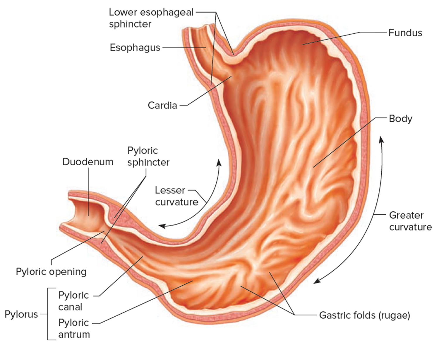
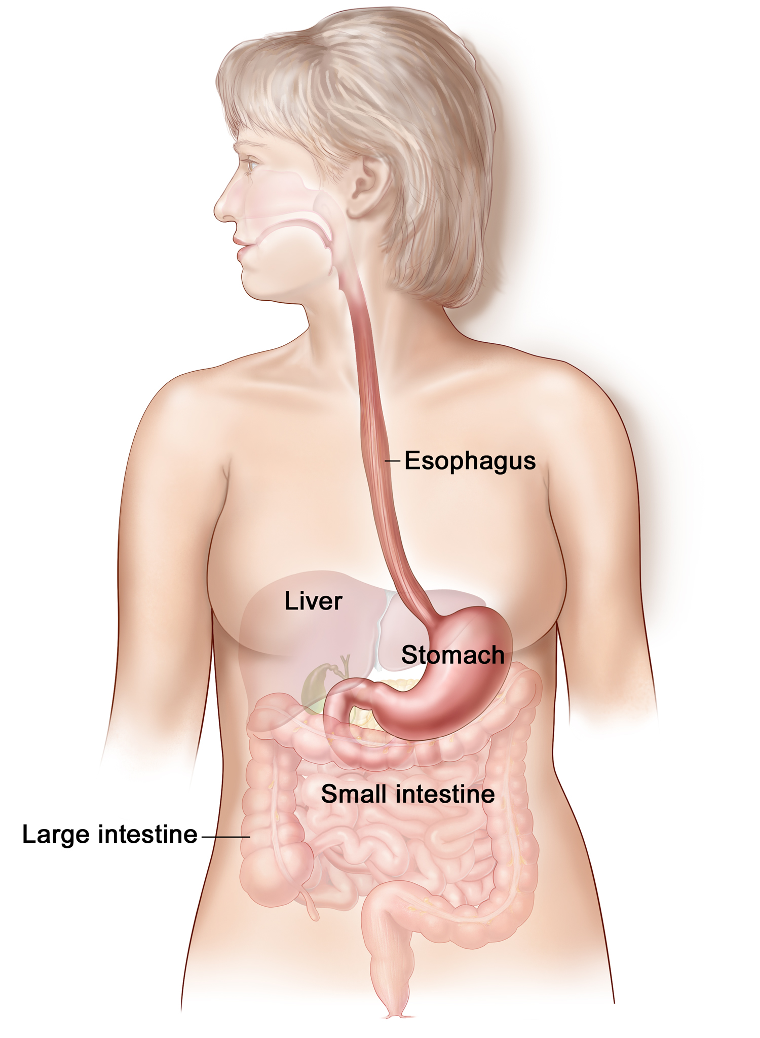
The stomach is a muscular J-shaped pouchlike hollow organ that hangs inferior to the diaphragm in the upper left portion of the abdominal cavity and has a capacity of about 1 liter or more (Figure 1) 1. The stomach’s shape and size vary from person to person, depending on things like people’s sex and build, but also on how much they eat.

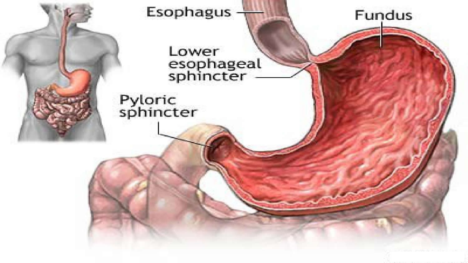
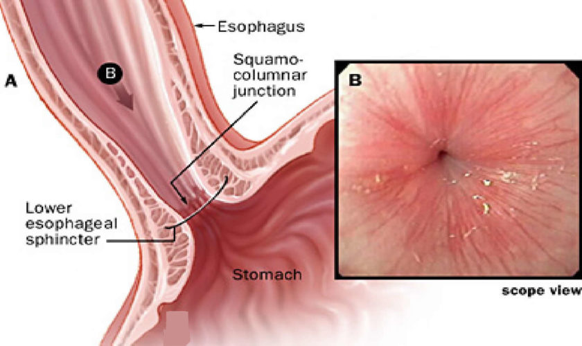
At the point where the esophagus leads into the stomach, the digestive tube is usually kept shut by muscles of the esophagus and diaphragm. When you swallow, these muscles relax and the lower end of the esophagus opens, allowing food to enter the stomach. If this mechanism does not work properly, acidic gastric juice might get into the esophagus, leading to heartburn or an inflammation (see Figure 2).

Thick folds (rugae) of mucosal and submucosal layers mark the stomach’s inner lining and disappear when the stomach wall is distended. The stomach receives food from the esophagus, mixes the food with gastric juice, initiates protein digestion, carries on limited absorption, and moves food into the small intestine.

Figure 1. Stomach

stomach

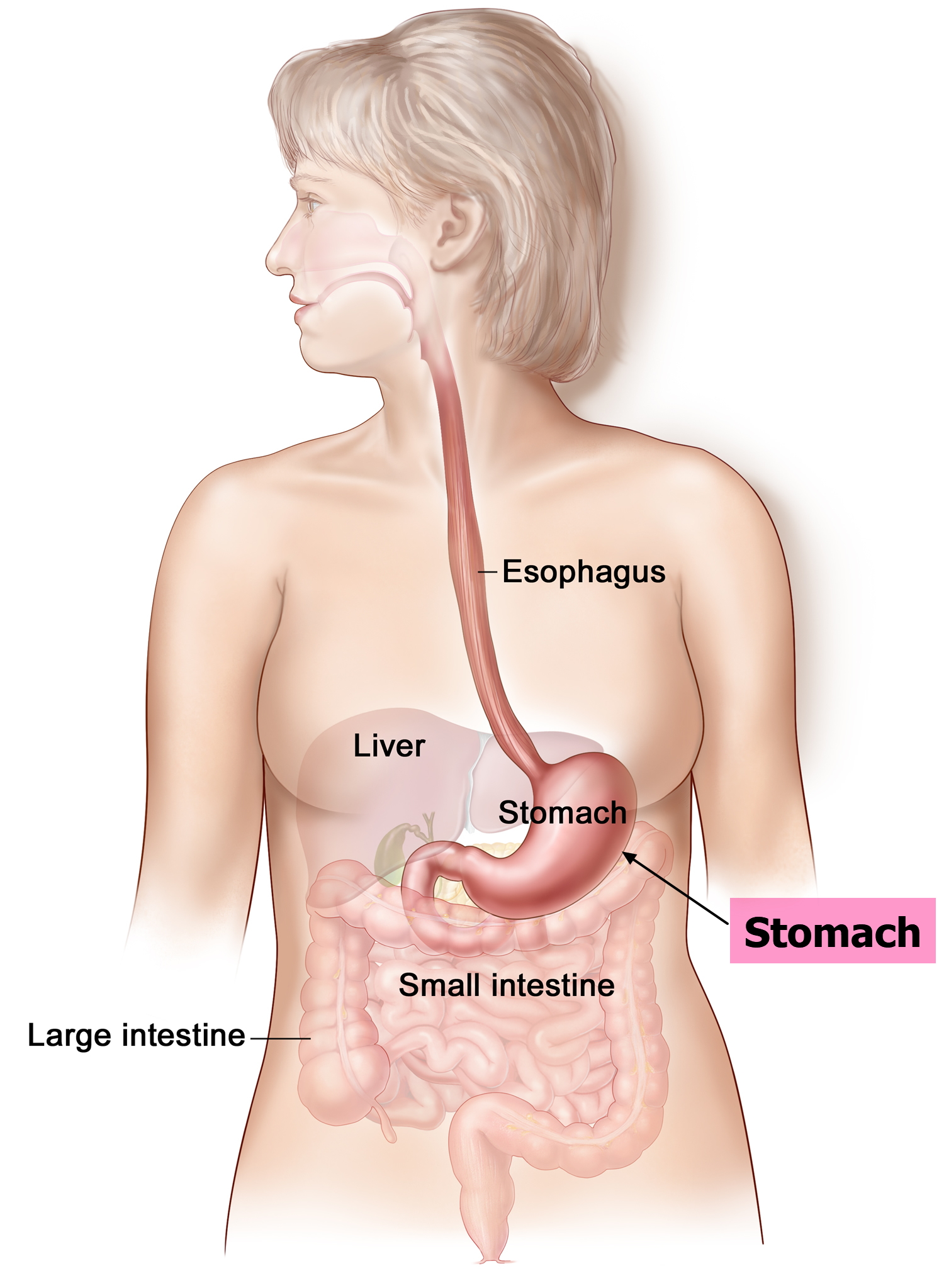
Figure 2. Gastroesophageal junction

lower esophageal sphincter

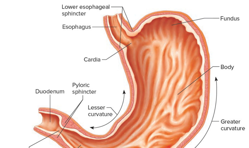
Parts of the Stomach

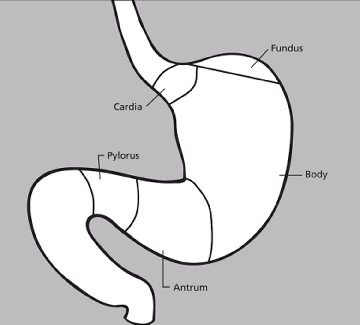
The stomach has 5 parts (Figure 3):

The cardia is a small area near the esophageal opening.

The fundus, which balloons superior to the cardia, is a temporary storage area. It is usually filled with air that enters the stomach when you swallow.

The dilated body region, called the body (corpus), which is the main part of the stomach, lies between the fundus and pylorus. In the body of the stomach food is churned and broken into smaller pieces, mixed with acidic gastric juice and enzymes, and pre-digested.

The antrum – the lower portion (near the intestine), where the food is mixed with gastric juice

The pylorus is the distal portion and the last part of the stomach where it approaches the small intestine. The pyloric canal is a narrowing of the pylorus as it approaches the small intestine. At the end of the pyloric canal the muscular wall thickens, forming a powerful circular muscle, the pyloric sphincter. This muscle is a valve that controls gastric emptying.

The first 3 parts of the stomach (cardia, fundus, and body) are sometimes called the proximal stomach. Some cells in these parts of the stomach make acid and pepsin (a digestive enzyme), the parts of the gastric juice that help digest food. They also make a protein called intrinsic factor, which the body needs to absorb vitamin B12.

The lower 2 parts (antrum and pylorus) are called the distal stomach. The stomach has 2 curves, which form its inner and outer borders. They are called the lesser curvature and greater curvature, respectively.

Figure 3. Parts of the stomach

parts of the stomach

parts of the stomach

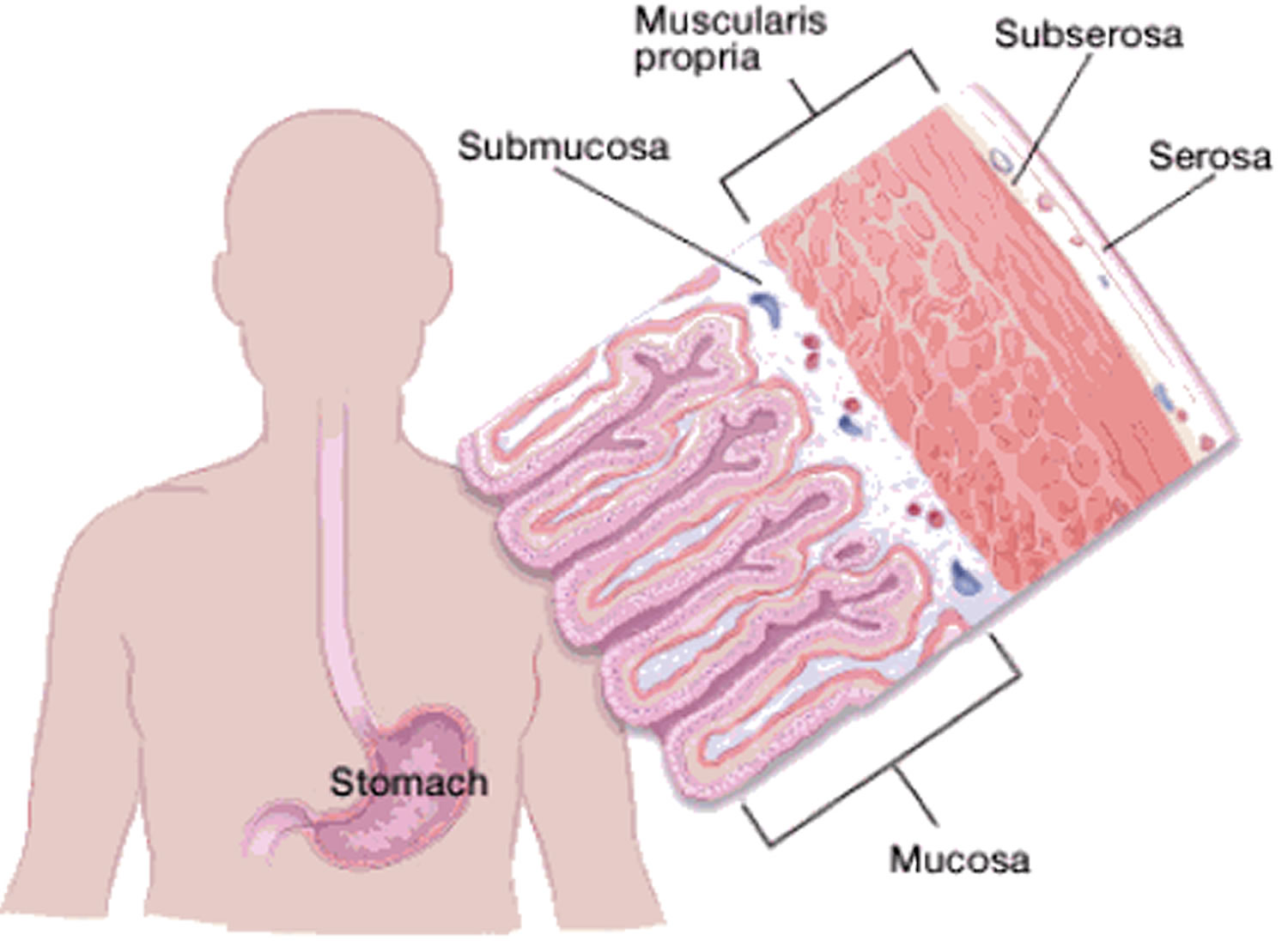
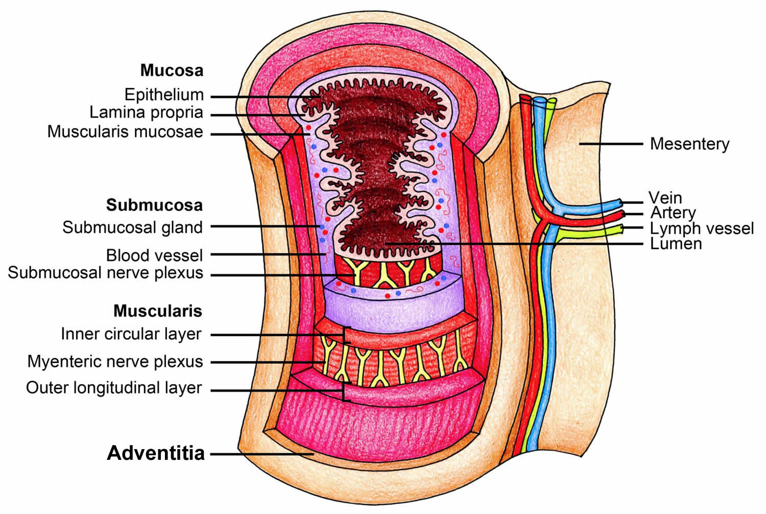
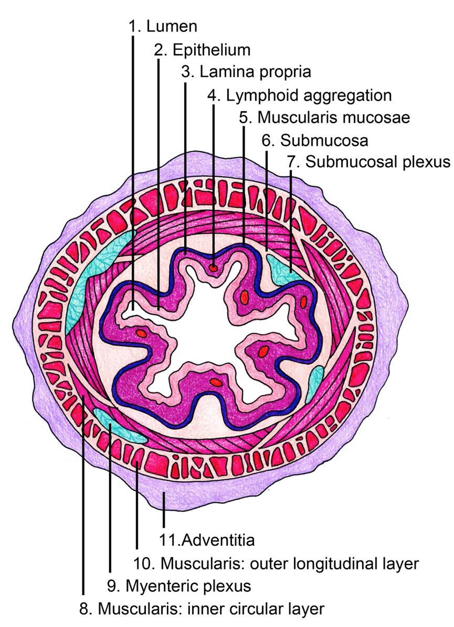
The stomach wall has 5 layers:

The stomach wall is made up of several layers of mucous membrane, connective tissue with blood vessels and nerves, and muscle fibers (see Figure 5).

  • The innermost layer is the mucosa. This is where stomach acid and digestive enzymes are made. Most stomach cancers start in this layer.
  • Next is a supporting layer called the submucosa.
  • Outside of this is the muscularis propria, a thick layer of muscle that moves and mixes the stomach contents.
  • The outer 2 layers, the subserosa and the outermost serosa, wrap the stomach.

The layers are important in determining the stage (extent) of the cancer and in helping to determine a person’s prognosis (outlook). As a cancer grows from the mucosa into deeper layers, the stage becomes more advanced and the prognosis is not as good.

The muscle layer alone has three different sub-layers. The muscles move the contents of the stomach around so vigorously that solid parts of the food are crushed and ground, and mixed into a smooth food pulp.

The inner mucous membrane (lining) has large folds that are visible to the naked eye. These folds run toward the exit of the stomach, providing “pathways” along which liquids can quickly flow through the stomach. If you look at the mucous membrane under a microscope, you can see lots of tiny glands. There are three different types of glands. These glands make digestive enzymes, hydrochloric acid, mucus and bicarbonate.

Gastric juice is made up of digestive enzymes, hydrochloric acid and other substances that are important for absorbing nutrients – about 3 to 4 liters of gastric juice are produced per day. The hydrochloric acid in the gastric juice breaks down the food and the digestive enzymes split up the proteins. The acidic gastric juice also kills bacteria. The mucus covers the stomach wall with a protective coating. Together with the bicarbonate, this ensures that the stomach wall itself is not damaged by the hydrochloric acid.

Figure 4. Stomach anatomy
stomach anatomy

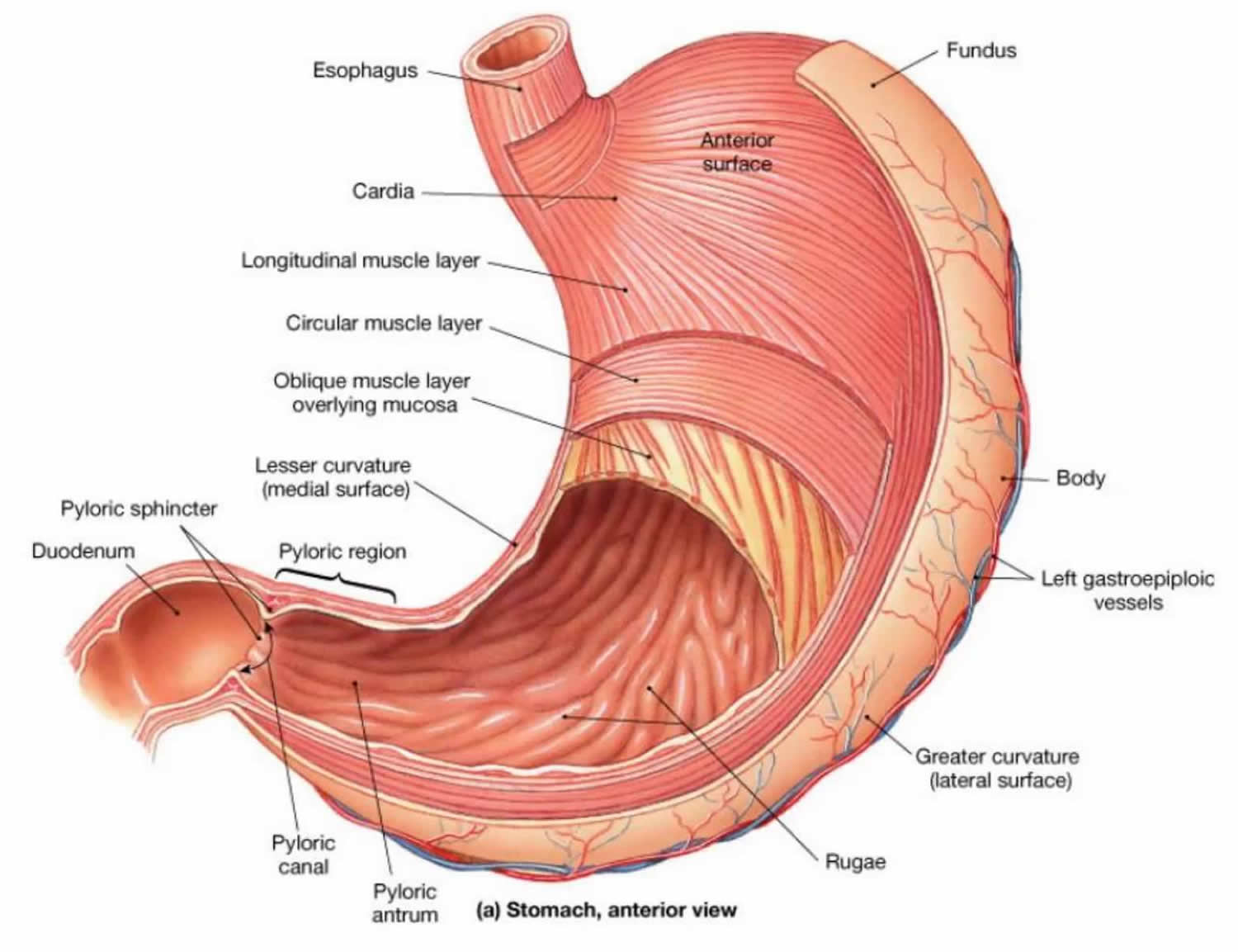
Figure 5. Layers of the stomach

layers of the stomach wall

Stomach function

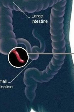
The stomach takes in food from the esophagus (gullet or food pipe), mixes it, breaks it down, and then passes it on to the small intestine in small portions. Following a meal, the mixing movements of the stomach wall aid in producing a semifluid paste of food particles and gastric juice called chyme. Peristaltic waves push the chyme toward the pylorus of the stomach. As chyme accumulates near the pyloric sphincter, the sphincter begins to relax. Stomach contractions push chyme a little at a time into the small intestine.

The rate at which the stomach empties depends on the fluidity of the chyme and the type of food present. Liquids usually pass through the stomach rapidly, but solids remain until they are well mixed with gastric juice. Fatty foods may remain in the stomach from three to six hours; foods high in proteins move through more quickly; carbohydrates usually pass through faster than either fats or proteins.

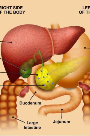
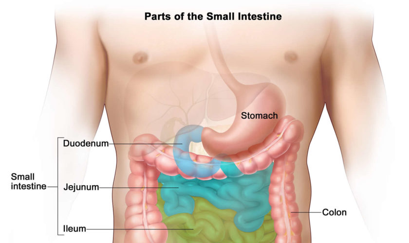
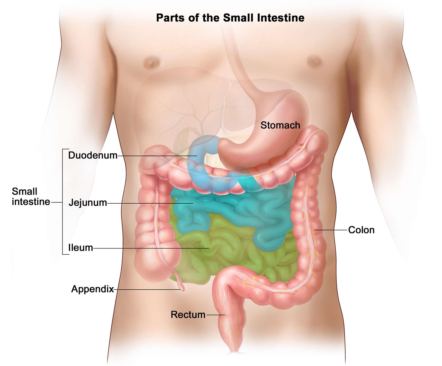
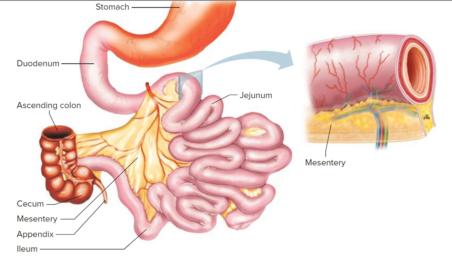
As chyme enters the duodenum (the proximal portion of the small intestine), accessory organs—the pancreas, liver, and gallbladder—add their secretions.

Gastric secretions

The mucous membrane that forms the inner lining of the stomach is thick. Its surface is studded with many small openings called gastric pits located at the ends of tubular gastric glands (Figure 6).

Gastric glands generally contain three types of secretory cells. Mucous cells, in the necks of the glands near the openings of the gastric pits, secrete mucus. Chief cells and parietal cells are in the deeper parts of the glands. The chief cells secrete digestive enzymes, and the parietal cells release a solution containing hydrochloric acid. The products of the mucous cells, chief cells, and parietal cells together form gastric juice.

Pepsin is by far the most important digestive enzyme in gastric juice. The chief cells secrete pepsin in the form of an inactive enzyme precursor called pepsinogen. When pepsinogen contacts hydrochloric acid from the parietal cells, it breaks down rapidly, forming pepsin. Pepsin begins the digestion of nearly all types of dietary protein into polypeptides. This enzyme is most active in an acidic environment, which is provided by the hydrochloric acid in gastric juice.

The mucous cells of the gastric glands (mucous neck cells) and the mucous cells associated with the stomach’s inner surface release a viscous, alkaline secretion that coats the inside of the stomach wall. This coating normally prevents the stomach from digesting itself.

Another component of gastric juice is intrinsic factor, which the parietal cells secrete. Intrinsic factor is necessary for the absorption of vitamin B12 in the small intestine. Table 1 summarizes the major components of gastric juice.

Figure 6. Stomach cells (gastric glands)

stomach cells

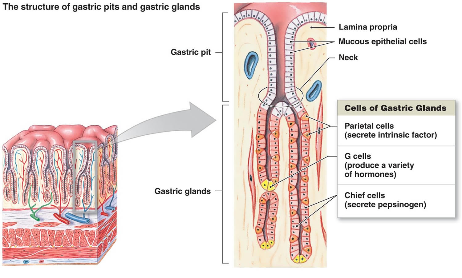
Note: Lining of the stomach. Gastric glands include mucous cells, parietal cells, and chief cells. The mucosa of the stomach is studded with gastric pits that are the openings of the gastric glands.

Table 1. Major Components of Gastric Juice

Component

Source

Function

Pepsinogen

Chief cells of the gastric glands

Inactive form of pepsin

Pepsin

Formed from pepsinogen in the presence of hydrochloric acid

A protein-splitting enzyme that digests nearly all types of dietary protein into polypeptides

Hydrochloric acid

Parietal cells of the gastric glands

Provides the acid environment needed for the production and action of pepsin

Mucus

Mucous cells

Provides a viscous, alkaline protective layer on the stomach’s inner surface

Intrinsic factor

Parietal cells of the gastric glands

Necessary for vitamin B12 absorption in the small intestine

Regulation of Gastric Secretions

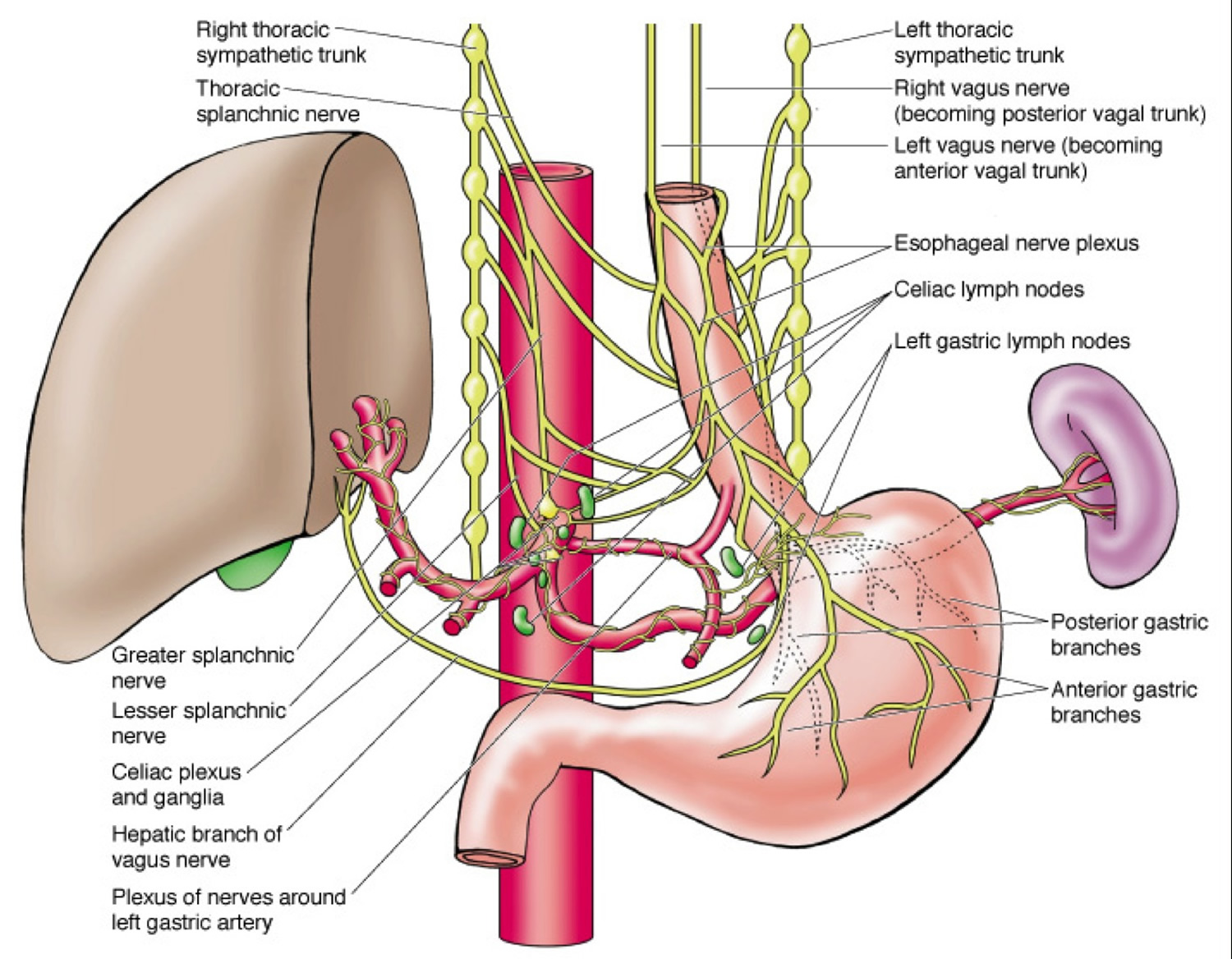
Gastric juice is produced continuously, but the rate varies considerably and is controlled both neurally and hormonally. When a person tastes, smells, or even sees appetizing food, or when food enters the stomach, parasympathetic impulses on the vagus nerves stimulate the release of the neurotransmitter acetylcholine (Ach). This acetylcholine (Ach) stimulates gastric glands to secrete abundant gastric juice, which is rich in hydrochloric acid and pepsinogen. These parasympathetic impulses also stimulate certain stomach cells to release the peptide hormone gastrin, which increases the secretory activity of gastric glands (Figure 6). Gastrin stimulates cell division in the mucosa of the stomach and intestines, which replaces mucosal cells damaged by normal stomach function, disease, or medical treatments.

As food moves into the small intestine, acid triggers sympathetic impulses that inhibit gastric juice secretion. At the same time, proteins and fats in this region of the intestine cause the intestinal wall to release the peptide hormone cholecystokinin. This hormonal action decreases gastric motility as the small intestine fills with food.

Figure 7. Nerve supply to the stomach

nerve supply to the stomach

Gastric Absorption

Gastric enzymes begin breaking down proteins, but the stomach wall is not well adapted to absorb digestive products. The stomach absorbs only small volumes of water and certain salts as well as certain lipid-soluble drugs. Alcohol, which is not a nutrient, is absorbed both in the small intestine and in the stomach.

References
  1. How does the stomach work ? National Center for Biotechnology Information, U.S. National Library of Medicine. https://www.ncbi.nlm.nih.gov/pubmedhealth/PMH0072488/
read more
Digestive SystemEsophagus

What is the esophagus ?

lower esophageal sphincter view

What is the esophagus ?

The esophagus, a straight, collapsible muscular tube, about 25 cm (10 in.) long, that lies posterior to the trachea. The esophagus is a food passageway from the pharynx to the stomach (see Figure 1). The esophagus begins at the base of the laryngopharynx and descends posterior to the trachea, passing through the mediastinum. It penetrates the diaphragm through an opening, the esophageal hiatus and is continuous with the stomach on the abdominal side of the diaphragm.

Mucous glands are scattered throughout the submucosa of the esophagus. Their secretions moisten and lubricate the tube’s inner lining.

Microscopic Anatomy of the Esophagus

Unlike the mouth and pharynx, the esophagus wall (Figure 2) contains all four layers of the alimentary canal: mucosa, submucosa, muscularis externa, and adventitia.

The following histological features are of interest:

  • The mucosal epithelium is a nonkeratinized stratified squamous epithelium. At the junction of the esophagus and stomach, this thick, abrasion-resistant layer changes abruptly to the thin simple columnar epithelium of the stomach, which is specialized for secretion (Figure 2).
  • When the esophagus is empty, its mucosa and submucosa are thrown into longitudinal folds, but during passage of a bolus, these folds flatten out (Figure 3).
  • The submucosa of the wall of the esophagus contains mucous glands, primarily compound tubuloalveolar glands, that extend to the lumen. As a bolus passes, it compresses these glands, causing them to secrete a lubricating mucus. This mucus helps the bolus pass through the esophagus.
  • The muscularis externa consists of skeletal muscle in the superior third of the esophagus, a mixture of skeletal and smooth muscle in the middle third, and smooth muscle in the inferior third. This arrangement is easy to remember if the esophagus is viewed as the zone where the skeletal
    muscle of the mouth and pharynx gives way to the smooth muscle of the stomach and intestines.
  • The most external esophageal layer is an adventitia, not a serosa, because the thoracic segment of the esophagus is not suspended in the peritoneal cavity.

Figure 1. Esophagus

esophagus

Figure 2. Esophagus anatomy

esophagus anatomy

Figure 3. Cross section view of the esophagus

cross section view of the esophagus

Some of the smooth muscle just superior to the point where the esophagus joins the stomach has increased muscle tone, forming the lower esophageal sphincter (LES), or cardiac sphincter (Figures 4 and 5). This smooth muscle usually remains contracted. The sphincter closes the entrance to the stomach, preventing the stomach contents from regurgitating into the esophagus. When peristaltic waves in the esophagus reach the stomach, the sphincter temporarily relaxes and allows the swallowed food to enter.

Figure 4. Lower esophageal sphincter 

lower esophageal sphincter

Figure 5. lower esophageal sphincter (endoscopic view)

lower esophageal sphincter view

What does the esophagus do

The esophagus is the muscular tube that carries food, and liquids from your mouth to the stomach. You may not be aware of your esophagus until you swallow something too large, too hot, or too cold. You may also notice it when something is wrong. You may feel pain or have trouble swallowing.

The most common problem with the esophagus is GERD (gastroesophageal reflux disease). With gastroesophageal reflux disease (GERD), a muscle at the end of your esophagus does not close properly. This allows stomach contents to leak back, or reflux, into the esophagus and irritate it. Over time, gastroesophageal reflux disease (GERD) can cause damage to the esophagus.

Other problems include heartburn, cancer, and esophagitis. Doctors may use various tests to make a diagnosis. These include imaging tests, an upper endoscopy, and a biopsy.

Treatment depends on the problem. Some problems get better with over-the-counter medicines or changes in diet. Others may need prescription medicines or surgery.

read more
Digestive SystemDrugsDrugs & SupplementsSmall and Large Intestine

Small intestine cancer

small intestine cancer

Small intestine cancer

Small intestine cancer is a rare disease in which malignant (cancer) cells form in the tissues of the small intestine 1.

The small intestine (also called small bowel) is part of the body’s digestive system, which also includes the esophagus, stomach, and large intestine. The digestive system removes and processes nutrients (vitamins, minerals, carbohydrates, fats, proteins, and water) from foods and helps pass waste material out of the body.

The small intestine is a long tube that connects the stomach to the large intestine. It folds many times to fit inside the abdomen. The small intestine consists of three parts: the duodenum, the jejunum, and the ileum (Figure 1). The duodenum, about 25 centimeters long and 5 centimeters in diameter, lies posterior to the parietal peritoneum and is the most fixed portion of the small intestine. It follows a C-shaped path as it passes anterior to the right kidney and the upper three lumbar vertebrae. The remainder of the small intestine is mobile and lies free in the peritoneal cavity. The proximal two-fifths of this portion of the small intestine is the jejunum, and the remainder is the ileum. The jejunum and ileum are not easily distinguished as separate parts; however, the diameter of the jejunum is typically greater than that of the ileum, and its wall is thicker, more vascular, and more active.

A double-layered fold of peritoneal membrane called mesentery suspends the jejunum and ileum from the posterior abdominal wall (Figure 2). The mesentery supports the blood vessels, nerves, and lymphatic vessels that supply the intestinal wall. A filmy, double fold of peritoneal membrane called the greater omentum drapes like an apron from the stomach over the transverse colon and the folds of the small intestine.

Estimated new cases and deaths from small intestine cancer in the United States in 2017 2:

  • New cases: 10,190.
  • Deaths: 1,390.

There are five types of small intestine cancer. The types of cancer found in the small intestine are:

  1. Adenocarcinoma (majority of cases).
  2. Lymphoma (uncommon), which is usually of the non-Hodgkin type.
  3. Sarcoma (most commonly leiomyosarcoma and more rarely angiosarcoma or liposarcoma).
  4. Gastrointestinal stromal tumor.
  5. Carcinoid tumors.

Approximately 25% to 50% of the primary malignant tumors in the small intestine are adenocarcinomas, and most occur in the duodenum 3. Small intestine carcinomas may occur synchronously (existing at the same time) or metachronously (multiple separate occurrences at different intervals) at multiple sites 4.

Leiomyosarcoma starts in the smooth muscle cells of the small intestine. Most of these tumors occur in the part of the small intestine near the large intestine most often in the ileum 4.

Some 20% of malignant lesions of the small intestine are carcinoid tumors, which occur more frequently in the ileum than in the duodenum or jejunum and may be multiple 4.

It is uncommon to find malignant lymphoma as a solitary small intestine lesion 4.

Together they account for the majority of small intestine malignancies, which, as a whole, account for only 1% to 2% of all gastrointestinal malignancies 5, 6, 7, 8.

Diet and health history can affect the risk of developing small intestine cancer

Anything that increases your risk of getting a disease is called a risk factor. Having a risk factor does not mean that you will get cancer; not having risk factors doesn’t mean that you will not get cancer. Talk with your doctor if you think you may be at risk. Risk factors for small intestine cancer include the following:

  • Eating a high-fat diet.
  • Having Crohn disease.
  • Having celiac disease.
  • Having familial adenomatous polyposis.

Small intestine cancer survival rate

The prognosis (chance of recovery) and treatment options depend on the following:

  • The type of small intestine cancer.
  • Whether the cancer is in the inner lining of the small intestine only or has spread into or beyond the wall of the small intestine.
  • Whether the cancer has spread to other places in the body, such as the lymph nodes, liver, or peritoneum (tissue that lines the wall of the abdomen and covers most of the organs in the abdomen).
  • Whether the cancer can be completely removed by surgery.
  • Whether the cancer is newly diagnosed or has recurred.

As in other gastrointestinal malignancies, the predominant modality of treatment is surgery when resection is possible, and cure relates to the ability to completely resect the cancer. The overall 5-year survival rate for resectable adenocarcinoma is only 20%. The 5-year survival rate for resectable leiomyosarcoma, the most common primary sarcoma of the small intestine, is approximately 50% 1.

Figure 1.  Parts of the small intestine

small intestine cancer

Figure 2. Small intestines

symptoms of small intestine cancer

Stage Information for Small Intestine Cancer

Staging is used to find out how far the cancer has spread, but treatment decisions are not based on stage.

There are three ways that cancer spreads in the body.

Cancer can spread through tissue, the lymph system, and the blood:

  1. Tissue. The cancer spreads from where it began by growing into nearby areas.
  2. Lymph system. The cancer spreads from where it began by getting into the lymph system. The cancer travels through the lymph vessels to other parts of the body.
  3. Blood. The cancer spreads from where it began by getting into the blood. The cancer travels through the blood vessels to other parts of the body.

The American Joint Committee on Cancer has designated staging by TNM classification to define small intestine cancer 3.

Table 1. Primary Tumor (T)

TXPrimary tumor cannot be assessed.
T0No evidence of primary tumor.
TisCarcinoma in situ.
T1aTumor invades lamina propria.
T1bTumor invades submucosa. (a)
T2Tumor invades muscularis propria.
T3Tumor invades through the muscularis propria into the subserosa or into the nonperitonealized perimuscular tissue (mesentery or retroperitoneum) with extension ≤2 cm. (a)
T4Tumor perforates the visceral peritoneum or directly invades other organs or structures (includes other loops of small intestine, mesentery, or retroperitoneum >2 cm, and abdominal wall by way of serosa; for duodenum only, invasion of pancreas or bile duct).

(a): The nonperitonealized perimuscular tissue is, for jejunum and ileum, part of the mesentery and, for duodenum in areas where serosa is lacking, part of the interface with the pancreas.

[Source 3]

Table 2. Regional Lymph Nodes (N)

NXRegional lymph nodes cannot be assessed.
N0No regional lymph node metastasis.
N1Metastasis in 1–3 regional lymph nodes.
N2Metastases in ≥4 regional lymph nodes.
[Source 3]

Table 3. Distant Metastasis (M)

M0No distant metastasis.
M1Distant metastasis.
[Source 3]

Table 4. Anatomic Stage/Prognostic Groups

StageTNM
0TisN0M0
IT1N0M0
T2N0M0
IIAT3N0M0
IIBT4N0M0
IIIAAny TN1M0
IIIBAny TN2M0
IVAny TAny NM1
[Source 3]

Cancer may spread from where it began to other parts of the body.

When cancer spreads to another part of the body, it is called metastasis. Cancer cells break away from where they began (the primary tumor) and travel through the lymph system or blood.

  • Lymph system. The cancer gets into the lymph system, travels through the lymph vessels, and forms a tumor (metastatic tumor) in another part of the body.
  • Blood. The cancer gets into the blood, travels through the blood vessels, and forms a tumor (metastatic tumor) in another part of the body.

The metastatic tumor is the same type of cancer as the primary tumor. For example, if small intestine cancer spreads to the liver, the cancer cells in the liver are actually small intestine cancer cells. The disease is metastatic small intestine cancer, not liver cancer.

Small intestine cancer is grouped according to whether or not the tumor can be completely removed by surgery.

Treatment depends on whether the tumor can be removed by surgery and if the cancer is being treated as a primary tumor or is metastatic cancer.

  • Recurrent Small Intestine Cancer

Recurrent small intestine cancer is cancer that has recurred (come back) after it has been treated. The cancer may come back in the small intestine or in other parts of the body.

Small intestine cancer signs and symptoms

Signs and symptoms of small intestine cancer include unexplained weight loss and abdominal pain.

These and other signs and symptoms may be caused by small intestine cancer or by other conditions. Check with your doctor if you have any of the following:

  • Pain or cramps in the middle of the abdomen.
  • Weight loss with no known reason.
  • A lump in the abdomen.
  • Blood in the stool.

Tests that examine the small intestine are used to detect (find), diagnose, and stage small intestine cancer

Procedures that make pictures of the small intestine and the area around it help diagnose small intestine cancer and show how far the cancer has spread. The process used to find out if cancer cells have spread within and around the small intestine is called staging.

In order to plan treatment, it is important to know the type of small intestine cancer and whether the tumor can be removed by surgery. Tests and procedures to detect, diagnose, and stage small intestine cancer are usually done at the same time. The following tests and procedures may be used:

  • Physical exam and history : An exam of the body to check general signs of health, including checking for signs of disease, such as lumps or anything else that seems unusual. A history of the patient’s health habits and past illnesses and treatments will also be taken.
  • Blood chemistry studies : A procedure in which a blood sample is checked to measure the amounts of certain substances released into the blood by organs and tissues in the body. An unusual (higher or lower than normal) amount of a substance can be a sign of disease.
  • Liver function tests : A procedure in which a blood sample is checked to measure the amounts of certain substances released into the blood by the liver. A higher than normal amount of a substance can be a sign of liver disease that may be caused by small intestine cancer.
  • Endoscopy : A procedure to look at organs and tissues inside the body to check for abnormal areas. There are different types of endoscopy:

Upper endoscopy : A procedure to look at the inside of the esophagus, stomach, and duodenum (first part of the small intestine, near the stomach). An endoscope is inserted through the mouth and into the esophagus, stomach, and duodenum. An endoscope is a thin, tube-like instrument with a light and a lens for viewing. It may also have a tool to remove tissue samples, which are checked under a microscope for signs of cancer.

Capsule endoscopy : A procedure to look at the inside of the small intestine. A capsule that is about the size of a large pill and contains a light and a tiny wireless camera is swallowed by the patient. The capsule travels through the digestive tract, including the small intestine, and sends many pictures of the inside of the digestive tract to a recorder that is worn around the waist or over the shoulder. The pictures are sent from the recorder to a computer and viewed by the doctor who checks for signs of cancer. The capsule passes out of the body during a bowel movement.

Double balloon endoscopy : A procedure to look at the inside of the small intestine. A special instrument made up of two tubes (one inside the other) is inserted through the mouth or rectum and into the small intestine. The inside tube (an endoscope with a light and lens for viewing) is moved through part of the small intestine and a balloon at the end of it is inflated to keep the endoscope in place. Next, the outer tube is moved through the small intestine to reach the end of the endoscope, and a balloon at the end of the outer tube is inflated to keep it in place. Then, the balloon at the end of the endoscope is deflated and the endoscope is moved through the next part of the small intestine. These steps are repeated many times as the tubes move through the small intestine. The doctor is able to see the inside of the small intestine through the endoscope and use a tool to remove samples of abnormal tissue. The tissue samples are checked under a microscope for signs of cancer. This procedure may be done if the results of a capsule endoscopy are abnormal. This procedure is also called double balloon enteroscopy.

  • Laparotomy : A surgical procedure in which an incision (cut) is made in the wall of the abdomen to check the inside of the abdomen for signs of disease. The size of the incision depends on the reason the laparotomy is being done. Sometimes organs or lymph nodes are removed or tissue samples are taken and checked under a microscope for signs of disease.
  • Biopsy : The removal of cells or tissues so they can be viewed under a microscope to check for signs of cancer. This may be done during an endoscopy or laparotomy. The sample is checked by a pathologist to see if it contains cancer cells.
  • Upper GI series with small bowel follow-through: A series of x-rays of the esophagus, stomach, and small bowel. The patient drinks a liquid that contains barium (a silver-white metallic compound). The liquid coats the esophagus, stomach, and small bowel. X-rays are taken at different times as the barium travels through the upper GI tract and small bowel.
  • CT scan (CAT scan): A procedure that makes a series of detailed pictures of areas inside the body, taken from different angles. The pictures are made by a computer linked to an x-ray machine. A dye may be injected into a vein or swallowed to help the organs or tissues show up more clearly. This procedure is also called computed tomography, computerized tomography, or computerized axial tomography.
  • MRI (magnetic resonance imaging): A procedure that uses a magnet, radio waves, and a computer to make a series of detailed pictures of areas inside the body. This procedure is also called nuclear magnetic resonance imaging (NMRI).

Small intestine cancer treatment

Different types of treatments are available for patients with small intestine cancer 9. Some treatments are standard (the currently used treatment), and some are being tested in clinical trials. A treatment clinical trial is a research study meant to help improve current treatments or obtain information on new treatments for patients with cancer. When clinical trials show that a new treatment is better than the standard treatment, the new treatment may become the standard treatment. Patients may want to think about taking part in a clinical trial. Some clinical trials are open only to patients who have not started treatment.

Three types of standard treatment are used:

Surgery

Surgery is the most common treatment of small intestine cancer. One of the following types of surgery may be done:

  • Resection: Surgery to remove part or all of an organ that contains cancer. The resection may include the small intestine and nearby organs (if the cancer has spread). The doctor may remove the section of the small intestine that contains cancer and perform an anastomosis (joining the cut ends of the intestine together). The doctor will usually remove lymph nodes near the small intestine and examine them under a microscope to see whether they contain cancer.
  • Bypass: Surgery to allow food in the small intestine to go around (bypass) a tumor that is blocking the intestine but cannot be removed.

Even if the doctor removes all the cancer that can be seen at the time of the surgery, some patients may be given radiation therapy after surgery to kill any cancer cells that are left. Treatment given after the surgery, to lower the risk that the cancer will come back, is called adjuvant therapy.

Radiation therapy

Radiation therapy is a cancer treatment that uses high-energy x-rays or other types of radiation to kill cancer cells or keep them from growing. There are two types of radiation therapy:

  • External radiation therapy uses a machine outside the body to send radiation toward the cancer.
  • Internal radiation therapy uses a radioactive substance sealed in needles, seeds, wires, or catheters that are placed directly into or near the cancer.

The way the radiation therapy is given depends on the type of the cancer being treated. External radiation therapy is used to treat small intestine cancer.

Chemotherapy

Chemotherapy is a cancer treatment that uses drugs to stop the growth of cancer cells, either by killing the cells or by stopping them from dividing. When chemotherapy is taken by mouth or injected into a vein or muscle, the drugs enter the bloodstream and can reach cancer cells throughout the body (systemic chemotherapy). When chemotherapy is placed directly into the cerebrospinal fluid, an organ, or a body cavity such as the abdomen, the drugs mainly affect cancer cells in those areas (regional chemotherapy). The way the chemotherapy is given depends on the type and stage of the cancer being treated.

New types of treatment are being tested in clinical trials

Not every new treatment being studied are listed here because new treatment modalities are constantly being added.

Biologic therapy

Biologic therapy is a treatment that uses the patient’s immune system to fight cancer. Substances made by the body or made in a laboratory are used to boost, direct, or restore the body’s natural defenses against cancer. This type of cancer treatment is also called biotherapy or immunotherapy.

Radiation therapy with radiosensitizers

Radiosensitizers are drugs that make tumor cells more sensitive to radiation therapy. Combining radiation therapy with radiosensitizers may kill more tumor cells.

Patients may want to think about taking part in a clinical trial.

For some patients, taking part in a clinical trial may be the best treatment choice. Clinical trials are part of the cancer research process. Clinical trials are done to find out if new cancer treatments are safe and effective or better than the standard treatment.

Many of today’s standard treatments for cancer are based on earlier clinical trials. Patients who take part in a clinical trial may receive the standard treatment or be among the first to receive a new treatment.

Patients who take part in clinical trials also help improve the way cancer will be treated in the future. Even when clinical trials do not lead to effective new treatments, they often answer important questions and help move research forward.
Patients can enter clinical trials before, during, or after starting their cancer treatment.

Some clinical trials only include patients who have not yet received treatment. Other trials test treatments for patients whose cancer has not gotten better. There are also clinical trials that test new ways to stop cancer from recurring (coming back) or reduce the side effects of cancer treatment.

Clinical trials are taking place in many parts of the country. Speak to your healthcare provider about the treatment options available including being involved as part of treatment clinical trials.

Follow-up tests may be needed.

Some of the tests that were done to diagnose the cancer or to find out the stage of the cancer may be repeated. Some tests will be repeated in order to see how well the treatment is working. Decisions about whether to continue, change, or stop treatment may be based on the results of these tests.

Some of the tests will continue to be done from time to time after treatment has ended. The results of these tests can show if your condition has changed or if the cancer has recurred (come back). These tests are sometimes called follow-up tests or check-ups.

Small Intestine Adenocarcinoma Treatment

When possible, treatment of small intestine adenocarcinoma will be surgery to remove the tumor and some of the normal tissue around it 10.

Treatment of small intestine adenocarcinoma that cannot be removed by surgery may include the following:

  • Surgery to bypass the tumor.
  • Radiation therapy as palliative therapy to relieve symptoms and improve the patient’s quality of life.
  • A clinical trial of radiation therapy with radiosensitizers, with or without chemotherapy.
  • A clinical trial of new anticancer drugs.
  • A clinical trial of biologic therapy.

Speak to your healthcare provider about the treatment options available including being involved as part of treatment clinical trials.

Small Intestine Leiomyosarcoma Treatment

When possible, treatment of small intestine leiomyosarcoma will be surgery to remove the tumor and some of the normal tissue around it.

Treatment of small intestine leiomyosarcoma that cannot be removed by surgery may include the following:

  • Surgery (to bypass the tumor) and radiation therapy.
  • Surgery, radiation therapy, or chemotherapy as palliative therapy to relieve symptoms and improve the patient’s quality of life.
  • A clinical trial of new anticancer drugs.
  • A clinical trial of biologic therapy.

Speak to your healthcare provider about the treatment options available including being involved as part of treatment clinical trials.

Recurrent Small Intestine Cancer

At the present time, no standard effective chemotherapy exists for patients with recurrent metastatic adenocarcinoma or leiomyosarcoma of the small intestine. These types of patients should be considered candidates for clinical trials evaluating the use of new anticancer drugs or biologic therapy in phase I and phase II trials 11.

Treatment of locally recurrent small intestine cancer may include the following:

  • Surgery.
  • Radiation therapy or chemotherapy as palliative therapy to relieve symptoms and improve the patient’s quality of life.
  • A clinical trial of radiation therapy with radiosensitizers, with or without chemotherapy.

Speak to your healthcare provider about the treatment options available including being involved as part of treatment clinical trials.

Gastrointestinal stromal tumor

Gastrointestinal stromal tumor is a disease in which abnormal cells form in the tissues of the gastrointestinal tract 12. Gastrointestinal stromal tumors may be malignant (cancer) or benign (not cancer) 12. They are most common in the stomach and small intestine but may be found anywhere in or near the gastrointestinal tract. Some scientists believe that gastrointestinal stromal tumors begin in cells called interstitial cells of Cajal, in the wall of the gastrointestinal tract 12.

Although they comprise fewer than 1% of all gastrointestinal tumors, gastrointestinal stromal tumor are the most common mesenchymal tumors of the gastrointestinal tract 13. It has been estimated that there are 3,300 to 6,000 new gastrointestinal stromal tumor cases per year in the United States 14. A study based on Surveillance, Epidemiology and End Results registry data found that the age-adjusted yearly incidence of gastrointestinal stromal tumor in the United States was 6.8 per million from 1992 to 2000 15. However, the true incidence is not known, in part because many tumors have not been tested for the characteristic KIT or platelet-derived growth factor receptor alpha gene mutations. In addition, small, indolent gastrointestinal stromal tumor, only a few millimeters in diameter, are common in the general population and are not included in cancer registries 16. Gastrointestinal stromal tumor are equally distributed across all geographic and ethnic groups and men and women are equally affected. Most patients present between the ages of 50 and 80 17. The vast majority of gastrointestinal stromal tumor are sporadic, but there are rare familial forms associated with the characteristic heritable mutations in the KIT gene (or, rarely, in succinate dehydrogenase genes in Carney-Stratakis syndrome). Familial gastrointestinal stromal tumor may present as multiple primary tumors.

Gastrointestinal stromal tumor may be part of a genetic syndrome, but this is rare. A genetic syndrome is a set of symptoms or conditions that occur together and is usually caused by abnormal genes. The following genetic syndromes have been linked to gastrointestinal stromal tumor:

  • Neurofibromatosis type 1 (NF1).
  • Carney triad.

Gastrointestinal stromal tumor can occur anywhere along the GI tract, but most often are found in the stomach or small intestine. The American Joint Committee on Cancer Cancer Staging Manual lists the following approximate distributions 18:

  • Stomach (60%).
  • Small intestine (30%).
  • Rectum (3%).
  • Colon (1–2%).
  • Esophagus (<1%).
  • Omentum/mesentery (rare).

Less frequently, gastrointestinal stromal tumor may arise in the appendix, gallbladder, pancreas, retroperitoneum, and paravaginal and periprostatic tissues 19. Approximately 20% to 25% of gastric gastrointestinal stromal tumor and 40% to 50% of small intestinal gastrointestinal stromal tumor are clinically aggressive 20. It has been estimated that approximately 10% to 25% of patients present with metastatic disease 21.

Signs and symptoms of gastrointestinal stromal tumors

The clinical presentation of patients with GIST varies depending on the anatomic location of the tumor and the tumor size and aggressiveness 22. The most common presentation of GIST is GI bleeding, which may be acute (melena or hematemesis) or chronic and results in anemia 20. Signs and symptoms of gastrointestinal stromal tumors include blood in the stool or vomit.

These and other signs and symptoms may be caused by a gastrointestinal stromal tumor or by other conditions. Check with your doctor if you have any of the following:

  • Blood (either bright red or very dark) in the stool or vomit.
  • Pain in the abdomen, which may be severe.
  • Feeling very tired.
  • Trouble or pain when swallowing.
  • Feeling full after only a little food is eaten.

Tests that examine the GI tract are used to detect (find) and diagnose gastrointestinal stromal tumors.

The following tests and procedures may be used:

  • Physical exam and history : An exam of the body to check general signs of health, including checking for signs of disease, such as lumps or anything else that seems unusual. A history of the patient’s health habits and past illnesses and treatments will also be taken.
  • CT scan (CAT scan): A procedure that makes a series of detailed pictures of areas inside the body, taken from different angles. The pictures are made by a computer linked to an x-ray machine. A dye may be injected into a vein or swallowed to help the organs or tissues show up more clearly. This procedure is also called computed tomography, computerized tomography, or computerized axial tomography.
  • MRI (magnetic resonance imaging): A procedure that uses a magnet, radio waves, and a computer to make a series of detailed pictures of areas inside the body. This procedure is also called nuclear magnetic resonance imaging (NMRI).
  • Endoscopic ultrasound and biopsy : Endoscopy and ultrasound are used to make an image of the upper GI tract and a biopsy is done. An endoscope (a thin, tube-like instrument with a light and a lens for viewing) is inserted through the mouth and into the esophagus, stomach, and first part of the small intestine. A probe at the end of the endoscope is used to bounce high-energy sound waves (ultrasound) off internal tissues or organs and make echoes. The echoes form a picture of body tissues called a sonogram. This procedure is also called endosonography. Guided by the sonogram, the doctor removes tissue using a thin, hollow needle. A pathologist views the tissue under a microscope to look for cancer cells.

If cancer is found, the following tests may be done to study the cancer cells:

  • Immunohistochemistry : A test that uses antibodies to check for certain antigens in a sample of tissue. The antibody is usually linked to a radioactive substance or a dye that causes the tissue to light up under a microscope. This type of test may be used to tell the difference between different types of cancer.
  • Mitotic rate : A measure of how fast the cancer cells are dividing and growing. The mitotic rate is found by counting the number of cells dividing in a certain amount of cancer tissue.

Certain factors affect prognosis (chance of recovery) and treatment options.

The prognosis (chance of recovery) and treatment options depend on the following:

  • How quickly the cancer cells are growing and dividing.
  • The size of the tumor.
  • Where the tumor is in the body.
  • Whether the tumor can be completely removed by surgery.
  • Whether the tumor has spread to other parts of the body.

Stages of Gastrointestinal Stromal Tumors

After a gastrointestinal stromal tumor has been diagnosed, tests are done to find out if cancer cells have spread within the gastrointestinal tract or to other parts of the body.

The process used to find out if cancer has spread within the gastrointestinal (GI) tract or to other parts of the body is called staging. The information gathered from the staging process determines the stage of the disease. The following tests and procedures may be used in the staging process:

  • PET scan (positron emission tomography scan): A procedure to find malignant tumor cells in the body. A small amount of radioactive glucose (sugar) is injected into a vein. The PET scanner rotates around the body and makes a picture of where glucose is being used in the body. Malignant tumor cells show up brighter in the picture because they are more active and take up more glucose than normal cells do.
  • CT scan (CAT scan): A procedure that makes a series of detailed pictures of areas inside the body, taken from different angles. The pictures are made by a computer linked to an x-ray machine. A dye may be injected into a vein or swallowed to help the organs or tissues show up more clearly. This procedure is also called computed tomography, computerized tomography, or computerized axial tomography.
  • MRI (magnetic resonance imaging): A procedure that uses a magnet, radio waves, and a computer to make a series of detailed pictures of areas inside the body. This procedure is also called nuclear magnetic resonance imaging (NMRI).
  • Chest x-ray : An x-ray of the organs and bones inside the chest. An x-ray is a type of energy beam that can go through the body and onto film, making a picture of areas inside the body.
  • Bone scan : A procedure to check if there are rapidly dividing cells, such as cancer cells, in the bone. A very small amount of radioactive material is injected into a vein and travels through the bloodstream. The radioactive material collects in the bones and is detected by a scanner.

There are three ways that cancer spreads in the body.

Cancer can spread through tissue, the lymph system, and the blood:

  • Tissue. The cancer spreads from where it began by growing into nearby areas.
  • Lymph system. The cancer spreads from where it began by getting into the lymph system. The cancer travels through the lymph vessels to other parts of the body.
  • Blood. The cancer spreads from where it began by getting into the blood. The cancer travels through the blood vessels to other parts of the body.

Cancer may spread from where it began to other parts of the body.

When cancer spreads to another part of the body, it is called metastasis. Cancer cells break away from where they began (the primary tumor) and travel through the lymph system or blood.

  • Lymph system. The cancer gets into the lymph system, travels through the lymph vessels, and forms a tumor (metastatic tumor) in another part of the body.
  • Blood. The cancer gets into the blood, travels through the blood vessels, and forms a tumor (metastatic tumor) in another part of the body.

The metastatic tumor is the same type of tumor as the primary tumor. For example, if a gastrointestinal stromal tumor (GIST) spreads to the liver, the tumor cells in the liver are actually GIST cells. The disease is metastatic GIST, not liver cancer.

The results of diagnostic and staging tests are used to plan treatment.

For many cancers it is important to know the stage of the cancer in order to plan treatment. However, the treatment of GIST is not based on the stage of the cancer. Treatment is based on whether the tumor can be removed by surgery and if the tumor has spread to other parts of the abdomen or to distant parts of the body.

Treatment is based on whether the tumor is:

  • Resectable: These tumors can be removed by surgery .
  • Unresectable: These tumors cannot be completely removed by surgery.
  • Metastatic and recurrent: Metastatic tumors have spread to other parts of the body. Recurrent tumors have recurred (come back) after treatment.
  • Recurrent GISTs may come back in the gastrointestinal tract or in other parts of the body. They are usually found in the abdomen, peritoneum, and/or liver.
  • Refractory: These tumors have not gotten better with treatment.

Treatment Options for Gastrointestinal stromal tumor

Different types of treatments are available for patients with gastrointestinal stromal tumors (GISTs). Some treatments are standard (the currently used treatment), and some are being tested in clinical trials. A treatment clinical trial is a research study meant to help improve current treatments or obtain information on new treatments for patients with cancer. When clinical trials show that a new treatment is better than the standard treatment, the new treatment may become the standard treatment. Patients may want to think about taking part in a clinical trial. Some clinical trials are open only to patients who have not started treatment.

Speak to your healthcare provider about the treatment options available including being involved as part of treatment clinical trials.

Four types of standard treatment are used:

  • Surgery

If the GIST has not spread and is in a place where surgery can be safely done, the tumor and some of the tissue around it may be removed. Sometimes surgery is done using a laparoscope (a thin, lighted tube) to see inside the body. Small incisions (cuts) are made in the wall of the abdomen and a laparoscope is inserted into one of the incisions. Instruments may be inserted through the same incision or through other incisions to remove organs or tissues.

  • Targeted therapy

Targeted therapy is a type of treatment that uses drugs or other substances to identify and attack specific cancer cells without harming normal cells.

Tyrosine kinase inhibitors (TKIs) are targeted therapy drugs that block signals needed for tumors to grow. TKIs may be used to treat GISTs that cannot be removed by surgery or to shrink GISTs so they become small enough to be removed by surgery. Imatinib mesylate and sunitinib are two TKIs used to treat GISTs. TKIs are sometimes given for as long as the tumor does not grow and serious side effects do not occur.

  • Watchful waiting

Watchful waiting is closely monitoring a patient’s condition without giving any treatment until signs or symptoms appear or change.

  • Supportive care

If a GIST gets worse during treatment or there are side effects, supportive care is usually given. The goal of supportive care is to prevent or treat the symptoms of a disease, side effects caused by treatment, and psychological, social, and spiritual problems related to a disease or its treatment. Supportive care helps improve the quality of life of patients who have a serious or life-threatening disease. Radiation therapy is sometimes given as supportive care to relieve pain in patients with large tumors that have spread.

  • New types of treatment are being tested in clinical trials

For some patients, taking part in a clinical trial may be the best treatment choice. Clinical trials are part of the cancer research process. Clinical trials are done to find out if new cancer treatments are safe and effective or better than the standard treatment.

Many of today’s standard treatments for cancer are based on earlier clinical trials. Patients who take part in a clinical trial may receive the standard treatment or be among the first to receive a new treatment.

Patients who take part in clinical trials also help improve the way cancer will be treated in the future. Even when clinical trials do not lead to effective new treatments, they often answer important questions and help move research forward.

Patients can enter clinical trials before, during, or after starting their cancer treatment.

Some clinical trials only include patients who have not yet received treatment. Other trials test treatments for patients whose cancer has not gotten better. There are also clinical trials that test new ways to stop cancer from recurring (coming back) or reduce the side effects of cancer treatment.

Clinical trials are taking place in many parts of the country. Speak to your healthcare provider about the treatment options available including being involved as part of treatment clinical trials.

Follow-up tests may be needed.

Some of the tests that were done to diagnose the cancer or to find out the stage of the cancer may be repeated. Some tests will be repeated in order to see how well the treatment is working. Decisions about whether to continue, change, or stop treatment may be based on the results of these tests.

Some of the tests will continue to be done from time to time after treatment has ended. The results of these tests can show if your condition has changed or if the cancer has recurred (come back). These tests are sometimes called follow-up tests or check-ups.

Follow-up for GISTs that were removed by surgery may include CT scan of the liver and pelvis or watchful waiting. For GISTs that are treated with tyrosine kinase inhibitors, follow-up tests, such as CT, MRI, or PET scans, may be done to check how well the targeted therapy is working.

Resectable Gastrointestinal Stromal Tumors

Resectable gastrointestinal stromal tumors (GISTs) can be completely or almost completely removed by surgery. Treatment may include the following:

  • Surgery to remove tumors that are 2 centimeters or larger. Laparoscopic surgery may be done if the tumor is 5 cm or smaller. If there are cancer cells remaining at the edges of the area where the tumor was removed, watchful waiting or targeted therapy with imatinib mesylate may follow.
  • A clinical trial of targeted therapy with imatinib mesylate following surgery, to decrease the chance the tumor will recur (come back).

Unresectable Gastrointestinal Stromal Tumors

Unresectable GISTs cannot be completely removed by surgery because they are too large or in a place where there would be too much damage to nearby organs if the tumor is removed. Treatment is usually a clinical trial of targeted therapy with imatinib mesylate to shrink the tumor, followed by surgery to remove as much of the tumor as possible.

Metastatic and Recurrent Gastrointestinal Stromal Tumors

Treatment of GISTs that are metastatic (spread to other parts of the body) or recurrent (came back after treatment) may include the following:

  • Targeted therapy with imatinib mesylate.
  • Targeted therapy with sunitinib, if the tumor begins to grow during imatinib mesylate therapy or if the side effects are too bad.
  • Surgery to remove tumors that have been treated with targeted therapy and are shrinking, stable (not changing), or that have slightly increased in size.
  • Targeted therapy may continue after surgery.
  • Surgery to remove tumors when there are serious complications, such as bleeding, a hole in the gastrointestinal (GI) tract, a blocked GI tract, or infection.
  • A clinical trial of a new treatment.

Refractory Gastrointestinal Stromal Tumors

Many GISTs treated with a tyrosine kinase inhibitor (TKI) become refractory (stop responding) to the drug after a while. Treatment is usually a clinical trial with a different TKI or a clinical trial of a new drug.

Gastrointestinal Carcinoid Tumors

A gastrointestinal carcinoid tumor is cancer that forms in the lining of the gastrointestinal tract 23.

Gastrointestinal carcinoid tumors form from a certain type of neuroendocrine cell (a type of cell that is like a nerve cell and a hormone -making cell). These cells are scattered throughout the chest and abdomen but most are found in the gastrointestinal tract. Neuroendocrine cells make hormones that help control digestive juices and the muscles used in moving food through the stomach and intestines. A gastrointestinal carcinoid tumor may also make hormones and release them into the body.

Gastrointestinal carcinoid tumors are rare and most grow very slowly. Most of them occur in the small intestine, rectum, and appendix. Sometimes more than one tumor will form.

Health history can affect the risk of gastrointestinal carcinoid tumors.

Anything that increases a person’s chance of developing a disease is called a risk factor. Having a risk factor does not mean that you will get cancer; not having risk factors doesn’t mean that you will not get cancer. Talk to your doctor if you think you may be at risk.

Risk factors for GI carcinoid tumors include the following:

  • Having a family history of multiple endocrine neoplasia type 1 (MEN1) syndrome or neurofibromatosis type 1 (NF1) syndrome.
  • Having certain conditions that affect the stomach’s ability to make stomach acid, such as atrophic gastritis, pernicious anemia, or Zollinger-Ellison syndrome.

Some gastrointestinal carcinoid tumors have no signs or symptoms in the early stages.

Signs and symptoms may be caused by the growth of the tumor and/or the hormones the tumor makes. Some tumors, especially tumors of the stomach or appendix, may not cause signs or symptoms. Carcinoid tumors are often found during tests or treatments for other conditions.

Carcinoid tumors in the small intestine (duodenum, jejunum, and ileum), colon, and rectum sometimes cause signs or symptoms as they grow or because of the hormones they make. Other conditions may cause the same signs or symptoms. Check with your doctor if you have any of the following:

Duodenum

Signs and symptoms of GI carcinoid tumors in the duodenum (first part of the small intestine, that connects to the stomach) may include the following:

  • Abdominal pain.
  • Constipation.
  • Diarrhea.
  • Change in stool color.
  • Nausea.
  • Vomiting.
  • Jaundice (yellowing of the skin and whites of the eyes).
  • Heartburn.

Jejunum and ileum

Signs and symptoms of GI carcinoid tumors in the jejunum (middle part of the small intestine) and ileum (last part of the small intestine, that connects to the colon) may include the following:

  • Abdominal pain.
  • Weight loss for no known reason.
  • Feeling very tired.
  • Feeling bloated
  • Diarrhea.
  • Nausea.
  • Vomiting.

Colon

Signs and symptoms of GI carcinoid tumors in the colon may include the following:

  • Abdominal pain.
  • Weight loss for no known reason.

Rectum

Signs and symptoms of GI carcinoid tumors in the rectum may include the following:

  • Blood in the stool.
  • Pain in the rectum.
  • Constipation.

Carcinoid syndrome may occur if the tumor spreads to the liver or other parts of the body.

The hormones made by gastrointestinal carcinoid tumors are usually destroyed by liver enzymes in the blood. If the tumor has spread to the liver and the liver enzymes cannot destroy the extra hormones made by the tumor, high amounts of these hormones may remain in the body and cause carcinoid syndrome. This can also happen if tumor cells enter the blood. Signs and symptoms of carcinoid syndrome include the following:

  • Redness or a feeling of warmth in the face and neck.
  • Abdominal pain.
  • Feeling bloated.
  • Diarrhea.
  • Wheezing or other trouble breathing.
  • Fast heartbeat.

These signs and symptoms may be caused by gastrointestinal carcinoid tumors or by other conditions. Talk to your doctor if you have any of these signs or symptoms.

Imaging studies and tests that examine the blood and urine are used to detect (find) and diagnose gastrointestinal carcinoid tumors.

The following tests and procedures may be used:

  • Physical exam and history : An exam of the body to check general signs of health, including checking for signs of disease, such as lumps or anything else that seems unusual. A history of the patient’s health habits and past illnesses and treatments will also be taken.
  • Blood chemistry studies : A procedure in which a blood sample is checked to measure the amounts of certain substances, such as hormones, released into the blood by organs and tissues in the body. An unusual (higher or lower than normal) amount of a substance can be a sign of disease. The blood sample is checked to see if it contains a hormone produced by carcinoid tumors. This test is used to help diagnose carcinoid syndrome.
  • Tumor marker test : A procedure in which a sample of blood, urine, or tissue is checked to measure the amounts of certain substances, such as chromogranin A, made by organs, tissues, or tumor cells in the body. Chromogranin A is a tumor marker. It has been linked to neuroendocrine tumors when found in increased levels in the body.
  • Twenty-four-hour urine test: A test in which urine is collected for 24 hours to measure the amounts of certain substances, such as 5-HIAA or serotonin (hormone). An unusual (higher or lower than normal) amount of a substance can be a sign of disease in the organ or tissue that makes it. This test is used to help diagnose carcinoid syndrome.
  • MIBG scan : A procedure used to find neuroendocrine tumors, such as carcinoid tumors. A very small amount of radioactive material called MIBG (metaiodobenzylguanidine) is injected into a vein and travels through the bloodstream. Carcinoid tumors take up the radioactive material and are detected by a device that measures radiation.
  • CT scan (CAT scan): A procedure that makes a series of detailed pictures of areas inside the body, taken from different angles. The pictures are made by a computer linked to an x-ray machine. A dye may be injected into a vein or swallowed to help the organs or tissues show up more clearly. This procedure is also called computed tomography, computerized tomography, or computerized axial tomography.
  • MRI (magnetic resonance imaging): A procedure that uses a magnet, radio waves, and a computer to make a series of detailed pictures of areas inside the body. This procedure is also called nuclear magnetic resonance imaging
  • PET scan (positron emission tomography scan): A procedure to find malignant tumor cells in the body. A small amount of radioactive glucose (sugar) is injected into a vein. The PET scanner rotates around the body and makes a picture of where glucose is being used in the body. Malignant tumor cells show up brighter in the picture because they are more active and take up more glucose than normal cells.
  • Endoscopic ultrasound: A procedure in which an endoscope is inserted into the body, usually through the mouth or rectum. An endoscope is a thin, tube-like instrument with a light and a lens for viewing. A probe at the end of the endoscope is used to bounce high-energy sound waves (ultrasound) off internal tissues or organs, such as the stomach, small intestine, colon, or rectum, and make echoes. The echoes form a picture of body tissues called a sonogram. This procedure is also called endosonography.
  • Upper endoscopy : A procedure to look at organs and tissues inside the body to check for abnormal areas. An endoscope is inserted through the mouth and passed through the esophagus into the stomach. Sometimes the endoscope also is passed from the stomach into the small intestine. An endoscope is a thin, tube-like instrument with a light and a lens for viewing. It may also have a tool to remove tissue or lymph node samples, which are checked under a microscope for signs of disease.
  • Colonoscopy : A procedure to look inside the rectum and colon for polyps, abnormal areas, or cancer. A colonoscope is inserted through the rectum into the colon. A colonoscope is a thin, tube-like instrument with a light and a lens for viewing. It may also have a tool to remove polyps or tissue samples, which are checked under a microscope for signs of cancer.
  • Capsule endoscopy : A procedure used to see all of the small intestine. The patient swallows a capsule that contains a tiny camera. As the capsule moves through the gastrointestinal tract, the camera takes pictures and sends them to a receiver worn on the outside of the body.
  • Biopsy : The removal of cells or tissues so they can be viewed under a microscope to check for signs of cancer. Tissue samples may be taken during endoscopy and colonoscopy.

Certain factors affect prognosis (chance of recovery) and treatment options.

The prognosis (chance of recovery) and treatment options depend on the following:

  • Where the tumor is in the gastrointestinal tract.
  • The size of the tumor.
  • Whether the cancer has spread from the stomach and intestines to other parts of the body, such as the liver or lymph nodes.
  • Whether the patient has carcinoid syndrome or has carcinoid heart syndrome.
  • Whether the cancer can be completely removed by surgery.
  • Whether the cancer is newly diagnosed or has recurred.

The plan for cancer treatment depends on where the carcinoid tumor is found and whether it can be removed by surgery.

For many cancers it is important to know the stage of the cancer in order to plan treatment. However, the treatment of gastrointestinal carcinoid tumors is not based on the stage of the cancer. Treatment depends mainly on whether the tumor can be removed by surgery and if the tumor has spread.

Treatment is based on whether the tumor:

  • Can be completely removed by surgery.
  • Has spread to other parts of the body.
  • Has come back after treatment. The tumor may come back in the stomach or intestines or in other parts of the body.
  • Has not gotten better with treatment.

There are different types of treatment for patients with gastrointestinal carcinoid tumors.

Different types of treatment are available for patients with gastrointestinal carcinoid tumor. Some treatments are standard (the currently used treatment), and some are being tested in clinical trials. A treatment clinical trial is a research study meant to help improve current treatments or obtain information on new treatments for patients with cancer. When clinical trials show that a new treatment is better than the standard treatment, the new treatment may become the standard treatment. Patients may want to think about taking part in a clinical trial. Some clinical trials are open only to patients who have not started treatment.

Four types of standard treatment are used:

Surgery

Treatment of GI carcinoid tumors usually includes surgery. One of the following surgical procedures may be used:

  • Endoscopic resection: Surgery to remove a small tumor that is on the inside lining of the GI tract. An endoscope is inserted through the mouth and passed through the esophagus to the stomach and sometimes, the duodenum. An endoscope is a thin, tube-like instrument with a light, a lens for viewing, and a tool for removing tumor tissue.
  • Local excision: Surgery to remove the tumor and a small amount of normal tissue around it.
  • Resection: Surgery to remove part or all of the organ that contains cancer. Nearby lymph nodes may also be removed.
  • Cryosurgery: A treatment that uses an instrument to freeze and destroy carcinoid tumor tissue. This type of treatment is also called cryotherapy. The doctor may use ultrasound to guide the instrument.
  • Radiofrequency ablation: The use of a special probe with tiny electrodes that release high-energy radio waves (similar to microwaves) that kill cancer cells. The probe may be inserted through the skin or through an incision (cut) in the abdomen.
  • Liver transplant: Surgery to remove the whole liver and replace it with a healthy donated liver.
  • Hepatic artery embolization: A procedure to embolize (block) the hepatic artery, which is the main blood vessel that brings blood into the liver. Blocking the flow of blood to the liver helps kill cancer cells growing there.

Radiation therapy

Radiation therapy is a cancer treatment that uses high-energy x-rays or other types of radiation to kill cancer cells or keep them from growing. There are two types of radiation therapy:

  • External radiation therapy uses a machine outside the body to send radiation toward the cancer.
  • Internal radiation therapy uses a radioactive substance sealed in needles, seeds, wires, or catheters that are placed directly into or near the cancer.

Radiopharmaceutical therapy is a type of internal radiation therapy. Radiation is given to the tumor using a drug that has a radioactive substance, such as iodine I 131, attached to it. The radioactive substance kills the tumor cells.

External and internal radiation therapy are used to treat gastrointestinal carcinoid tumors that have spread to other parts of the body.

Chemotherapy

Chemotherapy is a cancer treatment that uses drugs to stop the growth of cancer cells, either by killing the cells or by stopping the cells from dividing. When chemotherapy is taken by mouth or injected into a vein or muscle, the drugs enter the bloodstream and can reach cancer cells throughout the body (systemic chemotherapy). When chemotherapy is placed directly into the cerebrospinal fluid, an organ, or a body cavity such as the abdomen, the drugs mainly affect cancer cells in those areas (regional chemotherapy).

Chemoembolization of the hepatic artery is a type of regional chemotherapy that may be used to treat a gastrointestinal carcinoid tumor that has spread to the liver. The anticancer drug is injected into the hepatic artery through a catheter (thin tube). The drug is mixed with a substance that embolizes (blocks) the artery, and cuts off blood flow to the tumor. Most of the anticancer drug is trapped near the tumor and only a small amount of the drug reaches other parts of the body. The blockage may be temporary or permanent, depending on the substance used to block the artery. The tumor is prevented from getting the oxygen and nutrients it needs to grow. The liver continues to receive blood from the hepatic portal vein, which carries blood from the stomach and intestine.

The way the chemotherapy is given depends on the type and stage of the cancer being treated.

Hormone therapy

Hormone therapy with a somatostatin analogue is a treatment that stops extra hormones from being made. GI carcinoid tumors are treated with octreotide or lanreotide which are injected under the skin or into the muscle. Octreotide and lanreotide may also have a small effect on stopping tumor growth.
Treatment for carcinoid syndrome may also be needed.

Treatment of carcinoid syndrome may include the following:

  • Hormone therapy with a somatostatin analogue stops extra hormones from being made. Carcinoid syndrome is treated with octreotide or lanreotide to lessen flushing and diarrhea. Octreotide and lanreotide may also help slow tumor growth.
  • Interferon therapy stimulates the body’s immune system to work better and lessens flushing and diarrhea. Interferon may also help slow tumor growth.
  • Taking medicine for diarrhea.
  • Taking medicine for skin rashes.
  • Taking medicine to breathe easier.
  • Taking medicine before having anesthesia for a medical procedure.

Other ways to help treat carcinoid syndrome include avoiding things that cause flushing or difficulty breathing such as alcohol, nuts, certain cheeses and foods with capsaicin, such as chili peppers. Avoiding stressful situations and certain types of physical activity can also help treat carcinoid syndrome.

For some patients with carcinoid heart syndrome, a heart valve replacement may be done.

New types of treatment are being tested in clinical trials.

Targeted therapy

Targeted therapy is a type of treatment that uses drugs or other substances to identify and attack specific cancer cells without harming normal cells. Several types of targeted therapy are being studied in the treatment of GI carcinoid tumors.

Patients may want to think about taking part in a clinical trial.

For some patients, taking part in a clinical trial may be the best treatment choice. Clinical trials are part of the cancer research process. Clinical trials are done to find out if new cancer treatments are safe and effective or better than the standard treatment.

Many of today’s standard treatments for cancer are based on earlier clinical trials. Patients who take part in a clinical trial may receive the standard treatment or be among the first to receive a new treatment.

Patients who take part in clinical trials also help improve the way cancer will be treated in the future. Even when clinical trials do not lead to effective new treatments, they often answer important questions and help move research forward.

Patients can enter clinical trials before, during, or after starting their cancer treatment.

Some clinical trials only include patients who have not yet received treatment. Other trials test treatments for patients whose cancer has not gotten better. There are also clinical trials that test new ways to stop cancer from recurring (coming back) or reduce the side effects of cancer treatment.

Clinical trials are taking place in many parts of the country. Speak to your healthcare provider about the treatment options available including being involved as part of treatment clinical trials.

Follow-up tests may be needed.

Some of the tests that were done to diagnose the cancer or to find out the stage of the cancer may be repeated. Some tests will be repeated in order to see how well the treatment is working. Decisions about whether to continue, change, or stop treatment may be based on the results of these tests.

Some of the tests will continue to be done from time to time after treatment has ended. The results of these tests can show if your condition has changed or if the cancer has recurred (come back). These tests are sometimes called follow-up tests or check-ups.

Carcinoid Tumors in the Small Intestine

It is not clear what the best treatment is for GI carcinoid tumors in the duodenum (first part of the small intestine, that connects to the stomach). Treatment may include the following:

  • Endoscopic surgery (resection) for small tumors.
  • Surgery (local excision) to remove slightly larger tumors.
  • Surgery (resection) to remove the tumor and nearby lymph nodes.

Treatment of GI carcinoid tumors in the jejunum (middle part of the small intestine) and ileum (last part of the small intestine, that connects to the colon) may include the following:

  • Surgery (resection) to remove the tumor and the membrane that connects the intestines to the back of the abdominal wall. Nearby lymph nodes are also removed.
  • A second surgery to remove the membrane that connects the intestines to the back of the abdominal wall, if any tumor remains or the tumor continues to grow.
  • Hormone therapy.

Clinical trials are taking place in many parts of the country. Speak to your healthcare provider about the treatment options available including being involved as part of treatment clinical trials.

Metastatic Gastrointestinal Carcinoid Tumors

Distant metastases

Treatment of distant metastases of GI carcinoid tumors is usually palliative therapy to relieve symptoms and improve quality of life. Treatment may include the following:

  • Surgery (resection) to remove as much of the tumor as possible.
  • Hormone therapy.
  • Radiopharmaceutical therapy.
  • External radiation therapy for cancer that has spread to the bone, brain, or spinal cord.
  • A clinical trial of a new treatment.

Liver metastases

Treatment of cancer that has spread to the liver may include the following:

  • Surgery (local excision) to remove the tumor from the liver.
  • Hepatic artery embolization.
  • Cryosurgery.
  • Radiofrequency ablation.
  • Liver transplant.

Clinical trials are taking place in many parts of the country. Speak to your healthcare provider about the treatment options available including being involved as part of treatment clinical trials.

Recurrent Gastrointestinal Carcinoid Tumors

Treatment of recurrent GI carcinoid tumors may include the following:

  • Surgery (local excision) to remove part or all of the tumor.
  • A clinical trial of a new treatment.

Clinical trials are taking place in many parts of the country. Speak to your healthcare provider about the treatment options available including being involved as part of treatment clinical trials.

References
  1. Small Intestine Cancer Treatment. National Cancer Institute. https://www.cancer.gov/types/small-intestine/patient/small-intestine-treatment-pdq
  2. American Cancer Society: Cancer Facts and Figures 2017. Atlanta, Ga: American Cancer Society, 2017. https://www.cancer.org/content/dam/cancer-org/research/cancer-facts-and-statistics/annual-cancer-facts-and-figures/2017/cancer-facts-and-figures-2017.pdf
  3. Small intestine. In: Edge SB, Byrd DR, Compton CC, et al., eds.: AJCC Cancer Staging Manual. 7th ed. New York, NY: Springer, 2010, pp 127-32.
  4. Cellular Classification of Small Intestine Cancer. National Cancer Institute. https://www.cancer.gov/types/small-intestine/hp/small-intestine-treatment-pdq#section/_3
  5. Zureikat AH, Heller MT, Zeh HJ III: Cancer of the small intestine. In: DeVita VT Jr, Lawrence TS, Rosenberg SA: Cancer: Principles and Practice of Oncology. 9th ed. Philadelphia, Pa: Lippincott Williams & Wilkins, 2011, pp 1048-59.
  6. Serour F, Dona G, Birkenfeld S, et al.: Primary neoplasms of the small bowel. J Surg Oncol 49 (1): 29-34, 1992. https://www.ncbi.nlm.nih.gov/pubmed/1548877
  7. Matsuo S, Eto T, Tsunoda T, et al.: Small bowel tumors: an analysis of tumor-like lesions, benign and malignant neoplasms. Eur J Surg Oncol 20 (1): 47-51, 1994. https://www.ncbi.nlm.nih.gov/pubmed/8131869
  8. Chow JS, Chen CC, Ahsan H, et al.: A population-based study of the incidence of malignant small bowel tumours: SEER, 1973-1990. Int J Epidemiol 25 (4): 722-8, 1996. https://www.ncbi.nlm.nih.gov/pubmed/8921448
  9. North JH, Pack MS: Malignant tumors of the small intestine: a review of 144 cases. Am Surg 66 (1): 46-51, 2000. https://www.ncbi.nlm.nih.gov/pubmed/10651347
  10. Rose DM, Hochwald SN, Klimstra DS, et al.: Primary duodenal adenocarcinoma: a ten-year experience with 79 patients. J Am Coll Surg 183 (2): 89-96, 1996. https://www.ncbi.nlm.nih.gov/pubmed/8696551
  11. Recurrent Small Intestine Cancer. National Cancer Institute. https://www.cancer.gov/types/small-intestine/hp/small-intestine-treatment-pdq#section/_72
  12. Gastrointestinal Stromal Tumors. National Cancer Institute. https://www.cancer.gov/types/soft-tissue-sarcoma/hp/gist-treatment-pdq
  13. Judson I, Demetri G: Advances in the treatment of gastrointestinal stromal tumours. Ann Oncol 18 (Suppl 10): x20-4, 2007. https://www.ncbi.nlm.nih.gov/pubmed/17761719
  14. Corless CL, Heinrich MC: Molecular pathobiology of gastrointestinal stromal sarcomas. Annu Rev Pathol 3: 557-86, 2008. https://www.ncbi.nlm.nih.gov/pubmed/18039140
  15. Tran T, Davila JA, El-Serag HB: The epidemiology of malignant gastrointestinal stromal tumors: an analysis of 1,458 cases from 1992 to 2000. Am J Gastroenterol 100 (1): 162-8, 2005. https://www.ncbi.nlm.nih.gov/pubmed/15654796
  16. Agaimy A, Wünsch PH, Hofstaedter F, et al.: Minute gastric sclerosing stromal tumors (GIST tumorlets) are common in adults and frequently show c-KIT mutations. Am J Surg Pathol 31 (1): 113-20, 2007. https://www.ncbi.nlm.nih.gov/pubmed/17197927
  17. Nowain A, Bhakta H, Pais S, et al.: Gastrointestinal stromal tumors: clinical profile, pathogenesis, treatment strategies and prognosis. J Gastroenterol Hepatol 20 (6): 818-24, 2005. https://www.ncbi.nlm.nih.gov/pubmed/15946127
  18. Gastrointestinal stromal tumor. In: Edge SB, Byrd DR, Compton CC, et al., eds.: AJCC Cancer Staging Manual. 7th ed. New York, NY: Springer, 2010, pp 175-80.
  19. Corless CL, Fletcher JA, Heinrich MC: Biology of gastrointestinal stromal tumors. J Clin Oncol 22 (18): 3813-25, 2004. https://www.ncbi.nlm.nih.gov/pubmed/15365079
  20. Miettinen M, Lasota J: Gastrointestinal stromal tumors: review on morphology, molecular pathology, prognosis, and differential diagnosis. Arch Pathol Lab Med 130 (10): 1466-78, 2006. https://www.ncbi.nlm.nih.gov/pubmed/17090188
  21. DeMatteo RP, Lewis JJ, Leung D, et al.: Two hundred gastrointestinal stromal tumors: recurrence patterns and prognostic factors for survival. Ann Surg 231 (1): 51-8, 2000. https://www.ncbi.nlm.nih.gov/pubmed/10636102
  22. Demetri GD: Gastrointestinal stromal tumor. In: DeVita VT Jr, Lawrence TS, Rosenberg SA: Cancer: Principles and Practice of Oncology. 9th ed. Philadelphia, Pa: Lippincott Williams & Wilkins, 2011, pp 1060-73.
  23. Gastrointestinal Carcinoid Tumors. National Cancer Institute. https://www.cancer.gov/types/gi-carcinoid-tumors/hp/gi-carcinoid-treatment-pdq
read more
Conditions & DiseasesDigestive System

What are symptoms and signs of dehydration ?

dehydration

What is dehydration

Dehydration is a state of total body water deficit due to primary water losses (e.g., sweating or gastrointestinal losses) or to inadequate water intake 1. In dehydration, water output exceeds water intake. Dehydration may develop following excessive sweating or as a result of prolonged water deprivation accompanied by continued water output 2. The extracellular fluid becomes more concentrated, and water leaves cells by osmosis (Figure 1 A). Dehydration may also accompany prolonged vomiting or diarrhea that depletes body fluids. During dehydration, the skin and mucous membranes of the mouth feel dry, and body weight drops. Severe hyperthermia may develop as the body’s temperature-regulating mechanism falters due to lack of water for sweat production. Infants are more likely to become dehydrated because their kidneys are less efficient at conserving water than those of adults. Elderly people are also especially susceptible to developing water imbalances because the sensitivity of their thirst mechanism decreases with age, and physical disabilities may make it difficult for them to obtain adequate fluids. The treatment for dehydration is to replace the lost water and electrolytes. If only water is replaced, the extracellular fluid will become more dilute than normal, causing cells to swell (Figure 1 B). This may produce a condition called water intoxication.

Figure 1. The effect of dehydration on cells

dehydration

[Source 2]

The severity of acute or subacute dehydration is characterized by the percentage of body weight loss 3. However, there is no generally accepted definition of different degrees of dehydration in adults 4. Mild dehydration has been defined as 1–2% loss of body weight 5, 6, 7, while severe dehydration represented a body weight loss of more than 5% 8, 9, 10. Moderate dehydration in the adult was defined as a 2–5% loss of body weight 4.

Water makes up at least two-thirds of the human body 11. About 60% of our body weight is made of water 12. The body of an average adult female is about 52% water by weight and that of an average male is about 63% water by weight. The reason for this difference is that females generally have more adipose tissue, which contains little water 2. Males generally have proportionately more muscle tissue, which contains a great deal of water. Water in the adult human body (about 40 liters), with its dissolved electrolytes, is distributed into two major compartments: an intracellular fluid compartment and an extracellular fluid compartment. This water content varies with body composition (lean and fat mass) 13. In infants and young children, water as a percentage of body weight is higher than in adults. This is mainly due to higher water content in the extracellular compartment, whereas the water content in the intracellular compartment is lower in infants than in older children and adults 12. Body composition changes rapidly during the first year of life, with a decrease in the water content of the fat-free mass and an increase in the content of protein and minerals 12.

In adults, about two-thirds of total water is in the intracellular space, whereas one-third is extracellular water 12. The intracellular fluid compartment includes all the water and electrolytes that cell membranes enclose. In other words, intracellular fluid is the fluid inside cells. In an adult it accounts for about 63% by volume of total body water.

The extracellular fluid compartment includes all the fluid outside of cells—in tissue spaces (interstitial fluid), blood vessels (plasma), and lymphatic vessels (lymph). Transcellular fluid, a type of extracellular fluid, is found in cavities separated from other extracellular fluids by epithelial or connective tissue membranes. Transcellular fluid includes cerebrospinal fluid of the central nervous system, aqueous and vitreous humors of the eyes, synovial fluid of the joints, and serous fluid in the body cavities. The fluids of the extracellular compartment constitute about 37% by volume of total body water.

A 70kg human has about 42 liters of total body water, of which 28 liters is intracellular water and 14 liters is extracellular fluid (ECF) 14. Of the latter, 3 liters is in blood plasma, 1 liter is the transcellular fluid (cerebrospinal fluid, ocular, pleural, peritoneal and synovial fluids) and 10 liters is the interstitial fluid, including lymph, which provides an aqueous medium surrounding cells 12.

Figure 2. Components of total body water

components of total body water

Body Fluid Composition

  • Extracellular fluids (ECF) generally are similar in composition, including high concentrations of sodium, chloride, calcium, and bicarbonate ions and lesser concentrations of potassium, magnesium, phosphate, and sulfate ions. The blood plasma portion of extracellular fluid has considerably more protein than does either interstitial fluid or lymph.
  • Intracellular fluid (ICF) has high concentrations of potassium, phosphate, and magnesium ions. It includes a greater concentration of sulfate ions and lesser concentrations of sodium, chloride, calcium, and bicarbonate ions than does extracellular fluid. Intracellular fluid also has a greater concentration of protein than does plasma. Figure 2 shows these relative concentrations.

The constancy of the amount and composition of extracellular fluid (ECF) is a necessity for the function of cells. This constancy is due to the homeostatic mechanisms that monitor and regulate its composition, osmotic pressure, pH and temperature 15. These mechanisms rely on the function of the main systems of the body, such as the circulatory, respiratory, renal and alimentary systems. The monitoring and regulation of these systems are coordinated by the nervous and endocrine systems. The composition of the intracellular fluid is maintained by solute movement across the cell membrane by passive or active transports 15.

An average person on an average day needs about 3 liters of water. But if you’re out in the hot sun, you’ll need a lot more than that. Most healthy bodies are very good at regulating water. Elderly people, young children and some special cases – like people taking certain medications – need to be a little more careful.

Water plays a large part in your normal body functions, drinking enough water is essential for physiological processes such as circulation, metabolism, temperature regulation, and waste removal 16. Water is the main constituent of cells, tissues and organs and is vital for life 17. Water as a vital nutrient, a multifunctional constituent of the human body. Every day you lose water through your breath, perspiration, urine and bowel movements.

Water is essential for cellular homeostasis because it transports nutrients to cells and removes wastes from cells 18. It is the medium in which all transport systems function, allowing exchanges between cells, interstitial fluid and capillaries 19. Water maintains the vascular volume and allows blood circulation, which is essential for the function of all organs and tissues of the body 20. Thus, the cardiovascular and respiratory systems, the digestive tract, the reproductive system, the kidney and liver, the brain and the peripheral nervous system, all depend on adequate hydration to function effectively 18. Severe dehydration therefore affects the function of many systems and is a life-threatening condition 21.

For your body to function properly, you must replenish its water supply by consuming beverages and foods that contain water. Although excessive dehydration is associated with serious health problems, even mild dehydration can cause issues, including headaches, irritability, poorer physical performance, and reduced cognitive functioning. Once the water in your body is reduced, it needs to be replaced because an imbalance between the salts and sugar in your body can affect the way you will perform. Dehydration has been classically referred to the excessive loss of body water through conditions such as diarrhea, sweating, or urinary losses, but among the lay public dehydration may also refer to the loss of both water and salt leading to a hypovolemic state 22.

How you use water to keep every system in your body functioning properly

  • carrying nutrients and oxygen to your cells
  • flushing bacteria from your bladder
  • aiding digestion
  • preventing constipation
  • normalizing blood pressure
  • stabilizing the heartbeat
  • cushioning joints
  • protecting organs and tissues
  • regulating body temperature
  • maintaining electrolyte (sodium) balance.

Giving your body enough fluids to carry out those tasks means that you’re staying hydrated.

If you don’t drink enough water, you risk becoming dehydrated. Warning signs of dehydration include weakness, low blood pressure, dizziness, confusion, or urine that’s dark in color.

How much water should you drink each day ? It’s a simple question with no easy answers. Studies have produced varying recommendations over the years, but in truth, your water needs depend on many factors, including your health, how active you are and where you live.

Although no single formula fits everyone, knowing more about your body’s need for fluids will help you estimate how much water to drink each day.

If your body has lost one to two percent of its entire water content, you will feel thirsty. Thirst is normally just the brain’s way of warning that you’re dehydrated because you’re not drinking enough fluid. But excessive and persistent thirst (known as polydipsia) could be a sign of an underlying problem such as diabetes.

Dehydration happens when you’ve lost too much water in your body without replacing it, preventing your body to perform its normal functions. Mild dehydration can easily be treated but if it reaches extreme levels, it can be life-threatening and will require immediate medical attention.

It’s particularly important to stay well hydrated during hot weather, while exercising and while you’re unwell with vomiting and diarrhea.

Distribution of Body Fluids

Body fluids are not uniformly distributed. Instead, they occupy regions, or compartments, of different volumes that contain fluids of varying compositions. The movement of water and electrolytes between these compartments is regulated to stabilize both the distribution and the composition of body fluids.

Fluid Compartments

The body of an average adult female is about 52% water by weight and that of an average male is about 63% water by weight. The reason for this difference is that females generally have more adipose tissue, which contains little water 2. Males generally have proportionately more muscle tissue, which contains a great deal of water. Water in the adult human body (about 40 liters), with its dissolved electrolytes, is distributed into two major compartments: an intracellular fluid compartment and an extracellular fluid compartment. The intracellular fluid compartment includes all the water and electrolytes that cell membranes enclose. In other words, intracellular fluid is the fluid inside cells. In an adult it accounts for about 63% by volume of total body water.

The extracellular fluid compartment includes all the fluid outside of cells—in tissue spaces (interstitial fluid), blood vessels (plasma), and lymphatic vessels (lymph). Transcellular fluid, a type of extracellular fluid, is found in cavities separated from other extracellular fluids by epithelial or connective tissue membranes. Transcellular fluid includes cerebrospinal fluid of the central nervous system, aqueous and vitreous humors of the eyes, synovial fluid of the joints, and serous fluid in the body cavities. The fluids of the extracellular compartment constitute about 37% by volume of total body water.

Figure 3. Intracellular and extracellular fluid electrolyte concentrations

intrecellular and extracellular fluid electrolytes concentration

Note: Extracellular fluids have relatively high concentrations of sodium (Na+), calcium (Ca+2), chloride (Cl−), and bicarbonate (HCO3−) ions. Intracellular fluid has relatively high concentrations of potassium (K+), magnesium (Mg+2), phosphate (PO4−3), and sulfate (SO4−2) ions.

[Source 2]

Movement of Fluid Between Compartments

Two major factors contribute to the movement of fluid from one compartment to another: hydrostatic pressure and osmotic pressure (Figure 1 A & B and Figure 4). For example, blood vessels, fluid leaves the plasma at the arteriolar ends of capillaries and enters the interstitial spaces because of the net outward force of hydrostatic pressure (blood pressure). Fluid returns to the plasma from the interstitial spaces at the venular ends of capillaries because of the net inward force of colloid osmotic pressure due to the plasma proteins. Likewise, tissue fluid and lymph, fluid leaves the interstitial spaces and enters the lymph capillaries due to the hydrostatic pressure of the interstitial fluid. The circulation of lymph returns interstitial fluid to the plasma.

Hydrostatic pressure in the cells and surrounding interstitial fluid is ordinarily equal and remains stable. Therefore, any net fluid movement is likely to be the result of changes in osmotic pressure.

The total solute concentration in extracellular and intracellular fluids is normally equal. However, a decrease in extracellular sodium ion concentration causes a net movement of water from the extracellular compartment into the intracellular compartment by osmosis. The cells swell. Conversely, if the extracellular sodium ion concentration increases, cells shrink as they lose water by osmosis.

Figure 4. Net movements of fluids between compartments result from differences in hydrostatic and osmotic pressures

movement of fluid between intracellular and extracellular space

[Source 2]

Water Balance

Dehydration, water intoxication, and water retention (edema) are among the more common disorders that involve a water imbalance in body fluids.

For homeostasis, water balance and electrolyte balance must be maintained; that is, the quantities entering the body must equal the quantities leaving it 2. In other word, homeostasis requires control of both water intake and water output. Water balance exists when water intake equals water output. Therefore, the body requires mechanisms to:

  1. Replace lost water and electrolytes, and
  2. Excrete any excess water and electrolytes.

Levels of electrolytes in your body can become too low or too high. That can happen when the amount of water in your body changes, causing dehydration or overhydration. Causes include some medicines, vomiting, diarrhea, sweating or kidney problems 23. Problems most often occur with levels of sodium, potassium or calcium.

Water balance and electrolyte balance are interdependent because electrolytes are dissolved in the water of body fluids. Consequently, anything that alters the concentrations of the electrolytes will alter the concentration of the water, either by adding solutes to it or by removing solutes from it. Likewise, anything that changes the concentration of water will change concentrations of the electrolytes by concentrating or diluting them.

  • Water Intake

The volume of water gained each day varies among individuals. An average adult living in a moderate environment takes in about 2.5 liters. Probably 60% is obtained from drinking water or beverages, and another 30% comes from moist foods. The remaining 10% is a by-product of the oxidative metabolism of nutrients, called water of metabolism.

The primary regulator of water intake is thirst. The intense feeling of thirst derives from the osmotic pressure of extracellular fluids and both neural and hormonal input to the brain. As the body loses water, the osmotic pressure of extracellular fluids increases. Such a change stimulates osmoreceptors in the hypothalamus, and in response, the hypothalamus causes the person to feel thirsty and to seek water.

Thirst is a homeostatic mechanism, normally triggered when total body water decreases by as little as 1%. The act of drinking and the resulting distension of the stomach wall trigger impulses that inhibit the thirst mechanism. In this way, drinking stops even before the swallowed water is absorbed, preventing the person from drinking more than is required to replace the volume lost, avoiding development of an imbalance.

  • Water Output

Water normally enters the body only through the mouth, but it can be lost by a variety of routes. These include obvious losses in urine, feces, and sweat (sensible perspiration), as well as evaporation of water from the skin (insensible perspiration) and from the lungs during breathing.

If an average adult takes in 2.5 liters of water each day, then 2.5 liters must be eliminated to maintain water balance. Of this volume, perhaps 60% is lost in urine, 6% in feces, and 6% in sweat. About 28% is lost by evaporation from the skin and lungs. These percentages vary with such environmental factors as temperature
and relative humidity and with physical exercise.

The primary means of regulating water output is urine production. The renal distal convoluted tubules of the nephrons and collecting ducts are the effectors of the mechanism that regulates urine volume. The epithelial linings in these structures remain relatively impermeable to water unless antidiuretic hormone (ADH) is present. Antidiuretic hormone (ADH) increases the permeability of the distal convoluted tubule and collecting duct, thereby increasing water reabsorption and reducing urine production. In the absence of antidiuretic hormone (ADH), less water is reabsorbed and more urine is produced.

Figure 5. Water balance

water balance

Note: Water balance. (a) Major sources of body water. (b) Routes by which the body loses water.

[Source 2]

Electrolyte balance

Electrolyte balance exists when the quantities of electrolytes the body gains equal those lost. Homeostatic mechanisms maintain electrolyte balance. This involves keeping the associated ions in appropriate concentrations within the plasma and the interstitial fluid.

  • Electrolyte Intake

The electrolytes of greatest importance to cellular functions dissociate to release sodium, potassium, calcium, magnesium, chloride, sulfate, phosphate, bicarbonate, and hydrogen ions. These electrolytes are primarily obtained from foods, but they may also be found in drinking water and other beverages. In addition, some electrolytes are by-products of metabolic reactions.

Ordinarily, a person obtains sufficient electrolytes by responding to hunger and thirst. However, a severe electrolyte deficiency may cause salt craving, which is a strong desire to eat salty foods.

  • Electrolyte Output

The body loses some electrolytes by perspiring, with more lost in sweat on warmer days and during strenuous exercise. Varying amounts of electrolytes are lost in the feces. The greatest electrolyte output occurs as a result of kidney function and urine production. The kidneys alter electrolyte output to maintain the proper  composition of body fluids, thereby promoting homeostasis.

Precise concentrations of positively charged ions, such as sodium (Na+), potassium (K+), and calcium (Ca+2), are required for impulse conduction along an axon, muscle fiber contraction, and maintenance of cell membrane potential. Sodium ions account for nearly 90% of positively charged ions in extracellular fluids. The kidneys and the hormone aldosterone regulate these ions. Aldosterone, which the adrenal cortex secretes, increases sodium ion reabsorption in the distal convoluted tubules of the kidneys’ nephrons and in the collecting ducts.

Aldosterone also regulates potassium ion concentration. A rising potassium ion concentration directly stimulates cells of the adrenal cortex to secrete aldosterone. This hormone enhances tubular secretion of potassium ions at the same time that it causes tubular reabsorption of sodium ions.

The calcium ion concentration dropping below normal directly stimulates the parathyroid glands to secrete parathyroid hormone. This hormone returns the concentration of calcium in extracellular fluids toward normal.

Generally the regulatory mechanisms that control positively charged ions secondarily control the concentrations of negatively charged ions. For example, chloride ions (Cl−), the most abundant negatively charged ions in extracellular fluids, are passively reabsorbed in response to the active tubular reabsorption of sodium ions. That is, the negatively charged chloride ions are electrically attracted to positively charged sodium ions and accompany them as they are reabsorbed. Water is then reabsorbed by osmosis.

Active transport mechanisms with limited transport capacities partially regulate some negatively charged ions, such as phosphate ions (PO4−3) and sulfate ions (SO4−2). Therefore, if the extracellular phosphate ion concentration is low, renal tubules reabsorb phosphate ions. On the other hand, if the renal plasma threshold is exceeded, excess phosphate is excreted in urine.

Types of Dehydration

There are three types of dehydration 24: (1) Isotonic dehydration in which net salt and water loss is equal, (2) Hypertonic dehydration characterized by loss of water in excess of salt and (3) Hypotonic dehydration, characterized by loss of salt in excess of water.

Isotonic dehydration

In isotonic dehydration, salt may be lost isotonically from the gastrointestinal tract, such as after profuse diarrhea 25. Only the extracellular fluid (ECF) volume is reduced, and treatment is the prescription of isotonic salt solutions, such as the World health Organization’s rehydration solution for the treatment of diarrhea, a solution that is widely used in developing countries.

Hypertonic dehydration

Inadequate water intake and excessive water loss are the two mechanisms responsible for the development of hypertonic dehydration 25. Insufficient water intake may be caused by defective thirst or impaired consciousness, or by a lack of available water. Large water loss may result from osmotic diuresis or diabetes insipidus. Vomiting is accompanied by a loss of hydrochloric acid, which is almost equivalent to the loss of pure water because NaHCO3 (which is finally absorbed and passes into the blood) replaces it. Sweating can represent an important hypotonic fluid loss when exercising in a hot environment.

Hypotonic dehydration

Hypotonic dehydration occurs when losses of gastrointestinal fluids (which are either hypotonic or isotonic in relation to plasma) are replaced by water, or by a solution that contains less Na+ and K+ than the fluid that has been lost 26. The reduced osmolarity of extracellular fluid (ECF) causes a shift of water into the intracellular fluid to reach osmotic balance. Hence, cell volume increases in spite of a reduction in extracellular fluid (ECF). Treatment of hypotonic dehydration may need both hypertonic saline to restore the osmolarity of body fluids and isotonic saline to compensate the loss of extracellular fluid (ECF).

Measurement of Hydration Status

A normal hydration status is the condition of healthy individuals who maintain their water balance. It is of practical importance to be able to assess the degree of hydration in individuals exposed to ambient conditions that can induce dehydration 27. In particular, elderly persons are prone to water deficit during the summer period because of blunted thirst and less efficient renal urinary concentrating mechanisms. Mild dehydration of 1 or 2% of body water can impair cognitive functions, alertness and capacity for exercise. Young infants are also prone to dehydration because they cannot express their sensation of thirst.

Body weight

The commonly used technique to measure changes in hydration status is the measurement of body weight changes that occur during short periods of time 28. When an individual is in a caloric balance, a body weight loss essentially equals water loss. Measurements of body weight must be carried out under standard conditions, preferably in the morning in the fasted state and after micturition and defecation.

Tracer techniques

Total body water can be measured by using tracer techniques, such as the use of deuterium oxide, which is a stable isotope of hydrogen 29. By determining the amount of tracer given and the equilibration concentration of the tracer in a body fluid, one can calculate the volume into which the tracer has been diluted. Tracer methods are mainly research tools and are not used in clinical practice.

Bioelectrical impedance analysis (BIA)

It is a technique that measures the resistance of body tissue and water to an electrical current that flows through the body 30. The method is easy to use, but many factors reduce the reliability and accuracy of this technique. These factors include the site placement of electrodes and problems of inadequate skin contact of electrodes, changes in plasma osmolarity and plasma sodium concentration and effect of posture. In spite of improvements in the initial technique that used a single frequency, the technological advancements, which allow impedance to be measured at numerous frequencies, have not brought about a significant improvement, and the bioelectrical impedance method remains inappropriate for measuring small changes in total body water in the range of 1 liter 31.

Plasma or serum osmolarity, plasma indices

The term osmolality (osmol/kg solvent) is also used instead of osmolarity (osmol/l solution). However, because in dilute aqueous solutions the molal concentrations (mol/kg water) closely approximate molar concentrations (mol/l solution), the terms osmolality and osmolarity are used interchangeably. Plasma or serum osmolarity is tightly controlled and rarely varies by more than 2% around a set point of 280–290 mosm/l. In well-hydrated individuals, a basal mean value of 287 mosm/l is maintained by hypothalamic osmoreceptors that control antidiuretic hormone (ADH) secretion (Figure 1 below). An osmolarity increase of 1% is sufficient to initiate a sensation of thirst and to increase the antidiuretic hormone (ADH) plasma concentration by 100% of the basal value. Therefore, measurement of changes in plasma osmolarity is the most widely used hematological index of hydration 26. However, if osmolarity is increased by solutes such as urea and glucose, which penetrate plasma membrane, antidiuretic hormone (ADH) release and thirst are not initiated. Although hypernatraemia is a sign of hypertonic dehydration, an increase in blood urea is not a valid indicator of a negative water balance, as its value depends on kidney function and protein intake.

Urine indices

Urinary indices of hydration, such as urine osmolarity 32, urine-specific gravity or 24-h urine volume, may be used, but urine variables often mirror the recent volume of fluid consumed rather than the state of hydration 33. For example, the intake of a large volume of water rapidly dilutes the plasma and the kidneys excrete diluted urine even if dehydration exists.

In conclusion, even if there is no real consensus with regard to the method by which to measure hydration status, for clinicians and general practitioners, the urine color chart can be used as an indicator 34. This is used, for example, in nursing home residents because it is a low-cost and rapid method for assessing hydration status, which can help in early intervention. Depending on the color of the urine sample that matches with the color on the chart, one can identify patients who are well hydrated or those who are poorly hydrated and who should consume fluids.

Finally, an approximation of hydration status can be obtained by measuring the sensation of thirst with a simple numerical scale. This approach is, however, of limited value in elderly individuals who have a blunted sensation of thirst.

how much water should I drink a day

Water Inputs

Water inputs are composed of three major sources (Table 1): the water you drink, the water you eat and the water you produce. The water you drink is essentially composed of water and other liquids with a high water content (85 to >90%). The water you eat comes from various foods with a wide range of water content (40 to >80%). The water you produce results from the oxidation of macronutrients (endogenous or metabolic water).

Table 1. Water balance in sedentary adults living in temperate climate

Water inputs (ml/day)Water outputs (ml/day)
MinMaxAverageMinMaxAverage
Beverages1400a1750a1575Urine120020001600
Foodsb600a750a675Skin450450450c
Subtotal2000d2500e2250Respiration250c350c300
Metabolic water250350300Faeces100300200c
Total225028502550Total200031002550
[Source 12]

a It is normally assumed that the contribution of food to total dietary water intake is 20–30%, whereas 70–80% are provided by beverages. This relationship is not fixed and depends on the type of beverages and on the choice of foods.

b Foods with a wide range of water content (<40 to >80%).

c (EFSA, 2008) 24.

d Average total water intakes in sedentary women (EFSA, 2008) 24.

e Average total water intakes in sedentary men (EFSA, 2008) 24.

It is normally assumed that the contribution of food to total water intake is 20–30%, whereas 70–80% is provided by beverages. This relationship is not fixed and depends on the type of beverages and on the choice of foods 24.

For an individual at rest under temperate conditions, the volume that might be drunk in a day is on an average 1.5 liters. This has to be adapted according to age, gender, climate and physical activity. The water content of food can vary within a wide range, and consequently the amount of water contributed by foods can vary between 500 ml and 1 liter a day. Endogenous or metabolic water represents about 250–350 ml a day in sedentary people.

The adequate total water intakes for sedentary adults are on an average between 2 and 2.5 liters per day (women and men, respectively) 24. In conclusion, the total water inputs for sedentary adults are on an average between 2 and 3 liters.

So how much fluid does the average, healthy adult living in a temperate climate need ?

The Institute of Medicine determined that an adequate intake (AI) for :

  • Men is roughly about 13 cups (3 liters) of total beverages a day.
  • The adequate intake for women is about 9 cups (2.2 liters) of total beverages a day.

You don’t need to rely only on what you drink to meet your fluid needs. What you eat also provides a significant portion of your fluid needs. On average, food provides about 20 percent of total water intake. For example, many fruits and vegetables, such as watermelon and spinach, are 90 percent or more water by weight.

Remember that sugary drinks due to their very high added sugar content can lead to weight gain and inflammation, which can increase your risk for developing diseases such as diabetes.

In general, though, drinking too much water resulting in low sodium levels in the blood, a condition called hyponatremia, is rare in healthy adults who eat an average American diet.

Hyponatremia: It’s possible to become overhydrated while exercising. This is caused by low sodium (salt) levels in the blood. It can occur if too much water is drunk over a short period of time. Hyponatremia sometimes affects athletes whose blood sodium level is reduced through sweat and then diluted by drinking large amounts of water. Symptoms of hyponatremia include nausea, vomiting and headache. In serious cases, the brain can swell, causing confusion, seizures, coma and, in rare cases, death.

Water Outputs

The main routes of water loss from the body are kidneys, skin and the respiratory tract and, at a very low level, the digestive system (Table 1).

Over a 24-h period, a sedentary adult produces 1–2 liters of urine.

Water is lost by evaporation through the skin; this is called insensible perspiration because it is an invisible water loss and it represents about 450 ml of water per day in a temperate environment.

Water is also lost by evaporation through the respiratory tract (250–350 ml per day).

Finally, a sedentary adult loses about 200 ml of water a day through faeces.

On an average, a sedentary adult loses 2–3 l of water per day. These water losses through the skin and lungs depend on the climate, air temperature and relative humidity.

When the internal body temperature rises, the only mechanism for increasing heat losses is the activation of sweat glands. Evaporation of water by way of sweat on the skin surface is a very efficient mechanism for removing heat from the body: 2.2 kJ is lost by the evaporation of 1 g of water. When exercising in a hot environment, the sweating rate can reach as much as 1–2 liters of water loss per hour 35. This can lead to dehydration and hyperosmolarity of extracellular fluid (ECF).

It is important to note that sweat is always hypotonic when compared with plasma or extracellular fluid (ECF). Sweat contains 20–50 mmol/l of Na+, whereas the extracellular Na+ concentration is 150 mmol/liter. Intense sweating therefore leads to greater water than electrolyte losses 35. The consequence is an increased extracellular osmolarity that draws water from cells into the ECF. Thus, the loss of water through sweating concerns both intracellular fluid and ECF, a situation that characterizes hypertonic dehydration. The need to drink hypotonic drinks during endurance exercise is well established. A person losing 4 liters of sweat with no fluid replacement loses about 10% of body water, but only 4% of extracellular sodium (about 120 mmol of Na+). This indicates that during exercise, fluid replacement is more important than salt replacement.

Dehydration and hyperosmolarity of extracellular fluid (ECF) can affect consciousness and are involved in the occurrence of heat stroke when internal temperature rises above critical thresholds. The latter can occur when exercising in a warm and humid environment 35.

Regulation of water balance

The intake of water is partially determined by thirst. When water losses exceed water intake, the osmotic pressure of extracellular fluid (ECF) increases. By activation of hypothalamic osmoreceptors, an antidiuretic hormone (ADH) is released from the posterior pituitary gland 15. Both the increased ECF osmotic pressure and antidiuretic hormone (ADH) elicit the feeling of thirst (Figure 2). The receptors that elicit thirst have an osmotic threshold higher than the osmoreceptors involved in ADH release. Thus, ADH can act on the kidneys to increase water reabsorption before thirst is elicited. Thirst is often blunted in elderly subjects who are at risk of having an insufficient water intake in conditions of elevated ambient temperature and humidity 36. Thirst is triggered by an increase in plasma and ECF osmolarity, by reductions in plasma volume at water deficits that correspond to a body weight loss of 1–3% 24. During rehydration, thirst can disappear before water balance is reached.

Figure 6. Water balance regulation in your body

water regulation

[Source 12]

Note: Feedback loops for water balance: main perturbations and physiological responses to hypertonic dehydration due to a negative water balance. Solid arrows show the responses induced by osmoreceptors when plasma osmotic pressure increases. Dashed arrows show the corrective mechanisms induced by insufficient water intake and a decreased blood volume to restore blood volume and blood pressure. Note that in the case of hypotonic dehydration due to a positive water balance, all perturbations and physiological responses that are induced occur in the reverse direction.

The set point of plasma osmolarity above which antidiuretic hormone (ADH) secretion is stimulated is about 280 mosm/l. Furthermore, the sensitivity of antidiuretic hormone (ADH) response to a rise in plasma osmolarity is enhanced when the circulating blood volume is lowered 15.

Kidneys are the main regulators of water losses. They have the unique property to modify the osmotic pressure of urine within a large range in response to minute changes in plasma osmotic pressure.

There are two conditions that induce the production of a large volume of urine, and therefore a large water loss. Water diuresis occurs when water is ingested in excess of body requirements. This leads to a small decrease in plasma osmolarity, with a suppression of ADH secretion. As a result, a large volume of hypotonic urine is produced. In contrast, osmotic diuresis results from a filtered load of a solute that exceeds the renal tubules’ maximum reabsorption capacity for this solute.

In summary, both deficit and excess water intakes are counterbalanced by subtle hormonal changes (ADH, aldosterone and atrial natriuretic peptide) that contribute to buffer the deleterious effects of these abnormal conditions. In the end, the final and precise regulation of water balance is dependent on thirst and on ADH release, with its predominant role in water reabsorption in the kidneys (Figure 1). Therefore, voluntary drinking of water is a key behavior for maintaining water balance. Consequently, drinking water before being thirsty is a good habit for maintaining a good body hydration status.

Factors that influence your water needs

You may need to modify your total fluid intake depending on how active you are, the climate you live in, your health status, and if you’re pregnant or breast-feeding.

  • Exercise. If you exercise or engage in any activity that makes you sweat, you need to drink extra water to compensate for the fluid loss. An extra 1.5 to 2.5 cups (400 to 600 milliliters) of water should suffice for short bouts of exercise, but intense exercise lasting more than an hour (for example, running a marathon) requires more fluid intake. How much additional fluid you need depends on how much you sweat during exercise, and the duration and type of exercise.
  • Intense exercise. During long bouts of intense exercise, it’s best to use a sports drink that contains sodium, as this will help replace sodium lost in sweat and reduce the chances of developing hyponatremia, which can be life-threatening. Also, continue to replace fluids after you’re finished exercising.
  • Environment. Hot or humid weather can make you sweat and requires additional intake of fluid. Heated indoor air also can cause your skin to lose moisture during wintertime. Further, altitudes greater than 8,200 feet (2,500 meters) may trigger increased urination and more rapid breathing, which use up more of your fluid reserves.
  • Illnesses or health conditions. When you have fever, vomiting or diarrhea, your body loses additional fluids. In these cases, you should drink more water. In some cases, your doctor may recommend oral rehydration solutions, such as Gatorade, Powerade or CeraLyte. You may also need increased fluid intake if you develop certain conditions, including bladder infections or urinary tract stones. On the other hand, some conditions, such as heart failure and some types of kidney, liver and adrenal diseases, may impair excretion of water and even require that you limit your fluid intake.
  • Pregnancy or breast-feeding. Women who are pregnant or breast-feeding need additional fluids to stay hydrated. Large amounts of fluid are used especially when nursing. The Institute of Medicine recommends that pregnant women drink about 10 cups (2.3 liters) of fluids daily and women who breast-feed consume about 13 cups (3.1 liters ) of fluids a day.

If you think you are not getting enough water, these tips may help:

  • Carry a water bottle for easy access when you are at work of running errands.
  • Freeze some freezer safe water bottles. Take one with you for ice-cold water all day long.
  • Choose water instead of sugar-sweetened beverages. This can also help with weight management. Substituting water for one 20-ounce sugar sweetened soda will save you about 240 calories. For example, during the school day students should have access to drinking water, giving them a healthy alternative to sugar-sweetened beverages.
  • Choose water when eating out. Generally, you will save money and reduce calories.
  • Add a wedge of lime or lemon to your water. This can help improve the taste and help you drink more water than you usually do.

Please Continue To Part 2 of Dehydration: https://healthjade.com/symptoms-signs-dehydration-part-2/

References
  1. Leaf A. Dehydration in the elderly. N Engl J Med 311: 791–792, 1984. http://www.nejm.org/doi/full/10.1056/NEJM198409203111209
  2. Hole’s Essentials of Human Anatomy and Physiology 13th edition. Published by McGraw-Hill Education, 2 Penn Plaza, New York, NY 10121.
  3. Phillips PA, Rolls BJ, Ledingham JGG, Forsling ML, Morton JJ, Crowe MB, and Wollner L. Reduced thirst after water deprivation in healthy elderly men. N Engl J Med 311: 753–759, 1984. https://www.ncbi.nlm.nih.gov/pubmed/6472364
  4. Effect of water deprivation on cognitive-motor performance in healthy men and women. American Journal of Physiology – Regulatory, Integrative and Comparative Physiology Jul 2005, 289 (1) R275-R280; DOI: 10.1152/ajpregu.00501.2004. http://ajpregu.physiology.org/content/289/1/R275.long
  5. Ekblom B, Greenleaf CJ, Greenleaf JE, and Hermansen L. Temperature regulation during exercise dehydration in man. Acta Physiol Scand 79: 475–483, 1970. https://www.ncbi.nlm.nih.gov/pubmed/5472114
  6. Kleiner SM. Water: an essential but overlooked nutrient. J Am Diet Assoc 99: 200–206, 1999. https://www.ncbi.nlm.nih.gov/pubmed/9972188
  7. Weinberg AD, Minaker KL, and the Council of Scientific Affairs AMA. Dehydration. JAMA 274: 1552–1556, 1995. https://www.ncbi.nlm.nih.gov/pubmed/7474224
  8. Holtzhausen LM, Noakes TD, Kroning B, De Klerk M, Roberts M, and Emsley R. Clinical and biochemical characteristics of collapsed ultramarathon runners. Med Sci Sports Exerc 26: 1095–1101, 1994. https://www.ncbi.nlm.nih.gov/pubmed/7808242
  9. Nielsen B. Effects of changes in plasma volume and osmolarity on thermoregulation during exercise. Acta Physiol Scand 90: 725–730, 1974. https://www.ncbi.nlm.nih.gov/pubmed/4832088
  10. Noakes TD, Adams BA, Myburgh KH, Greeff C, Lotz T, and Nathan M. The danger of inadequate water intake during prolonged exercise. Eur J Appl Physiol 57: 210–219, 1988.
  11. NHS. Dehydration – Introduction. http://www.nhs.uk/Conditions/Dehydration/Pages/Introduction.aspx
  12. Water as an essential nutrient: the physiological basis of hydration. European Journal of Clinical Nutrition (2010) 64, 115–123; doi:10.1038/ejcn.2009.111; published online 2 September 2009. http://www.nature.com/ejcn/journal/v64/n2/full/ejcn2009111a.html
  13. Dietary Reference Intakes (2006). The Essential Guide to Nutrients Requirements. Institute of Medicine of the National Academies: Washington DC, 543 pp.
  14. Wang ZM, Deurenberg P, Wang W, Pietrobelli A, Baumgartner RN, Heymsfield SB (1999). Hydration of fat-free body mass: a review and critique of a classic body-composition constant. Am J Clin Nutr 69, 833–841. https://www.ncbi.nlm.nih.gov/pubmed/10232621
  15. Ganong WF (2005). Review of Medical Physiology, 22nd edn, LANGE-Science: New York.
  16. Jequier E, Constant F. Water as an essential nutrient: the physiological basis of hydration. Eur J Clin Nutr. 2010;64(2):115–123. http://www.nature.com/ejcn/journal/v64/n2/full/ejcn2009111a.html
  17. Lang F, Waldegger S (1997). Regulating cell volume. Am Scientist 85, 456–463.
  18. Häussinger D (1996). The role of cellular hydration in the regulation of cell function. Biochem J 313, 697–710. https://www.ncbi.nlm.nih.gov/pmc/articles/PMC1216967/
  19. Grandjean AC, Campbell SM (2004). Hydration: Fluids for Life. A monograph by the North American Branch of the International Life Science Institute. ILSI North America: Washington DC.
  20. Ritz P, Berrut G (2005). The importance of good hydration for day-to-day health. Nutr Rev 63, S6–S13. https://academic.oup.com/nutritionreviews/article-abstract/63/suppl_1/S6/1927737/The-Importance-of-Good-Hydration-for-Day-to-Day
  21. Szinnai G, Schachinger H, Arnaud MJ, Linder L, Keller U (2005). Effect of water deprivation on cognitive-motor performance in healthy men and women. Am J Physiol Regul Integr Comp Physiol 289, R275–R280. http://ajpregu.physiology.org/content/289/1/R275.long
  22. Parikh C, Berl T: Disorders of Water Metabolism. In: Floege J, Johnson RJ, Feehally J, eds. Comprehensive Clinical Nephrology. 4th edition ed. Philadelphia, Elsevier, 2010.
  23. U.S. National Library of Medicine. Fluid and Electrolyte Balance. https://medlineplus.gov/fluidandelectrolytebalance.html
  24. EFSA (2008). Draft dietary reference values for water. Scientific Opinion of the Panel on Dietetic Products, Nutrition and Allergies.
  25. Grandjean AC, Campbell SM (2004). Hydration: Fluids for Life. A monograph by the North American Branch of the International Life Science Institute. ILSI North America: Washington DC.
  26. Francesconi RP, Hubbard RW, Szlyk PC, Schnakenberg D, Carlson D, Leva N et al. (1987). Urinary and hematologic indexes of hypohydration. J Appl Physiol 62, 1271–1276. https://www.ncbi.nlm.nih.gov/pubmed/3571082
  27. Armstrong LE (2005). Hydration assessment techniques. Nut Rev 63, S40–S54. https://academic.oup.com/nutritionreviews/article-abstract/63/suppl_1/S40/1927763/Hydration-Assessment-Techniques
  28. Shirreffs SM (2003). Markers of hydration status. Eur J Clin Nutr 57 (Suppl 2), S6–S9. https://www.ncbi.nlm.nih.gov/pubmed/14681707
  29. Grandjean AC, Reimers CJ, Buyckx ME (2003). Hydration: issues for the 21st century. Nutr Rev 61, 261–271. https://www.ncbi.nlm.nih.gov/pubmed/13677588
  30. Pialoux V, Mischler I, Mounier R, Gachon P, Ritz P, Coudert J et al. (2004). Effect of equilibrated hydration changes on total body water estimates by bioelectrical impedance analysis. Br J Nutr 91, 153–159. https://www.ncbi.nlm.nih.gov/pubmed/14748949
  31. Mathie JR (2005). Second generation mixture theory equation for estimating intracellular water using bioimpedance spectroscopy. J Appl Physiol 99, 780–781. http://jap.physiology.org/content/99/2/780
  32. Oppliger RA, Magnes SA, Popowski LA, Gisolfi CV (2005). Accuracy of urine specific gravity and osmolality as indicators of hydration status. Int J Sport Nutr Exerc Metab 15, 236–251. https://www.ncbi.nlm.nih.gov/pubmed/16131695
  33. Armstrong LE, Soto JA, Hacker Jr FT, Casa DJ, Kavouras SA, Maresh CM (1998). Urinary indices during dehydration, exercise, and rehydration. Int J Sport Nutr 8, 345–355. https://www.ncbi.nlm.nih.gov/pubmed/9841955
  34. Mentes JC, Wakefield B, Culp K (2006). Use of a urine color chart to monitor hydration status in nursing home residents. Biol Res Nurs 7, 197–203. https://www.ncbi.nlm.nih.gov/pubmed/16552947
  35. Sawka MN, Cheuvront SN, Carter III R (2005). Human water needs. Nutr Rev 63, S30–S39. https://www.ncbi.nlm.nih.gov/pubmed/16028570
  36. Phillips PA, Rolls BJ, Ledingham JG, Forsling ML, Morton JJ, Crowe MJ et al. (1984). Reduced thirst after water deprivation in healthy elderly men. N Engl J Med 311, 753–759. https://www.ncbi.nlm.nih.gov/pubmed/6472364
read more
1 2
Page 2 of 2
Health Jade