close

Skin System

HairSkin System

What is hair ?

hair

What is hair

Hair is a slender filament of keratinized cells that grows from an oblique tube in the skin called a hair follicle. Each hair is composed of columns of dead, keratinized epidermal cells bonded together by extracellular proteins. The hair shaft is the superficial portion of the hair, which projects above the surface of the skin. The hair root is the portion of the hair deep to the shaft that penetrates into the dermis, and sometimes into the subcutaneous layer.

Hairs project beyond the surface of the skin almost everywhere except the sides and soles of the feet, the palms of the hands, the sides of the fingers and toes, the lips, and portions of the external genitalia. There are about 5 million hairs on the human body, and 98 percent of them are on the general body surface, not the head. Hairs are nonliving structures that form in organs called hair follicles.

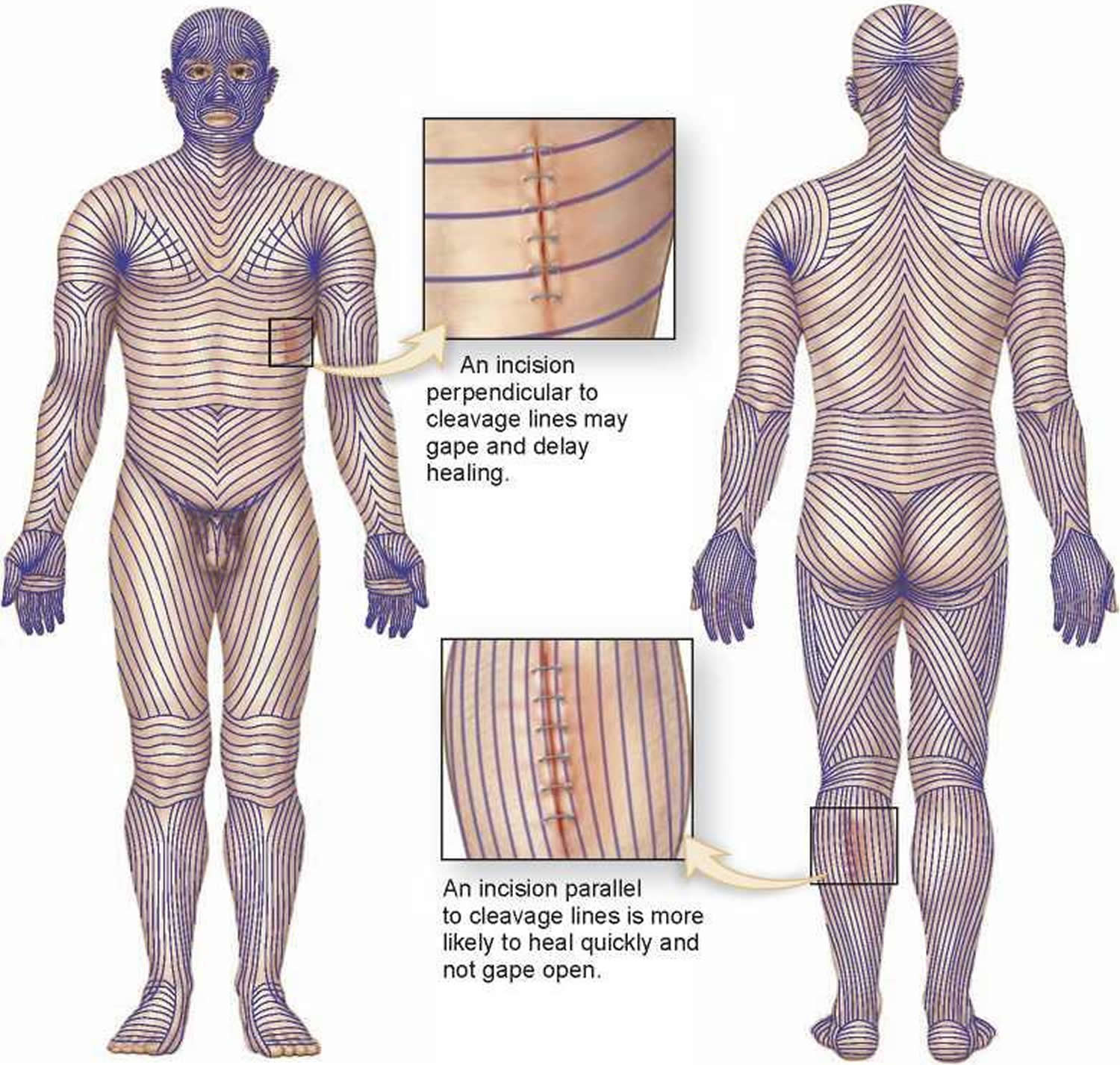
The density of hair does not differ much from one person to another or even between the sexes; indeed, it is virtually the same in humans, chimpanzees, and gorillas. Differences in apparent hairiness are due mainly to differences in texture and pigmentation.

Types of Hairs

Hairs first appear after about three months of embryonic development. These hairs, collectively known as lanugo, are extremely fine and unpigmented. Most lanugo hairs are shed before birth.

The two types of hairs in the adult skin are vellus hairs and terminal hairs:

  1. Vellus hairs are the fine “peach fuzz” hairs found over much of the body surface.
  2. Terminal hairs are heavy, more deeply pigmented, and sometimes curly. The hairs on your head, including your eyebrows and eyelashes, are terminal hairs. After puberty, it also forms the armpit and pubic hair, male facial hair, and some of the hair on the trunk and limbs.

Hair follicles may alter the structure of the hairs they produce in response to circulating hormones.

Figure 1. Hair structure

Hair structure

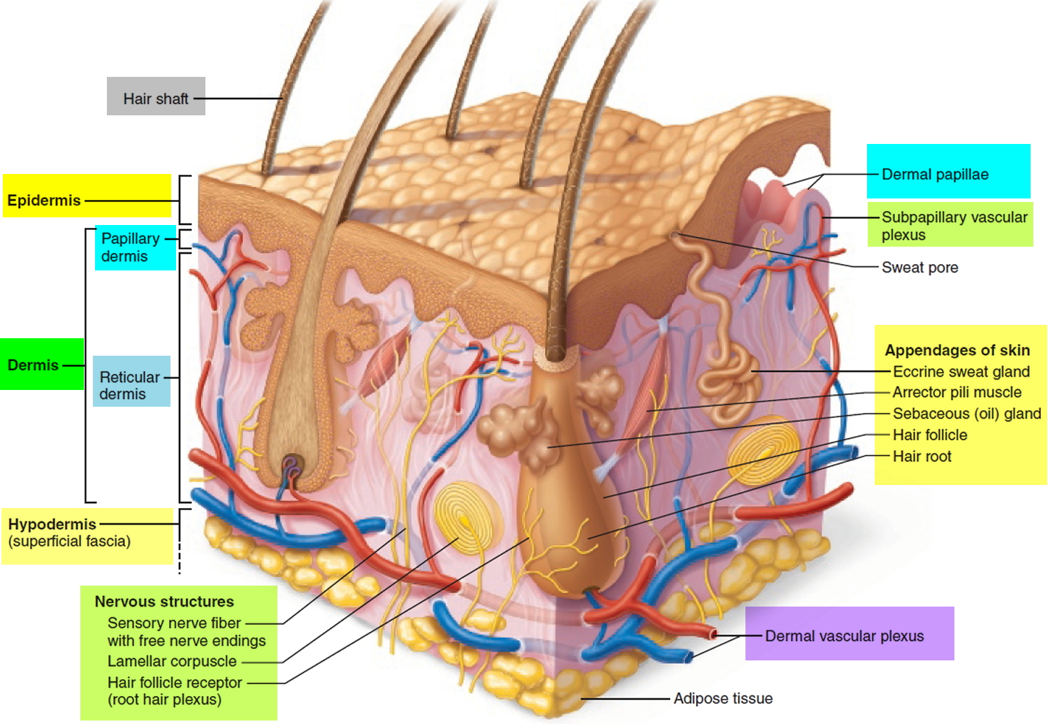
Figure 2. Hair follicle

structure of hair and hair follicle

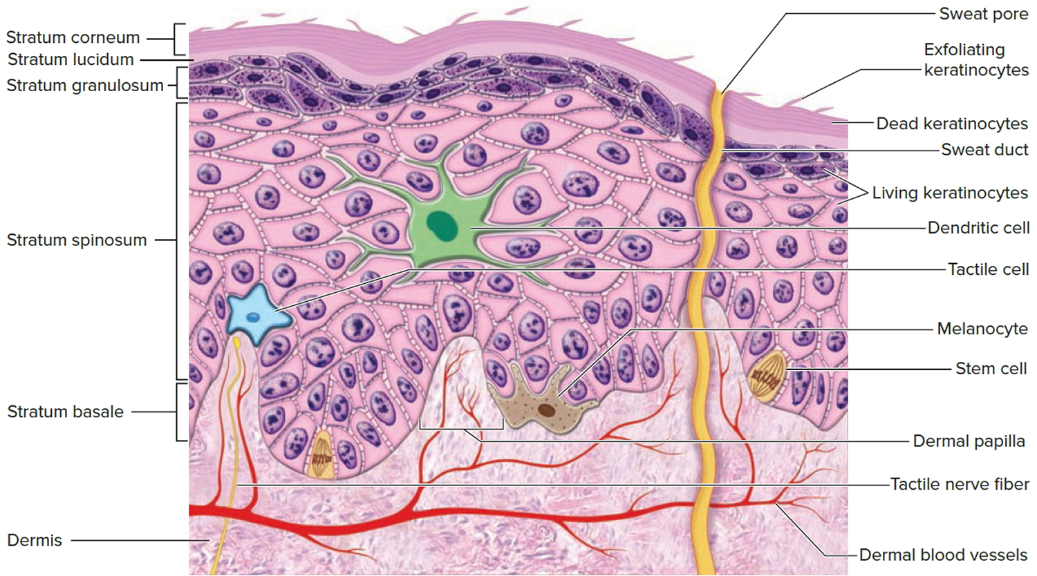
Structure of Hair Follicle

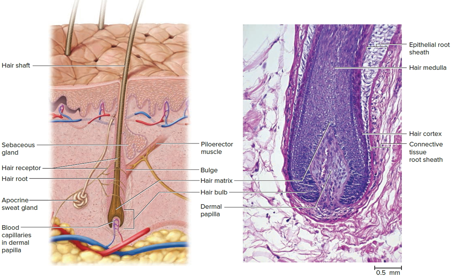
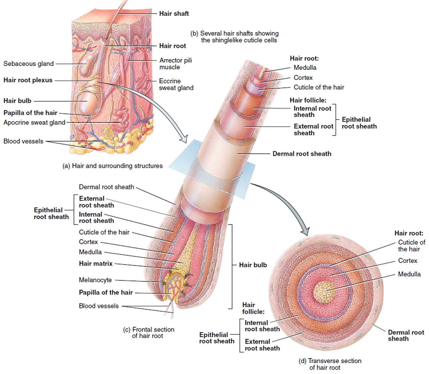
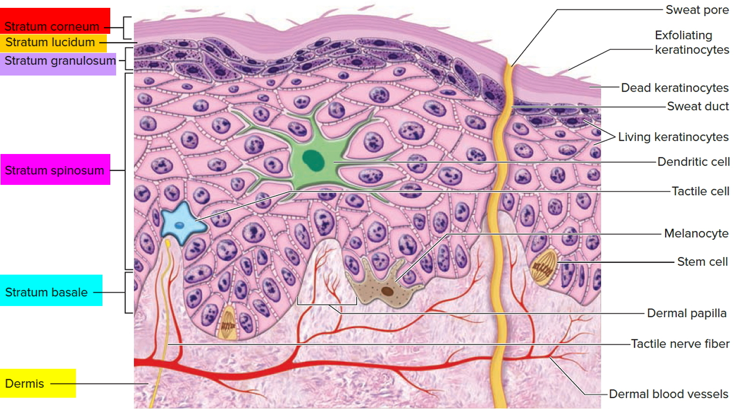
The portion of a hair above the skin is called the shaft, and all that beneath the surface is the root. The root penetrates deeply into the dermis or hypodermis and ends with a dilation called the hair bulb. The only living cells of a hair are in and near the hair bulb. The hair bulb grows around a bud of vascular connective tissue called the dermal papilla, which provides the hair with its sole source of nutrition. Immediately above the papilla is a region of mitotically active cells, the hair matrix, which is the hair’s growth center. All cells higher up are dead.

Figure 3. Hair follicle and hair structure

Hair structure

Hair Structure

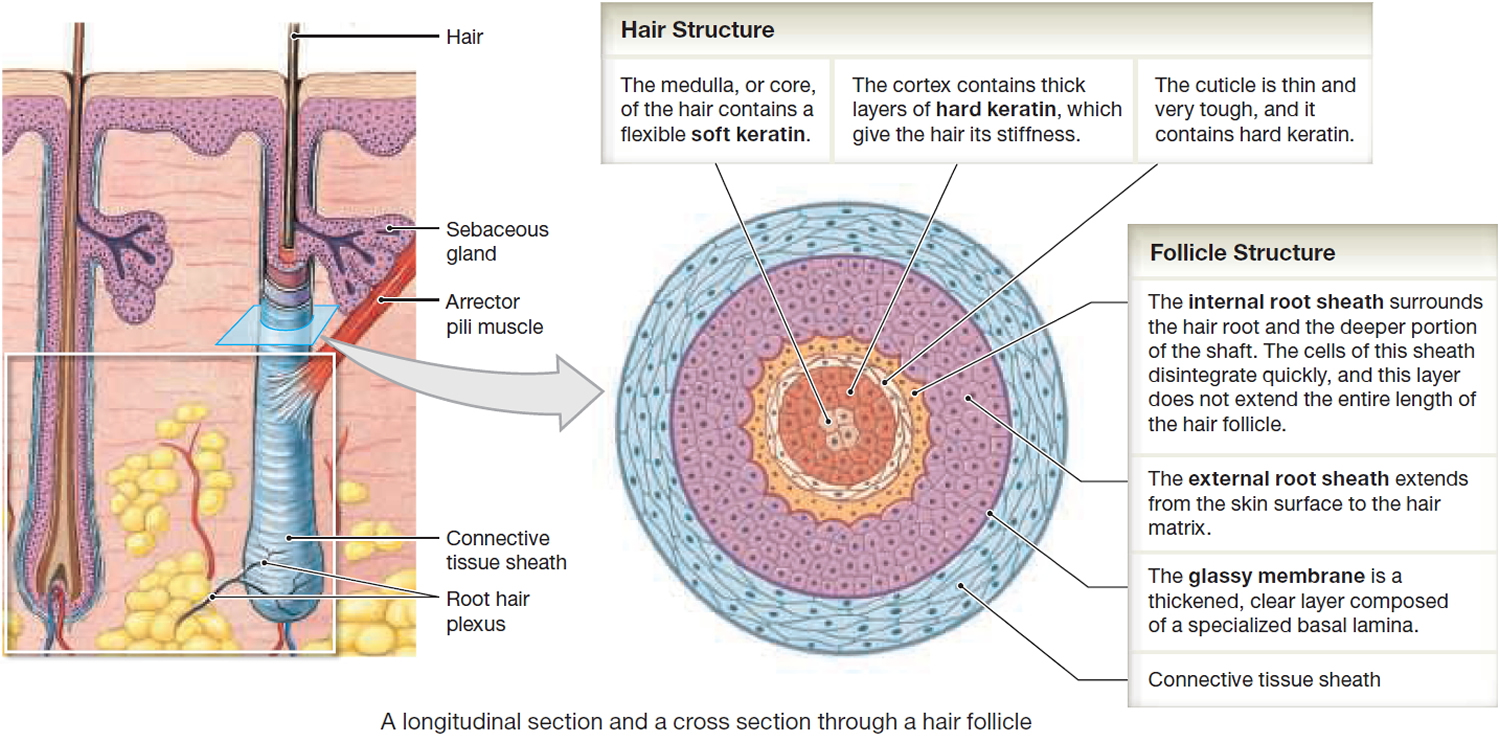
In cross section, a hair reveals up to three layers. From the inside out, these are the medulla, cortex, and cuticle.

The medulla is a core of loosely arranged cells and air spaces. It is most prominent in thick hairs such as those of the eyebrows, but narrower in hairs of medium thickness and absent from the thinnest hairs of the scalp and elsewhere.

The cortex constitutes most of the bulk of a hair. It consists of several layers of elongated keratinized cells that appear cuboidal to flattened in cross sections.

The cuticle is composed of multiple layers of very thin, scaly cells that overlap each other like roof shingles with their free edges directed upward.

Hair Follicle

Cells lining the hair follicle are like shingles facing in the opposite direction. They interlock with the scales of the hair cuticle and resist pulling on the hair. When a hair is pulled out, this layer of follicle cells comes with it.

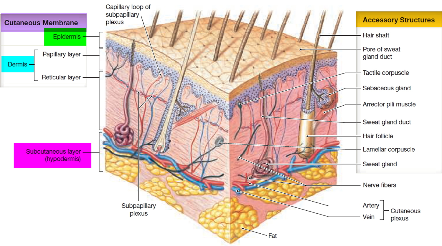
The hair follicle is a diagonal tube that contains the hair root. It has two principal layers: an epithelial root sheath and a connective tissue root sheath. The epithelial root sheath is an extension of the epidermis; it consists of stratified squamous epithelium and lies immediately adjacent to the hair root. Toward the deep end of the follicle, it widens to form a bulge, a source of stem cells for follicle growth. The connective tissue root sheath, which is derived from the dermis and composed of collagenous connective tissue, surrounds the epithelial sheath and is somewhat denser than the adjacent dermis.

Associated with the hair follicle are nerve and muscle fibers. Nerve fibers called hair receptors entwine each hair follicle and respond to hair movements. You can feel their effect by carefully moving a single hair with a pin or by lightly running your finger over the hairs of your forearm without touching the skin.

Each hair has a piloerector muscle—also known as a pilomotor muscle or arrector pili—a bundle of smooth muscle cells extending from dermal collagen fibers to the connective tissue root sheath of the follicle. In response to cold, fear, touch, or other stimuli, the sympathetic nervous system stimulates the piloerector to contract, making the hair stand on end and wrinkling the skin in such areas as the scrotum and areola. In humans, it pulls the follicles into a vertical position and causes “goose bumps,” but serves no useful purpose.

Figure 4. Hair structure

Hair follicle

Hair Production

Hair follicles extend deep into the dermis, often projecting into the underlying subcutaneous layer. The epithelium at the follicle base surrounds a small hair papilla, a peg of connective tissue containing capillaries and nerves. The hair bulb consists of epithelial cells that surround the papilla.

Hair production involves a specialized keratinization process. The hair matrix is the epithelial layer involved in hair production. When the superficial basal cells divide, they produce daughter cells that are pushed toward the surface as part of the developing hair. Most hairs have an inner medulla and an outer cortex. The medulla contains relatively soft and flexible soft keratin. Matrix cells closer to the edge of the developing hair form the relatively hard cortex. The cortex contains
hard keratin, which gives hair its stiffness. A single layer of dead, keratinized cells at the outer surface of the hair overlap and form the cuticle that coats the hair.

The hair root anchors the hair into the skin. The root begins at the hair bulb and extends distally to the point where the internal organization of the hair is complete, about halfway to the skin surface. The hair shaft extends from this halfway point to the skin surface, where we see the exposed hair tip.

The size, shape, and color of the hair shaft are highly variable.

Growth and Replacement of Hair

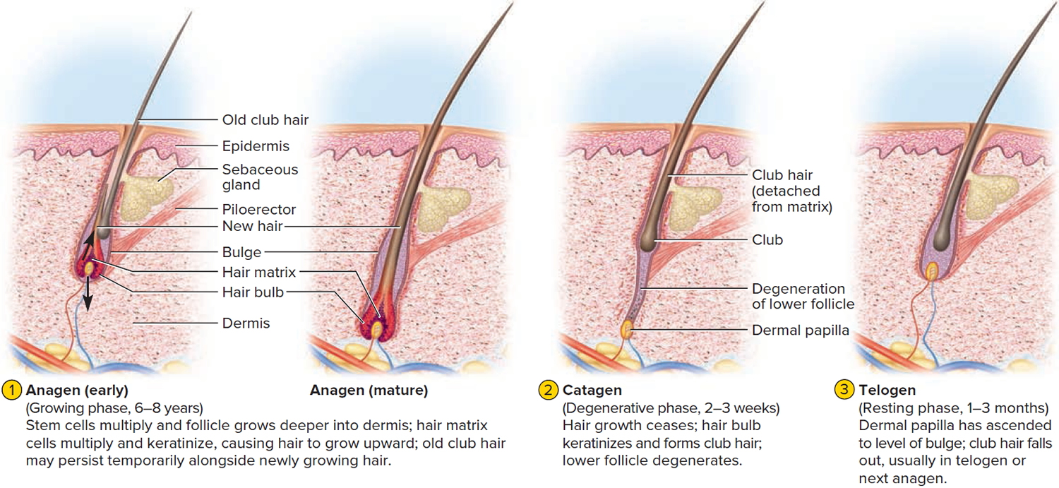
A hair in the scalp grows for two to five years, at a rate of around 0.33 mm/day (about 1/64 inch). Variations in hair growth rate and the duration of the hair growth cycle account for individual differences in uncut hair length. A given hair goes through a hair cycle consisting of three developmental stages (see Figure 5):

  • Anagen.
  • Catagen.
  • Telogen.

At any given time, about 90% of the scalp follicles are in the anagen stage. In this stage, stem cells from the bulge in the follicle multiply and travel downward, pushing the dermal papilla deeper into the skin and forming the epithelial root sheath. Root sheath cells directly above the papilla form the hair matrix. Here, sheath cells transform into hair cells, which synthesize keratin and then die as they are pushed upward away from the papilla. The new hair grows up the follicle, often alongside an old club hair left from the previous cycle.

In the catagen stage, mitosis in the hair matrix ceases and sheath cells below the bulge die. The follicle shrinks and the dermal papilla draws up toward the bulge. The base of the hair keratinizes into a hard club and the hair, now known as a club hair, loses its anchorage. Club hairs are easily pulled out by brushing the hair, and the hard club can be felt at the hair’s end. When the papilla reaches the bulge, the hair goes into a resting period called the telogen stage. Eventually, anagen begins anew and the cycle repeats itself. A club hair may fall out during catagen or telogen, or as it is pushed out by the new hair in the next anagen phase.

You lose about 50 to 100 scalp hairs daily. In a young adult, scalp follicles typically spend 6 to 8 years in anagen, 2 to 3 weeks in catagen, and 1 to 3 months in telogen. Scalp hairs grow at a rate of about 1 mm per 3 days (10–18 cm/yr) in the anagen phase.

Hair grows fastest from adolescence until the 40s. After that, an increasing percentage of follicles are in the catagen and telogen phases rather than the growing anagen phase. Hair follicles also shrink and begin producing wispy vellus hairs instead of thicker terminal hairs. Thinning of the hair, or baldness, is called alopecia. It occurs to some degree in both sexes and may be worsened by disease, poor nutrition, fever, emotional stress, radiation, or chemotherapy. In the great majority of cases, however, it is simply a matter of aging.

Pattern baldness is the condition in which hair is lost unevenly across the scalp rather than thinning uniformly. It results from a combination of genetic and hormonal influences. The relevant gene has two alleles: one for uniform hair growth and a baldness allele for patchy hair growth. The baldness allele is dominant in males and is expressed only in the presence of the high level of testosterone characteristic of men. In men who are either heterozygous or homozygous for the baldness allele, testosterone causes terminal hair to be replaced by vellus hair, beginning on top of the head and later the sides. In women, the baldness allele is recessive. Homozygous dominant and heterozygous women show normal hair distribution; only homozygous recessive women are at risk of pattern baldness. Even then, they exhibit the trait only if their testosterone levels are abnormally high for a woman (for example, because of a tumor of the adrenal gland, a woman’s principal source of testosterone). Such characteristics in which an allele is dominant in one sex and recessive in the other are called sex-influenced traits.

Excessive or undesirable hairiness in areas that are not usually hairy, especially in women and children, is called hirsutism. It tends to run in families and usually results from either masculinizing ovarian tumors or hypersecretion of testosterone by the adrenal cortex. It is often associated with menopause.

Contrary to popular misconceptions, hair and nails do not continue to grow after a person dies, cutting hair does not make it grow faster or thicker, and emotional stress cannot make the hair turn white overnight.

Figure 5. Hair growth cycle

hair growth cycle

Hair Color

Genes determine hair color by directing the type and amount of pigment that epidermal melanocytes produce. Variations in hair color reflect differences in hair structure and in the pigment produced by melanocytes at the papilla. Hormonal or environmental factors may influence the condition of your hair. Whether your hair is black or brown depends on the density of melanin in your cortex. Dark hair has more of the brownish-black eumelanin, while blonde hair and red hair have more of the reddish-yellow pheomelanin. The white hair of a person with the inherited condition albinism lacks melanin altogether. A mixture of pigmented and unpigmented hairs appears gray. As pigment production decreases with age, hair color lightens toward gray. White hair results from the combination of a lack of pigment and the presence of air bubbles within the medulla of the hair shaft. Because the hair itself is dead and inert, changes in coloration are gradual; your hair can’t “turn white overnight,” as some horror stories suggest.

Hair Texture

The texture of hair is related to differences in cross-sectional shape—straight hair is round, wavy hair is oval, and tightly curled hair is relatively flat.

Function of Hair

The 5 million hairs on the human body have important functions. Most hair of the human trunk and limbs is probably best interpreted as vestigial, with little present function. The roughly 100,000 hairs on the head protect the scalp from ultraviolet light and bumps to the head and insulate the skull. The hairs guarding the entrances to your nostrils and external auditory canals (ear canals) help block foreign particles and insects, and eyelashes perform a similar function for the surface of the eye.

A root hair plexus of sensory nerves surrounds the base of each hair follicle. As a result, you can feel the movement of even a single hair. This sensitivity gives an early-warning system that may help prevent injury. For example, stimulation of the hair receptors, however, alerts people to parasites crawling on the skin, such as fleas and ticks, and to remove them. Thus, you are less likely to become unknowingly infested with parasites.

A ribbon of smooth muscle, the arrector pili muscle, extends from the papillary layer of the dermis to the connective tissue sheath surrounding the hair follicle. When stimulated, the arrector pili muscle pulls on the follicle and raises the hair. Contraction may be due to emotional state, such as fear or rage, or to cold temperatures that produce characteristic “goose bumps.” In a furry mammal, this action thickens the insulating coat, rather like putting on an extra sweater. Although we do not receive any comparable insulating benefits, the reflex persists.

read more
SkinSkin System

Dermis

dermis

What is dermis

The dermis, the second major region of the skin, is a strong, flexible connective tissue. The cells of the dermis are typical of any connective tissue proper: fibroblasts, macrophages, mast cells, and scattered white blood cells. The fiber types—collagen, elastic, and reticular—also are typical.

The function of the dermis is to bind the entire body together like a body stocking. It is your “hide” and corresponds to animal hides used to make leather products.

The dermis is also the site where all the accessory structures of the skin – your hair, nails, and a variety of multicellular exocrine glands originate. These structures are located in the dermis and protrude through the epidermis to the surface.

The dermis has two regions:

  1. the Papillary Dermis and
  2. the Reticular Dermis.

Figure 1. Dermis layers

dermis layers

Papillary layer of Dermis

The papillary dermis, the superficial 20% of the dermis, is areolar connective tissue containing very thin collagen and elastic fibers. It includes the dermal papillae (“nipples”), fingerlike projections that extend into the overlying epidermis. These projections of the dermal papillae into the epidermis increase the surface area for exchange of gases, nutrients, and waste products between these layers. Recall that the epidermis is avascular and depends on the diffusion of these materials from the underlying dermis.

The inter-digitation of these layers also strengthens the dermal-epidermal junction and thus reduces blister formation.

On the palms of the hands and the soles of the feet, the dermal papillae lie atop larger mounds called dermal ridges. These elevate the overlying epidermis into epidermal ridges or friction ridges, which create fingerprints, palm-prints, and footprints. Epidermal ridges increase friction and enhance the gripping ability of the hands and feet.

Patterns of these ridges are genetically determined and unique to each person. Because sweat pores open along the crests of the friction ridges, they leave distinct fingerprints on almost anything they touch. Thus, fingerprints are “sweat films.”

Reticular layer of Dermis

The deeper reticular dermis, which accounts for about 80% of the thickness of the dermis, is dense irregular connective tissue. Its extracellular matrix contains thick bundles of interlacing collagen and elastic fibers that run in many different planes. However, most run parallel to the skin surface. The reticular layer is named for its networks of collagen fibers (reticulum = network); the name does not imply any special abundance of reticular fibers. Separations or less dense regions between the collagen bundles form the cleavage lines or tension lines of the skin.

These invisible lines occur over the entire body: They run longitudinally in the skin of the limbs and head and in circular patterns around the neck and trunk. A knowledge of cleavage lines is important to surgeons. Incisions made parallel to these lines tend to gape less and heal more readily than incisions made across cleavage lines.

Figure 2. Cleavage or tension lines of the skin

cleavage lines of skin

The collagen fibers of the dermis give skin its strength and resilience. Thus, many jabs and scrapes do not penetrate this tough layer. Furthermore, elastic fibers in the dermis provide the skin with stretch-recoil properties. Extreme stretching of the skin, as occurs in obesity and pregnancy, can tear the collagen in the dermis. Such dermal tearing results in silvery white scars called striae (“streaks”), which is commonly known as “stretch marks.” The dermis is also the receptive site for the pigments used in tattoos.

From the deep part of the dermis arise the skin surface markings called flexure lines. Observe, for example, the deep skin creases on your palm. These result from
a continual folding of the skin, often over joints, where the dermis attaches tightly to underlying structures. Flexure lines are also visible on the wrists, soles, fingers, and toes.

The dermis is richly supplied with nerve fiber and blood vessels. The dermal blood vessels consist of two vascular plexuses (a plexus is a network of converging and diverging vessels). The deep dermal plexus is located between the hypodermis and the dermis. It nourishes the hypodermis and the structures located within the deeper portions of the dermis. The more superficial subpapillary plexus, located just below the dermal papillae, supplies the more superficial dermal structures, the dermal papillae, and the epidermis.

Dermal blood vessels do more than just nourish the dermis and overlying epidermis; they also perform a critical role in temperature regulation. These vessels are so extensive that they can hold 5% of all blood in the body. When internal organs need more blood or more heat, nerves stimulate the dermal vessels to constrict, shunting more blood into the general circulation and making it available to the internal organs. By contrast, on hot days the dermal vessels engorge with warm blood, cooling the body by radiating heat away from it.

Below the dermis is another connective tissue layer, the hypodermis, which is not part of the skin but is customarily studied in conjunction with it. Most of the skin is 1 to 2 mm thick, but it ranges from less than 0.5 mm on the eyelids to 6 mm between the shoulder blades. The difference is due mainly to variation in thickness of the dermis, although skin is classified as thick or thin based on the relative thickness of the epidermis alone.

The Epidermis

The skin consists of two main parts, the most superficial part of the skin is the epidermis. The epidermis is a thinner portion of the skin, which is composed of
epithelial tissue. While the epidermis is avascular, the dermis is vascular. For this reason, if you cut the epidermis there is no bleeding, but if the cut penetrates to the dermis there is bleeding.

The epidermis is composed of keratinized stratified squamous epithelium. It contains five principal types of cells: stem cells, keratinocytes, melanocytes, Merkel cells (Tactile cells) and Dendritic cells (Langerhans cells). About 90% of epidermal cells are keratinocytes, which are arranged in four or five layers and produce the protein keratin. Keratin is a tough, fibrous protein that helps protect the skin and underlying tissues from abrasions, heat, microbes, and chemicals.

Keratinocytes also produce lamellar granules, which release a water-repellent sealant that decreases water entry and loss and inhibits the entry of foreign materials.

About 8% of the epidermal cells are melanocytes, which produce the pigment melanin. Their long, slender projections extend between the keratinocytes and transfer melanin granules to them. Melanin is a yellowred or brown-black pigment that contributes to skin color and absorbs damaging ultraviolet (UV) light. Once inside keratinocytes, the melanin granules cluster to form a protective veil over the nucleus, on the side toward the skin surface. In this way, they shield the nuclear DNA from damage by UV light. Although their melanin granules effectively protect keratinocytes, melanocytes themselves are particularly susceptible to damage by UV light.

Intraepidermal macrophages or Langerhans cells (Dendritic cells) arise from red bone marrow and migrate to the epidermis, where they constitute a small fraction of the epidermal cells. They participate in immune responses mounted against microbes that invade the skin, and are easily damaged by UV light. Their role in the immune response is to help other cells of the immune system recognize an invading microbe and destroy it.

Tactile epithelial cells, or Merkel cells, are the least numerous of the epidermal cells. They are located in the deepest layer of the epidermis, where they contact the flattened process of a sensory neuron (nerve cell), a structure called a tactile disc or Merkel disc. Tactile epithelial cells and their associated tactile discs detect touch sensations.

Several distinct layers of keratinocytes in various stages of development form the epidermis. In most regions of the body the epidermis has four strata or layers —stratum basale, stratum spinosum, stratum granulosum, and a thin stratum corneum. This is called thin skin. Where exposure to friction is greatest, such as in the fingertips, palms, and soles, the epidermis has five layers—stratum basale, stratum spinosum, stratum granulosum, stratum lucidum, and a thick stratum corneum. This is called thick skin.

read more
SkinSkin System

Epidermis

epidermis

What is epidermis

The epidermis is a keratinized stratified squamous epithelium. That is, the epidermis outermost layer consists of dead cells packed with the tough protein keratin. Like other epithelia, the epidermis lacks blood vessels and depends on the diffusion of nutrients from the underlying connective tissue. It has sparse nerve endings for touch and pain, but most sensations of the skin are due to nerve endings in the dermis.

Figure 1. Skin structure

skin structure and layers

There are 5 cell types in the epidermis: stem cells, keratinocytes, melanocytes, Merkel cells (Tactile cells) and Dendritic cells (Langerhans cells).

Cells of the Epidermis

The epidermis is composed of five types of cells (Figure 2):

  1. Stem cells are undifferentiated cells that divide and give rise to the keratinocytes described next. They are found only in the deepest layer of the epidermis, called the stratum basale.
  2. Keratinocytes are the great majority of epidermal cells. They are named for their role in synthesizing keratin. In ordinary histological specimens, nearly all visible epidermal cells are keratinocytes.
  3. Melanocytes also occur only in the stratum basale, amid the stem cells and deepest keratinocytes. They synthesize the brown to black pigment melanin. They have branching processes that spread among the keratinocytes and continually shed melanin-containing fragments (melanosomes) from their tips. The keratinocytes phagocytize these and gather the melanin granules on the “sunny side” of their nucleus. Like a parasol, the dark granules shield the DNA from ultraviolet rays.
  4. Merkel cells (Tactile cells), relatively few in number, are receptors for touch. They, too, are found in the basal layer of the epidermis and are associated with an underlying dermal nerve fiber. The tactile cell and its nerve fiber are collectively called a tactile disc.
  5. Langerhans cells (Dendritic cells) are found in two layers of the epidermis called the stratum spinosum and stratum granulosum (described in the next section). They are immune cells that originate in the bone marrow but migrate to the epidermis and epithelia of the oral cavity, esophagus, and vagina. The epidermis has as many as 800 dendritic cells per square millimeter. They stand guard against toxins, microbes, and other pathogens that penetrate into the skin. When they detect such invaders, they alert the immune system so the body can defend itself.

Figure 2. 5 layers of epidermis

layers of epidermis

Layers of the Epidermis

The epidermis of thick skin has five layers. Beginning at the basal lamina and traveling superficially toward the epithelial surface, we find the stratum basale, stratum spinosum, stratum granulosum, stratum lucidum, and stratum corneum. Refer to Figure 2 as we describe the layers in a section of thick skin.

Stratum Basale

The deepest epidermal layer is the stratum basale or stratum germinativum. This single layer of cells is firmly attached to the basal lamina, which separates the epidermis from the loose connective tissue of the adjacent dermis. Large stem cells, termed basal cells, dominate the stratum basale. As basal cells undergo mitosis, new keratinocytes are formed and move into the more superficial layers of the epidermis. This upward migration of cells replaces more superficial keratinocytes that are shed at the epithelial surface.

The brown tones of the skin result from the pigment-producing cells called melanocytes. Melanocytes are scattered among the basal cells of the stratum basale. They have numerous cytoplasmic processes that inject melanin—a black, yellow-brown, or brown pigment—into the basal cells in this layer and into the keratinocytes of more superficial layers. The ratio of melanocytes to stem cells ranges between 1:4 and 1:20 depending on the region examined. Melanocytes are most abundant in the cheeks, forehead, nipples, and genital region.

Differences in skin color result from varying levels of melanocyte activity, not varying numbers of melanocytes. Albinism is an inherited disorder characterized by deficient melanin production; individuals with this condition have a normal distribution of melanocytes, but the cells cannot produce melanin. It affects approximately one person in 10,000.

Skin surfaces that lack hair contain specialized epithelial cells known as Merkel cells (tactile cells). These cells are found among the cells of the stratum basale and are most abundant in skin where sensory perception is most acute, such as fingertips and lips. Merkel cells are sensitive to touch and, when compressed, release chemicals that stimulate sensory nerve endings, providing information about objects touching the skin. There are many other kinds of touch receptors,
but they are located in the dermis and will be introduced in later sections.

Stratum Spinosum

Each time a basal cell divides, one of the daughter cells is pushed into the next, more superficial layer, the stratum spinosum. The stratum spinosum is several cells thick. Each keratinocyte in the stratum spinosum contains bundles of protein filaments that extend from one side of the cell to the other. These bundles, called tonofibrils, begin and end at a desmosome (macula adherens) that connects the keratinocyte to its neighbors. The tonofibrils act as cross braces, strengthening and supporting the cell junctions. This interlocking network of desmosomes and tonofibrils ties all the cells in the stratum spinosum together.

The deepest cells within the stratum spinosum are mitotically active and continue to divide, making the epithelium thicker. Melanocytes are common in this layer, as are Langerhans cells (also termed dendritic cells). Langerhans cells, which account for 3–8 percent of the cells in the epidermis, are most common in the superficial portion of the stratum spinosum. These cells play an important role in triggering an immune response against epidermal cancer cells and pathogens that have penetrated the superficial layers of the epidermis.

Stratum Granulosum

Superficial to the stratum spinosum is the stratum granulosum (granular layer). This is the most superficial layer of the epidermis in which all the cells still possess a nucleus. The stratum granulosum consists of keratinocytes that have moved out of the stratum spinosum. By the time cells reach this layer, they have begun to manufacture large quantities of the proteins keratohyalin and keratin. Keratohyalin accumulates in electron dense keratohyalin granules. These granules form an intracellular matrix that surrounds the keratin filaments. Cells of this layer also contain membrane-bound granules that release their contents by exocytosis, which forms sheets of a lipid-rich substance that begins to coat the cells of the stratum granulosum. In more superficial layers, this substance forms a complete water resistant layer around the cells that protects the epidermis, but also prevents the diffusion of nutrients and wastes into and out of the cells. As a result, cells in the more superficial layers of the epidermis die.

Environmental factors often influence the rate at which keratinocytes synthesize keratohyalin and keratin. Increased friction against the skin, for example, stimulates increased synthesis, thickening the skin and forming a callus (also termed a clavus).

In humans, keratin forms the basic structural component of hair and nails. It is a very versatile material, however, and it also forms the claws of dogs and cats, the horns of cattle and rhinos, the feathers of birds, the scales of snakes, the baleen of whales, and a variety of other interesting epidermal structures.

Stratum Lucidum

The stratum lucidum is a thin zone superficial to the stratum granulosum, seen only in thick skin. Here, the keratinocytes are densely packed with a clear protein named eleidin. The cells have no nuclei or other organelles. This zone has a pale, featureless appearance with indistinct cell boundaries.

Stratum Corneum

The stratum corneum is the most superficial layer of both thick and thin skin. It consists of numerous layers of flattened, dead cells that possess a thickened plasma membrane. These dehydrated cells lack organelles and a nucleus, but still contain many keratin filaments. Because the interconnections established in the stratum spinosum remain intact, the cells of this layer are usually shed in large groups or sheets, rather than individually.

An epithelium containing large amounts of keratin is termed a keratinized or cornified epithelium.

Normally, the stratum corneum is relatively dry, which makes the surface unsuitable for the growth of many microorganisms. Maintenance of this barrier involves coating the surface with the secretions of sebaceous and sweat glands (discussed in a later section). The process of keratinization occurs everywhere on exposed skin surfaces except over the anterior surface of the eyes. Although the stratum corneum is water resistant, it is not waterproof. Water from the interstitial fluids slowly penetrates the surface and evaporates into the surrounding air. This process, called insensible perspiration, accounts for a loss of roughly 500 ml (about 1 pint) of water per day.

It takes 15–30 days for a cell to move superficially from the stratum basale to the stratum corneum. The dead cells in the exposed stratum corneum layer usually remain for two weeks before they are shed or washed away. Thus, the deeper portions of the epithelium—and all underlying tissues—are always protected by a barrier composed of dead, durable, and expendable cells.

The Life History of a Keratinocyte

Dead cells constantly flake off the skin surface. Because you constantly lose these epidermal cells, they must be continually replaced. Keratinocytes are produced deep in the epidermis by the mitosis of stem cells in the stratum basale. Some of the deepest keratinocytes in the stratum spinosum also continue dividing. Mitosis requires an abundant supply of oxygen and nutrients, which these deep cells acquire from the blood vessels in the nearby dermis.

Once the epidermal cells migrate more than two or three cells away from the dermis, their mitosis ceases. As new keratinocytes form, they push the older ones toward the surface. In 30 to 40 days, a keratinocyte makes its way to the surface and flakes off. This migration is slower in old age and faster in skin that has been injured or stressed. Injured epidermis regenerates more rapidly than any other tissue in the body. Mechanical stress from manual labor or tight shoes accelerates keratinocyte multiplication and results in calluses or corns, thick accumulations of dead keratinocytes on the hands or feet.

As keratinocytes are shoved upward by the dividing cells below, they flatten and produce more keratin filaments and lipid-filled membrane-coating vesicles. In the stratum granulosum, four important developments occur: (1) Keratohyalin granules release a protein called filaggrin that binds the cytoskeletal keratin filaments together into coarse, tough bundles. (2) The cells produce a tough layer of envelope proteins just beneath the plasma membrane, resulting in a nearly indestructible protein sac around the keratin bundles. (3) Membrane-coating vesicles release a lipid mixture that spreads out over the cell surface and waterproofs it. (4) Finally, as these barriers cut the keratinocytes off from the supply of nutrients from below, their organelles degenerate and the cells die, leaving just the tough waterproof sac enclosing coarse bundles of keratin. These processes, along with the tight junctions between keratinocytes, result in an epidermal water barrier that is crucial to the retention of body water.

Thin and Thick Skin

Most of the body is covered by thin skin, which has only four layers because the stratum lucidum is typically absent. In thin skin, the epidermis is a mere 0.08 mm thick and the stratum corneum is only a few cell layers deep. Thick skin, found only on the palms of the hands and soles of the feet, contains all five layers and may be covered by 30 or more layers of keratinized cells. As a result, the epidermis in these locations is up to six times thicker than the epidermis covering the general body surface.

Dermal Ridges

The stratum basale of the epidermis forms dermal ridges (also known as friction ridges) that extend into the dermis, increasing the area of contact between the two regions. Projections from the dermis toward the epidermis, called dermal papillae (singular, papilla), extend between adjacent ridges (Figure 1 and 2).

The contours of the skin surface follow the ridge patterns, which vary from small conical pegs (in thin skin) to the complex whorls seen on the thick skin of the palms and soles. Ridges on the palms and soles increase the surface area of the skin and promote friction, ensuring a secure grip. Ridge shapes are genetically determined: Those of each person are unique and do not change during a lifetime. Ridge patterns on the fingertips can therefore identify individuals.

Epidermis function


The skin is much more than a container for the body. It has a variety of very important functions that go well beyond appearance, as you shall see here.

  1. Resistance to trauma and infection. The skin suffers the most physical injuries to the body, but it resists and recovers from trauma better than other organs do. The epidermal cells are packed with the tough protein keratin and linked by strong desmosomes that give this epithelium its durability. Few infectious organisms can penetrate the intact skin. Bacteria and fungi colonize the surface, but their numbers are kept in check by its relative dryness, its slight acidity (pH 4–6), and defensive antimicrobial peptides called dermcidin and defensins. The protective acidic film is called the acid mantle.
  2. Other barrier functions. The skin is important as a barrier to water. It prevents the body from absorbing excess water when you are swimming or bathing, but even more importantly, it prevents the body from losing excess water. The epidermis is also a barrier to ultraviolet (UV) rays, blocking much of this cancer causing radiation from reaching deeper tissue layers; and it is a barrier to many potentially harmful chemicals. It is, however, permeable to several drugs and poisons.
  3. Vitamin D synthesis. The skin carries out the first step in the synthesis of vitamin D, which is needed for bone development and maintenance. The liver and kidneys complete the process.
  4. Sensation. The skin is our most extensive sense organ. It is equipped with a variety of nerve endings that react to heat, cold, touch, texture, pressure, vibration, and tissue injury. These sensory receptors are especially abundant on the face, palms, fingers, soles, nipples, and genitals. There are relatively few on the back and in skin overlying joints such as the knees and elbows.
  5. Thermoregulation. The skin receives 10 times as much blood flow as it needs for its own maintenance, and is richly supplied with nerve endings called thermoreceptors, which monitor the body surface temperature. All of this relates to its great importance in regulating body temperature. In response to chilling, the body retains heat by constricting blood vessels of the dermis (cutaneous vasoconstriction), keeping warm blood deeper in the body. In response to overheating, it loses excess heat by dilating those vessels (cutaneous vasodilation), allowing more blood to flow close to the surface and lose heat through the skin. If this is insufficient to restore normal temperature, sweat glands secrete perspiration. The evaporation of sweat can have a powerful cooling effect. Thus, the skin plays roles in both warming and cooling the body.
  6. Nonverbal communication. The skin is an important means of nonverbal communication. Humans, like most other primates, have much more expressive faces than other mammals. Complex skeletal muscles insert in the dermis and pull on the skin to create subtle and varied facial expressions. The general appearance of the skin, hair, and nails is also important to social acceptance and to a person’s self-image and emotional state—whether the ravages of adolescent acne, the presence of a birthmark or scar, or just a “bad hair day.”

The Dermis

Beneath the epidermis is a connective tissue layer, the dermis. It ranges from 0.2 mm thick in the eyelids to about 4 mm thick in the palms and soles. It is composed mainly of collagen, but also contains elastic and reticular fibers, fibroblasts, and the other cells typical of fibrous connective tissue. It is well supplied with blood vessels, cutaneous glands, and nerve endings. The hair follicles and nail roots are embedded in the dermis. In the face, skeletal muscles attach to dermal collagen fibers and produce such expressions as a smile, a wrinkle of the forehead, or the lifting of an eyebrow.

The boundary between the epidermis and dermis is histologically conspicuous and usually wavy. The upward waves are fingerlike extensions of the dermis called dermal papillae and the downward epidermal waves between the papillae are called epidermal ridges. The dermal and epidermal boundaries thus interlock like corrugated cardboard, an arrangement that resists slippage of the epidermis across the dermis. If you look closely at your hand and wrist, you will see delicate furrows that divide the skin into tiny rectangular to rhomboidal areas. The dermal papillae produce the raised areas between the furrows. On the fingertips, this wavy boundary forms the friction ridges that produce fingerprints. In highly sensitive areas such as the lips and genitals, exceptionally tall dermal papillae allow blood capillaries and nerve fibers to reach close to the surface. This imparts a redder color and more sensitivity to touch in such areas.

read more
SkinSkin System

Skin

skin

The Skin

The skin (integument) is the body’s largest and heaviest organ. In adults, the skin covers an area of 1.5 to 2.0 m2 and accounts for about 15% of the body weight.

The skin consists of two layers:

  1. a stratified squamous epithelium called the Epidermis and
  2. a deeper connective tissue layer called the Dermis (Figure 1).

Figure 1. Skin structure

skin structure and layers

Below the dermis is another connective tissue layer, the hypodermis, which is not part of the skin but is customarily studied in conjunction with it. Most of the skin is 1 to 2 mm thick, but it ranges from less than 0.5 mm on the eyelids to 6 mm between the shoulder blades. The difference is due mainly to variation in thickness of the dermis, although skin is classified as thick or thin based on the relative thickness of the epidermis alone.

Thick skin covers the palms, soles, and corresponding surfaces of the fingers and toes. Its epidermis alone is about 0.5 mm thick, due to a very thick surface layer of dead cells called the stratum corneum (see Figure 2). Thick skin has sweat glands but no hair follicles or sebaceous (oil) glands. The rest of the body is covered with thin skin, which has an epidermis about 0.1 mm thick, with a thin stratum corneum. It possesses hair follicles, sebaceous glands, and sweat glands.

The accessory structures include hair, nails, and a variety of multicellular exocrine glands. These structures are located in the dermis and protrude through the epidermis to the surface.

Figure 2. Structure and skin cells of the Epidermis

structure of epidermis

Functions of the skin

The skin is much more than a container for the body. It has a variety of very important functions that go well beyond appearance, as you shall see here.

  1. Resistance to trauma and infection. The skin suffers the most physical injuries to the body, but it resists and recovers from trauma better than other organs do. The epidermal cells are packed with the tough protein keratin and linked by strong desmosomes that give this epithelium its durability. Few infectious organisms can penetrate the intact skin. Bacteria and fungi colonize the surface, but their numbers are kept in check by its relative dryness, its slight acidity (pH 4–6), and defensive antimicrobial peptides called dermcidin and defensins. The protective acidic film is called the acid mantle.
  2. Other barrier functions. The skin is important as a barrier to water. It prevents the body from absorbing excess water when you are swimming or bathing, but even more importantly, it prevents the body from losing excess water. The epidermis is also a barrier to ultraviolet (UV) rays, blocking much of this cancer causing radiation from reaching deeper tissue layers; and it is a barrier to many potentially harmful chemicals. It is, however, permeable to several drugs and poisons.
  3. Vitamin D synthesis. The skin carries out the first step in the synthesis of vitamin D, which is needed for bone development and maintenance. The liver and kidneys complete the process.
  4. Sensation. The skin is our most extensive sense organ. It is equipped with a variety of nerve endings that react to heat, cold, touch, texture, pressure, vibration, and tissue injury. These sensory receptors are especially abundant on the face, palms, fingers, soles, nipples, and genitals. There are relatively few on the back and in skin overlying joints such as the knees and elbows.
  5. Thermoregulation. The skin receives 10 times as much blood flow as it needs for its own maintenance, and is richly supplied with nerve endings called thermoreceptors, which monitor the body surface temperature. All of this relates to its great importance in regulating body temperature. In response to chilling, the body retains heat by constricting blood vessels of the dermis (cutaneous vasoconstriction), keeping warm blood deeper in the body. In response to overheating, it loses excess heat by dilating those vessels (cutaneous vasodilation), allowing more blood to flow close to the surface and lose heat through the skin. If this is insufficient to restore normal temperature, sweat glands secrete perspiration. The evaporation of sweat can have a powerful cooling effect. Thus, the skin plays roles in both warming and cooling the body.
  6. Nonverbal communication. The skin is an important means of nonverbal communication. Humans, like most other primates, have much more expressive faces than other mammals. Complex skeletal muscles insert in the dermis and pull on the skin to create subtle and varied facial expressions. The general appearance of the skin, hair, and nails is also important to social acceptance and to a person’s self-image and emotional state—whether the ravages of adolescent acne, the presence of a birthmark or scar, or just a “bad hair day.”

The Epidermis

The epidermis is a keratinized stratified squamous epithelium. That is, its surface consists of dead cells packed with the tough protein keratin. Like other epithelia, the epidermis lacks blood vessels and depends on the diffusion of nutrients from the underlying connective tissue. It has sparse nerve endings for touch and pain, but most sensations of the skin are due to nerve endings in the dermis.

There are 5 cell types in the epidermis: stem cells, keratinocytes, melanocytes, Merkel cells (Tactile cells) and Dendritic cells (Langerhans cells).

Cells of the Epidermis

The epidermis is composed of five types of cells (Figure 2):

  1. Stem cells are undifferentiated cells that divide and give rise to the keratinocytes described next. They are found only in the deepest layer of the epidermis, called the stratum basale.
  2. Keratinocytes are the great majority of epidermal cells. They are named for their role in synthesizing keratin. In ordinary histological specimens, nearly all visible epidermal cells are keratinocytes.
  3. Melanocytes also occur only in the stratum basale, amid the stem cells and deepest keratinocytes. They synthesize the brown to black pigment melanin. They have branching processes that spread among the keratinocytes and continually shed melanin-containing fragments (melanosomes) from their tips. The keratinocytes phagocytize these and gather the melanin granules on the “sunny side” of their nucleus. Like a parasol, the dark granules shield the DNA from ultraviolet rays.
  4. Merkel cells (Tactile cells), relatively few in number, are receptors for touch. They, too, are found in the basal layer of the epidermis and are associated with an underlying dermal nerve fiber. The tactile cell and its nerve fiber are collectively called a tactile disc.
  5. Langerhans cells (Dendritic cells) are found in two layers of the epidermis called the stratum spinosum and stratum granulosum (described in the next section). They are immune cells that originate in the bone marrow but migrate to the epidermis and epithelia of the oral cavity, esophagus, and vagina. The epidermis has as many as 800 dendritic cells per square millimeter. They stand guard against toxins, microbes, and other pathogens that penetrate into the skin. When they detect such invaders, they alert the immune system so the body can defend itself.

Layers of the Epidermis

The epidermis of thick skin has five layers. Beginning at the basal lamina and traveling superficially toward the epithelial surface, we find the stratum basale, stratum spinosum, stratum granulosum, stratum lucidum, and stratum corneum. Refer to Figure 2 as we describe the layers in a section of thick skin.

Stratum Basale

The deepest epidermal layer is the stratum basale or stratum germinativum. This single layer of cells is firmly attached to the basal lamina, which separates the epidermis from the loose connective tissue of the adjacent dermis. Large stem cells, termed basal cells, dominate the stratum basale. As basal cells undergo mitosis, new keratinocytes are formed and move into the more superficial layers of the epidermis. This upward migration of cells replaces more superficial keratinocytes that are shed at the epithelial surface.

The brown tones of the skin result from the pigment-producing cells called melanocytes. Melanocytes are scattered among the basal cells of the stratum basale. They have numerous cytoplasmic processes that inject melanin—a black, yellow-brown, or brown pigment—into the basal cells in this layer and into the keratinocytes of more superficial layers. The ratio of melanocytes to stem cells ranges between 1:4 and 1:20 depending on the region examined. Melanocytes are most abundant in the cheeks, forehead, nipples, and genital region.

Differences in skin color result from varying levels of melanocyte activity, not varying numbers of melanocytes. Albinism is an inherited disorder characterized by deficient melanin production; individuals with this condition have a normal distribution of melanocytes, but the cells cannot produce melanin. It affects approximately one person in 10,000.

Skin surfaces that lack hair contain specialized epithelial cells known as Merkel cells (tactile cells). These cells are found among the cells of the stratum basale and are most abundant in skin where sensory perception is most acute, such as fingertips and lips. Merkel cells are sensitive to touch and, when compressed, release chemicals that stimulate sensory nerve endings, providing information about objects touching the skin. There are many other kinds of touch receptors,
but they are located in the dermis and will be introduced in later sections.

Stratum Spinosum

Each time a basal cell divides, one of the daughter cells is pushed into the next, more superficial layer, the stratum spinosum. The stratum spinosum is several cells thick. Each keratinocyte in the stratum spinosum contains bundles of protein filaments that extend from one side of the cell to the other. These bundles, called tonofibrils, begin and end at a desmosome (macula adherens) that connects the keratinocyte to its neighbors. The tonofibrils act as cross braces, strengthening and supporting the cell junctions. This interlocking network of desmosomes and tonofibrils ties all the cells in the stratum spinosum together.

The deepest cells within the stratum spinosum are mitotically active and continue to divide, making the epithelium thicker. Melanocytes are common in this layer, as are Langerhans cells (also termed dendritic cells). Langerhans cells, which account for 3–8 percent of the cells in the epidermis, are most common in the superficial portion of the stratum spinosum. These cells play an important role in triggering an immune response against epidermal cancer cells and pathogens that have penetrated the superficial layers of the epidermis.

Stratum Granulosum

Superficial to the stratum spinosum is the stratum granulosum (granular layer). This is the most superficial layer of the epidermis in which all the cells still possess a nucleus. The stratum granulosum consists of keratinocytes that have moved out of the stratum spinosum. By the time cells reach this layer, they have begun to manufacture large quantities of the proteins keratohyalin and keratin. Keratohyalin accumulates in electron dense keratohyalin granules. These granules form an intracellular matrix that surrounds the keratin filaments. Cells of this layer also contain membrane-bound granules that release their contents by exocytosis, which forms sheets of a lipid-rich substance that begins to coat the cells of the stratum granulosum. In more superficial layers, this substance forms a complete water resistant layer around the cells that protects the epidermis, but also prevents the diffusion of nutrients and wastes into and out of the cells. As a result, cells in the more superficial layers of the epidermis die.

Environmental factors often influence the rate at which keratinocytes synthesize keratohyalin and keratin. Increased friction against the skin, for example, stimulates increased synthesis, thickening the skin and forming a callus (also termed a clavus).

In humans, keratin forms the basic structural component of hair and nails. It is a very versatile material, however, and it also forms the claws of dogs and cats, the horns of cattle and rhinos, the feathers of birds, the scales of snakes, the baleen of whales, and a variety of other interesting epidermal structures.

Stratum Lucidum

The stratum lucidum is a thin zone superficial to the stratum granulosum, seen only in thick skin. Here, the keratinocytes are densely packed with a clear protein named eleidin. The cells have no nuclei or other organelles. This zone has a pale, featureless appearance with indistinct cell boundaries.

Stratum Corneum

The stratum corneum is the most superficial layer of both thick and thin skin. It consists of numerous layers of flattened, dead cells that possess a thickened plasma membrane. These dehydrated cells lack organelles and a nucleus, but still contain many keratin filaments. Because the interconnections established in the stratum spinosum remain intact, the cells of this layer are usually shed in large groups or sheets, rather than individually.

An epithelium containing large amounts of keratin is termed a keratinized or cornified epithelium.

Normally, the stratum corneum is relatively dry, which makes the surface unsuitable for the growth of many microorganisms. Maintenance of this barrier involves coating the surface with the secretions of sebaceous and sweat glands (discussed in a later section). The process of keratinization occurs everywhere on exposed skin surfaces except over the anterior surface of the eyes. Although the stratum corneum is water resistant, it is not waterproof. Water from the interstitial fluids slowly penetrates the surface and evaporates into the surrounding air. This process, called insensible perspiration, accounts for a loss of roughly 500 ml (about 1 pint) of water per day.

It takes 15–30 days for a cell to move superficially from the stratum basale to the stratum corneum. The dead cells in the exposed stratum corneum layer usually remain for two weeks before they are shed or washed away. Thus, the deeper portions of the epithelium—and all underlying tissues—are always protected by a barrier composed of dead, durable, and expendable cells.

The Life History of a Keratinocyte

Dead cells constantly flake off the skin surface. Because you constantly lose these epidermal cells, they must be continually replaced. Keratinocytes are produced deep in the epidermis by the mitosis of stem cells in the stratum basale. Some of the deepest keratinocytes in the stratum spinosum also continue dividing. Mitosis requires an abundant supply of oxygen and nutrients, which these deep cells acquire from the blood vessels in the nearby dermis.

Once the epidermal cells migrate more than two or three cells away from the dermis, their mitosis ceases. As new keratinocytes form, they push the older ones toward the surface. In 30 to 40 days, a keratinocyte makes its way to the surface and flakes off. This migration is slower in old age and faster in skin that has been injured or stressed. Injured epidermis regenerates more rapidly than any other tissue in the body. Mechanical stress from manual labor or tight shoes accelerates keratinocyte multiplication and results in calluses or corns, thick accumulations of dead keratinocytes on the hands or feet.

As keratinocytes are shoved upward by the dividing cells below, they flatten and produce more keratin filaments and lipid-filled membrane-coating vesicles. In the stratum granulosum, four important developments occur: (1) Keratohyalin granules release a protein called filaggrin that binds the cytoskeletal keratin filaments together into coarse, tough bundles. (2) The cells produce a tough layer of envelope proteins just beneath the plasma membrane, resulting in a nearly indestructible protein sac around the keratin bundles. (3) Membrane-coating vesicles release a lipid mixture that spreads out over the cell surface and waterproofs it. (4) Finally, as these barriers cut the keratinocytes off from the supply of nutrients from below, their organelles degenerate and the cells die, leaving just the tough waterproof sac enclosing coarse bundles of keratin. These processes, along with the tight junctions between keratinocytes, result in an epidermal water barrier that is crucial to the retention of body water.

Thin and Thick Skin

Most of the body is covered by thin skin, which has only four layers because the stratum lucidum is typically absent. In thin skin, the epidermis is a mere 0.08 mm thick and the stratum corneum is only a few cell layers deep. Thick skin, found only on the palms of the hands and soles of the feet, contains all five layers and may be covered by 30 or more layers of keratinized cells. As a result, the epidermis in these locations is up to six times thicker than the epidermis covering the general body surface.

Dermal Ridges

The stratum basale of the epidermis forms dermal ridges (also known as friction ridges) that extend into the dermis, increasing the area of contact between the two regions. Projections from the dermis toward the epidermis, called dermal papillae (singular, papilla), extend between adjacent ridges (Figure 1 and 2).

The contours of the skin surface follow the ridge patterns, which vary from small conical pegs (in thin skin) to the complex whorls seen on the thick skin of the palms and soles. Ridges on the palms and soles increase the surface area of the skin and promote friction, ensuring a secure grip. Ridge shapes are genetically determined: Those of each person are unique and do not change during a lifetime. Ridge patterns on the fingertips can therefore identify individuals.

The Dermis

Beneath the epidermis is a connective tissue layer, the dermis. It ranges from 0.2 mm thick in the eyelids to about 4 mm thick in the palms and soles. It is composed mainly of collagen, but also contains elastic and reticular fibers, fibroblasts, and the other cells typical of fibrous connective tissue. It is well supplied with blood vessels, cutaneous glands, and nerve endings. The hair follicles and nail roots are embedded in the dermis. In the face, skeletal muscles attach to dermal collagen fibers and produce such expressions as a smile, a wrinkle of the forehead, or the lifting of an eyebrow.

The boundary between the epidermis and dermis is histologically conspicuous and usually wavy. The upward waves are fingerlike extensions of the dermis called dermal papillae and the downward epidermal waves between the papillae are called epidermal ridges. The dermal and epidermal boundaries thus interlock like corrugated cardboard, an arrangement that resists slippage of the epidermis across the dermis. If you look closely at your hand and wrist, you will see delicate furrows that divide the skin into tiny rectangular to rhomboidal areas. The dermal papillae produce the raised areas between the furrows. On the fingertips, this wavy boundary forms the friction ridges that produce fingerprints. In highly sensitive areas such as the lips and genitals, exceptionally tall dermal papillae allow blood capillaries and nerve fibers to reach close to the surface. This imparts a redder color and more sensitivity to touch in such areas.

There are two zones of dermis called the papillary and reticular layers:

  1. The papillary layer is a thin zone of areolar tissue in and near the dermal papillae. This loosely organized tissue allows for mobility of leukocytes and other defenses against organisms introduced through breaks in the epidermis. This layer is especially rich in small blood vessels. The reticular layer of the dermis is deeper and much thicker. It consists of dense irregular connective tissue. The boundary between the papillary and reticular layers is often vague.
  2. In the reticular layer, the collagen forms thicker bundles with less room for ground substance, and there are often small clusters of adipocytes. Stretching of the skin in obesity and pregnancy can tear the collagen fibers and produce striae or stretch marks. These occur especially in areas most stretched by weight gain: the thighs, buttocks, abdomen, and breasts.

There are extensive plexuses of blood vessels at the dermal–epidermal boundary, in mid-dermis, and between the dermis and hypodermis. When dermal blood vessels are damaged by such causes as burns and friction from tight shoes, serous fluid can seep out of the vessels and accumulate as a blister, separating the epidermis from the dermis until the fluid is either reabsorbed or expelled by rupture of the blister.

The Hypodermis

Beneath the skin is a layer called the hypodermis9 or subcutaneous tissue. The boundary between the dermis and hypodermis is indistinct, but the hypodermis generally has more areolar and adipose tissue. It pads the body and binds the skin to the underlying tissues. Drugs are introduced into the hypodermis by injection because the subcutaneous tissue is highly vascular and absorbs them quickly.

Subcutaneous fat is hypodermis composed predominantly of adipose tissue. It serves as an energy reservoir and thermal insulation. It is not uniformly distributed; for example, it is virtually absent from the scalp but relatively abundant in the abdomen, hips, thighs, and female breasts. The subcutaneous fat averages about 8% thicker in women than in men, and varies with age. Infants and elderly people have less subcutaneous fat than other people and are therefore more sensitive to cold.

Skin Color

The color of the epidermis is due to a combination of factors: circulation in the dermis and variable quantities of two epidermal pigments, carotene and melanin.

Dermal Blood Supply

Blood contains red blood cells that carry the protein hemoglobin. When hemoglobin is bound to oxygen, it has a bright red color, giving blood vessels in the dermis a reddish tint that is most visible in fair-skinned people. When those vessels dilate, as during inflammation, the red tones become much more pronounced.

When circulation in the dermis is temporarily reduced, the skin becomes relatively pale. A frightened Caucasian may “turn white” because of a sudden drop in blood flow to the skin. During a sustained reduction in oxygen content in the blood, hemoglobin becomes a darker red. Seen from the surface, the skin takes on a bluish coloration called cyanosis. In people of any skin color, cyanosis is most apparent in areas of thin skin, such as the lips or beneath the nails. It can be a response to extreme cold or the result of circulatory or respiratory disorders, such as heart failure or severe asthma.

Epidermal Pigments

Two pigments determine skin color: carotene and melanin.

Carotene is an orange-yellow pigment also found in various orange- or yellow-colored vegetables, such as carrots, corn, and squashes. It can be converted to vitamin A, which is required for epithelial maintenance and the synthesis of photoreceptor pigments in the eye. Carotene normally accumulates in the subcutaneous fat as well as inside keratinocytes, becoming especially evident in the dehydrated cells of the stratum corneum.

Melanin is produced and stored in melanocytes of thin skin, and the amount produced is genetically determined. The black, yellow-brown, or brown melanin forms in intracellular vesicles called melanosomes. These vesicles, which are transferred to keratinocytes, color the keratinocytes temporarily, until lysosomes destroy the melanosomes. As a result, the cells in more superficial layers of the epidermis have less melanin, and therefore are lighter in color, than cells within the deeper layers of the epithelium. In light-skinned individuals, melanosome transfer occurs in the stratum basale and stratum spinosum, and the cells of more superficial layers lose their pigmentation. In dark-skinned individuals, the melanosomes are larger and the transfer may occur in the stratum granulosum as well, making the pigmentation darker and more persistent.

Little or no melanin is produced in the thick skin of the palms of the hands and soles of the feet. Melanin produced in thick skin is difficult to see because of the thickness of the stratum corneum.

Melanin pigments help protect the underlying dermis and also prevent skin damage by absorbing ultraviolet (UV) radiation in sunlight. A little ultraviolet radiation is necessary because the skin requires it to form vitamin D. The small intestine needs vitamin D to absorb calcium and phosphorus; inadequate vitamin D impairs bone maintenance and growth. However, too much UV radiation may damage chromosomes and cause widespread tissue damage similar to that caused by mild to moderate burns.

Within each keratinocyte, the melanosomes are most abundant around the cell’s nucleus, helping absorb the UV radiation before it can damage the nuclear DNA. Melanocytes respond to UV exposure by synthesizing and transferring more melanin. The skin “tans,” but this response is not quick enough to prevent a sunburn on the first day at the beach; it takes about 10 days. Anyone can get a sunburn, but dark-skinned individuals have greater initial protection against the effects of UV radiation. Repeated UV exposure sufficient to stimulate tanning will result in long-term skin damage in the dermis and epidermis. In the dermis, damage to fibrocytes causes abnormal connective tissue structure and premature wrinkling. In the epidermis, chromosomal damage in basal cells or melanocytes can lead to skin cancer.

Blood Supply to the Skin

Arteries and veins supplying the skin form an interconnected network at the junction between the reticular layer of the dermis and the subcutaneous layer. This network is called the cutaneous plexus. Branches of the arteries supply the adipose tissue of the subcutaneous layer as well as more superficial tissues of the skin. As small arteries travel toward the epidermis, branches supply the hair follicles, sweat glands, and other structures in the dermis.

Upon reaching the papillary layer, these small arteries enter another branching network, the subpapillary plexus. From there, capillary loops follow the contours of the epidermal–dermal boundary. These capillaries empty into a network of delicate veins that rejoin the papillary plexus. From there, larger veins drain into a network of veins in the deeper cutaneous plexus. Circulation to the skin must be tightly regulated. Skin plays a key role in thermoregulation, the control of body temperature. When body temperature increases, increased circulation to the skin permits the loss of excess heat. When body temperature decreases, reduced circulation to the skin promotes retention of body heat.

Total blood volume in the body is relatively constant. Thus, increased blood flow to the skin means a decreased blood flow to some other organ(s). The nervous, cardiovascular, and endocrine systems interact to regulate blood flow to the skin while maintaining adequate blood flow to other organs and systems.

Nerve Supply to the Skin

Nerve fibers in the skin control blood flow, adjust gland secretion rates, and monitor sensory receptors in the dermis and the deeper layers of the epidermis – the presence of Merkel cells in the deeper layers of the epidermis. These cells are touch receptors monitored by sensory nerve endings known as tactile discs. The epidermis also contains sensory nerves that are believed to respond to pain and temperature. The dermis contains similar receptors as well as other, more specialized receptors.

read more
Conditions & DiseasesSkinSkin System

Pustular psoriasis

Pustular psoriasis

What is pustular psoriasis

Pustular psoriasis is a severe form of a skin disorder called psoriasis. Pustular psoriasis is characterized by white pustules (blisters of noninfectious pus) surrounded by red skin 1. The pus consists of white blood cells. It is not an infection, nor is it contagious.Pustular psoriasis and other forms of psoriasis are caused by immune-mediated skin disease that causes itchy or sore patches of thick, raised, red skin with silvery scales to appear on the skin 2. Inflammation is a normal immune system response to injury and foreign invaders (such as bacteria). However, when inflammation is abnormal and uncontrolled, it can damage the body’s tissues and organs. Individuals with pustular psoriasis have repeated episodes in which large areas of skin become red and inflamed and develop small pus-filled blisters (pustules). The skin problems can be accompanied by fever, extreme tiredness (fatigue), muscle weakness, an increased number of white blood cells, and other signs of inflammation throughout the body (systemic inflammation). The inflammation problems subside and reappear often. Episodes can be triggered by infection, exposure to or withdrawal from certain medications, menstruation, or pregnancy, although the trigger is often unknown. Generalised pustular psoriasis can be life-threatening if not treated.

  • People with generalised pustular psoriasis should be referred immediately for same-day specialist assessment and treatment.

While many affected individuals have features only of generalised pustular psoriasis (called generalised pustular psoriasis alone), some develop features of another skin condition called plaque psoriasis (psoriasis vulgaris), either before or after generalised pustular psoriasis appears. Plaque psoriasis, the most common form of psoriasis, is characterized by red, scaly patches of skin (plaques) on parts of the body.

Types of Pustular Psoriasis

Von Zumbusch pustular psoriasis

Von Zumbusch pustular psoriasis can appear abruptly on the skin. It is characterized by widespread areas of reddened skin, which become painful and tender. Within hours, the pustules appear. Over the next 24 to 48 hours, the pustules dry, leaving the skin with a glazed and smooth appearance. Children rarely develop Von Zumbusch pustular psoriasis, but when it does happen it is often the first psoriasis flare and may have a better outcome than in adults. Von Zumbusch pustular psoriasis is associated with fever, chills, severe itching, dehydration, a rapid pulse rate, exhaustion, anemia, weight loss and muscle weakness.

  • Von Zumbusch pustular psoriasis can be life-threatening and requires immediate medical care. People with von Zumbusch pustular psoriasis often need to be hospitalized for rehydration and start topical and systemic treatment, which typically includes antibiotics.

Palmoplantar pustular psoriasis

Palmoplantar pustular psoriasis causes pustules on the palms of the hand and soles of the feet. It commonly affects the base of the thumb and the sides of the heels. Pustules initially appear in a studded pattern on top of red plaques of skin, but then turn brown, peel and become crusted. Palmoplantar pustular psoriasis is usually cyclical, with new crops of pustules followed by periods of low activity.

Acropustulosis

Acropustulosis (acrodermatitis continua of Hallopeau) is a rare type of psoriasis characterized by skin lesions on the ends of the fingers and sometimes on the toes. The eruption occasionally starts after an injury to the skin or infection. The lesions can be painful and disabling, and cause deformity of the nails. Occasionally bone changes occur in severe cases.

Figure 1. Palmoplantar pustular psoriasis

Pustular psoriasis

Symptoms of Pustular psoriasis

Pustular psoriasis is primarily seen in adults. It may be limited to certain areas of the body — for example, the hands and feet. Generalized pustular psoriasis also can cover most of the body. It tends to go in a cycle with reddening of the skin followed by pustules and scaling.

Triggers for Pustular psoriasis

A number of factors may trigger pustular psoriasis, including:

  • Internal medications
  • Irritating topical agents
  • Overexposure to UV light
  • Pregnancy
  • Systemic steroids
  • Infections
  • Emotional stress
  • Sudden withdrawal of systemic medications or potent topical steroids.

Treatment of pustular psoriasis

It is not unusual for doctors to combine or rotate treatments for pustular psoriasis due to the potential side effects of systemic medications and phototherapy. More than one study indicates a combination of acitretin (brand name Soriatane) and methotrexate can send pustular psoriasis into rapid remission and eventual clear the skin; however these medications do not need to be combined to be effective for pustular psoriasis. Treatments for specific types of pustular psoriasis include:

  • Generalized pustular psoriasis: The goal of treatment is to prevent infection and fluid loss, stabilize the body’s temperature and restore the skin’s chemical balance. Acitretin, cyclosporine, methotrexate, oral PUVA (the light-sensitizing drug psoralen plus ultraviolet light A) and TNF-alpha blockers, such as infliximab, are often prescribed.
  • Localized pustular psoriasis: This form can be stubborn to treat. Topical treatments are usually prescribed first. Your doctor may prescribe PUVA, ultraviolet light B (UVB), acitretin, methotrexate or cyclosporine.
  • Von Zumbusch pustular psoriasis: Treatment often consists of acitretin, cyclosporine or methotrexate. Some doctors may prescribe oral steroids for those who do not respond to other treatments or who have become very ill, but their use is controversial because sudden withdrawal of steroids can trigger von Zumbusch pustular psoriasis. PUVA may be used once the severe stage of pustule development and redness has passed.
  • Palmoplantar pustular psoriasis: Because PP often is stubborn to treat, doctors usually prescribe topical treatments first, and then consider other options, including PUVA, UVB, acitretin, methotrexate or cyclosporine.
  • Acropustulosis: Traditionally this form of pustular psoriasis has been hard to treat. Occlusion of topical preparations may help some people. Some people have had success using systemic medications to clear lesions and restore the nails.

Prognosis

Psoriasis is a chronic (long-lasting) disease of the immune system. It cannot be cured. This means that most people have psoriasis for life. Pustular psoriasis flare-ups that may be painful and disabling.

References
  1. Pustular Psoriasis. National Psoriasis Foundation. https://www.psoriasis.org/about-psoriasis/types/pustular
  2. About Psoriasis. National Psoriasis Foundation. https://www.psoriasis.org/about-psoriasis
read more
1 2
Page 2 of 2
Health Jade