close

Skin System

Conditions & DiseasesSkin

Skin cancer in children

Skin cancer in children

Skin cancer in children

Skin cancer is very rare in children. Skin cancer is a type of cancer that grows in the cells of the skin. Skin cancer can spread to and damage nearby tissue and spread to other parts of the body.

There are 3 main types of skin cancer:

  1. Basal cell carcinoma (BCC). The majority of skin cancers are basal cell carcinoma. It’s a very treatable cancer. It starts in the basal cell layer of the skin (epidermis) and grows very slowly. The cancer usually appears as a small, shiny bump or nodule on the skin. It occurs mainly on areas exposed to the sun, such as the head, neck, arms, hands, and face. It more often occurs among people with light-colored eyes, hair, and skin.
  2. Squamous cell carcinoma (SCC). This cancer is less common. It grows faster than basal cell carcinoma, but it’s also very treatable. Squamous cell carcinoma may appear as nodules or red, scaly patches of skin, and may be found on the face, ears, lips, and mouth. It can spread to other parts of the body, but this is rare. This type of skin cancer is most often found in people with light skin.
  3. Melanoma. This type of skin cancer is a small portion of all skin cancers, but it causes the most deaths. It starts in the melanocyte cells that make pigment in the skin. It may begin as a mole that turns into cancer. This cancer may spread quickly. Melanoma most often appears on fair-skinned people, but is found in people of all skin types.

The term non-melanoma skin cancer refers to all types of skin cancer apart from melanoma. BCC and SCC are also called keratinocyte cancer.

Each subtype of skin cancer has unique characteristics.

Skin cancer most commonly affects older adults. Your risk goes up as you get older.

Skin cancer in children key points:

  • Skin cancer is rare in children.
  • Skin cancer is more common in people with light skin, light-colored eyes, and blond or red hair.
  • Bring your child to see a doctor if you see any unusual changes in your child’s skin.
  • Follow the ABCDE rule to tell the difference between a normal mole and melanoma.
  • Biopsy is used to diagnose skin cancer.
  • Skin cancer can be treated with surgery, medicine, and radiation.
  • Staying out of the sun is the best way to prevent skin cancer.

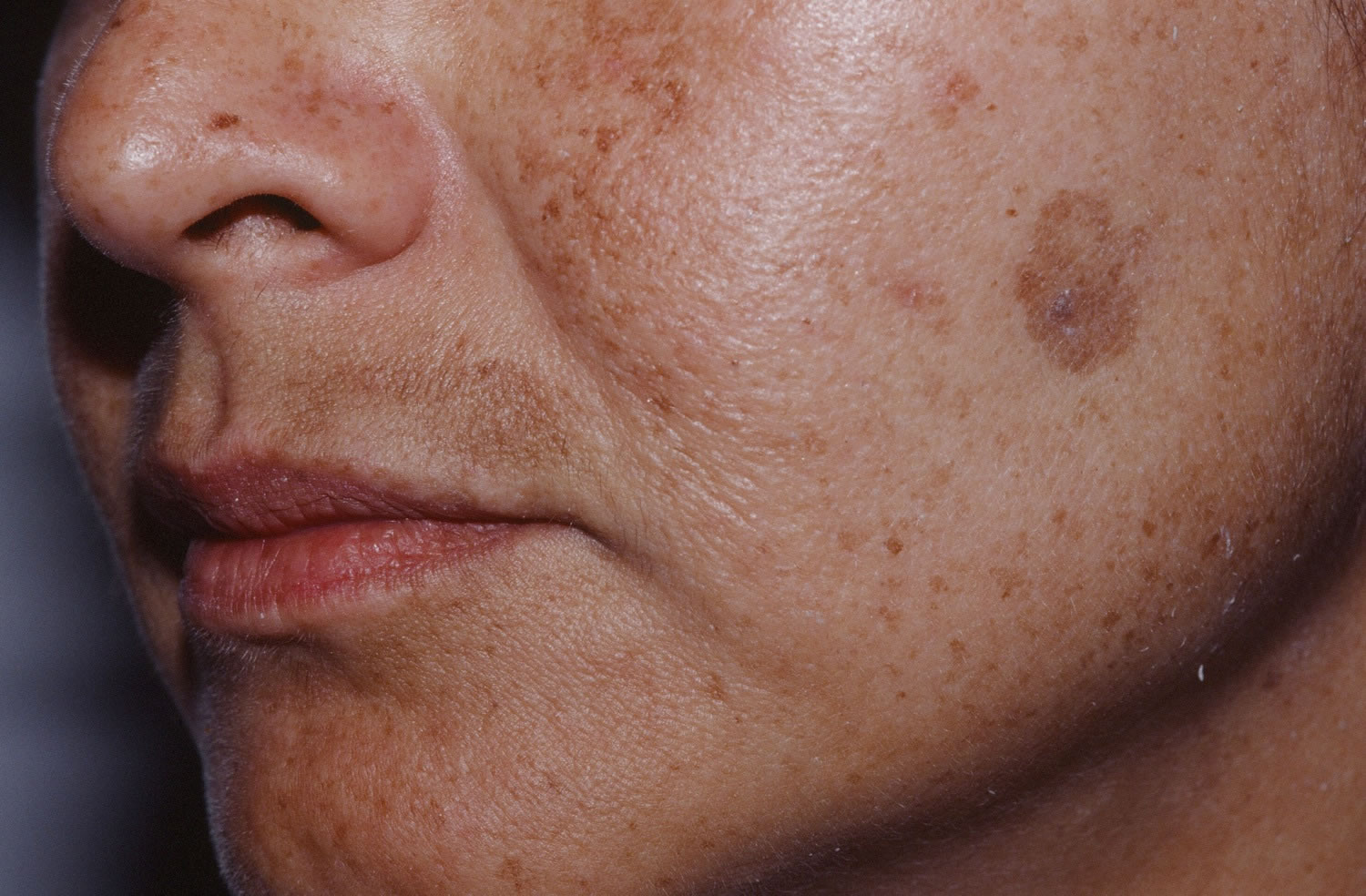
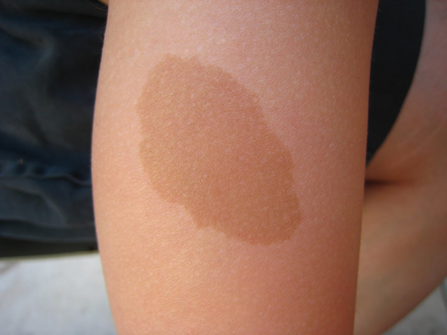
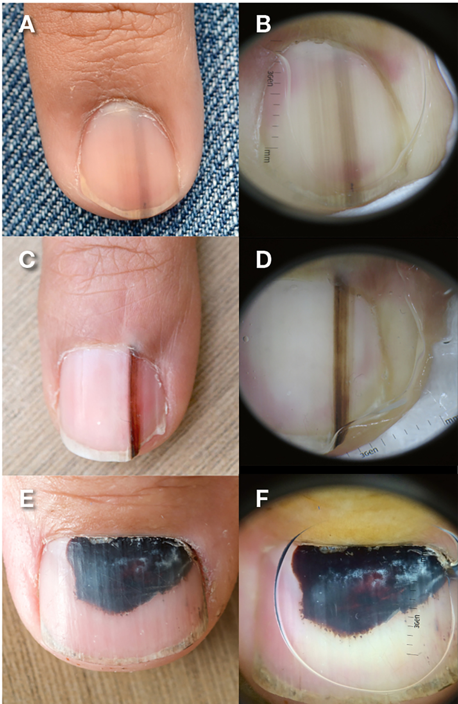
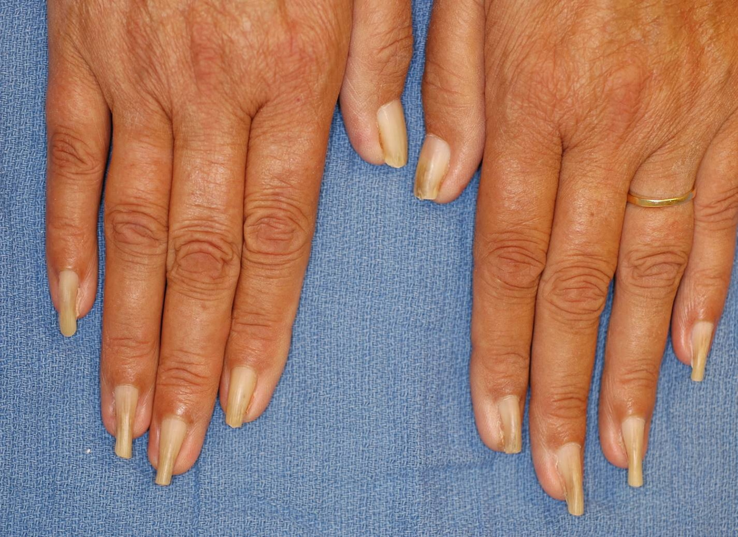
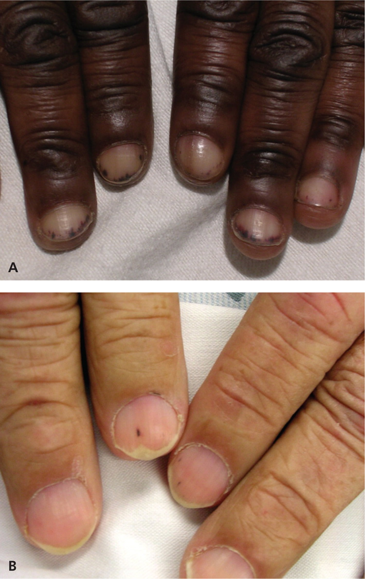
Figure 1. Melanoma in children

Melanoma in children

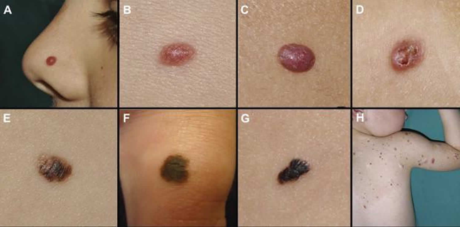
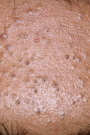
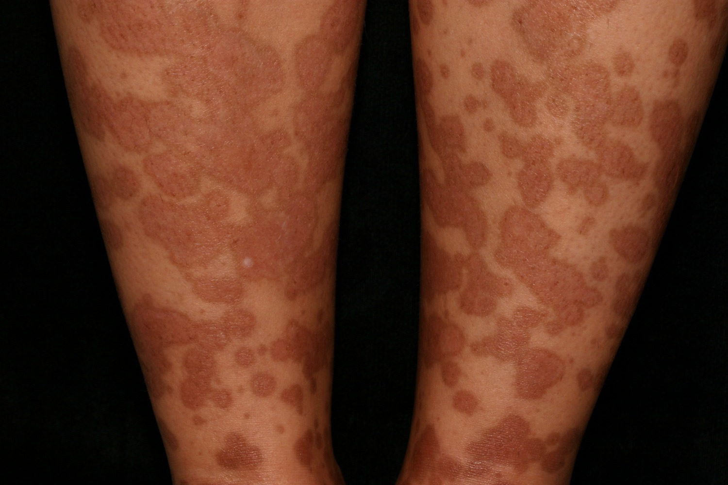
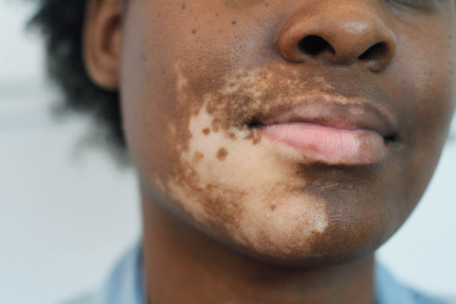
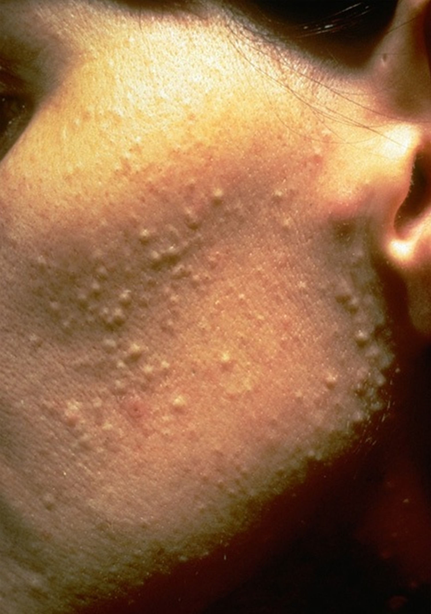
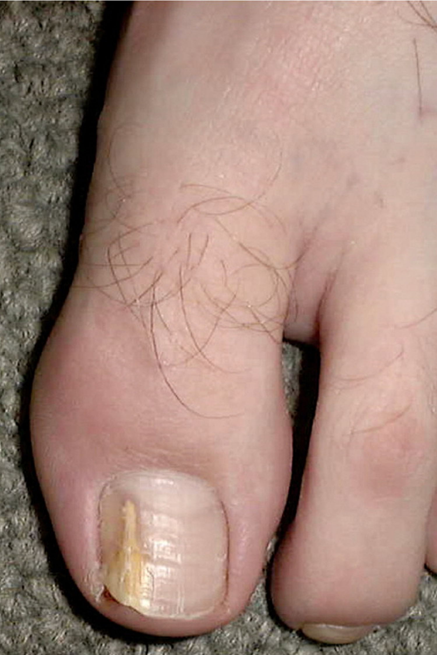
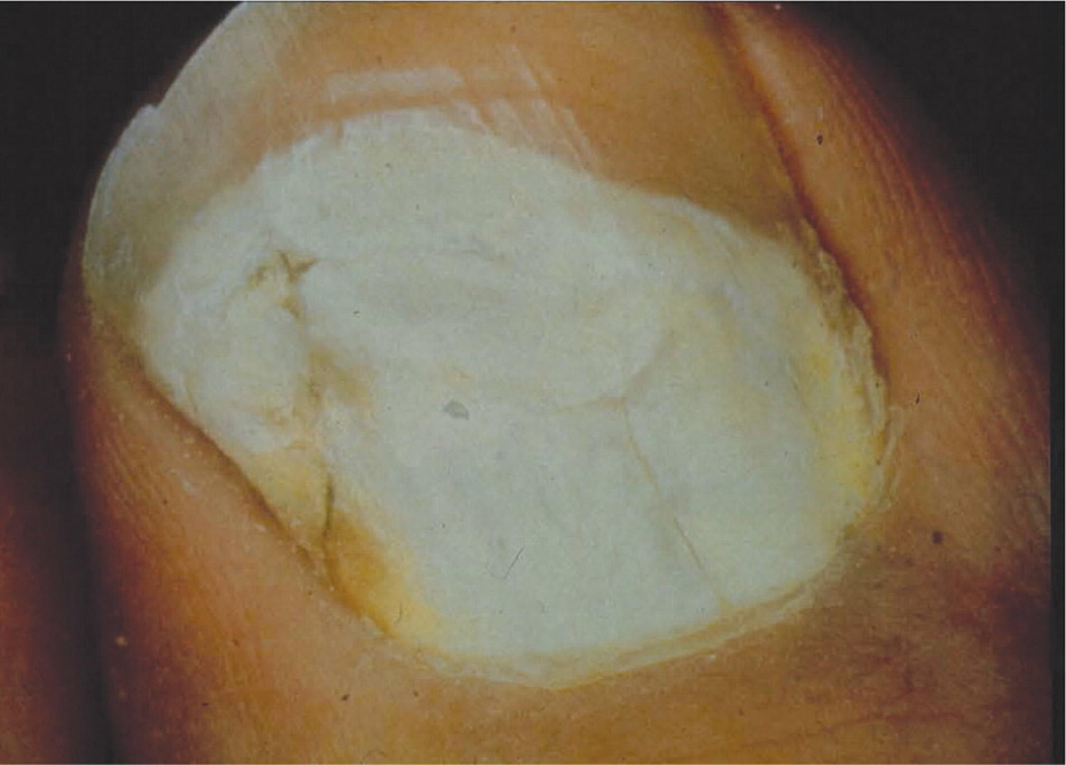
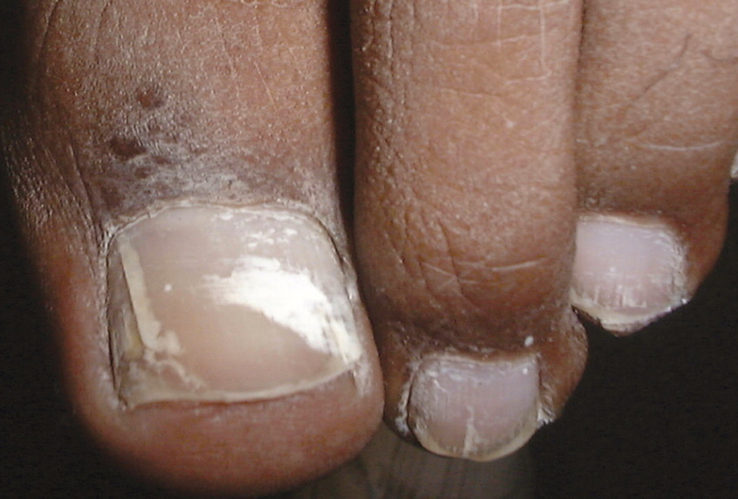
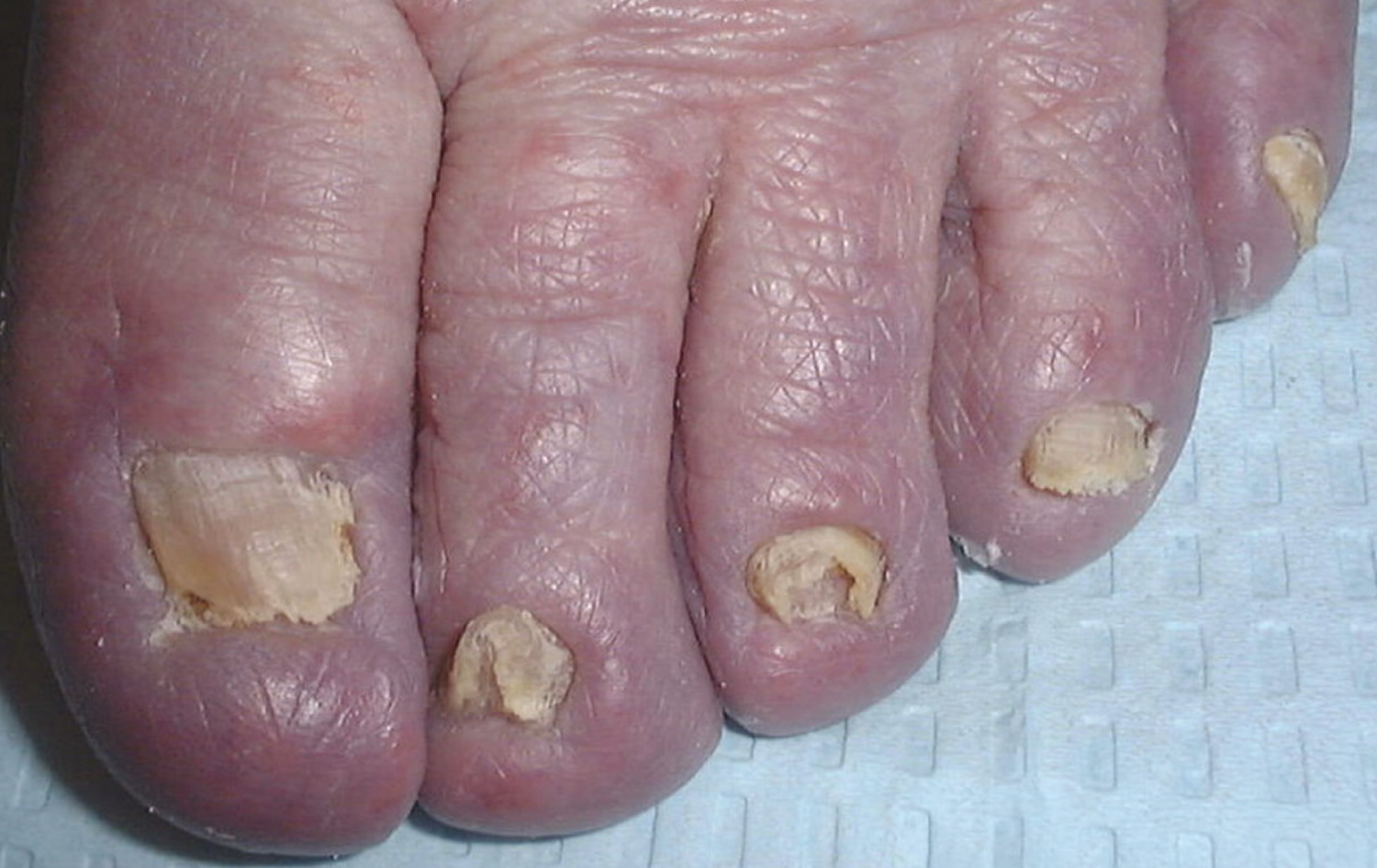
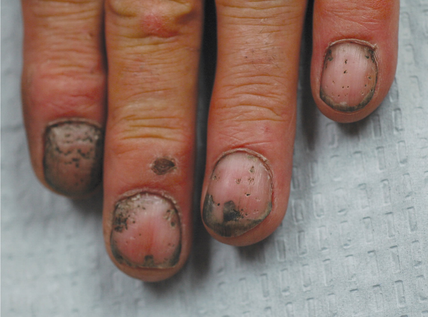
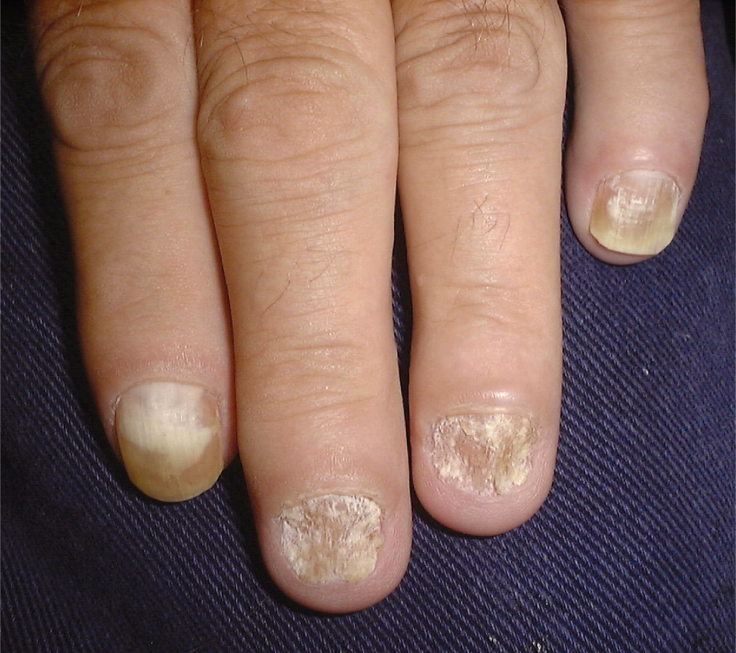
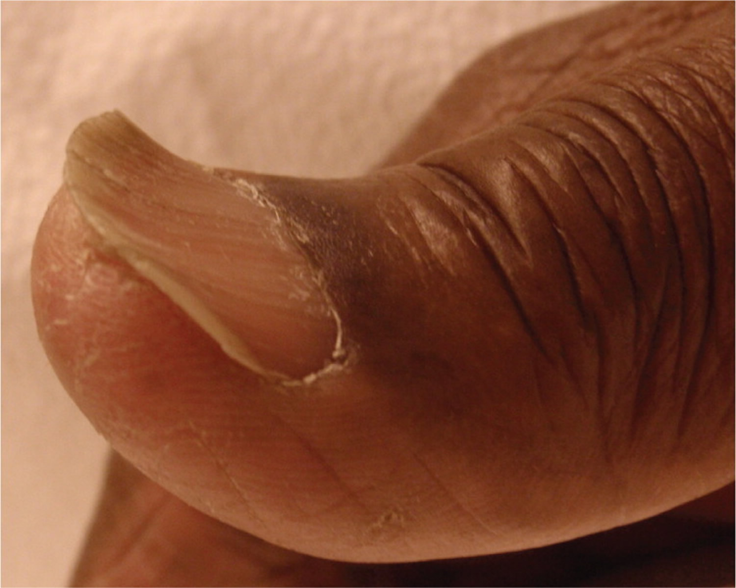
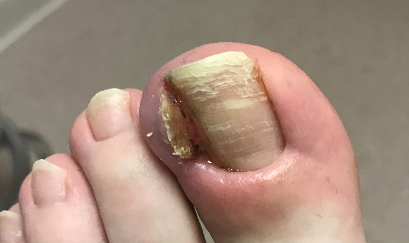
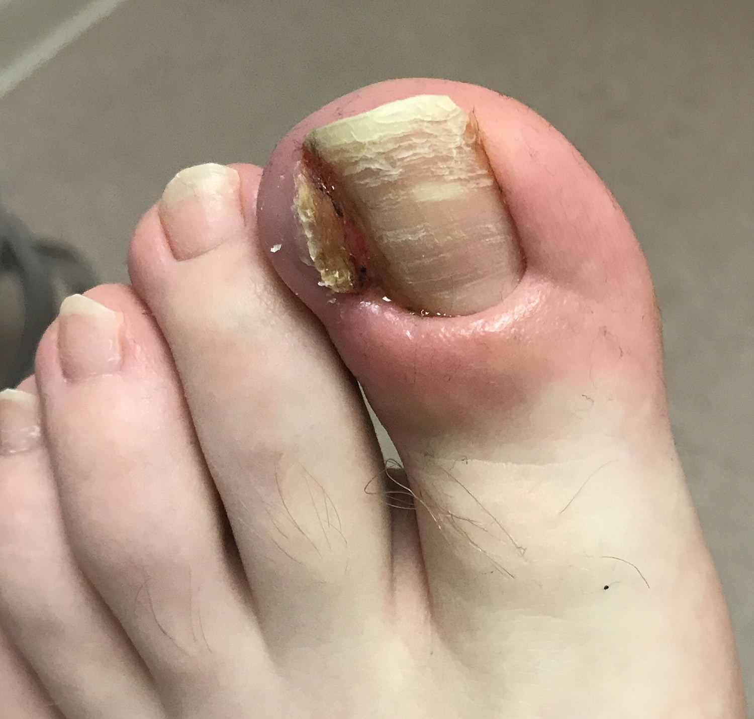
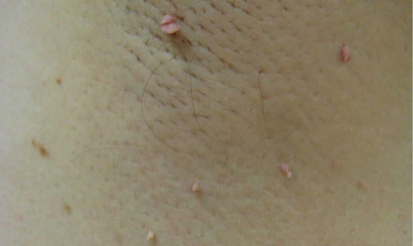
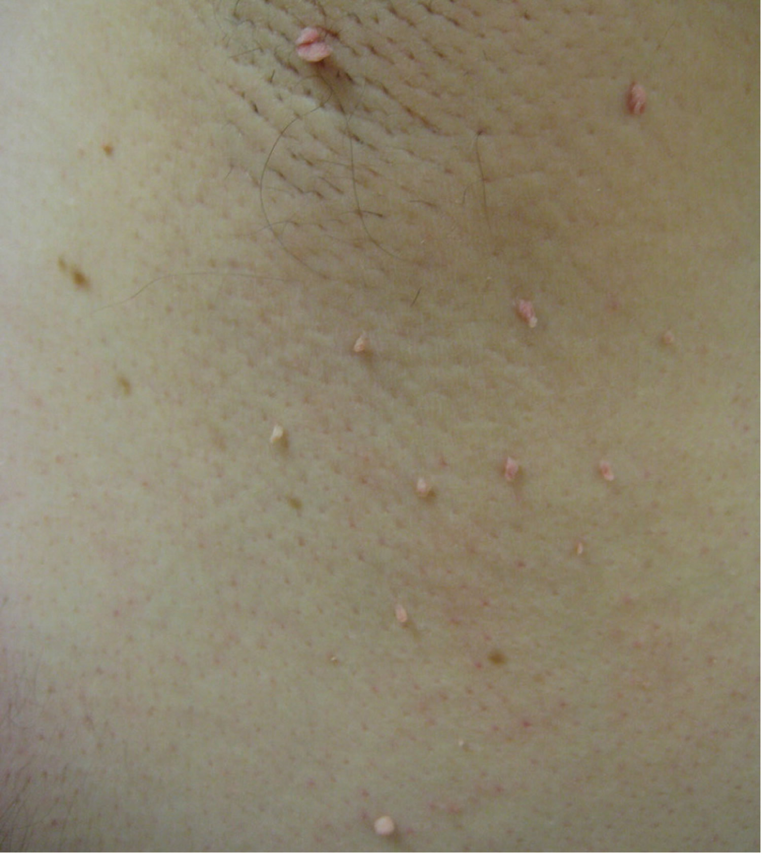
Figure 2. Squamous cell carcinoma in children

Squamous cell carcinoma in children

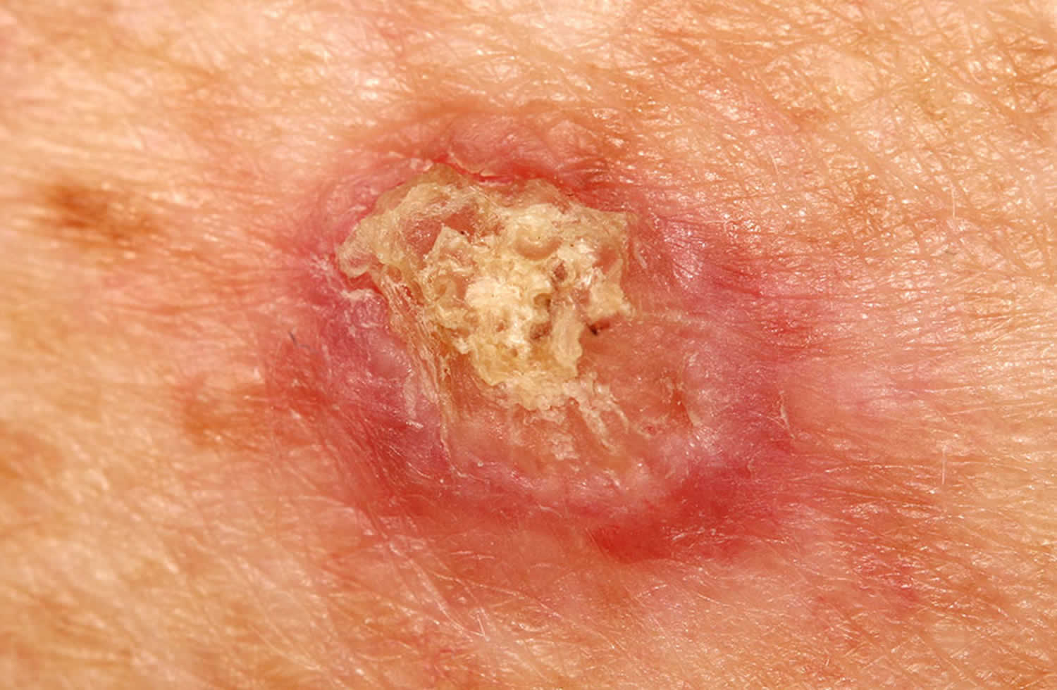
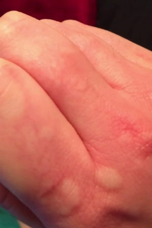
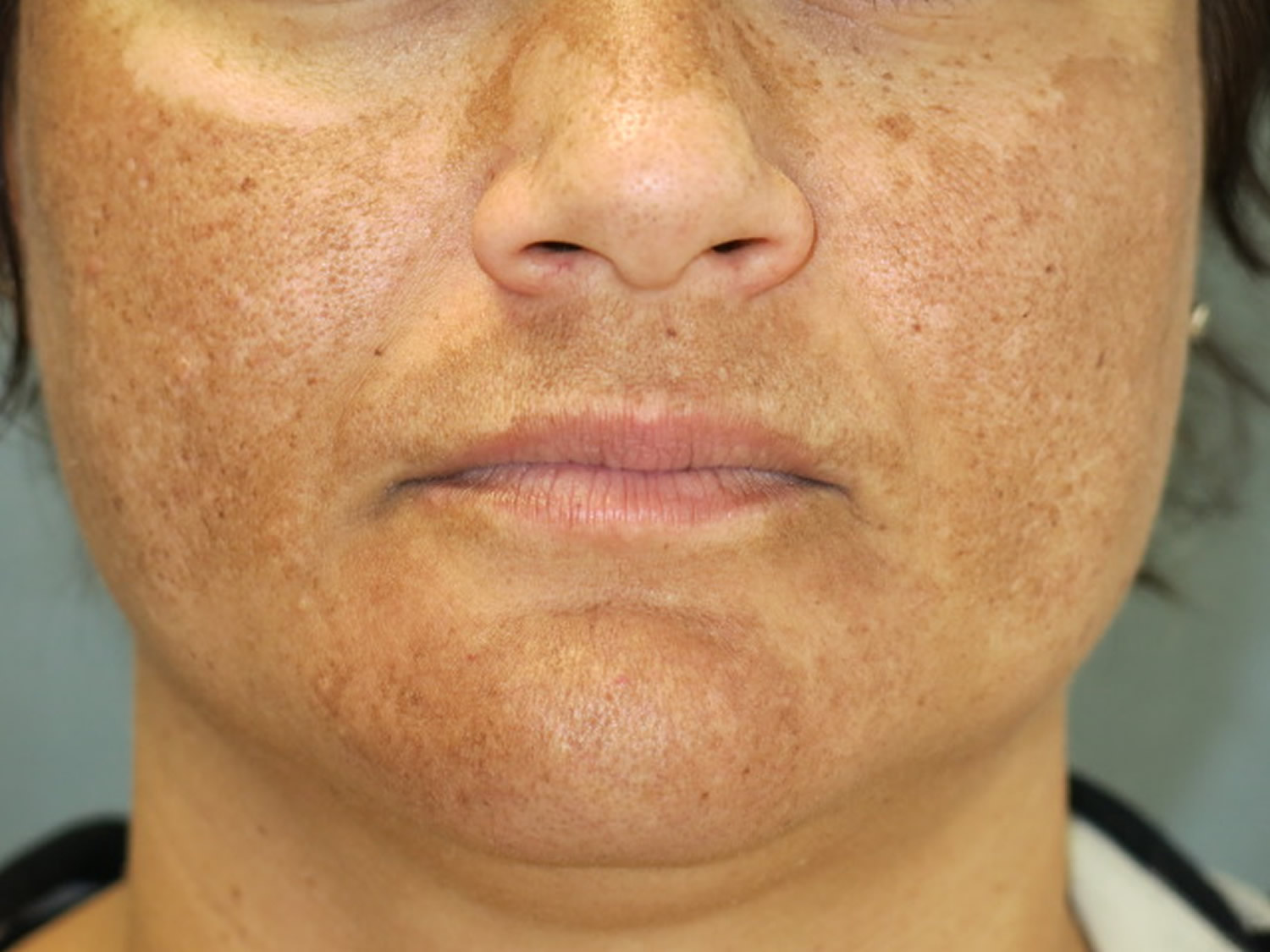
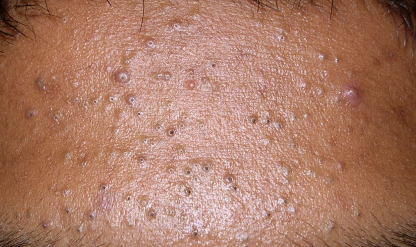
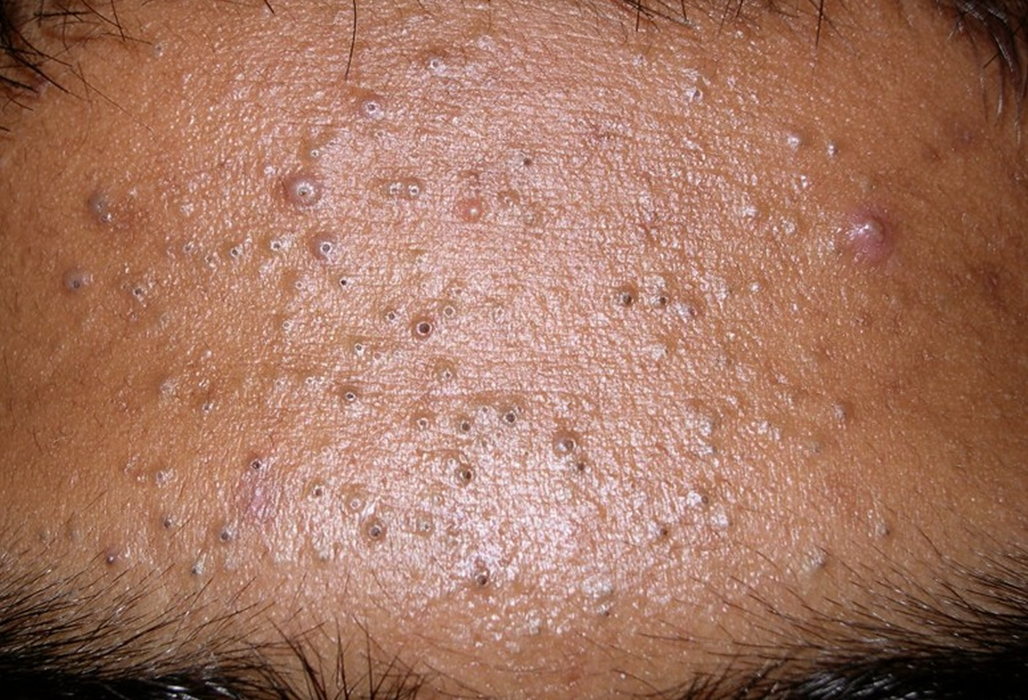
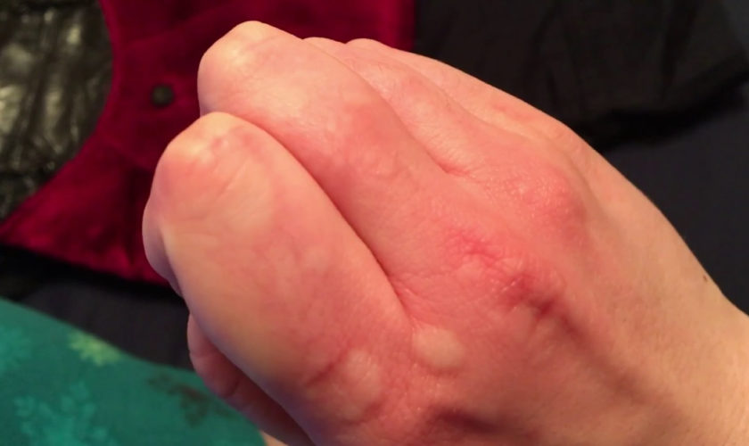
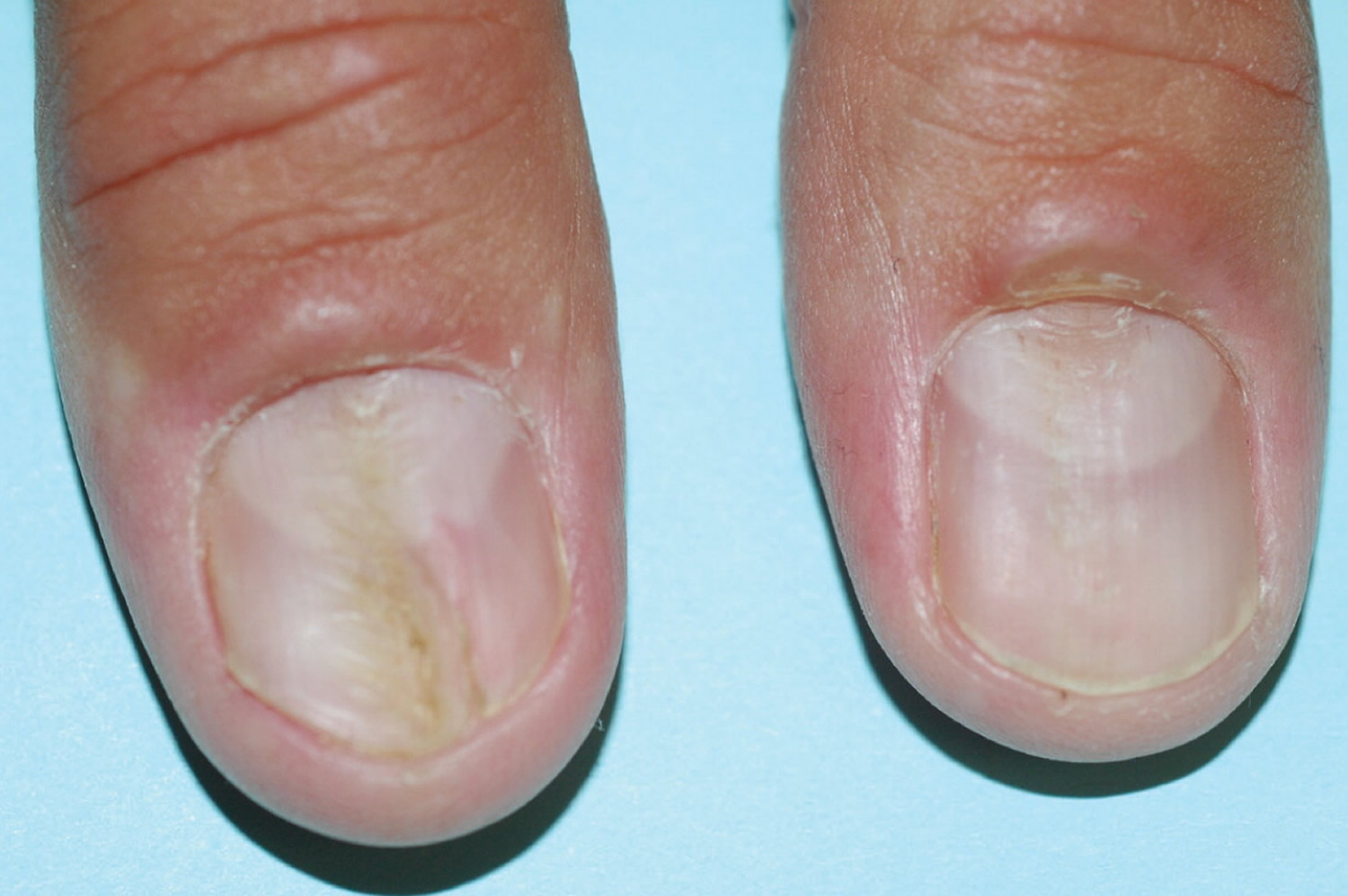
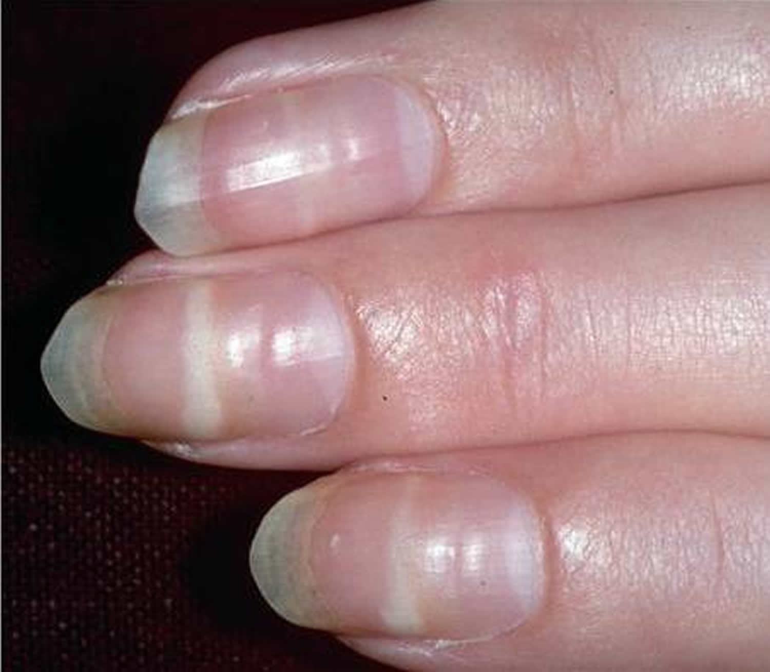
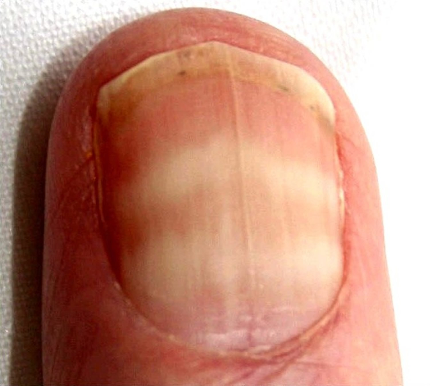
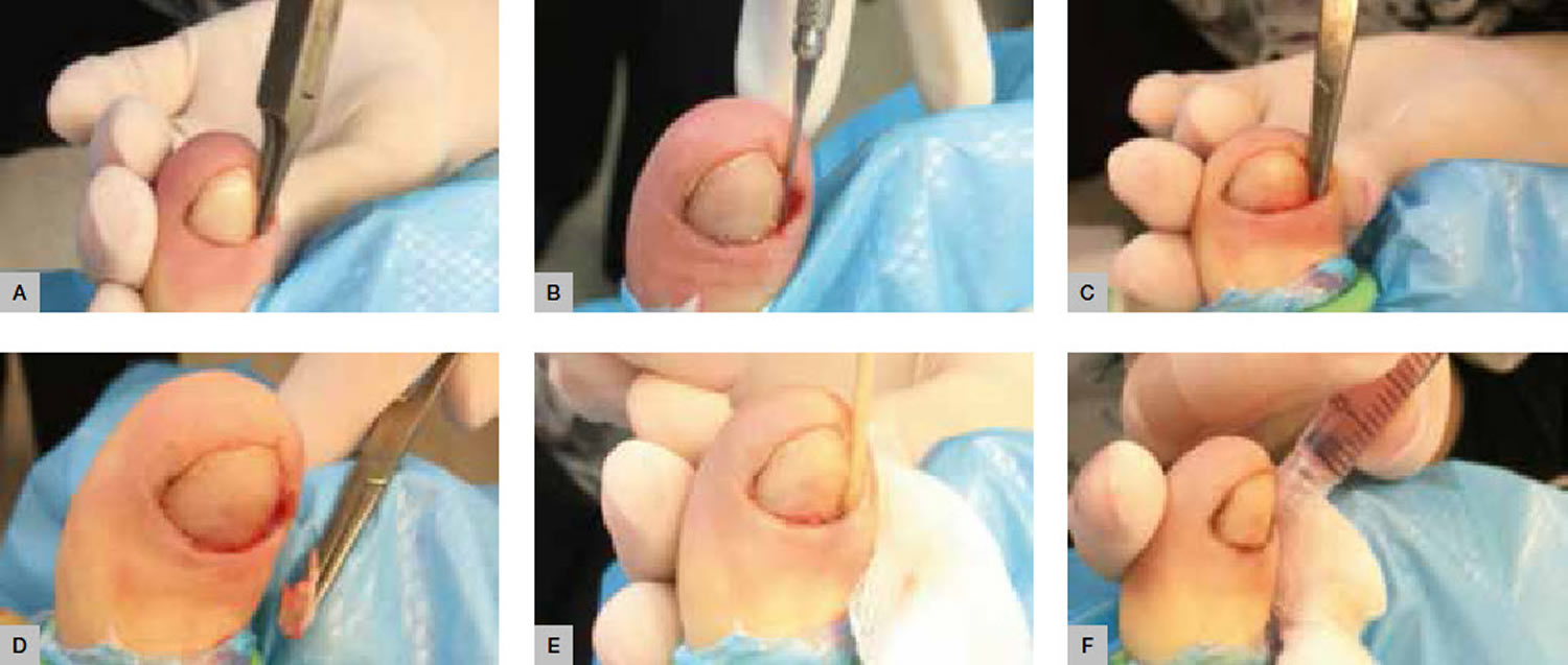
Figure 3. Basal cell carcinoma in children

Basal cell carcinoma in children

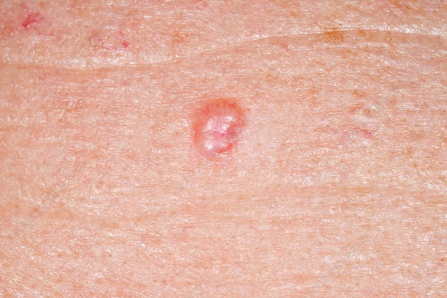
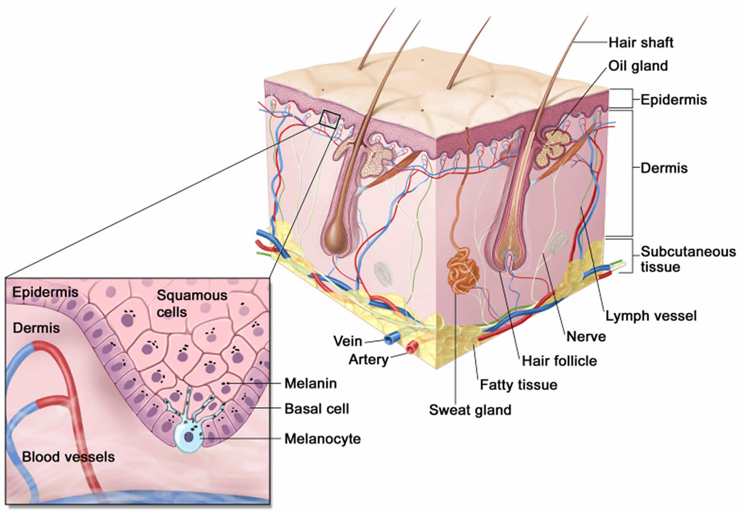
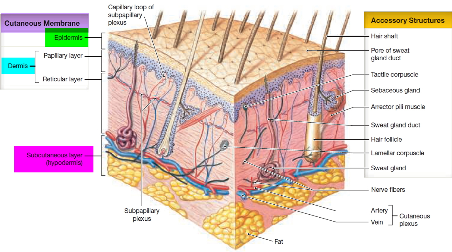
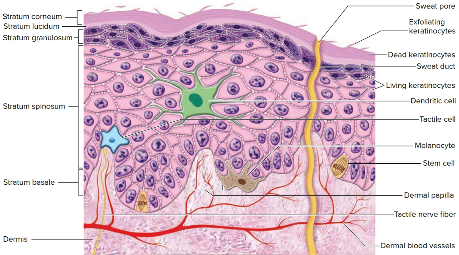
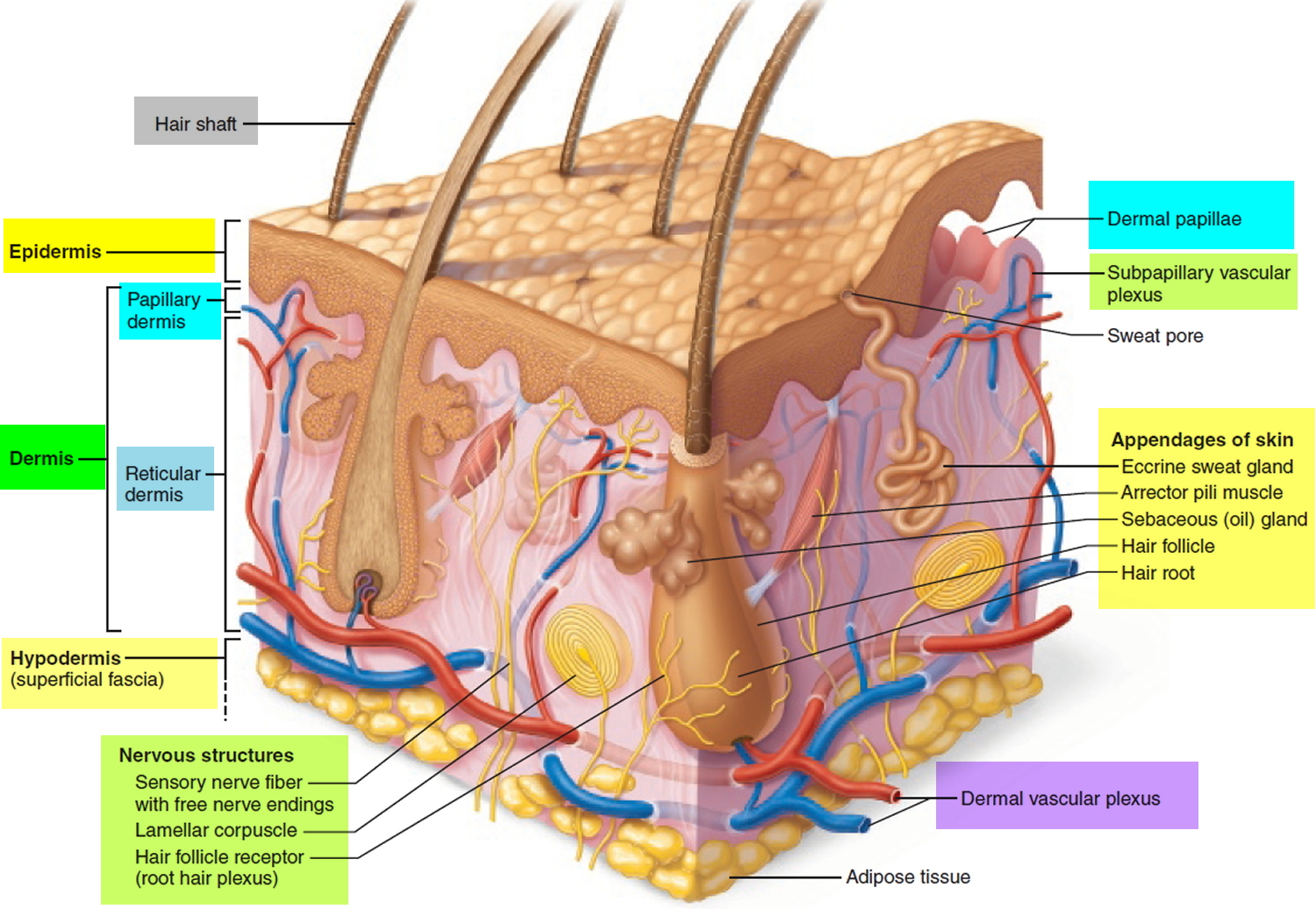
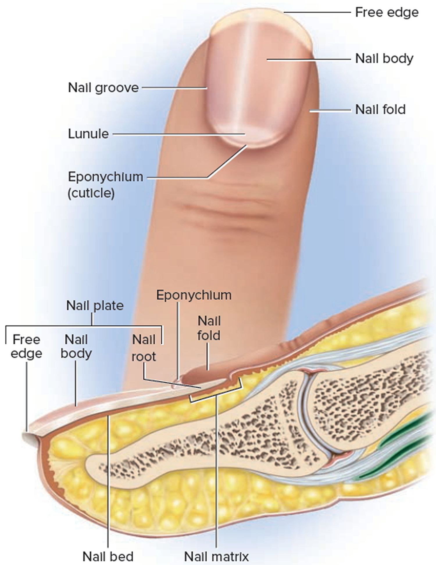
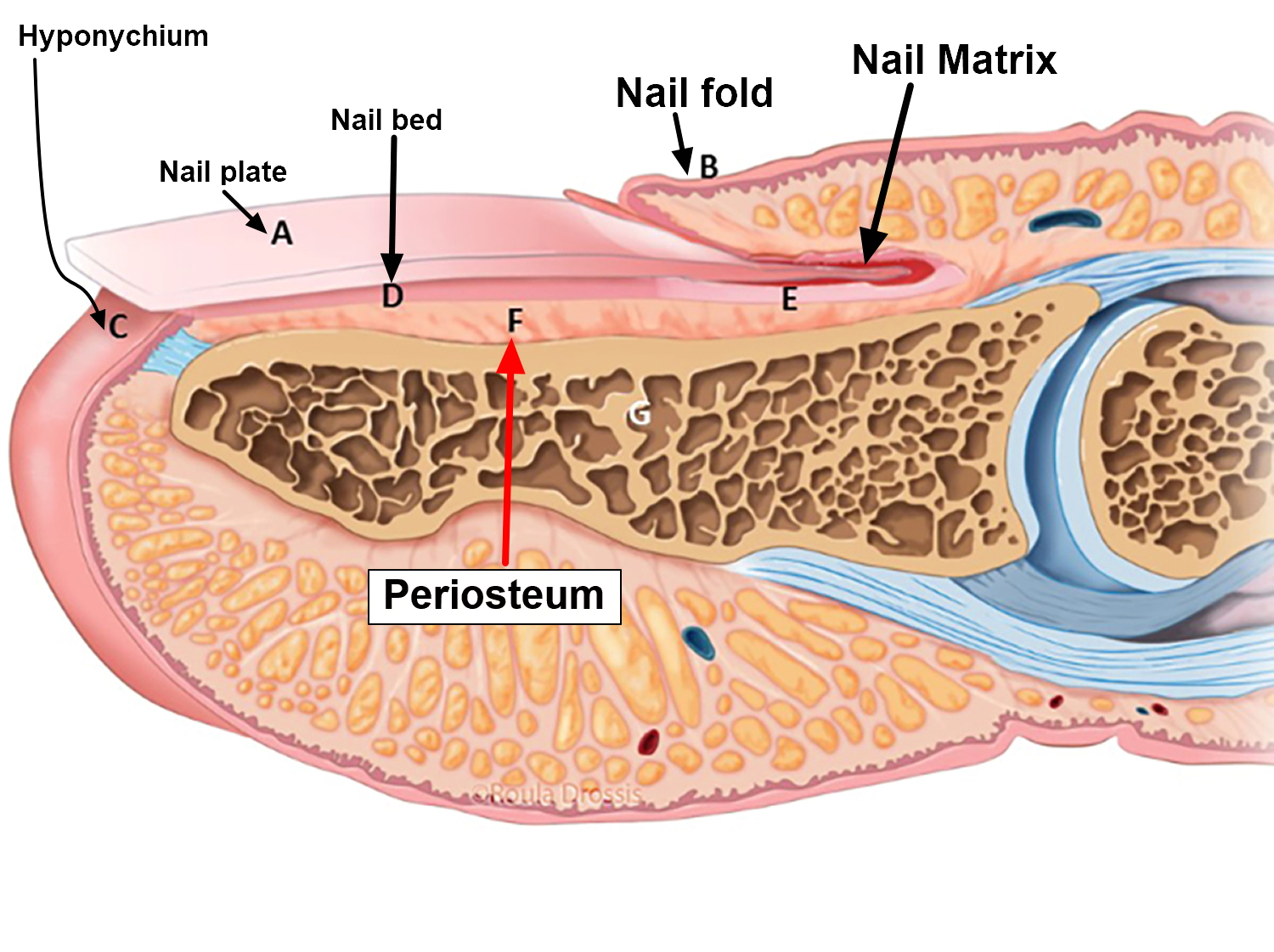
Figure 4. Skin anatomy

Skin anatomy

Basal cell carcinoma in children

Basal cell carcinoma (BCC) is a common, locally invasive, keratinocyte cancer also known as non-melanoma cancer. Basal cell carcinoma is very rarely a threat to life. Basal cell carcinoma (BCC) is the most common form of skin cancer. Basal cell carcinoma is also known as rodent ulcer and basalioma. Patients with basal cell carcinoma often develop multiple primary tumors over time.

Basal cell carcinoma main characteristics are:

  • Slowly growing plaque or nodule
  • Skin coloured, pink or pigmented
  • Varies in size from a few millimetres to several centimetres in diameter
  • Spontaneous bleeding or ulceration

A tiny proportion of basal cell carcinomas grow rapidly, invade deeply, and/or metastasise to local lymph nodes.

The cause of basal cell carcinoma is multifactorial:

  • Most often, there are DNA mutations in the patched (PTCH) tumour suppressor gene, part of hedgehog signalling pathway
  • These may be triggered by exposure to ultraviolet radiation
  • Various spontaneous and inherited gene defects predispose to basal cell carcinoma

Risk factors for basal cell carcinoma include:

  • Age and sex: basal cell carcinomas are particularly prevalent in elderly males. However, they also affect females and younger adults
  • Previous basal cell carcinoma or other form of skin cancer (squamous cell carcinoma, melanoma)
  • Sun damage (photoaging, actinic keratosis)
  • Repeated prior episodes of sunburn
  • Fair skin, blue eyes and blond or red hair—note; basal cell carcinoma can also affect darker skin types
  • Previous cutaneous injury, thermal burn, disease (eg cutaneous lupus, sebaceous nevus)
  • Inherited syndromes: basal cell carcinoma is a particular problem for families with basal cell naevus syndrome (Gorlin syndrome), Bazex-Dupré-Christol syndrome, Rombo syndrome, Oley syndrome and xeroderma pigmentosum
  • Other risk factors include ionizing radiation, exposure to arsenic, and immune suppression due to disease or medicines

Types of basal cell carcinoma

There are several distinct clinical types of basal cell carcinoma, and over 20 histological growth patterns of basal cell carcinoma.

Nodular basal cell carcinoma

  • Also known as nodulocystic carcinoma
  • Most common type of facial basal cell carcinoma
  • Shiny or pearly nodule with a smooth surface
  • May have central depression or ulceration, so its edges appear rolled
  • Blood vessels cross its surface
  • Cystic variant is soft, with jelly-like contents
  • Micronodular, microcystic and infiltrative types are potentially aggressive subtypes

Superficial basal cell carcinoma

  • Most common type in younger adults
  • Most common type on upper trunk and shoulders
  • Slightly scaly, irregular plaque
  • Thin, translucent rolled border
  • Multiple microerosions

Morphoeic basal cell carcinoma

  • Also known as morpheic, morphoeiform or sclerosing basal cell carcinoma
  • Usually found in mid-facial sites
  • Waxy, scar-like plaque with indistinct borders
  • Wide and deep subclinical extension
  • May infiltrate cutaneous nerves (perineural spread)

Basosquamous carcinoma

  • Also known as basisquamous carcinoma and mixed basal-squamous cell carcinoma
  • Mixed basal cell carcinoma (basal cell carcinoma) and squamous cell carcinoma (SCC)
  • Infiltrative growth pattern
  • Potentially more aggressive than other forms of basal cell carcinoma

Primary basal cell carcinoma treatment

The treatment for a basal cell carcinoma depends on its type, size and location, the number to be treated, patient factors, and the preference or expertise of the doctor. Most basal cell carcinomas are treated surgically. Long-term follow-up is recommended to check for new lesions and recurrence; the latter may be unnecessary if histology has reported wide clear margins.

Excision biopsy

Excision means the lesion is cut out and the skin stitched up.

  • Most appropriate treatment for nodular, infiltrative and morphoeic basal cell carcinomas
  • Should include 3 to 5 mm margin of normal skin around the tumor
  • Very large lesions may require flap or skin graft to repair the defect
  • Pathologist will report deep and lateral margins
  • Further surgery is recommended for lesions that are incompletely excised

Mohs micrographically controlled excision

Mohs micrographically controlled surgery involves examining carefully marked excised tissue under the microscope, layer by layer, to ensure complete excision.

  • Very high cure rates achieved by trained Mohs surgeons
  • Used in high-risk areas of the face around eyes, lips and nose
  • Suitable for ill-defined, morphoeic, infiltrative and recurrent subtypes
  • Large defects are repaired by flap or skin graft

Superficial skin surgery

Superficial skin surgery comprises shave, curettage, and electrocautery. It is a rapid technique using local anaesthesia and does not require sutures.

  • Suitable for small, well-defined nodular or superficial basal cell carcinomas
  • Lesions are usually located on trunk or limbs
  • Wound is left open to heal by secondary intention
  • Moist wound dressings lead to healing within a few weeks
  • Eventual scar quality variable

Cryotherapy

Cryotherapy is the treatment of a superficial skin lesion by freezing it, usually with liquid nitrogen.

  • Suitable for small superficial basal cell carcinomas on covered areas of trunk and limbs
  • Best avoided for basal cell carcinomas on head and neck, and distal to knees
  • Double freeze-thaw technique
  • Results in a blister that crusts over and heals within several weeks.
  • Leaves permanent white mark

Photodynamic therapy

Photodynamic therapy (PDT) refers to a technique in which basal cell carcinoma is treated with a photosensitising chemical, and exposed to light several hours later.

  • Topical photosensitisers include aminolevulinic acid lotion and methyl aminolevulinate cream
  • Suitable for low-risk small, superficial basal cell carcinomas
  • Best avoided if tumor in site at high risk of recurrence
  • Results in inflammatory reaction, maximal 3–4 days after procedure
  • Treatment repeated 7 days after initial treatment
  • Excellent cosmetic results

Imiquimod cream

Imiquimod is an immune response modifier.

  • Best used for superficial basal cell carcinomas less than 2 cm diameter
  • Applied three to five times each week, for 6–16 weeks
  • Results in a variable inflammatory reaction, maximal at three weeks
  • Minimal scarring is usual

Fluorouracil cream

5-Fluorouracil cream is a topical cytotoxic agent.

  • Used to treat small superficial basal cell carcinomas
  • Requires prolonged course, eg twice daily for 6–12 weeks
  • Causes inflammatory reaction
  • Has high recurrence rates

Radiotherapy

Radiotherapy or X-ray treatment can be used to treat primary basal cell carcinomas or as adjunctive treatment if margins are incomplete.

  • Mainly used if surgery is not suitable
  • Best avoided in young patients and in genetic conditions predisposing to skin cancer
  • Best cosmetic results achieved using multiple fractions
  • Typically, patient attends once-weekly for several weeks
  • Causes inflammatory reaction followed by scar
  • Risk of radiodermatitis, late recurrence, and new tumors

Advanced or metastatic basal cell carcinoma treatment

Locally advanced primary, recurrent or metastatic basal cell carcinoma requires multidisciplinary consultation. Often a combination of treatments is used.

  • Surgery
  • Radiotherapy
  • Targeted therapy

Targeted therapy refers to the hedgehog signalling pathway inhibitors, vismodegib and sonidegib. These drugs have some important risks and side effects.

Basal cell carcinoma prognosis

Most basal cell carcinomas are cured by treatment. Cure is most likely if treatment is undertaken when the lesion is small.

Death from basal and squamous cell skin cancers is uncommon. It’s thought that about 2,000 people in the US die each year from these cancers, and that this rate has been dropping in recent years. Most people who die from these cancers are elderly and may not have seen a doctor until the cancer had already grown quite large. Other people more likely to die of these cancers are those whose immune system is suppressed, such as people who have had organ transplants.

About 50% of people with basal cell carcinoma develop a second one within 3 years of the first. They are also at increased risk of other skin cancers, especially melanoma. Regular self-skin examinations and long-term annual skin checks by an experienced health professional are recommended.

Squamous cell carcinoma in children

Cutaneous squamous cell carcinoma (SCC) is a common type of keratinocyte cancer, or non-melanoma skin cancer. It is derived from cells within the epidermis that make keratin — the horny protein that makes up skin, hair and nails.

Cutaneous squamous cell carcinoma is an invasive disease, referring to cancer cells that have grown beyond the epidermis. Squamous cell carcinoma can sometimes metastasise (spread) and may prove fatal.

Cutaneous squamous cell carcinomas present as enlarging scaly or crusted lumps. They usually arise within pre-existing actinic keratosis or intraepidermal carcinoma.

  • They grow over weeks to months
  • They may ulcerate
  • They are often tender or painful
  • Located on sun-exposed sites, particularly the face, lips, ears, hands, forearms and lower legs
  • Size varies from a few millimetres to several centimetres in diameter.

Types of cutaneous squamous cell carcinoma

Distinct clinical types of invasive cutaneous squamous cell carcinoma include:

  • Cutaneous horn — the horn is due to excessive production of keratin
  • Keratoacanthoma — a rapidly growing keratinising nodule that may resolve without treatment
  • Carcinoma cuniculatum (‘verrucous carcinoma’), a slow-growing, warty tumour on the sole of the foot.
  • Multiple eruptive squamous cell carcinoma/keratoacanthoma-like lesions arising in syndromes, such as multiple self-healing squamous epitheliomas of Ferguson-Smith and Grzybowski syndrome

The histopathologist may classify a tumor as well differentiated, moderately well differentiated, poorly differentiated or anaplastic cutaneous squamous cell carcinoma. There are other variants.

Causes cutaneous squamous cell carcinoma

More than 90% of cases of squamous cell carcinoma are associated with numerous DNA mutations in multiple somatic genes. Mutations in the p53 tumour suppressor gene are caused by exposure to ultraviolet radiation (UV), especially UVB (known as signature 7). Other signature mutations relate to cigarette smoking, ageing and immune suppression (eg, to drugs such as azathioprine). Mutations in signalling pathways affect the epidermal growth factor receptor, RAS, Fyn, and p16INK4a signalling.

Beta-genus human papillomaviruses (wart virus) are thought to play a role in squamous cell carcinoma arising in immune-suppressed populations. β-HPV and HPV subtypes 5, 8, 17, 20, 24, and 38 have also been associated with an increased risk of cutaneous squamous cell carcinoma in immunocompetent individuals.

Risk factors for cutaneous squamous cell carcinoma include:

  • Age and sex: squamous cell carcinomas are particularly prevalent in elderly males. However, they also affect females and younger adults.
  • Previous squamous cell carcinoma or another form of skin cancer (basal cell carcinoma, melanoma) are a strong predictor for further skin cancers.
  • Actinic keratosis
  • Outdoor occupation or recreation
  • Smoking
  • Fair skin, blue eyes and blond or red hair
  • Previous cutaneous injury, thermal burn, disease (eg cutaneous lupus, epidermolysis bullosa, leg ulcer)
  • Inherited syndromes: squamous cell carcinoma is a particular problem for families with xeroderma pigmentosum and albinism
  • Other risk factors include ionising radiation, exposure to arsenic, and immune suppression due to disease (eg chronic lymphocytic leukaemia) or medicines. Organ transplant recipients have a massively increased risk of developing squamous cell carcinoma.

Cutaneous squamous cell carcinoma treatment

Cutaneous squamous cell carcinoma is nearly always treated surgically. Most cases are excised with a 3–10 mm margin of normal tissue around a visible tumor. A flap or skin graft may be needed to repair the defect.

Other methods of removal include:

  • Shave, curettage, and electrocautery for low-risk tumours on trunk and limbs
  • Aggressive cryotherapy for very small, thin, low-risk tumors
  • Mohs micrographic surgery for large facial lesions with indistinct margins or recurrent tumours
  • Radiotherapy for an inoperable tumour, patients unsuitable for surgery, or as adjuvant

Advanced or metastatic squamous cell carcinoma treatment

Locally advanced primary, recurrent or metastatic squamous cell carcinoma requires multidisciplinary consultation. Often a combination of treatments is used.

  • Surgery
  • Radiotherapy
  • Cemiplimab
  • Experimental targeted therapy using epidermal growth factor receptor inhibitors

The exact number of people who develop or die from basal and squamous cell skin cancers each year isn’t known for sure. Death from basal and squamous cell skin cancers is uncommon. It’s thought that about 2,000 people in the US die each year from these cancers, and that this rate has been dropping in recent years. Most people who die from these cancers are elderly and may not have seen a doctor until the cancer had already grown quite large. Other people more likely to die of these cancers are those whose immune system is suppressed, such as people who have had organ transplants.

Cutaneous squamous cell carcinoma prognosis

Most squamous cell carcinomas are cured by treatment. A cure is most likely if treatment is undertaken when the lesion is small. The risk of recurrence or disease-associated death is greater for tumours that are > 20 mm in diameter and/or > 2 mm in thickness at the time of surgical excision.

About 50% of people at high risk of squamous cell carcinoma develop a second one within 5 years of the first. They are also at increased risk of other skin cancers, especially melanoma. Regular self-skin examinations and long-term annual skin checks by an experienced health professional are recommended.

Melanoma in children

Melanoma is a skin cancer that arises from melanocytes (pigment-producing cells). Childhood melanoma usually refers to melanoma diagnosed in individuals under the age of 18 years.

Cutaneous melanoma in children is rare, and extremely rare before puberty 1. Melanoma comprises 3% of all pediatric cancers 2.

Melanoma arising in children has been classified into the following types 3:

  • Melanoma present at birth (congenital melanoma)
  • Melanoma developing in congenital melanocytic nevus (brown birthmark)
  • Melanoma arising in patients with dysplastic or atypical nevi (most often superficial spreading melanoma arising de novo)
  • Malignant blue nevus
  • Nodular melanoma (40-50% of malignant melanoma in children)
  • Spitzoid melanoma

Risk factors for childhood melanoma include 4:

  • Giant congenital nevus
  • Fitzpatrick skin phototypes I-II (i.e. fair skin that burns easily and tans poorly, freckles)
  • Immunodeficiency or immunosuppression
  • History of retinoblastoma
  • Familial atypical naevi (dysplastic naevus syndrome)
  • Many moles
  • Xeroderma pigmentosum (a very rare disorder with extreme sensitivity to sunlight)

Like the adult population, melanoma mainly affects Caucasian children and is associated with sun exposure. There is a slight female preponderance 1.

Melanoma in a congenital melanocytic nevus

Small congenital nevi arise in 1 in 100 births. Melanoma is a rare complication of small to medium congenital nevi. It tends to appear on the edge of the birthmark and is recognized by change within the mole and the ABCDE criteria.

The risk of melanoma is higher in larger congenital nevi 5. Melanoma arises in about 4% of children 10 years or younger that have a giant congenital melanocytic nevus >40 cm in diameter. Giant congenital melanocytic nevi are very rare, arising in 1 in 20,000 births. In giant congenital melanocytic nevi:

  • Melanoma may arise within the centre of the melanoma
  • It tends to arise within deeper dermal naevus cells rather than within superficial naevus cells
  • The melanoma may also arise within the central nervous system due to neurocutaneous melanocytosis
  • The risk of melanoma is greater in giant naevi that cross the midline of the spine and in children with satellite naevi
  • Prophylactic removal of the naevi does not appear to reduce the risk of melanoma

These melanomas can be difficult to detect early. Excision may also be difficult or impossible.

Melanoma in children aged 11 and older

Melanoma in older children appears similar to melanoma in adults; it presents as a growing lesion that looks different from the child’s other lesions. Most are pigmented. About 60% have the ABCDE criteria.

  • A: Asymmetry
  • B: Border irregularity
  • C: Color variation
  • D: Diameter >6 mm
  • E: Evolving

Melanoma in children aged 10 or younger

Superficial spreading melanoma is less common in younger children and melanoma has the ABCDE criteria in 40% of cases.

Melanoma in young children is more commonly amelanotic (red coloured), nodular, and tends to be thicker at diagnosis than in older children and adults.

Cordoro et al 6 have suggested adding additional ABCD detection criteria for skin lesions in children:

  • A: Amelanotic (the lesion is skin colored or red)
  • B: Bleeding, Bump
  • C: Color uniformity
  • D: De novo, any Diameter

Childhood melanoma treatment

Treatment of childhood melanoma is the same as in adults.

  • Lesions that are suspicious for melanoma are completely removed by initial diagnostic excision biopsy, usually with a 2-mm clinical margin.
  • If melanoma is confirmed, a second surgical procedure is undertaken to remove a wider margin of normal skin. This is called wide local excision. The size of the margin depends on the Breslow thickness of the melanoma.
  • If the melanoma has thickness >1 mm or other features of concern, sentinel node biopsy may be offered. However, its role in the pediatric population is not well established.
  • Follow-up is arranged to look for recurrence and new lesions of concern 7.

Metastatic melanoma or advanced melanoma is melanoma that has spread to lymph nodes or elsewhere in the body. Treatment is individualized but may include surgery, radiotherapy, chemotherapy or targeted therapy.

Childhood melanoma prognosis

Prognosis of melanoma depends on the stage of melanoma, ie whether it has spread beyond its original site in the skin. Spread of melanoma to lymph nodes and elsewhere is more likely in thicker tumors (measured by Breslow thickness at the time of removal of a primary tumor).

Survival rates are similar in older children and adults 2. However, melanomas in children under 11 years of age appear to have a less aggressive behavior than those detected in adults 1.

What causes skin cancer in kids?

The common forms of skin cancer listed above are related to exposure to ultraviolet (UV) radiation from sunlight or tanning beds or lamps and the effects of ageing. Skin cancer is more common in people with light skin, light-colored eyes, and blond or red hair.

Other risk factors include:

  • Smoking (especially for squamous cell carcinoma)
  • Human papillomavirus infection (genital warts), particularly for mucosal sites such as oral mucosa, lips and genitals
  • Immune suppression, for example in patients who have received an organ transplant and are on azathioprine and ciclosporin
  • Human immunodeficiency virus infection (HIV)
  • Exposure to ionizing radiation or radiation therapy in the past
  • Exposure to certain chemicals, such as arsenic and coal tar
  • Longstanding skin diseases such as lichen sclerosus, lupus erythematosus, linear porokeratosis or cutaneous tuberculosis
  • A longstanding wound or scar, for example, from a thermal burn (a Marjolin ulcer).
  • Age.
  • Time spent in the sun
  • History of sunburns
  • Actinic keratoses or Bowen disease. These are rough or scaly red or brown patches on the skin.
  • Family history of skin cancer
  • Having many freckles
  • Having many moles
  • Having skin cancer in the past
  • Having atypical moles (dysplastic nevi). These large, oddly shaped moles run in families.
  • Taking a medicine that suppresses the immune system.

Some skin cancers are due to genetic conditions, such as:

  • Albinism
  • Basal cell nevus syndrome (Gorlin syndrome)
  • Bazex–Dupré–Christol syndrome
  • Bloom syndrome
  • Brooke-Spiegler syndrome
  • Cowden syndrome
  • Dyskeratosis congenita
  • Epidermolysis bullosa
  • Epidermodysplasia verruciformis
  • Familial atypical mole-melanoma syndrome (FAMM)
  • Premature ageing syndromes (progeria)
  • Rothmund-Thomson syndrome
  • Torré-Muir syndrome
  • Xeroderma pigmentosum (XP).

Skin cancer in children prevention

The American Academy of Dermatology and the Skin Cancer Foundation advise you to:

  • Limit how much sun your child gets between the hours of 10 a.m. and 4 p.m.
  • Use broad-spectrum sunscreen with an SPF 30 or higher that protects against both UVA and UVB rays. Put it on the skin of children older than 6 months of age who are exposed to the sun.
  • Reapply sunscreen every 2 hours, even on cloudy days. Reapply after swimming.
  • Use extra caution near water, snow, and sand. They reflect the damaging rays of the sun. This can increase the chance of sunburn.
  • Make sure your child wears clothing that covers the body and shades the face. Hats should provide shade for both the face, ears, and back of the neck. Wearing sunglasses will reduce the amount of rays reaching the eye and protect the lids of the eyes, as well as the lens.
  • Don’t let your child use or be around sunlamps or tanning beds.

The American Academy of Pediatrics approves of the use of sunscreen on babies younger than 6 months old if adequate clothing and shade are not available. You should still try to keep your baby out of the sun. Dress the baby in lightweight clothing that covers most surface areas of skin. But you also may use a small amount of sunscreen on the baby’s face and back of the hands.

Skin cancer in children symptoms

Skin cancers generally appear as a lump or nodule, an ulcer, or a changing lesion.

Symptoms of basal cell carcinoma (BCC) appear on areas exposed to the sun, such as the head, face, neck, arms, and hands. The symptoms can include:

  • A small, raised bump that is shiny or pearly, and may have small blood vessels
  • A small, flat spot that is scaly, irregularly shaped, and pale, pink, or red
  • A spot that bleeds easily, then heals and appears to go away, then bleeds again in a few weeks
  • A growth with raised edges, a lower area in the center, and brown, blue, or black areas

Symptoms of squamous cell carcinoma (SCC) appear on areas exposed to the sun, such as the head, face, neck, arms, and hands. They can also appear on other parts of the body, such as skin in the genital area. The symptoms can include:

  • A rough or scaly bump that grows quickly
  • A wart-like growth that may bleed or crust over.
  • Flat, red patches on the skin that are irregularly shaped, and may or may not bleed

Symptoms of melanoma include a change in a mole, or a new mole that has ABCDE traits such as:

  • Asymmetry. One half of the mole does not match the other half.
  • Border irregularity. The edges of the mole are ragged or irregular.
  • Color. The mole has different colors in it. It may be tan, brown, black, red, or other colors. Or it may have areas that appear to have lost color.
  • Diameter. The mole is bigger than 6 millimeters across, about the size of a pencil eraser. But some melanomas can be smaller.
  • Evolving. A mole changes in size, shape, or color.

Other symptoms of melanoma can include a mole that:

  • Itches or hurts
  • Oozes, bleeds, or becomes crusty
  • Turns red or swells
  • Looks different from your child’s other moles.

Complications of skin cancer in children

Skin cancer can usually be treated and cured before complications occur. Signs of advanced, aggressive or neglected skin cancer may include:

  • Ulceration
  • Bleeding
  • Spread of a tumor to lymph glands and other organs such as liver and brain (metastasis).

Possible complications depend on the type and stage of skin cancer. Melanoma is more likely to cause complications. And the more advanced the cancer, the more likely there will be complications.

Complications may result from treatment, such as:

  • Loss of large areas of skin and underlying tissue
  • Scarring
  • Problems with the area healing
  • Infection in the area
  • Damage to nerves
  • Return of the skin cancer after treatment

Melanoma may spread to organs throughout the body and cause death.

Skin cancer in children diagnosis

Skin cancers are generally diagnosed clinically by a dermatologist or family doctor, when learning of an enlarging, crusting or bleeding lesion. The lesion will be inspected carefully, and ideally, a full skin examination will also be conducted. Dermatoscopy (a special magnifying light) may be used to confirm the diagnosis, to detect early skin cancers, and to exclude benign lesions.

Your doctor will examine your child’s skin. Tell your doctor:

  • When you first noticed the skin problem
  • If it oozes fluid or bleeds, or gets crusty
  • If it’s changed in size, color, or shape
  • If your child has pain or itching

Tell your doctor if your child has had skin cancer in the past, and if other your family members have had skin cancer.

Your child’s doctor will likely take a small piece of tissue (biopsy) from a mole or other skin mark that may look like cancer to confirm the diagnosis. The tissue is sent to a lab. A doctor called a histopathologist looks at the tissue under a microscope. He or she may do other tests to see if cancer cells are in the sample. It can take a few days for the report to be issued, or longer if special tests are required. The biopsy results will likely be ready in a few days or a week. Your child’s doctor will tell you the results. He or she will talk with you about other tests that may be needed if cancer is found.

Complete excision is usually undertaken to make a diagnosis if melanoma is suspected, as a partial biopsy can be misleading in melanocytic tumors. Genetic testing for melanoma and blood-based melanoma detection may be available in some centers.

Skin cancer in children treatment

Early treatment of skin cancer usually cures it. The majority of skin cancers are treated surgically, using a local anesthetic to numb the skin. Surgical techniques include:
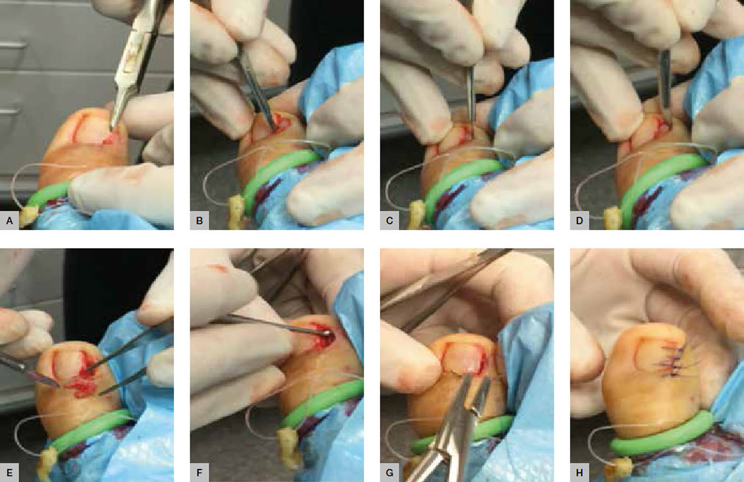
  • Excision biopsy
    • Simple excision: This is done to cut the cancer from the skin, along with some of the healthy tissue around it. Your child is given a local anesthetic. Then, the doctor uses a scalpel to remove the tumor from the skin. The doctor may also remove some of the normal skin around the tumor. This is called a margin. Stitches or a bandage strip may be used to close the wound. The tissue that was removed is sent to a lab for testing. If the report shows that not all the cancer was removed, your child will likely need another procedure to remove the rest of the cancer.
    • Shave excision: This method is used for cancer that is only in the top layers of the skin. Your child is given a local anesthetic. Then, the doctor uses a small blade to shave off the tumor. The goal is to remove the tumor at its base.
  • Mohs surgery: This procedure removes the cancer and a small amount of normal tissue. It’s done on sensitive areas, such as the face. During Mohs surgery, your child is given a local anesthetic to numb the area being treated. The cancer is removed from the skin one layer at a time. Each layer is checked under a microscope for cancer. If cancer cells are seen, another layer of skin is removed. Layers are removed until the doctor doesn’t see any more cancer. The procedure may take several hours, depending on how many layers need to be removed. After this surgery, the cancer is fully removed and the wound can be repaired.

Treatment options for superficial skin cancers include:

  • Minor surgery including curettage and diathermy/cautery and electrosurgery: This procedure removes tissue and burns (cauterizes) the area. Your child is given a local anesthetic to numb the area. The doctor then uses a sharp spoon-shaped tool called a curette to remove the cancer. This is called curettage. After curettage, the doctor passes an electric needle over the surface of the scraped area to stop bleeding, and destroy any other cancer cells. After it heals, a flat white scar may remain.
  • Cryotherapy: This method uses cold to destroy the cancer cells. This method is best for very small cancers near the skin’s surface. The doctor uses a device that sprays liquid nitrogen onto the tumor. This freezes the cells and destroys them. The dead skin then falls off. Your child may have some swelling and blistering in the area after treatment. A white scar is usually left behind. The procedure may need to be repeated.
  • Topical chemotherapy such as fluorouracil cream, imiquimod cream or ingenol mebutate gel. This kind of medicine is only used if the cancer is just in the top layers of the skin. The medicine is applied several times a week for a few weeks.
  • Photodynamic therapy (photosensitising cream plus light)
  • Radiotherapy (x-ray treatment): This is treatment with high-energy X-rays. Electron beam radiation is often used for skin cancer. This type of radiation doesn’t go deeper than the skin. This helps limit side effects. The radiation damages the cancer cells and stops them from growing. Radiation therapy is a local therapy. This means that it affects the cancer cells only in the treated area.
  • Lasers

Treatment for advanced or metastatic basal cell carcinoma may include targeted therapies vismodegib and sonidegib.

Treatment for advanced and metastatic melanoma may include:

  • Systemic immunotherapy using ipilimumab or checkpoint inhibitors pembrolizumab or nivolumab
  • Topical and intralesional immunotherapy for melanoma metastases
  • Targeted therapy against BRAF mutations using vemurafenib or dabrafenib or MEK inhibition with trametinib. The goal of targeted therapy is to shrink advanced melanoma tumors. This type of therapy is done with medicines that target specific parts of melanoma cells. For example, medicines called BRAF inhibitors target cells with a change in the BRAF gene. This gene is found in about half of all melanomas.
  • Combination medications, such as cometinib.

Patients with skin cancer may be at increased risk of developing other skin cancers. They may be advised to:

  • Practice careful sun protection, including the regular application of sunscreens
  • Learn and practice self-skin examination
  • Have regular skin checks
  • Undergo digital dermatoscopic surveillance (mole mapping), especially if they have many moles or atypical moles
  • Seek medical attention if they notice any changing or enlarging skin lesions
  • Take nicotinamide (vitamin B3) to reduce the numbers of squamous cell carcinomas.

Living with skin cancer in kids

If your child has skin cancer, you can help him or her during treatment in these ways:

  • Your child may have trouble eating. A dietitian or nutritionist may be able to help.
  • Your child may be very tired. He or she will need to learn to balance rest and activity.
  • Get emotional support for your child. Counselors and support groups can help.
  • Keep all follow-up appointments.
  • Keep your child out of the sun.

After treatment, check your child’s skin every month or as often as advised.

References
  1. Paradela S, Fonseca E, Pita-Fernández S, et al. Prognostic factors for melanoma in children and adolescents: a clinicopathologic, single-center study of 137 Patients. Cancer. 2010;116(18):4334-4344. doi:10.1002/cncr.25222
  2. Han D, Zager JS, Han G, et al. The unique clinical characteristics of melanoma diagnosed in children. Ann Surg Oncol. 2012;19(12):3888-3895. doi:10.1245/s10434-012-2554-5
  3. Mehregan AH, Mehregan DA. Malignant melanoma in childhood. Cancer. 1993;71(12):4096-4103. doi:10.1002/1097-0142(19930615)71:12<4096::aid-cncr2820711248>3.0.co;2-z
  4. Paradela S, Fonseca E, Prieto VG. Melanoma in children. Arch Pathol Lab Med. 2011;135(3):307-316. doi:10.1043/2009-0503-RA.1
  5. Vourc’h-Jourdain M, Martin L, Barbarot S; aRED. Large congenital melanocytic nevi: therapeutic management and melanoma risk: a systematic review. J Am Acad Dermatol. 2013;68(3):493-8.e14. doi:10.1016/j.jaad.2012.09.039
  6. Cordoro KM, Gupta D, Frieden IJ, McCalmont T, Kashani-Sabet M. Pediatric melanoma: results of a large cohort study and proposal for modified ABCD detection criteria for children. J Am Acad Dermatol. 2013;68(6):913-925. doi:10.1016/j.jaad.2012.12.953
  7. Rao BN, Hayes FA, Pratt CB, et al. Malignant melanoma in children: its management and prognosis. J Pediatr Surg. 1990;25(2):198-203. doi:10.1016/0022-3468(90)90402-u
read more
HairNatural RemediesSupplementsVitamins and Minerals

Hair growth supplements

supplements for hair growth

Vitamins and supplements for hair growth

Hair loss also known as alopecia is a common problem that affects up to 50 percent of men and women throughout their lives 1. Hair loss can also affect children. Hair loss can affect people of all ages. Hair loss can occur anywhere on your body, but more commonly affecting just your scalp where it can cause concerns about the cosmetic effect. Studies have shown that hair loss can be associated with low self-esteem, anxiety, depression, introversion, and feelings of unattractiveness 2, 3.

This is reinforced by attitudes in Western society, which place great value on youthful appearance and attractiveness. Some studies have shown that based on appearance alone, men with hair loss are seen as less attractive, less assertive, less likeable, and less successful than men without hair loss.

Hair loss can occur in different patterns, depending on the cause. The hair loss can be temporary or permanent.

Hair loss is not usually anything to be worried about, but occasionally it can be a sign of a medical condition.

Some types of hair loss are permanent, like male and female pattern baldness also known as androgenic alopecia or androgenetic alopecia. This type of hair loss usually runs in the family.

Hair loss occurs in:

  • Men and women
  • Children and adults
  • People with any color or type of hair.

Hair loss can be an isolated problem or associated with another disease or condition, including:

  • A family history of balding on your mother’s or father’s side. Androgenic alopecia also known as androgenetic alopecia is the modern medical term for either male pattern hair loss or female pattern baldness caused by a combination of genetic and hormonal factors 4. Androgenic alopecia represents close to 95% of all hair loss. This hair loss causes a receding hairline and lack of hair on the top of the head. This type of hair loss can be defined in two parts. First, andro- means to consist of androgens which are various hormones that control the appearance and development of masculine characteristics such as testosterone. Second is genetics, or the inheritance of genes from either the mother or father. Age added to genetics creates a time clock that signals the hair follicle to produce an enzyme named 5-alpha reductase. When testosterone is present in the hair follicle and it combines with the enzyme 5-alpha reductase, it produces dihydrotestosterone (DHT). DHT (dihydrotestosterone) attacks the hair follicle, causing it to shrink, finally causing the hair to fall out and not grow back. Hair follicle receptors are sensitive to DHT and thereby start the process of male or female pattern hair loss 5.
  • Age
  • Significant weight loss
  • Prolonged fever
  • Certain medical conditions, such as diabetes and lupus
  • Stressful conditions, physical or emotional, such as illness or surgery
  • Poor nutrition
  • Drug treatment for cancer
  • Autoimmune disease
  • A localized infection, such as tinea capitis
  • Severe local skin disease, such as psoriasis, seborrheic dermatitis, atopic dermatitis, pityriasis rubra pilaris, cutaneous lupus erythematosus, cutaneous T-cell lymphoma
  • Generalized skin disease (erythroderma)
  • Traumatic causes
  • Other causes of hair loss include certain medicines (e.g., chemotherapy drugs, contraceptives, anticoagulants, anticonvulsants), low levels of iron in your blood (iron deficiency), pregnancy (after childbirth), syphilis, thyroid disease, systemic lupus erythematosus (SLE) and repeated hair twisting
  • Unknown causes.

Hair loss can be temporary or permanent, depending on the cause.

  • Hair loss may be localized or diffuse.
  • Hair loss can affect the scalp or other parts of the body.
  • Hair loss may be due to hair shedding, poor quality hair, or hair thinning.
  • There may be areas of skin that are completely bald.
  • There may be associated skin disease or scarring.

Local hair loss in one or more small parts of the scalp can be caused by any of the following:

  • Alopecia areata (patchy hair loss; the cause is unknown)
  • Traction alopecia (tight hairstyles such as cornrows or pigtails)
  • Trichotillomania (repeated hair pulling or nervous hair twisting or twirling)
  • Tinea capitis (ringworm or fungal infection)

Since hair loss may be an early sign of a disease, it is important to find the cause so that it can be treated.

If you suspect that you may have excessive hair loss, talk to your doctor. He or she will probably ask you some questions about your diet, any medicines you’re taking, and whether you’ve had a recent illness, and how you take care of your hair. If you’re a woman, your doctor may ask questions about your menstrual cycle, pregnancies, and menopause. Your doctor may want to do a physical exam to look for other causes of hair loss. Finally, your doctor may order blood tests to measure hormone levels, serum ferritin and thyroid function or a biopsy (taking a small sample of cells to examine under a microscope). Your doctor will usually diagnose androgenetic alopecia by examining the pattern of hair loss on the scalp.

Treatment for hair loss depends on the cause. In some cases, treating the underlying cause will correct the problem. With some conditions, such as patchy hair loss (alopecia areata), hair may regrow without treatment within a year.

If a medicine is causing your hair loss, your doctor may be able to prescribe a different medicine. Recognizing and treating an infection may help stop the hair loss. Correcting a hormone imbalance may prevent further hair loss.

Sometimes changing how you style or treat your hair can help. Getting rid of stress in your life can also help. Other treatments include changing your diet, correcting any hormone imbalances, switching medicines, treating infections, or getting shots into your scalp.

Medicines may also help slow or prevent the development of common baldness. One medicine that is used to slow hair loss, minoxidil (brand name: Rogaine), is available without a prescription. It is applied to the scalp. Both men and women can use it. Minoxidil (Rogaine) is the only US Food and Drug Administration (FDA) approved topical treatment for male or female pattern hair loss. Although minoxidil (Rogaine) is not effective in stimulating new hair growth in many males, it appears to be more effective in retarding hair loss in a substantial amount of both male and females.

Another medicine, finasteride (type II 5-alpha-reductase inhibitor), is available with a prescription. It comes in pills and is only for men. 5 alpha reductase converts testosterone into dihydrotestosterone (DHT). DHT (dihydrotestosterone) binding to the scalp hair follicle androgen receptors produces male pattern hair loss. For men, finasteride tablets reduce levels of dihydrotestosterone (DHT), which may slow hair loss and possibly help regrowth of hair 6. A daily oral finasteride dose of one milligram reduces scalp dihydrotestosterone by 64% and serum dihydrotestosterone by 68% 7. Continuous use for 3 to 6 months is required before a benefit is usually seen. It may take up to 6 months before you can tell if one of these medicines is working. When you stop taking these medicines, any beneficial effects on hair growth will be lost within 6 to 12 months of discontinuing treatment. Decreased libido and erectile problems are recognized side-effects of this treatment.

If treatment doesn’t work or is not available for your type of hair loss, you may want to consider wearing a wig, hairpiece, hair weave, or artificial hair replacement.

Despite the widespread use of supplements and vitamins for hair growth or hair loss, the safety and effectiveness of available products remain unclear 8. Studies of nutritional interventions with the highest-quality evidence showed the potential benefit of Viviscal, Nourkrin, Nutrafol, Lambdapil, Pantogar, capsaicin and isoflavone, omegas 3 and 6 with antioxidants, apple nutraceutical, total glucosides of paeony and compound glycyrrhizin tablets, zinc, tocotrienol, and pumpkin seed oil 8. Kimchi and cheonggukjang, vitamin D3, and Forti5 had low-quality evidence for hair growth 8.

Figure 1. Male pattern hair loss

male baldness pattern

Figure 2. Female pattern hair loss

hair loss in women

When to see a doctor

See your doctor if you are distressed by persistent hair loss in you or your child and want to pursue treatment. For women who are experiencing a receding hairline (frontal fibrosing alopecia), talk with your doctor about early treatment to avoid significant permanent baldness.

Also talk to your doctor if you notice sudden or patchy hair loss or more than usual hair loss when combing or washing your or your child’s hair. Sudden hair loss can signal an underlying medical condition that requires treatment.

Also if you are having significant, persistent hair loss or if there is redness, itching, or skin changes associated with the hair loss, seek medical advice, as there are sometimes other causes for hair loss that can be treated.

Lastly, if you have hair loss that is cosmetically concerning and other causes have been ruled out, you might consult a surgical specialist in hair replacement.

What is male pattern hair loss?

Male pattern hair loss also known as androgenic alopecia or androgenetic alopecia, is the most common type of diffuse thinning of the hair and balding that occurs in adult males.

  • Male pattern hair loss is due to a combination of hormones (androgens) and a genetic predisposition.
  • Male pattern hair loss is characterized by a receding hairline and hair loss on the top and front of the head.
  • A similar type of hair loss in women, female pattern hair loss, results in thinning hair on the mid-frontal area of the scalp and is generally less severe than occurs in males.

What causes male pattern baldness?

Male pattern hair loss is an inherited condition, caused by a genetically determined sensitivity to the effects of dihydrotestosterone (DHT) in some areas of the scalp. DHT (dihydrotestosterone) is believed to shorten the growth, or anagen phase of the hair cycle, from a usual duration of 3–6 years to just weeks or months. This occurs together with miniaturisation of the follicles and progressively produces fewer and finer hairs. The production of DHT is regulated by an enzyme called 5-alpha reductase.

Male pattern hair loss occurs in men who are genetically predisposed to be more sensitive to the effects of DHT. Researchers now believe that the condition can be inherited from either side of the family.

Several genes are involved, accounting for differing age of onset, progression, pattern and severity of hair loss in family members. The susceptibility genes are inherited from both mother and father. At this time, genetic testing for prediction of balding is unreliable.

A few women present with male pattern hair loss because they have excessive levels of androgens as well as genetic predisposition. These women also tend to suffer from acne, irregular menses and excessive facial and body hair. These symptoms are characteristic of polycystic ovarian syndrome (PCOS) although the majority of women with PCOS do not experience hair loss. Less often, congenital adrenal hyperplasia may be responsible. Females that are losing their hair with age are more likely to present with female pattern hair loss, in which hormone tests are normal.

Is male pattern hair loss hereditary?

Yes. It is believed male pattern hair loss can be inherited from either or both parents.

How common is male pattern hair loss?

Male pattern hair loss affects nearly all men at some point in their lives. It affects different populations at different rates, probably because of genetics. Up to half of male Caucasians will experience some degree of hair loss by age 50, and possibly as many as 80% by the age of 70 years, while other population groups such as Japanese and Chinese men are far less affected.

Can male pattern hair loss be cured?

No, there is no cure. However, it tends to progress very slowly, from several years to decades. An earlier age of onset may lead to quicker progression.

Can my hairstyle cause hair loss?

Wigs, tight braids, hair weaves, and the use of hair curlers can damage hair and lead to hair loss. Hair processing (such as bleaching, coloring, and perming) can also damage hair and cause hair loss. Your hair will usually grow back once you stop stressing your hair. In certain cases, it can lead to scarring and permanent hair loss.

What treatments are available for male pattern hair loss?

Current male pattern hair loss treatment options include:

  • Hair replacement / transplantation
  • Cosmetics
  • Micropigmentation (tattoo) to resemble shaven scalp
  • Hairpieces
  • Minoxidil solution
  • Finasteride tablets (type II 5-alpha-reductase inhibitor)
  • Dutasteride (type I and type II 5-alpha-reductase inhibitor).

A phase 2 randomized placebo-controlled study of dutasteride versus finasteride showed that the effect of dutasteride was dose dependent and 2.5mg of dutasteride was superior to 5mg finasteride in improving scalp hair growth in men between the ages of 21 and 45 years 9. It was also able to produce hair growth earlier than finasteride. This was evidenced by target area hair counts and clinical assessment at 12 and 24 weeks. In addition, a recent randomized, double blind, placebo-controlled study on the efficacy of dutasteride 0.5mg/day in identical twins demonstrated that dutasteride was able to significantly reduce hair loss progression in men with male pattern hair loss 10. A single case report showed improvement of hair loss with dutasteride 0.5mg in a woman who had failed to show any response to finasteride 11, 12.

In one phase 3 study dutasteride 0.5 mg daily showed significantly higher efficacy than placebo based on subject self-assessment and by investigator and panel photographic assessment 13. There was no major difference in adverse events between two groups the treatment and placebo groups. However, this study was limited to only 6 months. Another more recent phase 3 trial found that dutasteride 0.5 mg was statistically superior to finasteride 1 mg and placebo at 24 weeks 14.

There is some evidence that ketoconazole shampoo may also be of benefit, perhaps because it is effective in seborrheic dermatitis and dandruff 15, 16. Low-dose oral minoxidil (off label) can increase hair growth on the scalp, but may also result in generalized hypertrichosis and other adverse effects 17.

Low-level laser therapy (LLLT) is of unproven benefit in male pattern balding; the Capillus® laser cap and Hairmax® Lasercomb/Laserband are two low‐level laser therapy (LLLT) devices have been approved by the FDA for the management of androgenetic alopecia 18, 19. Minimal side effects were reported. Small number of participants reported adverse events of acne, mild paresthesia such as burning sensation, dry skin, headache, and itch 20.

Light‐emitting diode (LED) devices. In contrast with low-level laser therapy (LLLT) that delivers a single, collimated wavelength of light, light‐emitting diode (LED) devices may emit a small band of wavelengths. In particular, an all‐LED device that delivers dual dark orange (620 nm) and red light (660 nm) (Revian Red) to promote blood flow, reduce inflammation, and inhibit DHT via 5-alpha-reductase downregulation 21. In a prospective, randomized, double‐blind, controlled study, 18 male pattern hair loss subjects were treated with Revian Red cap vs. 18 male pattern hair loss subjects were treated with a sham light device for 10 min daily for 16 weeks total 22. Preliminary photographic assessments revealed increased mean hair count in the active group as compared to placebo group. Specifically, active group participants demonstrated approximately 26.3 more hairs per cm² compared to the placebo group. Overall, literature has suggested light therapy to be a safe treatment modality for androgenic alopecia (androgenetic alopecia) in both male and female patients when used independently or in combination with topical/oral therapies 20, 23. Light therapy has an excellent side effect profile, and there are no contraindications for use, although caution may be taken when administering in patients with dysplastic lesions on the scalp 24.

Platelet-rich plasma injections are also under investigation 25. Platelet‐rich plasma treatment can be administered alone or in combination with other therapies for androgenetic alopecia, although better results are obtained if platelet‐rich plasma administration is used in association with topical (such as minoxidil) or oral therapies (finasteride) 25. Further studies are required to determine the magnitude of the benefit if any.

Platelet‐rich plasma is generally indicated for patients with early‐stage androgenic alopecia, as intact hair follicles are present and a more significant hair restorative effect can be achieved 26. During the procedure, approximately 10–30 mL of blood are drawn from the patient’s vein and centrifuged for 10 min in order to separate the plasma from red blood cells. The platelet‐rich plasma, containing numerous growth factors, is then injected into the deep dermis or subcutaneous tissue at a volume of 4–8 mL per session. Mild side effects include scalp pain, headache, and burning sensation, but these effects usually subside in 10–15 minutes post‐injection and do not warrant use of topical anesthesia or pain medications 27. Vibration or cool air is typically sufficient to alleviate any significant pain that a patient may feel from the treatment. Patients can resume regular activities immediately after treatment but should avoid strenuous physical activity 24 hour post‐treatment to allow for optimal absorption of platelet‐rich plasma into tissue.

Hausauer and Jones 28 conducted a single center, blinded, randomized controlled trial investigating the efficacy of two platelet‐rich plasma regimens in 40 androgenic alopecia subjects. Participants received either subdermal platelet‐rich plasma injections with 3 monthly sessions and booster 3 months later (group 1) or 2 sessions every 3 months (group 2). Folliscope hair count and shaft caliber, global photography, and patient satisfaction questionnaires were completed at baseline, 3‐month, and 6‐month visits. The authors reported statistically significant increases in hair count and shaft caliber in both groups at 6 months. Importantly, improvements occurred more rapidly and profoundly in group 1, indicating that platelet‐rich plasma injections should be administered first monthly 28. Alves and Grimalt 29 demonstrated significant differences in mean anagen hair and telogen hair count as well as telogen and overall hair density when compared to baseline. In a review of 16 studies comprising a total of 389 patients with androgenic alopecia, the majority demonstrated efficacy in promoting successful hair growth after 3–4 sessions on a monthly basis, followed by quarterly maintenance sessions 30.

Platelet‐rich plasma is not curative for hair loss and must be continued long term for hair sustenance. However, patient satisfaction is typically very high and 60–70% of patients continue to undergo maintenance treatments. Due to the relatively recent introduction of platelet‐rich plasma injections for androgenic alopecia, there are no long‐term studies evaluating its effectiveness. Additionally, it is difficult to compare the efficacy with other remedies due to the lack of standardization in regard to platelet‐rich plasma kits, treatment fractions, and regimens, including the use of newer multi‐needle injectors.

While platelet‐rich plasma injections are considered safe when performed by a trained medical provider, these treatments are not suitable for everyone. Platelet‐rich plasma may not be appropriate for those with a history of bleeding disorders, autoimmune disease, or active infection, or those currently taking an anticoagulant medication. Although the majority of patients seem to tolerate the pain associated with scalp injections, some patients may prefer to avoid it.

Clascoterone gained FDA approval in August 2020 as the first topical antiandrogen agent to treat hormonal acne. The clascoterone molecule resembles DHT and spironolactone in molecular structure and works by antagonizing androgen receptors on dermal papillae and inhibiting DHT’s effect on hair miniaturization and dermal inflammation 31. Due to its mechanism of action, clascoterone has potential in treating androgenic alopecia. In a 6‐month dose‐ranging study, patients with androgenic alopecia who received clascoterone 7.5% twice a day showed a significant improvement in hair loss from baseline and compared to those who received placebo 32.

What is female pattern hair loss?

Female pattern hair loss also known as androgenic alopecia or androgenetic alopecia, is a distinctive form of diffuse hair loss that occurs in women. Many women are affected by female pattern hair loss. Around 40% of women by age 50 show signs of hair loss and less than 45% of women reach the age of 80 with a full head of hair.

In female pattern hair loss, there is diffuse thinning of hair on the scalp due to increased hair shedding or a reduction in hair volume, or both. It is normal to lose up to 50-100 hairs a day. Another condition called chronic telogen effluvium also presents with increased hair shedding and is often confused with female pattern hair loss. It is important to differentiate between these conditions as management for both conditions differ.

Female pattern hair loss presents quite differently from the more easily recognizable male pattern baldness, which usually begins with a receding frontal hairline that progresses to a bald patch on top of the head. It is very uncommon for women to bald following the male pattern unless there is excessive production of androgens in the body.

What causes female pattern hair loss?

Female pattern hair loss has a strong genetic predisposition. The mode of inheritance is polygenic, indicating that there are many genes that contribute to female pattern hair loss, and these genes could be inherited from either parent or both. Genetic testing to assess the risk of balding is currently not recommended, as it is unreliable.

Currently, it is not clear if androgens (male sex hormones) play a role in female pattern hair loss, although androgens have a clear role in male pattern baldness. The majority of women with female pattern hair loss have normal levels of androgens in their bloodstream. Due to this uncertain relationship, the term female pattern hair loss is preferred to ‘female androgenetic alopecia’.

The role of estrogen is uncertain. female pattern hair loss is more common after the menopause suggesting estrogens may be stimulatory for hair growth. But laboratory experiments have also suggested estrogens may suppress hair growth.

What treatments are available for female pattern hair loss?

A Cochrane systematic review published in 2012 33 concluded that minoxidil solution was effective for female pattern hair loss. Minoxidil is available as 2% and 5% solutions; the stronger preparation is more likely to irritate and may cause undesirable hair growth unintentionally on areas other than the scalp.

Hormonal treatment, i.e. anti-androgen medicines are oral medications that block the effects of androgens (e.g. spironolactone, cyproterone acetate, finasteride and flutamide) is also often tried. These medicines help stop hair loss and may also stimulate hair regrowth. Spironolactone has been shown to stop the loss of hair in 90 per cent women with androgenetic alopecia. In addition, partial hair regrowth occurs in almost half of treated women. The effects of treatment generally only last while you continue to take the medicine – stopping the medicine will mean that your hair loss will return. Spironolactone and cyproterone acetate should not be taken during pregnancy. Effective contraception must be used while you are being treated with these medicines, as it can affect a developing baby. These medicines should also not be taken if you are breast feeding.

A combination of low dose oral minoxidil (eg, 2.5 mg daily) and spironolactone (25 mg daily) has been shown to significantly improve hair growth, reduce shedding and improve hair density.

Once started, treatment needs to continue for at least six months before the benefits can be assessed, and it is important not to stop treatment without discussing it with your doctor first. Long term treatment is usually necessary to sustain the benefits.

It takes about 4 months of using minoxidil to see any obvious effect. You might have some hair loss for the first couple of weeks as hair follicles in the resting phase are stimulated to move to the growth phase. You need to keep using minoxidil to maintain its effect – once you stop treatment the scalp will return to its previous state of hair loss within 3 to 4 months. Also, be aware that minoxidil is not effective for all women, and the amount of hair regrowth will vary among women. Some women experience hair regrowth while in others hair loss is just slowed down. If there is no noticeable effect after 6 months, it’s recommended that treatment is stopped.

Always carefully follow the directions for use, making sure you use minoxidil only when your scalp and hair are completely dry. Take care when applying minoxidil near the forehead and temples to avoid unwanted excessive hair growth. Wash your hands after use.

The most common side effects of minoxidil include a dry, red and itchy scalp. Higher-strength solutions are more likely to cause scalp irritation.

Bear in mind that minoxidil is also used in tablet form as a prescription medicine to treat high blood pressure, and there is a small chance that minoxidil solution could possibly affect your blood pressure and heart function. For this reason, minoxidil is generally only recommended for people who do not have heart or blood pressure problems.

Minoxidil should not be used if you are pregnant or breast feeding.

Side effects of spironolactone can include:

  • irregular periods and spotting;
  • breast tenderness or lumpiness; and
  • tiredness.

Side effects of cyproterone acetate can include:

  • spotting and irregular periods;
  • tiredness;
  • weight gain;
  • reduced libido; and
  • depressed mood.

Cosmetic camouflages include colored hair sprays to cover thinning areas on the scalp, hair bulking fiber powder, and hair wigs. Hair transplantation for female pattern hair loss is becoming more popular although not everyone is suitable for this procedure. Your doctor can refer you to a hair transplant surgeon to assess whether hair transplant surgery may be a suitable option for you.

Hair transplant surgery involves follicular unit transplantation, where tiny clusters of hair-producing tissue (each containing up to 4 hairs) are taken from areas of the scalp where hair is growing well and surgically attached (grafted) onto thinning areas. However, if your hair is very thin all over your scalp, you may not have enough healthy hair to transplant.

Hair transplant surgery can be expensive and painful, and multiple procedures are sometimes needed. Side effects may include infection and scarring.

Low-level laser therapy (LLLT) is of unproven benefit in female pattern balding; the Capillus® laser cap and Hairmax® Lasercomb/Laserband are two low‐level laser therapy (LLLT) devices have been approved by the FDA for the management of androgenetic alopecia 34, 18, 19. In a randomized, double‐blind, placebo‐controlled trial comprising 42 female subjects with androgenetic alopecia, 24 active group subjects were treated with 655 nm low-level laser therapy (LLLT) vs. 18 placebo group subjects were treated with incandescent red lights (sham) 34. Subjects were treated on alternate days for 16 weeks, and photography and hair count assessments revealed a 37% increase in terminal hair counts in the active treatment group as compared to the control group. In a review of 11 trials, 10 demonstrated significant improvement in androgenetic alopecia compared to baseline or controls when treated with low-level laser therapy (LLLT) 20. Two of the trials demonstrated efficacy for low-level laser therapy (LLLT) in combination with topical minoxidil, and one trial showed efficacy in combination with finasteride 26. Small number of participants reported adverse events of acne, mild paresthesia such as burning sensation, dry skin, headache, and itch 20.

Overall, literature has suggested light therapy to be a safe treatment modality for androgenic alopecia (androgenetic alopecia) in both male and female patients when used independently or in combination with topical/oral therapies 20, 23. Light therapy has an excellent side effect profile, and there are no contraindications for use, although caution may be taken when administering in patients with dysplastic lesions on the scalp 24.

Platelet‐rich plasma (PRP) injections are also under investigation 25. Further studies are required to determine the magnitude of the benefit if any.

Platelet‐rich plasma is generally indicated for patients with early‐stage androgenic alopecia, as intact hair follicles are present and a more significant hair restorative effect can be achieved 26. During the procedure, approximately 10–30 mL of blood are drawn from the patient’s vein and centrifuged for 10 min in order to separate the plasma from red blood cells. The platelet‐rich plasma, containing numerous growth factors, is then injected into the deep dermis or subcutaneous tissue at a volume of 4–8 mL per session. Mild side effects include scalp pain, headache, and burning sensation, but these effects usually subside in 10–15 minutes post‐injection and do not warrant use of topical anesthesia or pain medications 27. Vibration or cool air is typically sufficient to alleviate any significant pain that a patient may feel from the treatment. Patients can resume regular activities immediately after treatment but should avoid strenuous physical activity 24 hour post‐treatment to allow for optimal absorption of platelet‐rich plasma into tissue.

Hausauer and Jones 28 conducted a single center, blinded, randomized controlled trial investigating the efficacy of two platelet‐rich plasma regimens in 40 androgenic alopecia subjects. Participants received either subdermal platelet‐rich plasma injections with 3 monthly sessions and booster 3 months later (group 1) or 2 sessions every 3 months (group 2). Folliscope hair count and shaft caliber, global photography, and patient satisfaction questionnaires were completed at baseline, 3‐month, and 6‐month visits. The authors reported statistically significant increases in hair count and shaft caliber in both groups at 6 months. Importantly, improvements occurred more rapidly and profoundly in group 1, indicating that platelet‐rich plasma injections should be administered first monthly 28. Alves and Grimalt 29 demonstrated significant differences in mean anagen hair and telogen hair count as well as telogen and overall hair density when compared to baseline. In a review of 16 studies comprising a total of 389 patients with androgenic alopecia, the majority demonstrated efficacy in promoting successful hair growth after 3–4 sessions on a monthly basis, followed by quarterly maintenance sessions 30. Platelet‐rich plasma is not curative for hair loss and must be continued long term for hair sustenance. However, patient satisfaction is typically very high and 60–70% of patients continue to undergo maintenance treatments. Due to the relatively recent introduction of platelet‐rich plasma injections for androgenic alopecia, there are no long‐term studies evaluating its effectiveness. Additionally, it is difficult to compare the efficacy with other remedies due to the lack of standardization in regard to platelet‐rich plasma kits, treatment fractions, and regimens, including the use of newer multi‐needle injectors.

While platelet‐rich plasma injections are considered safe when performed by a trained medical provider, these treatments are not suitable for everyone. Platelet‐rich plasma may not be appropriate for those with a history of bleeding disorders, autoimmune disease, or active infection, or those currently taking an anticoagulant medication. Although the majority of patients seem to tolerate the pain associated with scalp injections, some patients may prefer to avoid it.

How does hair grow?

Hair grows on most parts of the skin surface, except palms, soles, lips and eyelids. Hair thickness and length varies according to the site.

  • Vellus hair is fine, light in colour, and short in length
  • Terminal or androgenic hair is thicker, darker and longer

A hair shaft grows within a hair follicle at a rate of about 1 cm per month. It is due to cell division within the hair bulb at the base of the follicle. The cells produce the three layers of the hair shaft (medulla, cortex, cuticle), which are mainly made of the protein keratin (which is also the main structure of skin and nails).

The human scalp contains about 100,000 hair follicles. These anchor the hair to the skin and contain the cells that produce new hairs (Figure 3).

Hair grows in 3 phases. However, these phases are not synchronized, and any hair may be at a particular phase at random.

The 3 main phases of the hair growth cycle are:

  1. The anagen or follicle growing phase starts growing the new hair (about 90% of hairs). This phase is genetically determined and can vary from 2 to 6 years (the average is just under 3 years). Most hair follicles on the scalp are in the anagen phase.
  2. The catagen phase (less than 10% of hairs) is a transition stage between the growing and resting phases and lasts 1-2 weeks.
  3. The telogen or resting phase (5% to 10% of hairs) is a mature hair with a root, which is held very loosely in the follicle. The telogen phase generally lasts about 4-5 months. About 100 telogen hairs are lost from the human scalp each day.

Because each hair follicle passes independently through the three stages of growth, the normal process of hair loss usually is unnoticeable. At any one time, approximately 85 to 90 percent of scalp hair follicles are in the anagen phase of hair growth. Hair follicles remain in anagen phase for an average of three years (range, two to six years) 35. The transitional or catagen phase of follicular regression follows, usually affecting 2 to 3 percent of hair follicles. Finally, the telogen phase occurs, during which 10 to 15 percent of hair follicles undergo a rest period for about three months. At the conclusion of this phase, the inactive or dead hair is ejected from the skin, leaving a solid, hard, white nodule at its proximal hair shaft 36. The hair growth cycle is then repeated.

Different causes of hair loss affect the hair follicles in different phases of growth. See below for the different types of hair loss (What are causes of hair loss?).

What is normal hair growth and hair loss?

Hair normally grows in cycles of two to six years. Each hair grows about one centimeter per month during a cycle. About 90 percent of your hair is growing, and about 10 percent is resting. After two to three months, the resting hair falls out and new hair starts to grow in its place. It is normal to lose up to 100 hairs each day. But, some people may have more hair loss than normal. This can be stressful, can lead to depression, and may affect self-confidence.

The scalp contains, on average, 100,000 hair follicles 37. You lose up to 100 hairs from your scalp every day. That’s normal, and in most people, those hairs grow back. A hair shaft grows within a hair follicle at a rate of about 1 – 1.5 cm per month. It is due to cell division within the hair bulb at the base of the hair follicle. The cells produce the three layers of the hair shaft (medulla, cortex, cuticle), which are mainly made of the protein keratin (which is also the main structure of skin and nails). Hair growth follows a cycle and the hair growth cycle is divided into three phases: anagen (active growing stage, about 90 % of hairs), catagen (degeneration stage, less than 10% of hairs) and telogen (resting stage, 5% to 10% of hairs). Hair is shed during the telogen phase. When telogen hairs are shed, new anagen hairs grow to replace them, beginning a new cycle 38, 39. These phases are not synchronized, and any hair may be at a particular phase at random. Hair length depends on the duration of anagen. Short hairs (eyelashes, eyebrows, hair on arms and legs) have a short anagen phase of around one month. Anagen lasts up to 6 years or longer in scalp hair. In addition to the ratio of anagen hair to telogen hair, the diameter of the hair follicles determines scalp coverage. Vellus hairs have a hair-shaft diameter of less than 0.03 mm, whereas terminal hairs have a diameter greater than 0.06 mm. The optimal hairs for scalp-hair growth and scalp coverage are anagen and terminal hairs.

Timespan of the hair growth cycle

  • The anagen phase constitutes about 90% (1000 days or more) of the growth cycle. Anagen hairs are anchored deeply into the subcutaneous fat and cannot be pulled out easily.
  • The catagen phase (10 days) and telogen phase (100 days) constitute only 10% of the hair growth cycle.
  • During the catagen and telogen phase of the hair growth cycle, as hairs are at the shedding and rest-from-growth period, no bald spots are shown as hairs are randomly distributed over the scalp.

Anagen (active growing stage, about 90 % of hairs) stage

Your hair grows around 1 – 1.5 cm per month, faster in summer than in winter.

  • The anagen stage is the growing period of a hair follicle.
  • This stage typically lasts about 3 to 5 years. Asian hair can last 5-7 years
  • Full length hair can be upto 100 cm long

Catagen (degeneration stage, less than 10% of hairs) stage

At the end of the anagen phase, your hair enters the catagen phase.

The catagen stage is the intermediate period of hair growth.

  • Hair follicles prepare themselves for the resting phase.
  • It lasts around 1-2 weeks.
  • During this phase, the deeper portions of the hair follicles start to collapse.

Telogen (resting stage, 5% to 10% of hairs) stage

During the telogen phase each hair is released and falls out

  • The telogen stage is the resting and shedding period of the hair cycle.
  • The follicle remains inactive for 3 to 4 months.
  • At the end of this period, older hairs that have finished their life will fall out and newer hairs will begin to grow.
  • As compared with anagen hair, telogen hair is located higher in the skin and can be pulled out relatively easily. Normally, the scalp loses approximately 100 telogen hairs per day.

Hair loss, hair thinning and problems with hairgrowth occur when the growth cycle is interrupted/disrupted. This can be triggered by conditions such as nutritional and medical situations, illness or stress. For instance 6 weeks after intensive dieting or stress you can experience hair loss. This occurs because the growing stage (Anagen) is cut short and hairs enter the falling (Telogen) stage at the same time.

Figure 3. Hair growth cycle

hair growth cycle

What are causes of hair loss?

People typically lose 50 to 100 hairs a day. This usually isn’t noticeable because new hair is growing in at the same time. Hair loss occurs when new hair doesn’t replace the hair that has fallen out.

Hair loss is typically related to one or more of the following factors:

  • Family history (heredity). The most common cause of hair loss is a hereditary condition that happens with aging. This condition is called androgenic alopecia (androgenetic alopecia), male-pattern baldness and female-pattern baldness. It usually occurs gradually and in predictable patterns — a receding hairline and bald spots in men and thinning hair along the crown of the scalp in women.
  • Hormonal changes and medical conditions. A variety of conditions can cause permanent or temporary hair loss, including hormonal changes due to pregnancy, childbirth, menopause and thyroid problems. Medical conditions include alopecia areata, which is autoimmune hair loss and causes patchy hair loss, scalp infections such as ringworm, and a hair-pulling disorder called trichotillomania (traction alopecia or traumatic alopecia).
    • If your thyroid gland is overactive or underactive, your hair may fall out. This hair loss usually can be helped by treating your thyroid disease.
    • Hair loss may occur if male or female hormones, known as androgens and estrogens, are out of balance. Correcting the hormone imbalance may stop your hair loss.
    • Many women notice hair loss about 3 months after they’ve had a baby. This loss is also related to hormones. During pregnancy, high levels of certain hormones cause the body to keep hair that would normally fall out. When the hormones return to pre-pregnancy levels, that hair falls out and the normal cycle of growth and loss starts again.
    • Certain infections can cause hair loss. Fungal infections of the scalp (tinea capitis) can cause hair loss in adults and children. The infection is treated with antifungal medicines.
    • Systemic diseases resulting in reversible patchy hair thinning, poor hair quality and bald patches include:
      • Diabetes
      • Iron deficiency
      • Thyroid hormone deficiency (hypothyroidism)
      • Systemic lupus erythematosus (SLE)
      • Syphilis
      • Severe acute or chronic illness.
    • Dermatological disease resulting in reversible patchy hair thinning, poor hair quality and bald patches include:
      • Localized alopecia areata
      • A localized infection, such as tinea capitis
      • Severe local skin disease, such as psoriasis, seborrhoeic dermatitis, atopic dermatitis, pityriasis rubra pilaris, cutaneous lupus erythematosus, cutaneous T-cell lymphoma
      • Generalized skin disease (erythroderma).
  • Medications and supplements. Hair loss can be a side effect of certain drugs, such as those used for cancer, arthritis, depression (antidepressants), birth control pills, vitamin A (if you take too much of it), heart problems, gout, blood thinners (anticoagulants) and high blood pressure. This type of hair loss improves when you stop taking the medicine.
  • Radiation therapy to the head. The hair may not grow back the same as it was before.
  • A very stressful event. Many people experience a general thinning of hair several months after a physical or emotional shock. This type of hair loss is temporary.
  • Hairstyles and treatments. Excessive hairstyling or hairstyles that pull your hair tight, such as pigtails, cornrows or use tight hair rollers, the pull on your hair can cause a type of hair loss called traction alopecia. If the pulling is stopped before scarring of the scalp develops, your hair will grow back normally. However, scarring can cause permanent hair loss. Hot-oil hair treatments and chemicals used in permanents (also called “perms”) may cause inflammation (swelling) of the hair follicle cause the hair to fall out. If scarring occurs, hair loss could be permanent.

Hair loss can be subdivided into two main categories: scarring hair loss and non-scarring hair loss 40:

Non-scarring hair loss

Non-scarring hair loss falls into six major categories:

Androgenetic alopecia

Androgenetic alopecia is a pattern of hair loss that is affected by the genes and hormones (androgenic alopecia). Androgenetic alopecia is the most common form of hair loss in men and women and is a normal physiologic variant. Androgenetic alopecia is most prevalent in white men, with 30%, 40%, and 50% experiencing androgenetic alopecia at 30, 40, and 50 years of age, respectively (see Figure 1). Although androgenetic alopecia is less common in women, 38% of women older than 70 years may be affected (see Figure 2) 41. Androgenetic alopecia hair loss follows a gradual progressive course. Many patients with androgenetic alopecia have a family history of this condition. Hair thinning occurs in a sex-specific pattern.

Androgenic alopecia in men

Androgenic alopecia in men: bitemporal thinning of the frontal and vertex scalp, complete hair loss with some hair at the occiput and temporal fringes 42. Minoxidil and oral finasteride are the only treatments currently approved by the U.S. Food and Drug Administration for the treatment of androgenetic alopecia. Both of these drugs stimulate hair regrowth in some men, but are more effective in preventing progression of hair loss. Although there are a number of other treatments listed in various texts, there is not good evidence to support their use 43. Topical minoxidil (2% or 5% solution) is approved for the treatment of androgenetic alopecia in men. Hair regrowth is more robust at the vertex than in the frontal area, and will take six to 12 months to improve 42. Treatment should continue indefinitely because hair loss reoccurs when treatment is discontinued. Adverse effects include irritant and contact dermatitis. Finasteride (Propecia), 1 mg per day orally, is approved to treat androgenetic alopecia in men for whom topical minoxidil has been ineffective. Adverse effects of finasteride include decreased libido, erectile dysfunction, and gynecomastia (increase in the amount of breast gland tissue in men) 44.

Androgenic alopecia in women

Androgenic alopecia in women: diffuse hair thinning of the vertex with sparing of the frontal hairline. Treatment involves topical minoxidil (2% solution). Adverse effects include irritant or contact dermatitis.

Alopecia areata

Alopecia areata also called autoimmune alopecia or autoimmune hair loss, is a common autoimmune skin disease, where  your body’s immune system attacks your hair cells, causing patches of hair loss on the scalp, face and sometimes on other areas of your body 45. The term “alopecia” means hair loss and “areata” refers to the patchy nature of the hair loss that is typically seen with alopecia areata. Alopecia areata represent an attack on the hair roots by the body’s own immune system. Alopecia areata hair loss that can affect every part of the body, including the scalp, face, trunk, and extremities. When it affects only a portion of the body, it is called alopecia areata. When it affects an entire site, it is called alopecia totalis. When it involves the whole body, it is called alopecia universalis. The skin in these areas looks smooth. The cause is unknown, but it might be related to an autoimmune disease 46. The hair loss is usually fast, can happen at any age (mostly in young adults), and is more common in people with certain illnesses (such as diabetes and thyroid disease). In 80% of patients with a single bald patch, spontaneous regrowth occurs within a year. Even in the most severe cases of alopecia totalis and alopecia universalis, recovery may occur at some future date. This is an important difference between alopecia areata and the scarring forms of alopecia, which destroy the hair follicle and result in irreversible hair loss. Referral centers indicate that 34–50% of patients will recover spontaneously within 1 year, although most will experience multiple episodes of the alopecia, and 14–25% of patients will progress to alopecia totalis or alopecia universalis, from which full recovery is unusual (<10% of patients) 47.

Telogen effluvium

Telogen effluvium results from shifting of the hair cycle growth (anagen) phase towards the shedding (telogen) phase, you may lose 30 to 50 percent of your hair all at once. Clumps of hair come out in the shower or in hairbrush. Telogen effluvium is associated with physiologic or emotional stress. This stress may be a severe illness, injury, crash diet, or extreme mental stress. Your hair will usually grow back. Patients typically report significant hair loss and a decrease in hair volume (they commonly complain about their ponytail reducing in diameter) without well-defined alopecic patches. A pull test is typically positive 48. Telogen effluvium may result from an illness like hypothyroidism (underactive thyroid) or hyperthyroidism (overactive thyroid). Also, it can arise from stress like major surgery. A crash diet, poor feeding, and drugs can cause telogen effluvium 49. Telogen effluvium is usually self-limited and resolves within two to six months. Treatment involves removing the underlying cause and providing reassurance about the reversible nature of hair loss.

Traumatic alopecia

This is similar to traction alopecia, which results from forceful traction of the hair commonly seen in children. Also, trichotillomania is a type of traumatic alopecia in which the patient pulls on his/her hair repeatedly 50.

Tinea capitis (ringworm of the scalp)

Tinea capitis is a fungal infection of the scalp and hair shafts (scalp ringworm). It is caused primarily by the dermatophyte species Microsporum and Trichophyton 51. The fungi can penetrate the hair follicle outer root sheath and ultimately may invade the hair shaft. Tinea capitis also causes round patches of hair loss. The skin in these areas looks dry, red, and scaly. There may be swollen lymph nodes at the back of the lower head. Clinically, tinea capitis divides into inflammatory and non-inflammatory types. The non-inflammatory type usually will not be complicated by scarring alopecia. The inflammatory type may result in a kerion (painful nodules with pus) as well as scarring alopecia 52. Tinea capitis, a highly contagious infection, occurs primarily in children between 3 and 14 years of age, but it might affect any age group. It may also involve the eyelashes and eyebrows. The signs and symptoms of ringworm of the scalp may vary, but it usually appears as itchy, scaly, bald patches on the head. Tinea capitis can is treatable with systemic antifungal medications because topical antifungals do not penetrate hair follicles. The treatment is for 4 to 8 weeks. Topical treatment is not recommended, as it is ineffective 53.

  • Trichophyton species: oral terbinafine (Lamisil), itraconazole (Sporanox), fluconazole (Diflucan), or griseofulvin
  • Microsporum species: griseofulvin

Anagen effluvium

Anagen effluvium is a sudden loss of 80 to 90 percent of your hair that occurs during the anagen phase (growing phase) of the cell cycle due to an event that impairs the mitotic or metabolic activity of the hair follicle. Anagen effluvium often happens in people with cancer who are receiving chemotherapy or it can be an inherited or congenital condition, such as loose anagen syndrome. Patients typically present with diffuse hair loss that begins days to weeks after exposure to a chemotherapeutic agent and is most apparent after one or two months 54. In cancer patients who are receiving chemotherapeutic agents, short broken hairs and empty hair follicles may be observed. The incidence of anagen effluvium after chemotherapy is approximately 65% 55; it is most commonly associated with cyclophosphamide, nitrosoureas, and doxorubicin (Adriamycin). Other causative medications include tamoxifen, allopurinol, levodopa, bromocriptine (Parlodel), and toxins such as bismuth, arsenic, and gold. Other medical and inflammatory conditions, such as mycosis fungoides or pemphigus vulgaris, can lead to anagen effluvium 56. Anagen effluvium is usually reversible, with regrowth one to three months after cessation of the offending agent. Permanent alopecia is rare. No pharmacologic intervention has been proven effective. A large meta-analysis of clinical trials concluded that scalp cooling was the only intervention that significantly reduced the risk of chemotherapy-induced anagen effluvium 57. However, scalp cooling should be discouraged because it may minimize delivery of chemotherapeutic drugs to the scalp, leading to cutaneous scalp metastases 57. Minoxidil may help during regrowth period.

Scarring hair loss

Scarring hair loss is divided into four major types:

Tinea capitis

Tinea capitis: the inflammatory variety of tinea capitis (favus) may culminate with scarring hair loss.

Alopecia mucinosa

Alopecia mucinosa also known as follicular mucinosis: Alopecia mucinosa is a benign condition that occurs when mucinous material accumulates in the hair follicles and the sebaceous glands. The mucinous material causes an inflammatory response that hinders the growth of hair.

Alopecia neoplastica

Alopecia neoplastica: This is the metastatic infiltration of the scalp hair with malignant cells.

Frontal fibrosing alopecia

Frontal fibrosing alopecia is a form of scarring hair loss affecting the hair margin on the front of the scalp (i.e. the forehead and sideburns). This happens due to inflammation and destruction of the hair follicles. There may also be hair loss from the scalp near the ears and from the eyebrows. Sometimes hair loss can also occur from other parts of the body, but this is less common. Frontal fibrosing alopecia occurs mostly in white postmenopausal women but can occur in premenopausal women, men, and people of other ethnicities. Frontal fibrosing alopecia is thought to be a variant of another condition called lichen planopilaris. There are a number of treatments that are used for frontal fibrosing alopecia to help to slow down or halt further hair loss in some people. Unfortunately, their success is variable and some people cannot find a treatment that is effective for them. Treatments used to slow the progression of the condition include oral corticosteroids, intralesional steroid injections, anti-inflammatory antibiotics such as tetracyclines, or anti-malarial tablets (hydroxychloroquine). All these treatments aim to lower the activity of the immune system and slow down the attack on the hair follicles.

Hair loss prevention

There is no way to prevent male-pattern baldness or female-pattern baldness (androgenetic alopecia), because it is a genetic trait, meaning you inherited a gene for baldness from your parents. This type of hair loss is not preventable.

Some other causes of excessive hair loss can be prevented. These tips may help you avoid preventable types of hair loss:

  • Be gentle with your hair. Use a detangler and avoid tugging when brushing and combing, especially when your hair is wet. A wide-toothed comb might help prevent pulling out hair. Avoid harsh treatments such as hot rollers, curling irons, hot-oil treatments and permanents. Limit the tension on hair from styles that use rubber bands, barrettes and braids.
  • Ask your doctor about medications and supplements you take that might cause hair loss.
  • Protect your hair from sunlight and other sources of ultraviolet light.
  • Stop smoking. Some studies show an association between smoking and baldness in men.
  • If you’re being treated with chemotherapy, ask your doctor about a cooling cap. This cap can reduce your risk of losing hair during chemotherapy.

Hair loss diagnosis

Before making a diagnosis, your doctor will likely give you a physical exam and ask about your diet, your hair care routine, and your medical and family history. You might also have tests, such as the following:

  • Blood test. This might help uncover medical conditions that can cause hair loss.
  • Pull test. Your doctor gently pulls several dozen hairs to see how many come out. This helps determine the stage of the shedding process.
  • Scalp biopsy. Your doctor scrapes samples from the skin or from a few hairs plucked from the scalp to examine the hair roots under a microscope. This can help determine whether an infection is causing hair loss.
  • Light microscopy. Your doctor uses a special instrument to examine hairs trimmed at their bases. Microscopy helps uncover possible disorders of the hair shaft.

Best vitamins and supplements for hair growth

A variety of vitamins and supplements for hair growth have appeared in the market over the past few years 58, 8, 59. Several prior studies have demonstrated vitamins, omegas 3 and 6 fatty acids, and antioxidants promote hair growth, suggesting a role of adequate nutrition in hair growth and supporting the use of dietary supplements for hair loss 60, 61, 62.

Nutrafol

Nutrafol launched in 2016 and is currently the fastest growing nutraceutical supplement for hair growth on the market 59. Nutrafol is composed of 21 ingredients, including a proprietary Synergen Complex®, which includes standardized phytoactives with clinically tested anti-inflammatory, stress-adaptogenic, antioxidant and dihydrotestosterone (DHT)-inhibiting properties 59. The phytocompounds in this complex include curcumin, piperine, ashwagandha, saw palmetto, and tocotrienols 63, 64. In addition to the components detailed above, Nutrafol also contain amino acids, marine collagen, hyaluronic acid, organic kelp, and vitamins and minerals that have been identified to play a role in the stress response as well as gut, thyroid and hair health 64. There are currently four formulations available: Nutrafol Women, Nutrafol Women’s Balance, Nutrafol Postpartum, and Nutrafol Men, which has a higher concentration of saw palmetto.

Curcumin (a yellow pigment found primarily in turmeric) is a potent anti-inflammatory and immunomodulating agent that has been shown to inhibit NF-kb and decrease tumor necrosis factor (TNF)-alpha and interleukin (IL)-1, inflammatory cytokines involved in follicular regression 64. Curcumin also inhibits androgen receptor expression, which is known to be overexpressed in follicles in androgenetic alopecia 65, 66. Co-administration with botanical piperine, found in black pepper and long pepper, enhances curcumin bioavailability and has been shown to increase plasma levels up to 154 percent after ingestion 64, 67. The stress response is known to play an important role in hair loss pathology and is intrinsically linked to alopecia areata (autoimmune hair loss) and telogen effluvium (hair loss associated with physiologic or emotional stress), with recent studies indicating that cortisol and micro-inflammation at the level of the hair follicle also plays a role in androgenic alopecia 68, 69.

Ashwagandha is a botanical that contains steroidal lactones which modulate and reduce cortisol levels. In a randomized, double-blind, placebo-controlled study of 98 patients, daily supplementation with 10% withanolide ashwaghanda showed statistically significant reductions in serum cortisol, serum C-reactive protein, blood pressure, and subjective feelings of stress compared to placebo  68. Saw palmetto extract is a natural inhibitor of both types I and II 5-alpha reductase which prevents conversion of testosterone to active DHT. A study of 100 men with mild to moderate androgenic alopecia who were treated with either 320 mg of saw palmetto or 1 mg of finasteride daily for two years revealed a significant improvement in 38 percent of patients taking saw palmetto and 68 percent of patients taking finasteride 70. Despite the increased efficacy of finasteride, saw palmetto might be a desirable alternative to avoid side effects of erectile dysfunction and falsely reducing prostate specific antigen (PSA) levels.5 While there are no reports of teratogenicity, saw palmetto is considered functionally related to finasteride, and therefore, it is considered unsafe in pregnancy. Vitamin E isoforms consist of four tocopherols and four tocotrienols, which are potent free radical scavengers 64. A randomized, placebo-controlled study of 38 patients with hair loss showed a statistically significant increase in hair counts of 38 percent from baseline compared to placebo 71. The authors concluded that the effect was most likely due to antioxidant activity, inhibition of lipid peroxidation, and oxidative stress in the scalp 71, 72.

The efficacy of Nutrafol to promote hair growth was studied in a six-month randomized, double-blind, placebo-controlled trial. Forty healthy women between the ages of 21 and 65 years old with self-perceived hair thinning were randomized into two groups, with 26 subjects receiving four capsules of Nutrafol daily and 14 subjects receiving placebo. The number of terminal and vellus hairs was analyzed based on phototrichograms of a 1 cm² area along the frontalis bone at Day 0, Day 90, and Day 180. Subjects taking Nutrafol showed an increased terminal hair count of 6.8 percent and 10.4 percent at 90 and 180 days, respectively, compared to 0.07 percent and 3.5 percent in the placebo group 64. Vellus hair counts increased by 10.1 percent and 15.7 percent at days 90 and 180 in the Nutrafol group compared to a decrease in vellus hair counts of 2.9 percent and 2.2 percent at days 90 and 180 in the placebo group 73. There was no statistically significant difference in mean hair shaft diameter between treatment and placebo groups at any point in the study 73. Investigator scores for hair growth and hair quality increased significantly from baseline to day 180. Eighty percent of subjects in the Nutrafol group reported a significant improvement in hair growth compared with 46.2 percent of placebo-treated subjects. Subjects taking Nutrafol also reported improvement in overall hair volume, noticeable new hair, hair growth rate, stress and anxiety levels, sleep quality, skin smoothness and skin health 73. The majority of subjects taking Nutrafol (73.1%) would recommend it to friends with hair loss.

A recent case series demonstrated clinical improvement in four subjects taking Nutrafol as a monotherapy 64. A 52-year-old woman who had previously failed a several month course of topical minoxidil showed increased hair density after seven months. A 45-year-old woman with early signs of diffuse pattern hair loss showed improved hair density after four months. A 37-year-old man with early pattern hair loss and a strong family history of hair loss who had previously failed minoxidil showed improved hair growth and decreased shedding. Lastly, a 38-year-old woman with early diffuse thinning of the temple areas experienced increased density after three months of daily Nutrafol use. No patient reported any side effects, and all were satisfied with their improvement 59.

Viviscal

Viviscal® is an oral supplement based on a novel marine complex formulation designed to promote hair growth in both men and women 74, 75. The key ingredients in Viviscal include a proprietary blend of shark and mollusk powder derived from sustainable marine sources (AminoMar® C marine complex), Equisteum arvense (natural occurring form of silica), Malpighia glabra (acerola cherry providing vitamin C), biotin (vitamin B7), and zinc. Other ingredients include calcium, iron, horsetail stem extract, millet seed extract, flaxseed extract, procyanidin B-2 (i.e., apple fruit extract), L-cystine and L-methionine, depending on the formulation. In addition to the original formulation, other formulations include Viviscal® Professional Strength and Viviscal® Man. Early studies evaluating a similar oral formulation of marine extracts in women with photodamaged skin demonstrated improvements in skin thickness, elasticity and erythema, as well as improvements in hair and nail brittleness after 90 days of treatment 76, 77. Following an initial open-label pilot study, several randomized, placebo-controlled trials have demonstrated the current oral marine complex supplement to be effective in promoting hair growth in both men and women 75, 78, 79, 80.

In the first randomized-controlled trial, 15 healthy women with Fitzpatrick Skin Types I to IV and self-perceived hair thinning were randomized to receive the Viviscal® Maximum Strength oral supplement or placebo twice daily for 180 days 79. A 2 cm² area of the scalp was selected for hair counts, which were performed at baseline and after 90 and 180 days of treatment. Mean number of terminal hairs in the treatment group increased from 271.0 to 571 and 609.6 at 90 and 180 days, respectively. In contrast, the mean number of terminal hairs in the placebo group decreased at 90 and 180 days. The mean number of vellus hairs did not significantly change in either group. After 90 days, more subjects in the treatment group reported improvements in overall hair volume, scalp coverage and hair thickness. Additional improvements noted at 180 days in the treatment group included increased hair shine, skin moisture retention, and skin smoothness. No adverse events were reported.

In another double-blind, placebo-controlled trial, 60 healthy women with Fitzpatrick Skin Types I to IV with self-perceived hair thinning were randomized to receive either the Viviscal® Extra Strength oral supplement or placebo twice daily for 90 days 75. Similar to the prior study, a predesignated 4 cm² area of the scalp was selected for evaluation at baseline and after 90 days. Additionally, ten terminal hairs in the target area were chosen and cut at the surface of the scalp to evaluate hair growth. Digital photographs were obtained to measure hair diameter and the diameter for ten hairs was used to obtain the mean hair diameter within the target area. At 90 days, there was a significant increase in mean number of terminal hairs from 178.3 to 235.8 in the treatment group compared to a smaller increase of 178.2 to 180.9 in the placebo group. The number of vellus hairs also increased in the treatment group from 19.6 to 21.2 compared to 19.8 to 19.9 in the placebo group. A significant decrease in the mean number of shed hairs from 27.1 to 16.5 was also observed in the treatment group (vs. 23.4 to 21.9 in the placebo group). No significant change in terminal hair diameter was observed in either group. In addition to the improvements in objective measures observed, subjects in the treatment group also had higher scores on a self-assessment questionnaire that rated overall hair quality including hair growth, hair volume, hair thickness, hair strength, eyebrow hair growth and scalp coverage as well as overall skin health. No adverse events were reported.

A subsequent double-blind, placebo-controlled trial evaluated the efficacy of the Viviscal® Professional Strength Oral Tablets, an oral formulation of the marine supplement containing procyanidin B-2 (i.e., apple fruit extract), L-cystine and L-methionine for the treatment of hair loss in women 81. Forty healthy women with Fitzpatrick Skin Types I to IV and with self-perceived hair thinning were randomly assigned to receive either the oral supplement or placebo twice daily for 180 days 81. A predesignated target area on the scalp was assessed using phototrichogram analysis to determine change in number of terminal and vellus hairs at 180 days. Terminal hair diameter and response scores to quality of life and self-assessment questionnaires were also evaluated. In the treatment group, the mean number of terminal hairs increased from 189.9 to 297.4 and 341.0 at 90 and 180 days, respectively, and mean number of vellus hairs increased from 19.9 to 20.2 and 22.8 at 90 and 180 days, respectively. Mean hair diameter also significantly improved in the treatment group from 0.06 mm to 0.07 mm and 0.067 mm at 90 and 180 days, respectively, which had not been found in previous studies of the prior formulation. There were no significant improvements in any of these parameters in the placebo group. Subjects in the treatment group reported greater scores on the quality of life and self-assessment questionnaires compared to the placebo group, indicating greater improvements in overall hair volume, scalp coverage, hair strength, nail strength, eyelash growth, skin smoothness, and overall skin health.

In another study, 96 healthy women with Fitzpatrick Skin Types I to III and self-perceived hair thinning were randomly assigned to receive the Viviscal® Oral Supplement or placebo three times daily for 180 days 78. The aim of this study was to add to the results of the initial double-blind, placebo-controlled trial by evaluating shed hair count analysis and hair diameter analysis using a phototrichogram. A predesignated area of the scalp was selected for two-dimensional digital images and trichoanalysis, which was performed at baseline, 90 days and 180 days. Shed hair was collected during shampooing and counted at each visit. Overall, mean hair shedding was significantly reduced in the treatment group from 52.1 to 42.6 and 42.7 at 90 and 180 days, respectively. In the placebo group, an initial increase in mean hair shedding was seen at 90 days, followed by a small decrease at 180 days. Mean vellus hair diameter showed a small but significant increase in the treatment group at 90 and 180 days, but no change was observed in the placebo group at either time point.

The studies discussed thus far consistently demonstrated the effectiveness of Viviscal® in promoting hair growth in women with self-perceived hair thinning 59. However, the efficacy of the oral supplement in men with hair loss had not yet been evaluated. In a double-blind, placebo-controlled trial, Ablon 80 evaluated the efficacy of Viviscal® Man, a reformulation of the original marine complex supplement for use in men. Sixty healthy men with clinically diagnosed male pattern hair loss were randomized to receive the reformulated supplement or placebo twice daily for 180 days 80. A predesignated target area on the midline scalp was chosen for two-dimensional digital images and trichoanalysis at baseline, 90 days and 180 days. A hair pull test on the right and left parietal, frontal and occipital scalp was also performed at baseline and 180 days. After treatment, subjects in the treatment group experienced significant improvement in all efficacy measures. Mean total hair increased from 162.2 to 169.1 and 174.9 at 90 and 180 days, respectively, total hair density increased from 159.7 to 166.5 and 172.2 at 90 and 180 days, respectively, and terminal hair density increased from 121.9 to 127.7 and 130.3 at 90 and 180 days, respectively 80. The hair pull test was also significantly improved in the treatment group at 180 days. No improvements in any of these parameters were observed in the placebo group. Subjects in the treatment group also reported significant improvements in several quality of life measures and self-assessment items including overall hair growth, hair volume, hair and nail strength, and overall skin healthy. No adverse events were reported.

Based on the current literature, Viviscal® appears to be effective in promoting hair growth and decreasing hair shedding in both men and women 80, 75, 81. Studies have also demonstrated that Viviscal® may offer the additional benefit of subjective improvements in the appearance and quality of the skin, nails and eyebrows 80, 81, 79. Theories behind the benefit of Viviscal® primarily involve providing adequate nutrition and vitamins that promote hair growth, as inadequate nutrition and various vitamin deficiencies have been associated with hair loss 82.

Advantages of Viviscal over current pharmaceutical therapies, such as topical minoxidil and oral finasteride, include additional improvements to skin and nail health and a more favorable side effect profile. In all of the studies performed thus far, no significant adverse events associated with the oral supplement have been reported 59. Therefore, Viviscal would likely be appropriate to use in the majority of patients. This is in contrast to many of the current pharmaceutical treatments for hair loss, such as topical minoxidil, which may cause local irritation at the application site, or oral finasteride, which has been associated in rare instances with sexual dysfunction in men and is often not favorable to use in women. Several Viviscal formulations contain biotin (vitamin B7), which may be a cause for concern. Recently, the FDA issued a warning that biotin might interfere with certain laboratory testing results, including endocrine and cardiovascular laboratory tests 83, 84. Given this warning, patients taking Viviscal® regularly should be aware of this possibility. However, Viviscal supplement contains about 100 to 240 micrograms of biotin compared to 5,000 micrograms found in most biotin supplements, which might have a smaller impact on laboratory tests compared to supplements containing higher amounts of biotin. Of note, the formulation for men does not contain any biotin 59.

Nourkrin

Nourkrin® is a newly developed hair growth product containing marine proteins extract, vitamins, minerals, acerola cherry extract, silica kieselguhr, horsetail extract and immunoglobulins 85. The active ingredients in Nourkrin are meant tonourish the hair follicles and ‘awaken’ the dormant hair follicles, thereby stopping hairloss, stimulating regrowth of new hair and strengthening the existing hair 85. However, the mechanism of action of Nourkrin is not known, but it might be related to the production of dihydrotestosterone (DHT) in the hair follicle. Dihydrotestosterone (DHT) is known to cause hair loss. Studies have been initiated to investigate this aspect. Nourkrin has been shown, in open pilot studies carried out as part of the product development process, to have a favourable effect on hair loss 85. In a randomized, double-blind, placebo-controlled study involving 60 subjects (55 males and 4 females [there were 5/60 (8.3%) withdrawals/losses to follow‐up: 3/30 (10%) in Nourkrin® group, 2/30 (6.7%) placebo group]) taking 2 capsules/day (< 80 kg in body weight) or 3 capsules/day (> 80 kg) for 6 months, the average hair growth increase was 35.7% in the group using Nourkrin and 1.5% in the placebo group 85, The results of this study show that the subjects reported favourable effects on hair gain with Nourkrin® compared with placebo and the same effect was also detectable in the open long-term (6 months or more) study.

The results obtained in the present study compare favourably with results obtained in studies with the drugs minoxidil and finasteride 85. The positive effect of Nourkrin combined with its excellent tolerability may make this product an attractive alternative treatment for people with hair-loss problems.

Biotin (vitamin B7)

Biotin also known as vitamin B7 or vitamin H, is a B-complex water-soluble vitamin that helps turn the carbohydrates, fats, and proteins in the food you eat into the energy you need 86. Biotin (vitamin B7) is a cofactor for five carboxylase enzymes (propionyl-CoA carboxylase, pyruvate carboxylase, methylcrotonyl-CoA carboxylase [MCC], acetyl-CoA carboxylase 1, and acetyl-CoA carboxylase 2) that catalyze critical steps in fatty acid synthesis, amino acid catabolism, gluconeogenesis, and mitochondrial function in hair root cells 87, 88, 89. Biotin also plays key roles in histone modifications, gene regulation (by modifying the activity of transcription factors), and cell signaling 88.

Foods that contain the most biotin include organ meats, eggs, fish, meat, seeds, nuts, and certain vegetables (such as sweet potatoes) 89. The biotin content of food can vary; for example, plant variety and season can affect the biotin content of cereal grains, and certain processing techniques (e.g., canning) can reduce the biotin content of foods 90. Although overt biotin deficiency is very rare 91, 90 and severe biotin deficiency in healthy individuals eating a normal mixed diet has never been reported 92. The human requirement for dietary biotin has been demonstrated in three different situations: prolonged intravenous feeding (parenteral) without biotin supplementation, infants fed an elemental formula devoid of biotin, and consumption of raw egg white for a prolonged period (many weeks to years) 93.

The signs and symptoms of biotin deficiency typically appear gradually and can include thinning hair with progression to loss of all hair on the body; scaly, red rash around body openings (eyes, nose, mouth, and genital area); conjunctivitis; ketolactic acidosis (which occurs when lactate production exceeds lactate clearance) and aciduria (abnormal amounts of acid in urine); seizures; skin infection (candidiasis); brittle nails; neurological findings (e.g., depression, lethargy, hallucinations, ataxia and numbness and tingling of the extremities) in adults; and hypotonia, lethargy, hearing loss, vision loss and developmental delay in infants, older children and adolescents 94, 89, 88, 92. Once vision problems, hearing loss, and developmental delay occur, they are usually irreversible, even with biotin therapy 94. The rash and unusual distribution of facial fat in people with biotin deficiency is known as “biotin deficiency facies” 95, 92. Individuals with hereditary disorders of biotin metabolism resulting in functional biotin deficiency often have similar physical findings, as well as seizures and evidence of impaired immune system function and increased susceptibility to bacterial and fungal infections 96, 97.

Therefore, biotin supplements are often promoted for hair, skin, and nail health. However, these claims are supported, at best, by only a few case reports and small studies. Only case reports are available to support claims that biotin supplements can promote hair health, and these reports were only in children 98, 99. These studies found that 3–5 mg/day biotin in children with uncombable hair syndrome (a rare disorder of the hair shaft) significantly improved hair health after 3–4 months. The evidence supporting the use of biotin supplements to support skin health is equally limited to a small number of case reports, all in infants, showing that 100 mcg to 10 mg/day resulted in dramatic improvements in rash or dermatitis as well as hair loss 100, 101.

Biotin deficiency (< 100 ng/L) and suboptimal biotin levels (100–400 ng/L) were reported in 38% and 49% of healthy women complaining of hair loss, respectively. However, 11% of these patients were later found to have a secondary cause for low biotin levels, including use of antiepileptics, isotretinoin, antibiotics, or gastrointestinal disease altering the biotin-producing gut microflora 102.

Although highly popularized in the media for its beneficial effects on hair loss, there have been no randomized controlled trials to evaluate the effect of biotin supplementation in hair loss 103. Patients taking isotretinoin or valproic acid have decreased biotinidase levels, the enzyme responsible for releasing biotin from food. As a result, isotretinoin-associated telogen effluvium and alopecia secondary to valproic acid may benefit from biotin supplementation 104, 105. A recent review identified 11 cases of hair loss secondary to biotin deficiency, from either an inherited enzyme deficiency or medication, where biotin was an effective supplementation for hair regrowth 106. Current clinical evidence supports biotin supplementation as an effective therapy for hair loss only in cases secondary to biotin deficiency; however, apart from medications, this is rare in developed countries due to well-balanced dietary intake 86.

Clinicians should exercise caution in recommending biotin as an oral supplement for hair loss because it may interfere with thyrotropin and thyroid hormone assays, resulting in artificially high or low thyroid function results 83, 84. Many immunoassays used in diagnostic tests rely on the binding of biotin with streptavidin to improve test sensitivity; high levels of serum biotin can compete with these immunoassays. Multiple cases have been reported in which patients taking high levels of biotin had laboratory results indistinguishable from Graves disease (an autoimmune disorder that can cause hyperthyroidism, or overactive thyroid), and as a result were unnecessarily treated with antithyroid medications 107, 108. Biotin may also interact with troponin, N-terminal pro-brain natriuretic peptide, and parathyroid hormone assays, underscoring the need for a comprehensive medication history at every patient visit, including the use of over-the-counter supplements 109.

Vitamin D

Vitamin D also referred to as calciferol, is a fat-soluble vitamin that plays an important role in calcium and phosphorus homeostasis, as well as immune regulation 110. The flesh of fatty fish (such as trout, salmon, tuna, and mackerel) and fish liver oils are among the best sources of vitamin D 111. Vitamin D is also produced inside your body when ultraviolet (UV) rays from sunlight strike your skin and trigger vitamin D synthesis 112. Vitamin D dysregulation may contribute to autoimmune diseases, including rheumatoid arthritis, systemic lupus erythematosus, and multiple sclerosis 113. Vitamin D receptors are intracellular receptors expressed in hair follicles, essential for normal hair cycle and differentiation of the interfollicular epidermis. Homozygous knockout of vitamin D receptor in mice resulted in the development of hair loss and near total hair loss at 8 months 114.

Meta-analyses have reported that alopecia areata patients have a higher prevalence of vitamin D deficiency and lower 25-hydroxyvitamin D serum levels compared to healthy controls 115. One study reported the presence of vitamin D deficiency in 39% of alopecia areata patients compared to 12.79% of healthy controls 116. Given this association, multiple studies have investigated the efficacy of oral vitamin D supplementation for alopecia areata treatment; however, they failed to support its benefit in alopecia totalis or alopecia universalis or its ability to potentiate squaric acid dibutylester 117, 118. A case report of presumed alopecia areata associated with reduced vitamin D receptor expression reported a complete clinical remission after topical calcipotriol ointment 50 μg/mL applied once daily for 3 months 119. Alopecia areata patients using topical 0.005% calcipotriol cream twice daily for 12 weeks had greater than 50% hair regrowth in 65% of patients, greater than 75% hair regrowth in 62.5% of patients, and complete regrowth in 27.1% 120. Twice daily topical 0.005% calcipotriol in 22 patients with patchy alopecia areata resulted in 59.1% of patients demonstrating hair growth within 4.21 ± 2.13 weeks. Patients with lower baseline serum vitamin D levels responded faster and more robustly 121. Studies using topical vitamin D in alopecia are inconsistent and limited by small sample size or lack of appropriate controls. Preliminary results suggest a potential therapeutic benefit for topical vitamin D, with minor side effects 58.

Vitamin E derivatives (Tocotrienols)

Vitamin E consists of fat-soluble compounds known as tocopherols and tocotrienols that function as antioxidants by scavenging peroxyl radicals 122. Eight months of supplementation with 50 mg of mixed tocotrienols and 23 IU of alpha-tocopherol resulted in 34.5% increased hair count in 38 patients with hair loss, compared to a 0.1% decrease with placebo. The ability of the derivatives to inhibit lipid peroxidation may limit hair follicle oxidative stress, thus preventing hair loss; however, additional studies are needed 72.

Amino acids

A variety of amino acids have been studied for the treatment of hair loss. Most notably, cystine and lysine have been evaluated in humans 58. Other amino acids, including methionine and arginine, are often included in hair nutraceuticals, but have not yet been evaluated in clinical studies 58.

Cysteine plays a central role in hair health; it forms dimers that are oxidized to produce cystine, creating disulfide bridges that provide strength and rigidity between keratin strands. In disorders with decreased cysteine, such as trichothiodystrophy (is a rare inherited disease that affects many parts of the body and the hallmark of this condition is brittle hair that is sparse and easily broken), there is a deficiency of sulfur containing amino acids, and hair is brittle 123. In homocystinuria, patients have thin and hypopigmented hair 124. Many nutraceuticals contain cysteine rather than cystine, in particular N-acetyl-L-cysteine (NAC), as it is better absorbed than any other cysteine product 125.

Oral L-cystine (70 mg) in combination with retinol was evaluated for the treatment of diffuse hair loss, with increases seen in both hair density and anagen rate 126. Oral L-cystine (unknown dose) was also studied in combination with histidine, copper, and zinc taken 4 times daily, resulting in a significant mean change in total hair count after 50 weeks (29 vs. 11% for placebo) in 24 patients with androgenic alopecia 127. Millet seed containing amino acids, silicic acid, several B vitamins, and dietary minerals including manganese in combination with L-cystine (2 mg), and calcium pantothenate (Priorin®; Bayer Inc., Mississauga, ON, Canada) taken twice daily for 6 months showed a significantly increased anagen rate in 40 female patients 128. Supplementation with L-cystine (20 mg), medicinal yeast, pantothenic acid, thiamine, keratin, and para-aminobenzoic acid (Pantogar®; Merz Pharmaceuticals GmbH, Raleigh, NC, USA) in 30 women with telogen effluvium resulted in significant improvement and normalization of the mean anagen hair rate after 6 months compared with placebo 129.

Lysine is an essential amino acid found in meat and eggs, and is thought to play a role in the absorption of iron. In patients with chronic telogen effluvium, supplementation with L-lysine (1.5 g), iron (72 mg), vitamin B12, vitamin C, biotin, and selenium (Florisene®; Lambers Healthcare Ltd, Kent, UK) resulted in a significant 39% reduction in hair shedding after 6 months, as well as a significant increase in serum ferritin levels in women who had previously failed with iron supplementation alone 130.

Methionine is another essential amino acid that is vital for both keratin and procollagen synthesis. Studies have suggested a role for L-methionine in slowing the onset of grey hair in an in vitro (test tube) model by counteracting hydrogen peroxide-mediated oxidative stress and blunting of methionine sulfoxide repair. Currently, there are no in vivo studies in human subjects demonstrating a benefit for the treatment of hair loss 131.

Arginine is a nonessential amino acid that is protective against the negative effects of hydrogen peroxide on hair proteins and hair surface lipids secondary to oxidative coloring or bleaching 132. However, there are no studies that support arginine supplementation for the treatment of hair loss. Overall, the data regarding the use of amino acids to treat hair loss is limited. All of the trials reviewed here combined amino acids with various other supplements. Therefore, no definitive conclusions can be drawn regarding the effect of amino acids on hair loss.

Caffeine

Caffeine is an alkaloid methylxanthine and functions as a phosphodiesterase inhibitor, promoting cellular proliferation. In vitro studies (test tube studies) report that caffeine counteracts the inhibitory effects of testosterone on hair growth, promotes hair shaft elongation, prolongs anagen duration, and stimulates hair matrix keratinocyte proliferation 133. Caffeine also downregulates testosterone-induced transforming growth factor (TGF)-β1 expression, a hair growth inhibitor, and increases expression of insulin-like growth factor (IGF)-1, a hair growth promoter. Female hair follicles appear to have a higher sensitivity to caffeine 134.

Research has focused on the use of topical caffeine for the treatment of androgenetic alopecia. Six months of daily caffeine shampoo use (Alpecin caffeine shampoo C1, unknown concentration, 7 mL, left on scalp for 2 min) resulted in fewer hairs extracted on the hair-pull test and hairs shed during combing, with reduced speed of hair loss progression and overall hair loss intensity 135, 136, 137. Caffeine lotion (unknown concentration) has also been tested with similar results, including decrease in hairs released during the hair-pull test and positive treatment response in 75% of patients at 2 months and 83% at 4 months 138. Comparison of 0.2% caffeine topical liquid to 5% topical minoxidil demonstrated noninferiority (n = 210) with 10.59% improvement in anagen ratio compared to 11.68% using minoxidil 139.

Topical caffeine has also been studied in combination with conventional therapies. A topical solution containing both caffeine and 2.5% minoxidil was compared to 2.5% minoxidil alone in 60 patients with androgenetic alopecia. After 120 days of treatment, the combined solution was more effective than minoxidil alone, with 58.33% of patients satisfied versus 41.37% in the minoxidil cohort 140. Another topical solution containing 1% caffeine, 5% minoxidil, and 1.5% azelaic acid was more effective for hair regrowth and decreased shedding after 32 weeks of treatment compared to 5% minoxidil alone or placebo 141. Topical caffeine shows potential for hair loss; however, studies are limited by lack of quantitative, standardized evaluation 142.

Capsaicin

Capsaicin, via activation of vanilloid receptor-1 and release of calcitonin gene-related peptide from sensory neurons, upregulates IGF-1 and inhibits TGF-β, which induces apoptosis of keratinocytes through the phosphatidylinositol 3-kinase/Akt pathway 143. Oral capsaicin 6 mg and isoflavone 75 mg daily for 5 months increased serum IGF-I in patients with androgenetic alopecia and alopecia areata in comparison to those who received placebo. Hair growth occurred in 64.5% of treated patients versus 11.8% of controls. In androgenetic alopecia specifically, 88% observed hair growth following treatment 143.

Capsaicin cream 0.075% applied daily to affected scalp in patients with extensive alopecia areata resulted in growth of vellus hairs at day 21 144. In addition, half of patients (12 alopecia areata and 2 alopecia totalis) in a small prospective trial reported hair growth after three weeks of 0.075% topical capsaicin cream 58. Comparing topical capsaicin ointment to clobetasol 0.05% ointment in 50 alopecia areata patients showed an improvement in vellus hair growth, but no significant cosmetic hair regrowth 145. Further research shows that topical 0.01% raspberry ketone, which has a structure similar to capsaicin, also upregulates IGF-I and promotes hair growth in 50% of patients 146.

Curcumin

Curcumin also called diferuloylmethane (1,7-bis(4-hydroxy-3-methoxyphenyl)-1,6-heptadiene-3,5-dione), is the active ingredient of turmeric (Curcuma longa), has been used for centuries as an anti-inflammatory agent. Curcumin downregulates cyclooxygenase-2, lipoxygenase, and inducible nitric oxide synthetase enzymes and inhibits nuclear factor-kB signaling, thereby decreasing proinflammatory cytokines such as tumor necrosis factor (TNF)-α and interleukin (IL)-1. Additionally, TNF-α and IL-1 are involved in follicular regression. Curcumin also has antioxidant, antimicrobial, antineoplastic, and antiandrogenic properties 147.

A 5% topical hexane extract of Curcuma aeruginosa (pink and blue ginger) was compared to placebo, 5% minoxidil, and combination Curcuma aeruginosa and minoxidil in 87 patients. After 6 months, no significant improvement in total area hair count was noted in any group versus placebo. On photographic review, combination therapy of Curcuma aeruginosa and 5% minoxidil showed significant improvement, while subjective assessment of hair regrowth/shedding was only significantly improved in the combination group. While this study does not support the efficacy of Curcuma aeruginosa extract alone for hair growth, it does suggest a synergistic effect when used in combination with minoxidil. In fact, Curcuma aeruginosa (pink and blue ginger) has been shown to increase epidermal penetration of minoxidil, possibly improving drug delivery in androgenetic alopecia 148, 149.

Garlic Gel

Garlic (Allium sativum) belongs to the Allium genus along with onions, scallions, shallots, leeks, and chives. These plants produce organosulfur compounds, which have antimicrobial, immunomodulatory and anti-inflammatory effects 150. In a trial of 40 alopecia areata patients, topical 5% garlic gel in combination with betamethasone was evaluated in comparison to placebo. After 3 months, good to moderate responses were observed in 95% of those treated compared to 5% with placebo. No adverse effects were reported 151. Further investigation is needed to define the effect of topical garlic for hair loss.

Marine Proteins

Marine proteins, including extracellular matrix components from sharks and mollusks, have been produced for over 15 years to enhance hair growth. Initial studies with this marine complex supplement for the treatment of androgenetic alopecia were performed in the 1990s. Scandinavian researchers first described the exceptionally healthy skin and hair of the Inuit peoples to be a result of their fish- and protein-rich diet 152. Lassus and Eskelinen 152 conducted a 6‐month, controlled, randomized, double‐blind, parallel‐group study of 20 male subjects with hereditary androgenic alopecia receiving once daily marine complex supplement vs. 20 male subjects receiving once daily fish extract. The marine complex supplement group showed a mean increase in non‐vellus hair of 38% compared with a 2% increase in the fish extract group. Moreover, 19 subjects in the marine complex supplement group showed both clinical and histological improvement, while subjects in the fish extract group did not. More recently, in a 6‐month, double‐blind clinical trial, adult male subjects with thinning hair were randomized to marine complex supplement or placebo administration twice daily 80. Subjects taking the marine complex supplement experienced decreased shedding and increased hair growth (total hair count, total hair density, and terminal hair density) at 180 days. Digital photography, trichoanalysis, and investigator assessments demonstrated significant improvements in terminal and vellus hair count. Hair pull test results were also lower in the marine complex supplement group. Similar research conducted in female patients also has shown promising results 75, 79. These studies revealed that a marine complex supplement has efficacy in treating androgenic alopecia. Based on known side effects from its active ingredients, the marine proteins supplement may have the potential to cause arthralgias, bloating, constipation, diarrhea, nausea, and allergic reaction but none of these have been seen in clinical trials 153.

Originally marketed as Hairgain® (Parexel, Norway) and later Viviscal® (Hair Nourishment System: Lifes2good, Inc., Chicago, IL, USA), marine peptide complexes are thought to enhance the proliferation of dermal papillae cells and increase levels of alkaline phosphatase, an indicator of anagen phase 81. As summarized in a recent review, eight clinical trials and seven cohort studies demonstrated the ability of proprietary marine proteins, lipids, and glycosaminoglycans to promote growth of terminal and vellus hairs, increase hair shaft diameter, and decrease hair loss 154, 155, 156, 157, 158, 159, 160, 78, 75, 80, 152, 81.

Two recent trials by Ablon and colleagues 78, 80 reported a significant improvement in hair counts and hair volume with the use of oral marine protein supplement. Using oral marine protein supplement 3 times daily in 100 females with self-perceived hair loss for 6 months demonstrated a significant increase in the mean hair diameter of vellus-like hairs, as well as a reduction in shedding 78. Sixty males with androgenetic alopecia were treated with oral marine protein supplement twice daily for 6 months, resulting in significant increases in total hair count, total hair density, and terminal hair density, as well as fewer hairs extracted on the hair-pull test 80. Marine protein supplement appears to be an effective, well-tolerated for patients with hair loss with no adverse events reported. Shellfish allergy is a contraindication to marine protein supplement use and detailed allergy history of prospective patients is warranted 58.

Melatonin

Melatonin is a neurohormone secreted by the pineal gland that regulates mammalian circadian rhythm. Melatonin is also an antioxidant synthesized in hair follicles 161. The first study 162 to report the effects of melatonin on hair growth compared topical melatonin 0.1% solution daily for 6 months in 40 patients with androgenetic alopecia or diffuse alopecia; efficacy was evaluated by trichograms to assess anagen and telogen hair. After treatment with melatonin, patients with diffuse alopecia had a significant increase in anagen hair at the occiput versus the frontal hairline 162.

A follow-up study 163 evaluated a 0.0033% topical melatonin solution in an open-label study of 15 women and 15 men with androgenetic alopecia. After treatment, the degree of alopecia severity was significantly reduced. Using the same melatonin solution for 6 months in 35 men with androgenetic alopecia, TrichoScan evaluation demonstrated a 29.2% increase in the hair count in 54.8% of patients after 3 months, and a 42.7% increase in 58.1% of patients after 6 months. Hair density improved 29.1% in 54.8% of patients, and 40.9% in 58.1% of patients after 3 and 6 months, respectively 163. Continued research using 0.0033% topical melatonin solution, including a large multicenter study, demonstrated improvements in hair texture, decreased hair loss, and a reduction in seborrheic dermatitis 58.

Onion Juice

Although the mechanism of topical onion juice (Allium cepa L.) in alopecia areata is unknown, it is thought that sulfur and phenolic compounds cause an irritant contact dermatitis, stimulating hair regrowth through antigenic competition. The effectiveness of topical crude onion juice in the treatment of alopecia areata compared to tap water was evaluated in 62 patients (45 treatment, 17 placebo) 164. At 8 weeks 87% of patients treated with onion juice demonstrated full hair regrowth versus 13% using water. The most common reported adverse effect was unpleasant odor 164.

Procyanidin

Procyanidins are a class of flavonoids found mainly in plants, including apples, barley, cocoa, cinnamon, grapes, and tea, described to have antioxidant, anti-inflammatory and antifungal capabilities 165. Procyanidins also induce anagen phase in murine hair models 166. Topical 1% procyanidin B2, derived from apple juice, resulted in a significant increase in total and terminal hair counts at 4 months and 6 months in 29 patients with androgenetic alopecia compared to placebo 165, 167. Procyanidin 0.7% used to treat 43 men with androgenetic alopecia also demonstrated a significant increase in hair counts (3.3 vs. −3.6 for placebo) after 6 months, with a total increase of 23 hairs/cm² after 12 months 168.

Oral procyanidin supplementation (400 mg) was investigated in a double-blind, placebo-controlled randomized clinical trial conducted in 250 patients with androgenetic alopecia 169. This procyanidin combination is extracted from Annurca, an apple variety native to Southern Italy with one of the highest contents of oligomeric procyanidins (specifically procyanidin B2). The supplement can be prepared with the addition of biotin, zinc, and selenomethionine (AMSbzs) or without (AMS). Both supplements lead to improvement in all hair clinical parameters, with the biotin, zinc, and selenomethionine (AMSbzs) cohort demonstrating increased hair density by 125.2%, hair weight by 42.1%, and keratin content by 40.1% at 2 months 169. Procyanidins also upregulate MAPK/extracellular signal-related kinase kinase (MEK) in hair epithelial cells and counteract hair growth inhibitory effects of TGF-β in vitro, which may account for these clinical results 170, 171. Additional studies are warranted to further define the effect of procyanidins on hair growth.

Pumpkin Seed Oil

Pumpkin (Cucurbita pepo) is a member of the squash family native to North America. Pumpkin seed oil contains phytosterols known to inhibit 5-alpha-reductase, preventing the conversion of testosterone to active dihydrotestosterone (DHT) 172. Comparing 400 mg of oral pumpkin seed oil daily to placebo for 24 weeks in 76 male patients with androgenetic alopecia demonstrated a mean increase in hair count of 40 versus 10% with placebo, with improved patient-reported satisfaction scores 173. Mean hair count among the treatment group increased by 40% while mean hair count among the placebo group increased by 10% 173. However, this Korean pumpkin seed oil supplement (Octa Sabal Plus®; Serona Company, South Korea) contains additional ingredients derived from mixed vegetables, primrose, red clover, and tomatoes, making it unclear whether the effects are due mostly to the pumpkin seed oil component 173. In addition, this study showed that pumpkin seed oil is a promising treatment for androgenetic alopecia involving the vertex, but failed to address the supplement’s effect on frontal variants. Additional studies are required to confirm these preliminary results of the effects of pumpkin seed oil on hair growth.

Rosemary Oil

Rosemary (Rosmarinus officinalis L.) is an aromatic evergreen herb with antioxidant, antibacterial, antifungal, and anti-inflammatory properties such as increasing prostaglandin E2 production, and decreasing leukotriene B4 production 174, 175. Rosemary also enhances microcapillary perfusion.

In a randomized controlled trial with 100 androgenetic alopecia patients, topical rosemary oil lotion (3.7 mg/mL) applied daily was noninferior to topical 2% minoxidil 176. 50 subjects were assigned to each treatment group, and they were observed for a 6‐month period with microphotographic assessments. Both groups experienced a significant increase in hair count at the 6‐month endpoint compared to the baseline and 3‐month endpoint 176. The most common adverse effect reported was scalp itching, more frequent with minoxidil use 176. Rosemary oil appeared to be a safe nonprescription alternative for androgenetic alopecia, and the results of this study merit further investigation.

Tea tree oil

Tea tree oil also known as Melaleuca oil, Australian tea tree oil, tea tree essential oil or tea tree, is the essential oil extracted using steam distillation of the leaves and terminal branches of Melaleuca alternifolia (tea tree), a tree native to Australia 177. Herbal medicines containing tea tree oil preparations are usually available in liquid or semi-solid forms to be applied to the skin and in liquid forms to be applied to the lining of the mouth. Tea tree oil preparations may also be found in combination with other herbal substances in some herbal medicines. Tea tree oil is used as a topical antibacterial, antifungal, antiviral, anti-inflammatory and analgesic properties due to the presence of terpineol (terpinen-4-ol) and α-terpineol 178. Tea tree (Melaleuca alternifolia) is an Australian species from the northern coast with a high content of terpineol (terpinen-4-ol) (more than 30% of gross weight) and low content of cineole (lower than 15%) 179. Terpinen-4-ol is the main antimicrobial component but other components, such as α-terpineol, also have antimicrobial activities similar to those of terpinen-4-ol 180. Some of the chemical and physical properties of tea tree oil components are shown in Tables 1 and 2 below. The concentrations of tea tree oil found in commercially available products range from 2 to 5% 181. Although the oil comprises a variable and complex mixture of over 100 components, its composition has been described as being approximately a 50/50 blend of oxygenated and nonoxygenated monoterpenes 182. The essential oil from the Melaleuca leaves, because of its antinflammatory and antibacterial properties, is widely used in the traditional medicine among the native Australian population to treat infections of the urinary tract, fingernails, skin, and acne 183. Due to tea tree oil components, such as terpinen-4-ol, α-terpineol, linalool, α-pinene, β-pinene, β-myrcene and 1,8-cineole, tea tree essential oil has demonstrated an high degree of antibacterial effect against a wide range of bacteria (Staphylococcus aureus, Staphylococcus epidermidis, Streptococcus spp, vancomycin resistant enterococci [VRE], Acinetobacter baumanni, Escherichia coli, Klebisella pulmonae), fungi (Candida spp, Malassezia spp) and protozoa (Trichomonas vaginalis) 184. Tea tree oil has also been used in a spray form to control castor bean ticks (Ixodes ricinus) and poultry red mites (Dermanyssus gallinae) in poultry houses 184. The composition of tea tree oil may change considerably during storage, with ρ-cymene levels increasing and α- and γ-terpinene levels declining 185. Light, heat, exposure to air, and moisture all affect oil stability, and tea tree oil should be stored in dark, cool, dry conditions, preferably in a vessel that contains little air.

In a double‐blind, randomized, placebo‐controlled study, subjects receiving a microemulsion of minoxidil, diclofenac, and tea tree oil, vs. minoxidil alone or placebo, demonstrated an earlier response in androgenic alopecia treatment 186.

Saw Palmetto (Serenoa repens)

Saw palmetto (Serona repens) is an extract from the berries of the saw palmetto palm tree (American dwarf tree) containing phytosterols (beta-sitosterol), fatty acids, beta-carotene, and polysaccharides. Saw palmetto is a competitive, nonselective inhibitor of both forms of 5-alpha-reductase and was advertised as a regimen for benign prostatic hyperplasia and androgenic alopecia 187, 188, 189. Saw palmetto blocks nuclear uptake of dihydrotestosterone (DHT) in target cells and decreases DHT (dihydrotestosterone) binding to androgen receptors by approximately 50%. Additionally, the extract increases 3α-hydroxysteroid-dehydrogenase activity, increasing the conversion of DHT to its weaker metabolite, androstanediol. As a result, the pharmacodynamic profile of saw palmetto differs from finasteride due to multiple sites of action 190, 191.

Twenty-six males with androgenetic alopecia treated with either 50 mg of oral beta-sitosterol and 200 mg saw palmetto or placebo daily resulted in 60% of patients with “improved” outcomes compared to 11% of controls. Gastrointestinal side effects, including loss of appetite, flatulence, and diarrhea, were experienced by 3 patients 192. Another study applying topical saw palmetto extract in lotion and shampoo for 3 months led to 35% increase in hair density 189. In another study, 100 men with androgenetic alopecia were treated with 320 mg of oral saw palmetto extract or 1 mg of oral finasteride daily for 2 years 70. Although hair growth scores were higher in the finasteride group with 68% experiencing growth at the front and vertex of scalp, 38% of patients in the saw palmetto cohort also had an increase in hair growth, mainly on the vertex 70. The investigators also noted that finasteride affected the vertex and frontal scalp, while saw palmetto primarily affected the vertex scalp 70.

In addition to systemic therapy, saw palmetto has also been studied as a topical agent. A study evaluating the hair growth effect of 3.3 mL topical saw palmetto serum applied for 4 weeks and 2 mL lotion for 24 weeks, in 50 men with androgenetic alopecia, demonstrated increased average and terminal hair counts at 12 and 24 weeks 193. Although systemic saw palmetto has not demonstrated superiority to conventional systemic therapies, it does have clinical benefits and is an attractive alternative treatment for male androgenetic alopecia patients who are not interested in oral finasteride 58.

Side effects of saw palmetto are minimal. The most common side effect is gastric discomfort. Saw palmetto may reduce PSA levels by 50% after 6–12 months of treatment, thus possibly missing early detection of prostate cancer in patients self‐medicating with Serenoa repens (saw palmetto) 189.

Zinc

Zinc is an essential trace element involved in enzyme catalysis, protein folding, and gene expression. Signs of deficiency include growth retardation, delayed puberty, diarrhea, alopecia, glossitis, nail dystrophy, and decreased immunity among others 194. Low zinc levels have been identified in patients with alopecia areata, androgenetic alopecia, and telogen effluvium 195. Proposed mechanisms for zinc-associated hair regrowth include antimicrobial, anti-inflammatory, antioxidant, and anti-5alpha-reductase activity 196, 197, 198, 199. Zinc has been studied as both a topical and oral supplement.

Zinc chelates with pyrithione to create a coordination complex that acts as an antifungal for treatment of seborrheic dermatitis 200. Comparing the efficacy of 1% pyrithione zinc shampoo used daily, 5% topical minoxidil solution used twice daily, or a combination of both, for 9 weeks in 200 androgenetic alopecia patients resulted in increased hair counts in all groups compared to placebo. However, the 1% pyrithione zinc group had only a modest improvement in hair growth, with hair counts less than half of the minoxidil group. No increase in hair count was noted between the combination therapy versus minoxidil alone 201.

Oral supplementation of zinc has been studied for androgenetic alopecia and alopecia areata. Supplementation of 50 mg of zinc sulfate daily, 100 mg of calcium pantothenate (the calcium salt of vitamin B5) daily, or a combination of the two twice weekly was compared to 2% topical minoxidil solution in 73 women with androgenetic alopecia, demonstrating positive outcomes in all groups. Although 2% minoxidil results in a greater increase in hair density, oral zinc and pantothenate supplementation creates thicker hair shafts 202.

Alopecia areata may present with concomitant zinc deficiency, and oral supplementation may be beneficial at a dose of 50 mg daily, with positive results seen in 67% of patients in one study and complete resolution seen in a child with diffuse alopecia 203, 204. In a double-blinded, cross-over study, researchers systematically evaluated oral zinc sulfate (5 mg/kg/day) for the treatment of alopecia areata in 100 patients 205. The first cohort was initially treated with zinc and then placebo, resulting in complete hair regrowth in 60% of patients after 3 months and maintenance for 3 months after cessation. Conversely, the second cohort was first treated with placebo and then zinc. After 3 months of placebo, 10% of patients completely regrew hair, and subsequent supplementation with zinc resulted in 67% of patients with complete regrowth. Side effects were mild, including gastric upset 205. Topical and oral zinc supplementation may prove to be an efficacious adjuvant for both androgenetic alopecia and alopecia areata treatment 58.

References
  1. Price VH. Treatment of hair loss. N Engl J Med. 1999 Sep 23;341(13):964-73. doi: 10.1056/NEJM199909233411307
  2. Elsaie, LT, Elshahid, AR, Hasan, HM, Soultan, FAZM, Jafferany, M, Elsaie, ML. Cross sectional quality of life assessment in patients with androgenetic alopecia. Dermatologic Therapy. 2020; 33:e13799. https://doi.org/10.1111/dth.13799
  3. Rajabi, F., Drake, L.A., Senna, M.M. and Rezaei, N. (2018), Alopecia areata: a review of disease pathogenesis. Br J Dermatol, 179: 1033-1048. https://doi.org/10.1111/bjd.16808
  4. Chen S, Xie X, Zhang G, Zhang Y. Comorbidities in Androgenetic Alopecia: A Comprehensive Review. Dermatol Ther (Heidelb). 2022 Oct;12(10):2233-2247. doi: 10.1007/s13555-022-00799-7
  5. Girijala RL, Riahi RR, Cohen PR. Platelet-rich plasma for androgenic alopecia treatment: A comprehensive review. Dermatol Online J. 2018 Jul 15;24(7):13030/qt8s43026c https://doi.org/10.5070/D3247040910
  6. Motofei IG, Rowland DL, Tampa M, Sarbu MI, Mitran MI, Mitran CI, et al. Finasteride and androgenic alopecia; from therapeutic options to medical implications. J Dermatolog Treat. 2020;31:415–421. doi: 10.1080/09546634.2019.1595507
  7. Cranwell W, Sinclair R. Male Androgenetic Alopecia. [Updated 2016 Feb 29]. In: Feingold KR, Anawalt B, Boyce A, et al., editors. Endotext [Internet]. South Dartmouth (MA): MDText.com, Inc.; 2000-. Available from: https://www.ncbi.nlm.nih.gov/books/NBK278957
  8. Drake L, Reyes-Hadsall S, Martinez J, Heinrich C, Huang K, Mostaghimi A. Evaluation of the Safety and Effectiveness of Nutritional Supplements for Treating Hair Loss: A Systematic Review. JAMA Dermatol. Published online November 30, 2022. doi:10.1001/jamadermatol.2022.4867
  9. Olsen EA, Hordinsky M, Whiting D, Stough D, Hobbs S, Ellis ML, Wilson T, Rittmaster RS; Dutasteride Alopecia Research Team. The importance of dual 5alpha-reductase inhibition in the treatment of male pattern hair loss: results of a randomized placebo-controlled study of dutasteride versus finasteride. J Am Acad Dermatol. 2006 Dec;55(6):1014-23. doi: 10.1016/j.jaad.2006.05.007
  10. Debruyne F, Barkin J, van Erps P, Reis M, Tammela TL, Roehrborn C; ARIA3001, ARIA3002 and ARIB3003 Study Investigators. Efficacy and safety of long-term treatment with the dual 5 alpha-reductase inhibitor dutasteride in men with symptomatic benign prostatic hyperplasia. Eur Urol. 2004 Oct;46(4):488-94; discussion 495. doi: 10.1016/j.eururo.2004.05.008
  11. Olszewska M, Rudnicka L. Effective treatment of female androgenic alopecia with dutasteride. J Drugs Dermatol. 2005 Sep-Oct;4(5):637-40.
  12. Saceda-Corralo D, Moustafa F, Moreno-Arrones Ó, Jaén-Olasolo P, Vañó-Galván S, Camacho F. Mesotherapy With Dutasteride for Androgenetic Alopecia: A Retrospective Study in Real Clinical Practice. J Drugs Dermatol. 2022 Jul 1;21(7):742-747. https://jddonline.com/articles/mesotherapy-with-dutasteride-for-androgenetic-alopecia-a-retrospective-study-in-real-clinical-practice-S1545961622P0742X
  13. Eun HC, Kwon OS, Yeon JH, Shin HS, Kim BY, Ro BI, Cho HK, Sim WY, Lew BL, Lee WS, Park HY, Hong SP, Ji JH. Efficacy, safety, and tolerability of dutasteride 0.5 mg once daily in male patients with male pattern hair loss: a randomized, double-blind, placebo-controlled, phase III study. J Am Acad Dermatol. 2010 Aug;63(2):252-8. doi: 10.1016/j.jaad.2009.09.018
  14. Gubelin Harcha W, Barboza Martínez J, Tsai TF, Katsuoka K, Kawashima M, Tsuboi R, Barnes A, Ferron-Brady G, Chetty D. A randomized, active- and placebo-controlled study of the efficacy and safety of different doses of dutasteride versus placebo and finasteride in the treatment of male subjects with androgenetic alopecia. J Am Acad Dermatol. 2014 Mar;70(3):489-498.e3. doi: 10.1016/j.jaad.2013.10.049
  15. Inui S, Itami S. Reversal of androgenetic alopecia by topical ketoconzole: relevance of anti-androgenic activity. J Dermatol Sci. 2007 Jan;45(1):66-8. doi: 10.1016/j.jdermsci.2006.08.011
  16. Hugo Perez BS. Ketocazole as an adjunct to finasteride in the treatment of androgenetic alopecia in men. Med Hypotheses. 2004;62(1):112-5. doi: 10.1016/s0306-9877(03)00264-0
  17. Jimenez-Cauhe J, Saceda-Corralo D, Rodrigues-Barata R, Hermosa-Gelbard A, Moreno-Arrones OM, Fernandez-Nieto D, Vaño-Galvan S. Effectiveness and safety of low-dose oral minoxidil in male androgenetic alopecia. J Am Acad Dermatol. 2019 Aug;81(2):648-649. doi: 10.1016/j.jaad.2019.04.054
  18. Leavitt M, Charles G, Heyman E, Michaels D. HairMax LaserComb laser phototherapy device in the treatment of male androgenetic alopecia: A randomized, double-blind, sham device-controlled, multicentre trial. Clin Drug Investig. 2009;29(5):283-92. doi: 10.2165/00044011-200929050-00001
  19. Adil A, Godwin M. The effectiveness of treatments for androgenetic alopecia: A systematic review and meta-analysis. J Am Acad Dermatol. 2017 Jul;77(1):136-141.e5. doi: 10.1016/j.jaad.2017.02.054
  20. Darwin E, Heyes A, Hirt PA, Wikramanayake TC, Jimenez JJ. Low-level laser therapy for the treatment of androgenic alopecia: a review. Lasers Med Sci. 2018 Feb;33(2):425-434. doi: 10.1007/s10103-017-2385-5
  21. RED light treatment for hair loss ‐ the science behind REVIAN RED. https://revian.com/how-it-works
  22. Kernel Networks Inc . Modulated light therapy in participants with pattern hair loss. Case Med Res. 2019. 10.31525/ct1-nct04019795
  23. Esmat, S.M., Hegazy, R.A., Gawdat, H.I., Abdel Hay, R.M., Allam, R.S., El Naggar, R. and Moneib, H. (2017), Low level light-minoxidil 5% combination versus either therapeutic modality alone in management of female patterned hair loss: A randomized controlled study. Lasers Surg. Med., 49: 835-843. https://doi.org/10.1002/lsm.22684
  24. Frigo L, Luppi JS, Favero GM, Maria DA, Penna SC, Bjordal JM, Bensadoun RJ, Lopes-Martins RA. The effect of low-level laser irradiation (In-Ga-Al-AsP – 660 nm) on melanoma in vitro and in vivo. BMC Cancer. 2009 Nov 20;9:404. doi: 10.1186/1471-2407-9-404
  25. Alves R, Grimalt R. Platelet-Rich Plasma and its Use for Cicatricial and Non-Cicatricial Alopecias: A Narrative Review. Dermatol Ther (Heidelb). 2020 Aug;10(4):623-633. doi: 10.1007/s13555-020-00408-5
  26. Nestor MS, Ablon G, Gade A, Han H, Fischer DL. Treatment options for androgenetic alopecia: Efficacy, side effects, compliance, financial considerations, and ethics. J Cosmet Dermatol. 2021 Dec;20(12):3759-3781. doi: 10.1111/jocd.14537
  27. Girijala RL, Riahi RR, Cohen PR. Platelet-rich plasma for androgenic alopecia treatment: A comprehensive review. Dermatol Online J. 2018 Jul 15;24(7):13030/qt8s43026c https://escholarship.org/uc/item/8s43026c
  28. Hausauer AK, Jones DH. Evaluating the Efficacy of Different Platelet-Rich Plasma Regimens for Management of Androgenetic Alopecia: A Single-Center, Blinded, Randomized Clinical Trial. Dermatol Surg. 2018 Sep;44(9):1191-1200. doi: 10.1097/DSS.0000000000001567
  29. Alves R, Grimalt R. Randomized Placebo-Controlled, Double-Blind, Half-Head Study to Assess the Efficacy of Platelet-Rich Plasma on the Treatment of Androgenetic Alopecia. Dermatol Surg. 2016 Apr;42(4):491-7. doi: 10.1097/DSS.0000000000000665
  30. Hesseler MJ, Shyam N. Platelet-Rich Plasma and Its Utilities in Alopecia: A Systematic Review. Dermatol Surg. 2020 Jan;46(1):93-102. doi: 10.1097/DSS.0000000000001965
  31. Tobin DJ. The aging hair pigmentary unit. In: Trüeb, RM , Tobin, DJ , eds.. Aging Hair. Heidelberg: Springer‐Verlag; 2010:77‐89. 10.1007/978-3-642-02636-2_9
  32. Sun, H.Y. and Sebaratnam, D.F. (2020), Clascoterone as a novel treatment for androgenetic alopecia. Clin Exp Dermatol, 45: 913-914. https://doi.org/10.1111/ced.14292
  33. van Zuuren, E.J., Fedorowicz, Z. and Carter, B. (2012), Evidence-based treatments for female pattern hair loss: a summary of a Cochrane systematic review. British Journal of Dermatology, 167: 995-1010. https://doi.org/10.1111/j.1365-2133.2012.11166.x
  34. Lanzafame RJ, Blanche RR, Chiacchierini RP, Kazmirek ER, Sklar JA. The growth of human scalp hair in females using visible red light laser and LED sources. Lasers Surg Med. 2014 Oct;46(8):601-7. doi: 10.1002/lsm.22277
  35. Sperling LC, Mezebish DS. Hair diseases. Med Clin North Am. 1998 Sep;82(5):1155-69. doi: 10.1016/s0025-7125(05)70408-9
  36. Habif TP. Clinical dermatology: a color guide to diagnosis and therapy. 3d ed. St. Louis: Mosby, 1996.
  37. Hair Loss in Women. N Engl J Med 2007; 357:1620-1630. https://www.nejm.org/doi/full/10.1056/nejmcp072110
  38. Meephansan J, Thummakriengkrai J, Ponnikorn S, Yingmema W, Deenonpoe R, Suchonwanit P. Efficacy of topical tofacitinib in promoting hair growth in non-scarring alopecia: possible mechanism via VEGF induction. Arch Dermatol Res. 2017;309(9):729–738. doi:10.1007/s00403-017-1777-5
  39. Rojhirunsakool, S., & Suchonwanit, P. (2017). Parietal scalp is another affected area in female pattern hair loss: an analysis of hair density and hair diameter. Clinical, cosmetic and investigational dermatology, 11, 7–12. https://doi.org/10.2147/CCID.S153768
  40. Al Aboud AM, Zito PM. Alopecia. [Updated 2021 Aug 11]. In: StatPearls [Internet]. Treasure Island (FL): StatPearls Publishing; 2021 Jan-. Available from: https://www.ncbi.nlm.nih.gov/books/NBK538178
  41. Shapiro J. Clinical practice. Hair loss in women. N Engl J Med. 2007;357(16):1620–1630. DOI: 10.1056/NEJMcp072110
  42. Price VH. Treatment of hair loss. N Engl J Med. 1999;341(13):964–973.
  43. Banka N, Bunagan MJ, Shapiro J. Pattern hair loss in men: diagnosis and medical treatment. Dermatol Clin. 2013;31(1):129–140.
  44. Rittmaster RS. Finasteride. N Engl J Med. 1994;330(2):120–125.
  45. Pratt, C. H., King, L. E., Jr, Messenger, A. G., Christiano, A. M., & Sundberg, J. P. (2017). Alopecia areata. Nature reviews. Disease primers, 3, 17011. https://doi.org/10.1038/nrdp.2017.11
  46. Lee YB, Jun M, Lee WS. Alopecia areata and poliosis: A retrospective analysis of 258 cases. J Am Acad Dermatol. 2019 Jun;80(6):1776-1778. doi: 10.1016/j.jaad.2018.11.033
  47. Tosti A, Bellavista S, Iorizzo M. Alopecia areata: a long term follow-up study of 191 patients. J Am Acad Dermatol. 2006 Sep;55(3):438-41. doi: 10.1016/j.jaad.2006.05.008
  48. Asghar, F., Shamim, N., Farooque, U., Sheikh, H., & Aqeel, R. (2020). Telogen Effluvium: A Review of the Literature. Cureus, 12(5), e8320. https://doi.org/10.7759/cureus.8320
  49. Liu LY, King BA. Response to tofacitinib therapy of eyebrows and eyelashes in alopecia areata. J Am Acad Dermatol. 2019 Jun;80(6):1778-1779. doi: 10.1016/j.jaad.2018.11.037
  50. Chen P, Chen F, Zhou B. The risk of dermatological toxicities of combined BRAF and MEK inhibition versus BRAF inhibition alone in melanoma patients: a systematic review and meta-analysis. Cutan Ocul Toxicol. 2019 Jun;38(2):105-111. doi: 10.1080/15569527.2018.1553180
  51. Dei-Cas I, Carrizo D, Giri M, Boyne G, Domínguez N, Novello V, Acuña K, Dei-Cas P. Infectious skin disorders encountered in a pediatric emergency department of a tertiary care hospital in Argentina: a descriptive study. Int J Dermatol. 2019 Mar;58(3):288-295. doi: 10.1111/ijd.14234
  52. Souissi A, Ben Lagha I, Toukabri N, Mama M, Mokni M. Morse code-like hairs in tinea capitis disappear after successful treatment. Int J Dermatol. 2018 Dec;57(12):e150-e151. doi: 10.1111/ijd.14224
  53. Al Aboud AM, Crane JS. Tinea Capitis. [Updated 2021 Aug 11]. In: StatPearls [Internet]. Treasure Island (FL): StatPearls Publishing; 2021 Jan-. Available from: https://www.ncbi.nlm.nih.gov/books/NBK536909
  54. McGarvey EL, Baum LD, Pinkerton RC, Rogers LM. Psychological sequelae and alopecia among women with cancer. Cancer Pract. 2001;9(6):283–289.
  55. Trüeb RM. Chemotherapy-induced alopecia. Semin Cutan Med Surg. 2009;28(1):11–14.
  56. Kanwar AJ, Narang T. Anagen effluvium. Indian J Dermatol Venereol Leprol. 2013;79(5):604–612.
  57. Thiedke CC. Alopecia in women. Am Fam Physician. 2003;67(5):1007–1014.
  58. Hosking AM, Juhasz M, Atanaskova Mesinkovska N. Complementary and Alternative Treatments for Alopecia: A Comprehensive Review. Skin Appendage Disord. 2019 Feb;5(2):72-89. doi: 10.1159/000492035
  59. Ring C, Heitmiller K, Correia E, Gabriel Z, Saedi N. Nutraceuticals for Androgenetic Alopecia. J Clin Aesthet Dermatol. 2022 Mar;15(3):26-29. https://www.ncbi.nlm.nih.gov/pmc/articles/PMC8944288
  60. Trueb RM. Berlin Heidelberg: Springer- Verlag; 2008. Diffuse hair loss. In: Blume-Peytavi UTA, Whiting DA, Treub R, eds. Hair Growth and Disorders.
  61. Lengg N, Heidecker B, Seifert B, Trueb RM. Dietary supplement increases anagen hair rate in women with telogen effluvium: results of a double-blind, placebo-controlled trial. Therapy. 2007;4:59–65.
  62. Le Floc’h, C., Cheniti, A., Connétable, S., Piccardi, N., Vincenzi, C. and Tosti, A. (2015), Effect of a nutritional supplement on hair loss in women. J Cosmet Dermatol, 14: 76-82. https://doi.org/10.1111/jocd.12127
  63. Ablon G, Kogan S. A Six-Month, Randomized, Double-Blind, Placebo-Controlled Study Evaluating the Safety and Efficacy of a Nutraceutical Supplement for Promoting Hair Growth in Women With Self-Perceived Thinning Hair. J Drugs Dermatol. 2018 May 1;17(5):558-565. https://jddonline.com/articles/a-six-month-randomized-double-blind-placebo-controlled-study-evaluating-the-safety-and-efficacy-of-a-S1545961618P0558X/
  64. Farris PK, Rogers N, McMichael A, Kogan S. A Novel Multi-Targeting Approach to Treating Hair Loss, Using Standardized Nutraceuticals. J Drugs Dermatol. 2017 Nov 1;16(11):s141-s148. https://jddonline.com/articles/a-novel-multi-targeting-approach-to-treating-hair-loss-using-standardized-nutraceuticals-S1545961617S0141X
  65. Nakamura K, Yasunaga Y, Segawa T, Ko D, Moul JW, Srivastava S, Rhim JS. Curcumin down-regulates AR gene expression and activation in prostate cancer cell lines. Int J Oncol. 2002 Oct;21(4):825-30. https://doi.org/10.3892/ijo.21.4.825
  66. Shishodia, S. (2013), Molecular mechanisms of curcumin action: Gene expression. BioFactors, 39: 37-55. https://doi.org/10.1002/biof.1041
  67. Shoba G, Joy D, Joseph T, Majeed M, Rajendran R, Srinivas PS. Influence of piperine on the pharmacokinetics of curcumin in animals and human volunteers. Planta Med. 1998 May;64(4):353-6. doi: 10.1055/s-2006-957450
  68. Auddy B, Hazra J, Mitra A et al. A Standardized Withania Somnifera Extract Significantly Reduces Stress-Related Parameters in Chronically Stressed Humans: A Double-Blind, Randomized, Placebo-Controlled Study. Journal of American Nutraceutical Association. 2008;11:50–56.
  69. Thom E. Stress and the Hair Growth Cycle: Cortisol-Induced Hair Growth Disruption. J Drugs Dermatol. 2016 Aug 1;15(8):1001-4. https://jddonline.com/articles/stress-and-the-hair-growth-cycle-cortisol-induced-hair-growth-disruption-S1545961616P1001X
  70. Rossi A, Mari E, Scarno M, Garelli V, Maxia C, Scali E, Iorio A, Carlesimo M. Comparitive effectiveness of finasteride vs Serenoa repens in male androgenetic alopecia: a two-year study. Int J Immunopathol Pharmacol. 2012 Oct-Dec;25(4):1167-73. doi: 10.1177/039463201202500435
  71. Wang Y, Park NY, Jang Y, Ma A, Jiang Q. Vitamin E γ-Tocotrienol Inhibits Cytokine-Stimulated NF-κB Activation by Induction of Anti-Inflammatory A20 via Stress Adaptive Response Due to Modulation of Sphingolipids. J Immunol. 2015 Jul 1;195(1):126-33. doi: 10.4049/jimmunol.1403149
  72. Beoy LA, Woei WJ, Hay YK. Effects of tocotrienol supplementation on hair growth in human volunteers. Trop Life Sci Res. 2010 Dec;21(2):91-9. https://www.ncbi.nlm.nih.gov/pmc/articles/PMC3819075
  73. Ablon G, Kogan S. A Six-Month, Randomized, Double-Blind, Placebo-Controlled Study Evaluating the Safety and Efficacy of a Nutraceutical Supplement for Promoting Hair Growth in Women With Self-Perceived Thinning Hair. J Drugs Dermatol. 2018 May 1;17(5):558-565. https://jddonline.com/articles/a-six-month-randomized-double-blind-placebo-controlled-study-evaluating-the-safety-and-efficacy-of-a-S1545961618P0558X
  74. Hornfeldt, CS. Growing evidence of the beneficial effects of a marine protein-based dietary supplement for treating hair loss. J Cosmet Dermatol. 2018; 17: 209– 213. https://doi.org/10.1111/jocd.12400
  75. Ablon G. A 3-month, randomized, double-blind, placebo-controlled study evaluating the ability of an extra-strength marine protein supplement to promote hair growth and decrease shedding in women with self-perceived thinning hair. Dermatol Res Pract. 2015;2015:841570. doi: 10.1155/2015/841570
  76. Costa A, Pereira ESP, Favaro R et al. Treating cutaneous photoaging in women with an oral supplement based on marine protein, concentrated acerola, grape seed extract and tomato extract, for 360 days. Surg Cosmet Dermatol. 2011;3:302–311.
  77. Lassus A, Jeskanen L, Happonen HP, Santalahti J. Imedeen for the treatment of degenerated skin in females. J Int Med Res. 1991 Mar-Apr;19(2):147-52. doi: 10.1177/030006059101900208
  78. Rizer RL, Stephens TJ, Herndon JH, Sperber BR, Murphy J, Ablon GR. A Marine Protein-based Dietary Supplement for Subclinical Hair Thinning/Loss: Results of a Multisite, Double-blind, Placebo-controlled Clinical Trial. Int J Trichology. 2015 Oct-Dec;7(4):156-66. doi: 10.4103/0974-7753.171573
  79. Glynis A. A Double-blind, Placebo-controlled Study Evaluating the Efficacy of an Oral Supplement in Women with Self-perceived Thinning Hair. J Clin Aesthet Dermatol. 2012 Nov;5(11):28-34. https://www.ncbi.nlm.nih.gov/pmc/articles/PMC3509882
  80. Ablon, G. (2016), A 6-month, randomized, double-blind, placebo-controlled study evaluating the ability of a marine complex supplement to promote hair growth in men with thinning hair. J Cosmet Dermatol, 15: 358-366. https://doi.org/10.1111/jocd.12265
  81. Ablon G, Dayan S. A Randomized, Double-blind, Placebo-controlled, Multi-center, Extension Trial Evaluating the Efficacy of a New Oral Supplement in Women with Self-perceived Thinning Hair. J Clin Aesthet Dermatol. 2015 Dec;8(12):15-21. https://www.ncbi.nlm.nih.gov/pmc/articles/PMC4689507
  82. Hornfeldt, CS. Growing evidence of the beneficial effects of a marine protein-based dietary supplement for treating hair loss. J Cosmet Dermatol. 2018; 17: 209– 213. https://doi.org/10.1111/jocd.12400
  83. FDA In Brief: FDA provides draft recommendations to diagnostic manufacturers to help reduce risks of biotin interference with certain lab tests. https://www.fda.gov/news-events/fda-brief/fda-brief-fda-provides-draft-recommendations-diagnostic-manufacturers-help-reduce-risks-biotin
  84. FDA in Brief: FDA reminds patients, health care professionals and laboratory personnel about the potential for biotin interference with certain test results, especially specific tests to aid in heart attack diagnoses. https://www.fda.gov/news-events/fda-brief/fda-brief-fda-reminds-patients-health-care-professionals-and-laboratory-personnel-about-potential
  85. Thom E. Nourkrin: objective and subjective effects and tolerability in persons with hair loss. J Int Med Res. 2006 Sep-Oct;34(5):514-9. doi: 10.1177/147323000603400508
  86. Biotin. https://ods.od.nih.gov/factsheets/Biotin-HealthProfessional
  87. Zempleni J, Hassan YI, Wijeratne SS. Biotin and biotinidase deficiency. Expert Rev Endocrinol Metab. 2008 Nov 1;3(6):715-724. doi: 10.1586/17446651.3.6.715
  88. Zempleni J, Wijeratne SSK, Kuroishi T. Biotin. In: Erdman JW, Macdonald IA, Zeisel SH, eds. Present Knowledge in Nutrition. 10th ed. Washington, DC: Wiley-Blackwell; 2012:359-74.
  89. Mock DM. Biotin. In: Ross AC, Caballero B, Cousins RJ, Tucker KL, Ziegler TR, eds. Modern Nutrition in Health and Disease. 11th ed. Baltimore, MD: Lippincott Williams & Wilkins; 2014:390-8.
  90. Combs GF, Jr. Biotin. In: Combs GF, Jr., ed. The vitamins: fundamental aspects in nutrition and health. Third ed. Burlington, MA: Elsevier Academic Press; 2008:331-44.
  91. Perry CA, West AA, Gayle A, Lucas LK, Yan J, Jiang X, Malysheva O, Caudill MA. Pregnancy and lactation alter biomarkers of biotin metabolism in women consuming a controlled diet. J Nutr. 2014 Dec;144(12):1977-84. doi: 10.3945/jn.114.194472
  92. Mock DM. Biotin. In: Coates PM, Betz JM, Blackman MR, et al., eds. Encyclopedia of Dietary Supplements. 2nd ed. London and New York: Informa Healthcare; 2010:43-51.
  93. Mock DM. Biotin. In: Ross AC, Caballero B, Cousins RJ, Tucker KL, Ziegler TR, eds. Modern Nutrition in Health and Disease. 11th ed.: Lippincott Williams & Wilkins; 2014:390-398.
  94. Wolf B. Biotinidase Deficiency. 2000 Mar 24 [Updated 2016 Jun 9]. In: Adam MP, Everman DB, Mirzaa GM, et al., editors. GeneReviews® [Internet]. Seattle (WA): University of Washington, Seattle; 1993-2022. Available from: https://www.ncbi.nlm.nih.gov/books/NBK1322
  95. Food and Nutrition Board, Institute of Medicine. Biotin. Dietary Reference Intakes: Thiamin, Riboflavin, Niacin, Vitamin B6, Vitamin B12, Pantothenic Acid, Biotin, and Choline. Washington, D.C.: National Academy Press; 1998:374-389.
  96. Elrefai S, Wolf B. Disorders of biotin metabolism. In: Rosenberg RN, Pascual JM, eds. Rosenberg’s Molecular and Genetic basis of Neurological and Psychiatric Disease. 5th ed. United States of America: Elsevier; 2015:531-539.
  97. Regula Baumgartner, E. and Suormala, T. (1999), Inherited defects of biotin metabolism. BioFactors, 10: 287-290. https://doi.org/10.1002/biof.5520100229
  98. Shelley WB, Shelley ED. Uncombable hair syndrome: observations on response to biotin and occurrence in siblings with ectodermal dysplasia. J Am Acad Dermatol. 1985 Jul;13(1):97-102. doi: 10.1016/s0190-9622(85)70150-8
  99. Boccaletti, V., Zendri, E., Giordano, G., Gnetti, L. and De Panfilis, G. (2007), Familial Uncombable Hair Syndrome: Ultrastructural Hair Study and Response to Biotin. Pediatric Dermatology, 24: E14-E16. https://doi.org/10.1111/j.1525-1470.2007.00385.x
  100. Mock DM, Baswell DL, Baker H, Holman RT, Sweetman L. Biotin deficiency complicating parenteral alimentation: diagnosis, metabolic repercussions, and treatment. J Pediatr. 1985 May;106(5):762-9. doi: 10.1016/s0022-3476(85)80350-4
  101. Fujimoto, W., Inaoki, M., Fukui, T., Inoue, Y. and Kuhara, T. (2005), Biotin Deficiency in an Infant Fed with Amino Acid Formula. The Journal of Dermatology, 32: 256-261. https://doi.org/10.1111/j.1346-8138.2005.tb00758.x
  102. Trüeb RM. Serum Biotin Levels in Women Complaining of Hair Loss. Int J Trichology. 2016 Apr-Jun;8(2):73-7. doi: 10.4103/0974-7753.188040
  103. Soleymani T, Lo Sicco K, Shapiro J. The Infatuation With Biotin Supplementation: Is There Truth Behind Its Rising Popularity? A Comparative Analysis of Clinical Efficacy versus Social Popularity. J Drugs Dermatol. 2017 May 1;16(5):496-500. https://jddonline.com/articles/the-infatuation-with-biotin-supplementation-is-there-truth-behind-its-rising-popularity-a-comparativ-S1545961617P0496X
  104. Schulpis KH, Georgala S, Papakonstantinou ED, Michas T, Karikas GA. The effect of isotretinoin on biotinidase activity. Skin Pharmacol Appl Skin Physiol. 1999 Jan-Apr;12(1-2):28-33. doi: 10.1159/000029843
  105. Schulpis, K.H., Karikas, G.A., Tjamouranis, J., Regoutas, S. and Tsakiris, S. (2001), Low Serum Biotinidase Activity in Children with Valproic Acid Monotherapy. Epilepsia, 42: 1359-1362. https://doi.org/10.1046/j.1528-1157.2001.47000.x
  106. Patel DP, Swink SM, Castelo-Soccio L. A Review of the Use of Biotin for Hair Loss. Skin Appendage Disord. 2017 Aug;3(3):166-169. doi: 10.1159/000462981
  107. Thyroid News and Research Articles. January 2016: Thyroid Month: Beware of Biotin. https://endocrinenews.endocrine.org/january-2016-thyroid-month-beware-of-biotin
  108. Kummer S, Hermsen D, Distelmaier F. Biotin Treatment Mimicking Graves’ Disease. N Engl J Med. 2016 Aug 18;375(7):704-6. doi: 10.1056/NEJMc1602096
  109. Li D, Radulescu A, Shrestha RT, Root M, Karger AB, Killeen AA, Hodges JS, Fan SL, Ferguson A, Garg U, Sokoll LJ, Burmeister LA. Association of Biotin Ingestion With Performance of Hormone and Nonhormone Assays in Healthy Adults. JAMA. 2017 Sep 26;318(12):1150-1160. doi: 10.1001/jama.2017.13705
  110. Vitamin D. https://ods.od.nih.gov/factsheets/VitaminD-HealthProfessional
  111. Roseland JM, Phillips KM, Patterson KY, Pehrsson PR, Taylor CL. Vitamin D in foods: An evolution of knowledge. Pages 41-78 in Feldman D, Pike JW, Bouillon R, Giovannucci E, Goltzman D, Hewison M, eds. Vitamin D, Volume 2: Health, Disease and Therapeutics, Fourth Edition. Elsevier, 2018.
  112. Institute of Medicine, Food and Nutrition Board. Dietary Reference Intakes for Calcium and Vitamin D. Washington, DC: National Academy Press, 2010.
  113. Kriegel MA, Manson JE, Costenbader KH. Does vitamin D affect risk of developing autoimmune disease?: a systematic review. Semin Arthritis Rheum. 2011 Jun;40(6):512-531.e8. doi: 10.1016/j.semarthrit.2010.07.009
  114. Xie Z, Komuves L, Yu QC, Elalieh H, Ng DC, Leary C, Chang S, Crumrine D, Yoshizawa T, Kato S, Bikle DD. Lack of the vitamin D receptor is associated with reduced epidermal differentiation and hair follicle growth. J Invest Dermatol. 2002 Jan;118(1):11-6. doi: 10.1046/j.1523-1747.2002.01644.x
  115. Tsai TY, Huang YC. Vitamin D deficiency in patients with alopecia areata: A systematic review and meta-analysis. J Am Acad Dermatol. 2018 Jan;78(1):207-209. doi: 10.1016/j.jaad.2017.07.051
  116. Miller R, Conic RZ, Bergfeld W, Mesinkovska NA. Prevalence of Comorbid Conditions and Sun-Induced Skin Cancers in Patients with Alopecia Areata. J Investig Dermatol Symp Proc. 2015 Nov;17(2):61-2. doi: 10.1038/jidsymp.2015.44
  117. Berth-Jones J, Hutchinson PE. Alopecia totalis does not respond to the vitamin-D analogue calcipotriol. J Dermatol Treat. 2009;1:293–294.
  118. Orecchia G, Rocchetti GA. Topical use of calcipotriol does not potentiate squaric acid dibutylester effectiveness in the treatment of alopecia areata. J Dermatol Treat. 1995;6:21–23.
  119. Kim DH, Lee JW, Kim IS, Choi SY, Lim YY, Kim HM, Kim BJ, Kim MN. Successful treatment of alopecia areata with topical calcipotriol. Ann Dermatol. 2012 Aug;24(3):341-4. doi: 10.5021/ad.2012.24.3.341
  120. Çerman AA, Solak SS, Altunay İ, Küçükünal NA. Topical Calcipotriol Therapy for Mild-to-Moderate Alopecia Areata: A Retrospective Study. J Drugs Dermatol. 2015 Jun;14(6):616-20. https://jddonline.com/articles/topical-calcipotriol-therapy-for-mild-to-moderate-alopecia-areata-a-retrospective-study-S1545961615P0616X
  121. Narang, T, Daroach, M, Kumaran, MS. Efficacy and safety of topical calcipotriol in management of alopecia areata: A pilot study. Dermatologic Therapy. 2017; 30:e12464. https://doi.org/10.1111/dth.12464
  122. Serbinova E, Kagan V, Han D, Packer L. Free radical recycling and intramembrane mobility in the antioxidant properties of alpha-tocopherol and alpha-tocotrienol. Free Radic Biol Med. 1991;10(5):263-75. doi: 10.1016/0891-5849(91)90033-y
  123. Faghri S, Tamura D, Kraemer KH, Digiovanna JJ. Trichothiodystrophy: a systematic review of 112 published cases characterises a wide spectrum of clinical manifestations. J Med Genet. 2008 Oct;45(10):609-21. doi: 10.1136/jmg.2008.058743
  124. Sacharow SJ, Picker JD, Levy HL. Homocystinuria Caused by Cystathionine Beta-Synthase Deficiency. 2004 Jan 15 [Updated 2017 May 18]. In: Adam MP, Everman DB, Mirzaa GM, et al., editors. GeneReviews® [Internet]. Seattle (WA): University of Washington, Seattle; 1993-2022. Available from: https://www.ncbi.nlm.nih.gov/books/NBK1524
  125. Camfield DA. Cham: Springer; 2017. Nutritional-based nutraceuticals in the treatment of anxiety; in: Evidence-Based Herbal and Nutritional Treatments for Anxiety in Psychiatric Disorders; pp. pp 81–101.
  126. Hertel H, Gollnick H, Matthies C, Baumann I, Orfanos CE. Niedrig dosierte Retinol- und L-Cystin-Kombination bessern die Alopezie vom diffusen Typ nach peroraler Langzeitapplikation [Low dosage retinol and L-cystine combination improve alopecia of the diffuse type following long-term oral administration]. Hautarzt. 1989 Aug;40(8):490-5. German.
  127. Morganti P, Fabrizi P, James B, Bruno C. Effect of gelatin-cystine and Serenoa repens extract on free radicals level and hair growth. J Appl Cosmetol. 1998;16:57–64.
  128. Gehring W, Gloor M. Use of the phototrichogram to assess the stimulation of hair growth – an in vitro study of women with androgenetic alopecia. Z Hautkr. 2000;75:419–423.
  129. Lengg N, Heidecker B, Seifert B, Trüeb Dietary supplement increases anagen hair rate in women with telogen effluvium: results of a double-blind, placebo-controlled trial. Therapy. 2007;4:59–65.
  130. Rushton, D.H., Norris, M.J., Dover, R. and Busuttil, N. (2002), Causes of hair loss and the developments in hair rejuvenation. International Journal of Cosmetic Science, 24: 17-23. https://doi.org/10.1046/j.0412-5463.2001.00110.x
  131. Wood JM, Decker H, Hartmann H, Chavan B, Rokos H, Spencer JD, et al. Senile hair graying: H2O2-mediated oxidative stress affects human hair color by blunting methionine sulfoxide repair. FASEB J. 2009;23:2065–2075. https://faseb.onlinelibrary.wiley.com/doi/epdf/10.1096/fj.08-125435
  132. Oshimura E, Ino M. Effects of arginine on hair damage via oxidative coloring process. Journal of Cosmetic Science. 2004 ;55 Suppl:S155-70.
  133. Fischer, T.W., Hipler, U.C. and Elsner, P. (2007), Effect of caffeine and testosterone on the proliferation of human hair follicles in vitro. International Journal of Dermatology, 46: 27-35. https://doi.org/10.1111/j.1365-4632.2007.03119.x
  134. Fischer, T.W., Herczeg-Lisztes, E., Funk, W., Zillikens, D., Bíró, T. and Paus, R. (2014), Differential effects of caffeine on hair shaft elongation, matrix and outer root sheath keratinocyte proliferation, and transforming growth factor-β2/insulin-like growth factor-1-mediated regulation of the hair cycle in male and female human hair follicles in vitro. Br J Dermatol, 171: 1031-1043. https://doi.org/10.1111/bjd.13114
  135. Sisto T, Bussoletti C, Celleno L. Efficacy of a cosmetic caffeine shampoo in androgenetic alopecia management. J Appl Cosmetol. 2012;31:57–66.
  136. Bussoletti C, Mastropietro F, Tolani M. Use of a caffeine shampoo for the treatment of male androgenetic alopecia. J Appl Cosmetol. 2011;29:167–180.
  137. Bussoletti C, Tolaini MV, Celleno L. Efficacy of a cosmetic phyto-caffeine shampoo in female androgenetic alopecia. G Ital Dermatol Venereol. 2018 DOI: 10.23736/S0392-0488.18.05499-8
  138. Bussoletti C, Mastropietro F, Tolaini M, Celleno L. Use of a cosmetic caffeine lotion in the treatment of male androgenetic alopecia. J Appl Cosmetol. 2011;29:167–180.
  139. Dhurat R, Chitallia J, May TW, Jayaraaman AM, Madhukara J, Anandan S, Vaidya P, Klenk A. An Open-Label Randomized Multicenter Study Assessing the Noninferiority of a Caffeine-Based Topical Liquid 0.2% versus Minoxidil 5% Solution in Male Androgenetic Alopecia. Skin Pharmacol Physiol. 2017;30(6):298-305. doi: 10.1159/000481141
  140. Golpour M, Rabbani H, Farzin D, Azizi F. Comparing the effectiveness of local solution of minoxidil and caffeine 2.5% with local solution of minoxidil 2.5% in treatment of androgenetic alopecia. J Mazandaran Univ Med Sci. 2013;23:30–36.
  141. Pazoki-Toroudi H. The efficacy and safety of minoxidil 5% combination with azelaic acid 1/5% and caffeine 1% solution on male pattern hair loss (abstract) J Invest Dermatol. 2013;133:S84.
  142. Dressler, C., Blumeyer, A., Rosumeck, S., Arayesh, A. and Nast, A. (2017), Efficacy of topical caffeine in male androgenetic alopecia. JDDG: Journal der Deutschen Dermatologischen Gesellschaft, 15: 734-741. https://doi.org/10.1111/ddg.13271
  143. Harada N, Okajima K, Arai M, Kurihara H, Nakagata N. Administration of capsaicin and isoflavone promotes hair growth by increasing insulin-like growth factor-I production in mice and in humans with alopecia. Growth Horm IGF Res. 2007 Oct;17(5):408-15. doi: 10.1016/j.ghir.2007.04.009
  144. Hordinsky M, Ericson M. Autoimmunity: alopecia areata. J Investig Dermatol Symp Proc. 2004 Jan;9(1):73-8. doi: 10.1111/j.1087-0024.2004.00835.x
  145. Ehsani, A., Toosi, S., Seirafi, H., Akhyani, M., Hosseini, M., Azadi, R., Noormohamadpour, P. and Ghanadan, A. (2009), Capsaicin vs. clobetasol for the treatment of localized alopecia areata. Journal of the European Academy of Dermatology and Venereology, 23: 1451-1453. https://doi.org/10.1111/j.1468-3083.2009.03243.x
  146. Harada N, Okajima K, Narimatsu N, Kurihara H, Nakagata N. Effect of topical application of raspberry ketone on dermal production of insulin-like growth factor-I in mice and on hair growth and skin elasticity in humans. Growth Horm IGF Res. 2008 Aug;18(4):335-44. doi: 10.1016/j.ghir.2008.01.005
  147. Fadus MC, Lau C, Bikhchandani J, Lynch HT. Curcumin: An age-old anti-inflammatory and anti-neoplastic agent. J Tradit Complement Med. 2016 Sep 9;7(3):339-346. doi: 10.1016/j.jtcme.2016.08.002
  148. Pumthong G, Asawanonda P, Varothai S, Jariyasethavong V, Triwongwaranat D, Suthipinittharm P, Ingkaninan K, Leelapornpisit P, Waranuch N. Curcuma aeruginosa, a novel botanically derived 5α-reductase inhibitor in the treatment of male-pattern baldness: a multicenter, randomized, double-blind, placebo-controlled study. J Dermatolog Treat. 2012 Oct;23(5):385-92. doi: 10.3109/09546634.2011.568470
  149. Srivilai J, Waranuch N, Tangsumranjit A, Khorana N, Ingkaninan K. Germacrone and sesquiterpene-enriched extracts from Curcuma aeruginosa Roxb. increase skin penetration of minoxidil, a hair growth promoter. Drug Deliv Transl Res. 2018 Feb;8(1):140-149. doi: 10.1007/s13346-017-0447-7
  150. Arreola R, Quintero-Fabián S, López-Roa RI, Flores-Gutiérrez EO, Reyes-Grajeda JP, Carrera-Quintanar L, Ortuño-Sahagún D. Immunomodulation and anti-inflammatory effects of garlic compounds. J Immunol Res. 2015;2015:401630. doi: 10.1155/2015/401630
  151. Hajheydari Z, Jamshidi M, Akbari J, Mohammadpour R. Combination of topical garlic gel and betamethasone valerate cream in the treatment of localized alopecia areata: a double-blind randomized controlled study. Indian J Dermatol Venereol Leprol. 2007 Jan-Feb;73(1):29-32. doi: 10.4103/0378-6323.30648
  152. Lassus A, Eskelinen E. A comparative study of a new food supplement, ViviScal, with fish extract for the treatment of hereditary androgenic alopecia in young males. J Int Med Res. 1992 Nov;20(6):445-53. doi: 10.1177/030006059202000601. Erratum in: J Int Med Res 1993 Jan-Feb;21(1):following 65.
  153. Efficacy of the New Viviscal Professional Strength Oral Supplement in Females With Thinning Hair. https://clinicaltrials.gov/ct2/show/NCT02302053
  154. Hornfeldt CS, Holland M, Bucay VW, Roberts WE, Waldorf HA, Dayan SH. The Safety and Efficacy of a Sustainable Marine Extract for the Treatment of Thinning Hair: A Summary of New Clinical Research and Results from a Panel Discussion on the Problem of Thinning Hair and Current Treatments. J Drugs Dermatol. 2015 Sep;14(9):s15-22. https://jddonline.com/articles/the-safety-and-efficacy-of-a-sustainable-marine-extract-for-the-treatment-of-thinning-hair-a-summary-S1545961615S0015X/
  155. Lassus A, Santalahti J, Sellmann M. Treatment of hereditary androgenic alopecia in middle aged males by combined oral and topical administration of special marine extract-compound (Viviscal) for 8 months. Nouv Dermatol Anglo Fr Int Dermatol. 1994;13:254–255.
  156. Majass M, Puuste O, Prästbacka B, Brorsdotter-Johansson P. Treatment of alopecia areata, alopecia totalis and alopecia universalis with oral Viviscal for 12 months. Swedish Association for Alopecia. 1996 http://www.llogo.hu/viviscal/study4.html
  157. Pereira J. Treatment of androgenetic alopecia with a marine-based extract of proteins and polysaccharides. Rev Bras Med. 1997;54:144–149.
  158. Jackson B. A 4-month study evaluating the efficacy and tolerability of an oral supplement for the treatment of thinning hair in African American women. Viviscal Prof Clin Trials Conduct Res. 2011
  159. Thom E. Efficacy and tolerability of Hairgain in individuals with hair loss: a placebo-controlled, double-blind study. J Int Med Res. 2001 Jan-Feb;29(1):2-6. doi: 10.1177/147323000102900101
  160. Bloch L. Demonstrating the efficacy of a nutraceutical for promoting hair growth using a digital photography technique with posterior image analysis. 2015 World Hair Congress, Miami.
  161. Fischer, T.W., Slominski, A., Tobin, D.J. and Paus, R. (2008), Melatonin and the hair follicle. Journal of Pineal Research, 44: 1-15. https://doi.org/10.1111/j.1600-079X.2007.00512.x
  162. Fischer, T.W., Burmeister, G., Schmidt, H.W. and Elsner, P. (2004), Melatonin increases anagen hair rate in women with androgenetic alopecia or diffuse alopecia: results of a pilot randomized controlled trial. British Journal of Dermatology, 150: 341-345. https://doi.org/10.1111/j.1365-2133.2004.05685.x
  163. Fischer TW, Trüeb RM, Hänggi G, Innocenti M, Elsner P. Topical melatonin for treatment of androgenetic alopecia. Int J Trichology. 2012 Oct;4(4):236-45. doi: 10.4103/0974-7753.111199
  164. Sharquie, K.E. and Al-Obaidi, H.K. (2002), Onion Juice (Allium cepa L.), A New Topical Treatment for Alopecia Areata. The Journal of Dermatology, 29: 343-346. https://doi.org/10.1111/j.1346-8138.2002.tb00277.x
  165. Kamimura A, Takahashi T, Watanabe Y. Investigation of topical application of procyanidin B-2 from apple to identify its potential use as a hair growing agent. Phytomedicine. 2000 Dec;7(6):529-36. doi: 10.1016/S0944-7113(00)80040-9
  166. Takahashi T, Kamiya T, Hasegawa A, Yokoo Y. Procyanidin oligomers selectively and intensively promote proliferation of mouse hair epithelial cells in vitro and activate hair follicle growth in vivo. J Invest Dermatol. 1999 Mar;112(3):310-6. doi: 10.1046/j.1523-1747.1999.00532.x
  167. Takahashi, T., Kamimura, A., Yokoo, Y., Honda, S. and Watanabe, Y. (2001), The first clinical trial of topical application of procyanidin B-2 to investigate its potential as a hair growing agent. Phytother. Res., 15: 331-336. https://doi.org/10.1002/ptr.800
  168. Takahashi, T., Kamimura, A., Kagoura, M., Toyoda, M. and Morohashi, M. (2005), Investigation of the topical application of procyanidin oligomers from apples to identify their potential use as a hair-growing agent. Journal of Cosmetic Dermatology, 4: 245-249. https://doi.org/10.1111/j.1473-2165.2005.00199.x
  169. Tenore GC, Caruso D, Buonomo G, D’Avino M, Santamaria R, Irace C, Piccolo M, Maisto M, Novellino E. Annurca Apple Nutraceutical Formulation Enhances Keratin Expression in a Human Model of Skin and Promotes Hair Growth and Tropism in a Randomized Clinical Trial. J Med Food. 2018 Jan;21(1):90-103. doi: 10.1089/jmf.2017.0016
  170. Kamimura A, Takahashi T, Morohashi M, Takano Y. Procyanidin oligomers counteract TGF-beta1- and TGF-beta2-induced apoptosis in hair epithelial cells: an insight into their mechanisms. Skin Pharmacol Physiol. 2006;19(5):259-65. doi: 10.1159/000093981
  171. Kamimura, A. and Takahashi, T. (2002), Procyanidin B-3, isolated from barley and identified as a hair-growth stimulant, has the potential to counteract inhibitory regulation by TGF-β1. Experimental Dermatology, 11: 532-541. https://doi.org/10.1034/j.1600-0625.2002.110606.x
  172. Carbin BE, Larsson B, Lindahl O. Treatment of benign prostatic hyperplasia with phytosterols. Br J Urol. 1990 Dec;66(6):639-41. doi: 10.1111/j.1464-410x.1990.tb07199.x
  173. Cho YH, Lee SY, Jeong DW, Choi EJ, Kim YJ, Lee JG, Yi YH, Cha HS. Effect of pumpkin seed oil on hair growth in men with androgenetic alopecia: a randomized, double-blind, placebo-controlled trial. Evid Based Complement Alternat Med. 2014;2014:549721. doi: 10.1155/2014/549721
  174. Ribeiro-Santos R, Carvalho-Costa D, Cavaleiro C, Costa HS, Albuquerque TG, Castilho MC, et al. A novel insight on an ancient aromatic plant: the rosemary (Rosmarinus officinalis L.) Trends Food Sci Technol. 2015;45:355–368.
  175. Ezekwe N, King M, Hollinger JC. The Use of Natural Ingredients in the Treatment of Alopecias with an Emphasis on Central Centrifugal Cicatricial Alopecia: A Systematic Review. J Clin Aesthet Dermatol. 2020 Aug;13(8):23-27. https://www.ncbi.nlm.nih.gov/pmc/articles/PMC7595365
  176. Panahi Y, Taghizadeh M, Marzony ET, Sahebkar A. Rosemary oil vs minoxidil 2% for the treatment of androgenetic alopecia: a randomized comparative trial. Skinmed. 2015 Jan-Feb;13(1):15-21.
  177. Markham, J.L. (1999) Biological activity of tea tree oil. In Tea Tree, the Genus Melaleuca ed. Southwell, I. and Lowe, R. pp. 169 190. Amsterdam: Harwood Academic publishers.
  178. Carson, C. F., Riley, T. V. & Cookson, B. D. (1998). Efficacy and safety of tea tree oil as a topical antimicrobial agent. Journal of Hospital Infection 40, 175–8.
  179. Carson, C. F., Hammer, K. A., & Riley, T. V. (2006). Melaleuca alternifolia (Tea Tree) oil: a review of antimicrobial and other medicinal properties. Clinical microbiology reviews, 19(1), 50–62. https://doi.org/10.1128/CMR.19.1.50-62.2006
  180. Carson, C. F. & Riley, T. V. (1995). Antimicrobial activity of the major components of the essential oil of Melaleuca alternifolia. Journal of Applied Bacteriology 78, 264–9.
  181. Carson, C. F., Cookson, B. D., Farrelly, H. D. & Riley, T. V. (1995). Susceptibility of methicillin-resistant Staphylococcus aureus to the essential oil of Melaleuca alternifolia. Journal of Antimicrobial Chemotherapy 35, 421–4.
  182. Brophy, J.J., Davies, N.W., Southwell, I.A., Stiff, I.A., Williams, L.R. (1989) Gas chromatographic quality control for oil of Melaleuca Terpinen‐4‐ol Type (Australian Tea Tree). Journal of Agricultural and Food Chemistry 37, 1330 1335.
  183. EMA/HMPC. Assessment report on Melaleuca alternifolia (Maiden and Betch) Cheel, M.linarifolia Smith , M.dissitiflora F.Mueller and /or other species of Melaleuca, Aetheroleum. Eur Med Agency. 2013. https://www.ema.europa.eu/en/documents/herbal-report/draft-assessment-report-melaleuca-alternifolia-maiden-betch-cheel-m-linariifolia-smith-m/other-species-melaleuca-aetheroleum_en.pdf
  184. Puvača N, Čabarkapa I, Petrović A, Bursić V, Prodanović R, Soleša D, Lević J. Tea tree (Melaleuca alternifolia) and its essential oil: antimicrobial, antioxidant and acaricidal effects in poultry production. Worlds Poult Sci J. 2019;75(2):235–246. doi: 10.1017/S0043933919000229
  185. Brophy, J. J., N. W. Davies, I. A. Southwell, I. A. Stiff, and L. R. Williams. 1989. Gas chromatographic quality control for oil of Melaleuca terpinen-4-ol type (Australian tea tree). J. Agric. Food Chem. 37:1330-1335.
  186. Sakr FM, Gado AM, Mohammed HR, Adam AN. Preparation and evaluation of a multimodal minoxidil microemulsion versus minoxidil alone in the treatment of androgenic alopecia of mixed etiology: a pilot study. Drug Des Devel Ther. 2013 May 30;7:413-23. doi: 10.2147/DDDT.S43481
  187. Kalwat JI. The use of serenoa repens (Saw Palmetto) in hair care products. Biomed J Sci Tech Res. 2019;13(1):9725–9728. 10.26717/bjstr.2019.13.002348
  188. Dhariwala, MY, Ravikumar, P. An overview of herbal alternatives in androgenetic alopecia. J Cosmet Dermatol. 2019; 18: 966– 975. https://doi.org/10.1111/jocd.12930
  189. Murugusundram S. Serenoa Repens: Does It have Any Role in the Management of Androgenetic Alopecia? J Cutan Aesthet Surg. 2009 Jan;2(1):31-2. doi: 10.4103/0974-2077.53097
  190. Chaterjee S, Agrawala S. Saw palmetto (Serenoa repens) in androgenic alopecia – an effective phytotherapy. Nat Prod Radiance. 2003;2:302–305.
  191. Iehlé C, Délos S, Guirou O, Tate R, Raynaud JP, Martin PM. Human prostatic steroid 5 alpha-reductase isoforms–a comparative study of selective inhibitors. J Steroid Biochem Mol Biol. 1995 Sep;54(5-6):273-9. doi: 10.1016/0960-0760(95)00134-l
  192. Prager N, Bickett K, French N, Marcovici G. A randomized, double-blind, placebo-controlled trial to determine the effectiveness of botanically derived inhibitors of 5-alpha-reductase in the treatment of androgenetic alopecia. J Altern Complement Med. 2002 Apr;8(2):143-52. doi: 10.1089/acm.2002.8.143. Erratum in: J Altern Complement Med. 2006 Mar;12(2):199.
  193. Wessagowit, V., Tangjaturonrusamee, C., Kootiratrakarn, T., Bunnag, T., Pimonrat, T., Muangdang, N. and Pichai, P. (2016), AGA treatment with Serenoa repens. Australasian Journal of Dermatology, 57: e76-e82. https://doi.org/10.1111/ajd.12352
  194. Saper RB, Rash R. Zinc: an essential micronutrient. Am Fam Physician. 2009 May 1;79(9):768-72. https://www.ncbi.nlm.nih.gov/pmc/articles/PMC2820120
  195. Kil MS, Kim CW, Kim SS. Analysis of serum zinc and copper concentrations in hair loss. Ann Dermatol. 2013 Nov;25(4):405-9. doi: 10.5021/ad.2013.25.4.405
  196. Tenenbaum S, Opdyke DL. Antimicrobial properties of the pyrithione salts. VII. In vitro methods for comparing pyrithiones to standard antimicrobials. Food Cosmet Toxicol. 1969 May;7(3):223-32. doi: 10.1016/s0015-6264(69)80326-3
  197. Guéniche A, Viac J, Lizard G, Charveron M, Schmitt D. Protective effect of zinc on keratinocyte activation markers induced by interferon or nickel. Acta Derm Venereol. 1995 Jan;75(1):19-23. doi: 10.2340/00015555751923
  198. Rostan, E.F., DeBuys, H.V., Madey, D.L. and Pinnell, S.R. (2002), Evidence supporting zinc as an important antioxidant for skin. International Journal of Dermatology, 41: 606-611. https://doi.org/10.1046/j.1365-4362.2002.01567.x
  199. STAMATIADIS, D., BULTEAU-PORTOIS, M.-C. and MOWSZOWICZ, I. (1988), Inhibition of 5α-reductase activity in human skin by zinc and azelaic acid. British Journal of Dermatology, 119: 627-632. https://doi.org/10.1111/j.1365-2133.1988.tb03474.x
  200. Reeder NL, Kaplan J, Xu J, Youngquist RS, Wallace J, Hu P, Juhlin KD, Schwartz JR, Grant RA, Fieno A, Nemeth S, Reichling T, Tiesman JP, Mills T, Steinke M, Wang SL, Saunders CW. Zinc pyrithione inhibits yeast growth through copper influx and inactivation of iron-sulfur proteins. Antimicrob Agents Chemother. 2011 Dec;55(12):5753-60. doi: 10.1128/AAC.00724-11
  201. Berger, R.S., Fu, J.L., Smiles, K.A., Turner, C.B., Schnell, B.M., Werchowski, K.M. and Lammers, K.M. (2003), The effects of minoxidil, 1% pyrithione zinc and a combination of both on hair density: a randomized controlled trial. British Journal of Dermatology, 149: 354-362. https://doi.org/10.1046/j.1365-2133.2003.05435.x
  202. Siavash M, Tavakoli F, Mokhtari F. Comparing the Effects of Zinc Sulfate, Calcium Pantothenate, Their Combination and Minoxidil Solution Regimens on Controlling Hair Loss in Women: A Randomized Controlled Trial. J Res Pharm Pract. 2017 Apr-Jun;6(2):89-93. doi: 10.4103/jrpp.JRPP_17_17
  203. Park H, Kim CW, Kim SS, Park CW. The therapeutic effect and the changed serum zinc level after zinc supplementation in alopecia areata patients who had a low serum zinc level. Ann Dermatol. 2009 May;21(2):142-6. doi: 10.5021/ad.2009.21.2.142
  204. Alhaj E, Alhaj N, Alhaj NE. Diffuse alopecia in a child due to dietary zinc deficiency. Skinmed. 2007 Jul-Aug;6(4):199-200. doi: 10.1111/j.1540-9740.2007.05881.x
  205. Sharquie K, Noaimi A, Shwail E. Oral zinc sulphate in treatment of alopecia areata (double blind; cross-over study) J Clin Exp Dermatol Res. 2012;3:150.
read more
Hair

Photobiomodulation

Transcranial photobiomodulation

What is photobiomodulation therapy

Photobiomodulation, also known as low level laser (or light) therapy, low-intensity laser therapy, low-power laser therapy, cold laser, soft laser, red light therapy, biostimulation and photobiostimulation 1. Photobiomodulation describes the ability to stimulate or inhibit cellular functions by using light at specific wavelengths, intensities and dosing regimens. The classically described photobiomodulation treatment window is between 600 and 1,200 nm 2. Light in this portion of the spectrum readily penetrates skin and tissues via the so-called optical window. Light is absorbed by various structures and molecules, and primarily molecules that are instrumental in energy production and oxygen delivery. The primary site of light absorption in mammalian cells has been identified as the mitochondria and more specifically, cytochrome C oxidase (CCO). Photobiomodulation has been shown to influence a wide variety of cellular functions, including gene expression, growth and proliferation, survival, and differentiation 3. It is hypothesized cytochrome C oxidase appears to be the primary photoacceptor and transducer of photosignals in these regions of the light spectrum 4. The accepted light energy activates the cytochrome c oxidase and triggers a series of biochemical cascades that improve cellular functions 5. These functions are primarily mediated by raising the levels of adenosine triphosphate (ATP), which increases the mitochondrial membrane potential, cyclic adenosine monophosphate, calcium (Ca2+), and reactive oxygen species (ROS) and activates transcription factors 6. The mechanistic basis for the outcomes observed after using photobiomodulation therapy are a result of the upregulation of intracellular metabolism by increasing production of adenosine triphosphate (ATP), augmenting other metabolic pathways, and the induction or reduction of reactive oxygen species (ROS) and other free radical production 2.

The interaction of photons with cells and cellular structures is a necessary condition for photobiomodulation. Scientists have learned that all cells and tissues don’t respond to photobiomodulation and that one size does not fit all when determining the dose or course of treatment 7. Different photobiomodulatory effects have been described depending upon the specific cell lines and species being investigated. One laboratory demonstrated that cell proliferation and metabolism in vitro can be influenced by varying the dose frequency or treatment interval of the photobiomodulation therapy 8. Scientists have also demonstrated this same phenomenon as regards wound healing in a mice pressure ulcer model 7. These investigations underscore the concept that a unique dose frequency combination exists for tissues and cell lines and that this specific treatment paradigm must be determined to optimize outcomes and maximally stimulate cellular metabolism and proliferation.

Figure 1. Photobiomodulation mechanisms of action (hypothesis)

Mechanism-of-action-of-Photobiomodulation

The therapeutic use of light began with the invention of laser technology in the early 1960s 9. Mester et al. 10 noted that laser light at low doses demonstrated increased hair growth at an accelerated rate in mice and promoted excisional wound healing. However the use of the term low-level laser therapy has been replace with less ambiguous word photobiomodulation, because the words “low” and “level” are vague and not accurately definable, whereas the word “laser” is no longer appropriate, as other types of light devices such as LEDs and broadband light sources are currently used for photobiomodulation therapy 11.

Photobiomodulation (low level laser) therapy is a safe form of light/heat treatment under investigation for a variety of health indications. It is being used to treat the genetic forms of hair loss common in men and women, androgenetic alopecia or pattern balding 12, to reduce pain, inflammation, edema, and to regenerate damaged tissues such as wounds, bones and tendons 13, 14.

Figure 2. Photobiomodulation clinical effects

Photobiomodulation

Types of Photobiomodulation

Photobiomodulation can be classified into two modes by its continuity:

  1. Continuous wave (CW) and
  2. Pulsed wave light (PW).

Most previous studies have used continuous wave-photobiomodulation to aggressively promote the proliferation and differentiation of stem cells 15, beginning with dental treatment 16. Continuous wave-photobiomodulation typically uses low power density, from 5 mW/cm2 to 5 W/cm2  25, to prevent thermal effects in intracellular molecules. However, pulsed wave-photobiomodulation is more effective in maintaining an a-thermal environment due to the quenching periods, that is, OFF times. PW-PBM also enables the light to penetrate more deeply into a biological system than continuous wave-photobiomodulation because it uses higher peak power while keeping the total energy the same26. In addition, pulsed wave-photobiomodulation can promote light–biological system interactions. Some fundamental frequencies in biological systems, in the range of tens to hundreds Hz, are similar to the pulsing frequencies used in pulsed wave-photobiomodulation 17.

On the other side, the responsiveness of biological systems to photobiomodulation can be identified using delayed luminescence, which is measured in the form of optical photons emitted after the illumination source is switched off. Thus, delayed luminescence is a spectral emission from the optical range to near-infrared (780 to 1,100 nm), and its intensity is various orders of magnitude 18. Delayed luminescence demonstrates cellular reduction/oxidation (redox) states in relation to cytochrome C oxidase, which produces ROS in the mitochondrial respiratory chain28,29. Because the cellular redox state appears to differ in the proliferation and differentiation phases of a cell, delayed luminescence can be used to determine cellular phases 19. The cellular phase is associated with further transient increases in cellular ROS production, which also affects delayed luminescence 20.

How Photobiomodulation Therapy for Hair Loss is supposed to work ?

The hair growth cycle consists of three phases: growth (anagen phase), resting (telogen phase) and shedding (catagen phase). Hair loss in androgenetic alopecia depends on a testosterone derivative in the skin, dihydrotestosterone (DHT). Low level laser therapy is believed to increase blood flow in the scalp and stimulate metabolism in catagen or telogen follicles, resulting in the production of anagen hair. In theory:

  • The photons of light act on cytochrome C oxidase leading to the production of adenosine triphosphate (ATP). This is converted to cyclic AMP in the hair follicle cells, releasing energy and stimulating metabolic processes necessary for hair growth.
  • Release of nitric oxide from cells leads to increased vascularisation to the scalp distributing nutrients and oxygen to the hair roots.
  • Excessive build-up of dihydrotestosterone (DHT) is prevented.

What is the clinical evidence to show Photobiomodulation Therapy for Hair Loss is effective ?

Physicians have varying views on whether or not low level laser therapy is effective. While some physicians reject its use entirely, others believe that low level laser therapy can provide benefit for some men and women suffering from androgenic alopecia (genetic baldness). It has also been suggested that it may assist a hair transplant patient’s postoperative wound healing process and expedite hair growth.

  • Results of a double-blind, sham device-controlled, randomised multicentre trial have shown that 110 male patients with Norwood-Hamilton classes IIa-V androgenic alopecia, exhibited a significantly greater increase in mean terminal hair density compared with subjects in the sham device group after 26 weeks therapy with the laser light comb 21.
  • Macro photographs of the scalp were captured of all of the subjects at the beginning and end of the trial and sophisticated hair counting software was used to determine the number of normal-sized hairs that grew as a result of using either device.
  • Hair growth in subjects who used laser therapy increased by an average of 19 normal-size hairs per square centimeter, while it decreased by an average of 7 normal-size hairs per square centimeter in those using the placebo device.
  • Consistent with this evidence for primary effectiveness, significant improvements in overall hair regrowth were demonstrated in terms of patients’ subjective assessment at 26 weeks over baseline.
  • Treated subjects also experienced more favorable hair attributes, such as thicker, shinier, and more manageable hair compared to those who used the placebo device.
  • No statistical improvement was noted on global investigator assessment.
  • Similar study results have been reported in a double-blind device controlled study in women with androgenic alopecia 22.

In a second study of 103 males and 122 females with pattern alopecia that completed the study, HairMax® LaserComb (with 12, 9 and 7 beams) was reported to result in increase in terminal hair density compared to similar trial subjects treated with a sham device 23.

Trials are underway to study the efficacy of LaserCap™ (Transdermal Cap, Gates Mills, Ohio), TopHat 655 Rejuvenation System (Apira Science, Newport Beach, California) and Erchronia ML Scanner (Erchronia Corporation, McKinney, Texas) in pattern alopecia and other forms of hair loss 24.

However, published trials of low level laser light have been criticized as not being independent and anecdotal individual reports of using these devices appears disappointing.

Benefits of laser therapy for hair loss

  • Low level laser thereapy can be used in both men and women
  • No adverse effects have been reported
  • It is clean and painless
  • Low level laser hair therapy is relatively inexpensive
  • It requires minimal time commitment
  • Some low level laser therapy devices are portable
  • Hair growth may occur on the top of the head/crown and along the hairline of forehead

Improvement is reported in at least some users after 12 to 26 weeks of use, with reduced hair fall and noticeable hair growth.

How is low level laser hair therapy administered ?

Laser hair therapy may be delivered in a salon by professionals trained in its administration, or at home.

Two to three times weekly treatments are typically recommended, and consist of a 8 to 15-minute exposure of the scalp to light-emitting diodes under a bonnet or head cap or using a handheld comb or brush.

Scalp treatment and massages that promote blood circulation may be used additionally as part of the program.

Proprietors of low level laser therapy services speak about the importance of regularity, which includes frequent appointments (twice a week, more or less) over a long duration (typically one year).

Warnings and caution

Laser therapy should not be used concomitantly with medications or products that are photosensitising.

Transcranial photobiomodulation

Transcranial photobiomodulation is a non‐invasive low‐level laser therapy, where a laser with near‐infrared light (620–1,100 nm) is used to stimulate the brain is a novel form of non‐invasive photobiomodulation that has shown therapeutic potential in a variety of neurological and psychological conditions 25.

Figure 3. Transcranial photobiomodulation

Transcranial photobiomodulation

Note: Positions of the transcranial laser stimulation and configuration of the functional near‐infrared spectroscopy (fNIRS) probe in (A) Experiment I, and (B) Experiment II. In each graph, the pink circle indicates the approximate position where the treatment laser (in 4.16‐cm diameter) was shined on. The fNIRS probe consisted of two measurement channels (CH‐1 and CH‐2), one channel over each cerebral hemisphere.

The laser stimulation session was divided into 10 one‐minute cycles, 55‐second stimulation laser on and 5‐second stimulation laser off per cycle. The fNIRS data acquisition included 1‐minute baseline, 10‐minute laser stimulation, and 6‐minute recovery.

[Source 26]

In recent years, transcranial photobiomodulation has gained attention for its therapeutic potential in a variety of neurological and psychological conditions. Transcranial photobiomodulation has been shown to be safe for treating ischemic stroke patients in a few controlled clinical trials, but did not significantly improve patient outcomes 27. Two studies by Naeser et al. 28, 29 reported that daily use of near‐infrared light to the forehead may improve cognitive functions in patients with chronic traumatic brain injuries. Schiffer et al. 30 also found that a single near‐infrared light treatment to the forehead using LEDs may have psychological benefits in ten patients with major depression and anxiety. Stimulating with the same 0.25 W/cm2 irradiance as Schiffer et al. 30, but using a laser with a longer wavelength (1,064 nm), Barrett and Gonzalez‐Lima 31 conducted the first controlled study in 40 healthy human participants and demonstrated that transcranial laser stimulation improves cognitive and emotional functions. A subsequent controlled study by Blanco et al. 32 also demonstrated that transcranial laser stimulation with 0.25 W/cm2 irradiance and 1,064‐nm laser improves executive functions in healthy human participants.

The mechanism of action of near‐infrared light rests on photon absorption by cytochrome oxidase 33, which is the terminal enzyme in the mitochondrial respiratory chain that plays a key role in cerebral oxygen utilization for energy metabolism 34. The more the activity of cytochrome oxidase increases, the more oxygen consumption and metabolic energy is produced via mitochondrial oxidative phosphorylation 35. This photonics‐bioenergetics mechanism results in metabolic and hemodynamic alterations in the brain that facilitate both neuroprotection and cognitive enhancement 36. In 2012, Rojas et al. 37 were the first to report that near‐infrared light increased oxygen consumption in the rat prefrontal cortex in vivo. However, most of the human studies have evaluated the effects of low‐level laser therapy by observing the changes in behavioral and psychological measures and postulating the underlying neurophysiological mechanism that causes them. To date, only the study by Schiffer et al. 30 has looked at the effects of near‐infrared LEDs on human cerebral hemodynamics by measuring the total hemoglobin changes with a cerebral oximeter.

Functional near‐infrared spectroscopy (fNIRS) 38 is an emerging neuroimaging technology that measures the changes in cerebral hemodynamics and oxygenation related to neuronal activities. Because both fNIRS and transcranial laser stimulation use light in the near‐infrared range, they share similar optical pathways through the tissues. Thus, fNIRS is a suitable tool for in vivo mechanistic study of transcranial laser stimulation. Furthermore, both transcranial laser stimulation and fNIRS are safe, compact and easy to implement. A combination of these two non‐invasive, near‐infrared technologies can potentially provide an effective treatment‐with‐imaging approach for neurological and psychological applications.

However, due to the limitation in continuous‐wave fNIRS, the long‐term duration of effects of transcranial laser stimulation remains unknown. Some previous studies have suggested the benefits could last for several weeks. For example, Barrett and Gonzalez‐Lima 31 found a significant benefit as compared to the placebo group in positive and negative affective states in healthy volunteers two weeks after a single 8‐minute laser stimulation as described here. Schiffer et al. 30 reported psychological benefits at 2 and 4 weeks after a single treatment in patients with anxiety and depression. Light power density (0.25 W/cm2) and energy density (60 J/cm2) used in these two studies were the same, but Schiffer et al. 30 used 810‐nm LEDs instead of 1,064‐nm laser. Naeser et al. 29 used similar LEDs in patients with mild traumatic brain injury for 18 treatments (three treatments per week for 6 weeks), and measured cognitive performance after one week, and 1 and 2 months after the 18th treatment. They found a significant linear trend for the effect of LED treatment over time for various cognitive tests. While these pioneering studies are promising, there are no placebo‐controlled human studies investigating long‐term neuronal or cognitive effects after single or repeated transcranial photobiomodulation treatments.

References
  1. Anders JJ, Lanzafame RJ, Arany PR. Low-Level Light/Laser Therapy Versus Photobiomodulation Therapy. Photomedicine and Laser Surgery. 2015;33(4):183-184. doi:10.1089/pho.2015.9848. https://www.ncbi.nlm.nih.gov/pmc/articles/PMC4390214/
  2. Photobiomodulation, tissue effects and bystanders. Lanzafame RJ. Photomed Laser Surg. 2011 Aug; 29(8):519-20.
  3. Zhang, Y. et al. cDNA microarray analysis of gene expression profiles in human fibroblast cells irradiated with red light. J Invest Dermatol. 120, 849–57 (2003).
  4. Karu, T. I. Multiple roles of cytochrome c oxidase in mammalian cells under action of red and IR-A radiation. IUBMB Life 62, 607–10 (2010).
  5. Karu, T. Primary and secondary mechanisms of action of visible to near-IR radiation on cells. J Photochem Photobiol B. 49, 1–17 (1999).
  6. Wu, S. et al. Cancer phototherapy via selective photoinactivation of respiratory chain oxidase to trigger a fatal superoxide anion burst. Antioxid Redox Signal. 20, 733–46 (2014).
  7. Reciprocity of exposure time and irradiance on energy density during photoradiation on wound healing in a murine pressure ulcer model. Lanzafame RJ, Stadler I, Kurtz AF, Connelly R, Peter TA Sr, Brondon P, Olson D. Lasers Surg Med. 2007 Jul; 39(6):534-42.
  8. A study of the effects of phototherapy dose interval on photobiomodulation of cell cultures. Brondon P, Stadler I, Lanzafame RJ. Lasers Surg Med. 2005 Jun; 36(5):409-13.
  9. Schawlow AL. C. H. Townes infrared and optical masers physical review. Phys Rev X 1958;112:1940–1949
  10. [The effect of laser beams on the growth of hair in mice]. Mester E, Szende B, Gärtner P. Radiobiol Radiother (Berl). 1968; 9(5):621-6. https://www.ncbi.nlm.nih.gov/pubmed/5732466/
  11. Photobiomodulation: poised from the fringes. Arany PR. Photomed Laser Surg. 2012 Sep; 30(9):507-9. http://online.liebertpub.com/doi/abs/10.1089/pho.2012.9884
  12. https://www.dermnetnz.org/topics/low-dose-laser-therapy-for-hair-loss/
  13. Hamblin, M. R., Mechanisms and Mitochondrial Redox Signaling in Photobiomodulation. Photochem Photobiol. Accepted Author Manuscript. doi:10.1111/php.12864
  14. Pulse frequency dependency of photobiomodulation on the bioenergetic functions of human dental pulp stem cells. Scientific Reports 7, Article number: 15927 (2017). doi:10.1038/s41598-017-15754-2. https://www.nature.com/articles/s41598-017-15754-2
  15. Emelyanov, A. N. & Kiryanova, V. V. Photomodulation of proliferation and differentiation of stem cells by the visible and infrared light. Photomed Laser Surg. 33, 164–74 (2015).
  16. Arany, P. R. et al. Photoactivation of endogenous latent transforming growth factor-beta1 directs dental stem cell differentiation for regeneration. Sci Transl Med. 6, 238–69 (2014).
  17. Hashmi, J. T. et al. Effect of pulsing in low-level light therapy. Lasers in surgery and medicine 42, 450–466 (2010).
  18. Scordino, A. et al. Delayed luminescence to monitor programmed cell death induced by berberine on thyroid cancer cells. J Biomed Opt. 19, 117005 (2014).
  19. Tafur, J. & Mills, P. J. Low-intensity light therapy: exploring the role of redox mechanisms. Photomed Laser Surg. 26, 323–8 (2008).
  20. Tafur, J. et al. Biophoton detection and low-intensity light therapy: a potential clinical partnership. Photomed Laser Surg. 28, 23–30 (2010).
  21. Leavitt M, Charles G, Heyman E, Michaels D. HairMax LaserComb laser phototherapy device in the treatment of male androgenetic alopecia: A randomized, double-blind, sham device-controlled, multicentre trial. Clin Drug Investig. 2009;29(5):283-92
  22. Treatment of Androgenetic Alopecia in Females, 9 beam. https://clinicaltrials.gov/ct2/show/NCT00981461
  23. Efficacy and Safety of a Low-level Laser Device in the Treatment of Male and Female Pattern Hair Loss: A Multicenter, Randomized, Sham Device-controlled, Double-blind Study. Jimenez, J.J., Wikramanayake, T.C., Bergfeld, W. et al. Am J Clin Dermatol (2014) 15: 115. https://doi.org/10.1007/s40257-013-0060-6
  24. Kalia S, Lui H. Utilizing electromagnetic radiation for hair growth: a critical review of phototrichogenesis. Dermatol Clin. 2013 Jan;31(1):193-200.
  25. Eells JT, Wong‐Riley MT, VerHoeve J, Henry M, Buchman EV, Kane MP, Gould LJ, Das R, Jett M, Hodgson BD, Margolis D, Whelan HT. Mitochondrial signal transduction in accelerated wound and retinal healing by near‐infrared light therapy. Mitochondrion 2004; 4:559–567.
  26. Tian F, Hase SN, Gonzalez‐Lima F, Liu H. Transcranial laser stimulation improves human cerebral oxygenation. Lasers in Surgery and Medicine. 2016;48(4):343-349. doi:10.1002/lsm.22471. https://www.ncbi.nlm.nih.gov/pmc/articles/PMC5066697
  27. Zivin JA, Albers GW, Bornstein N, Chippendale T, Dahlof B, Devlin T, Fisher M, Hacke W, Holt W, Ilic S, Kasner S, Lew R, Nash M, Perez J, Rymer M, Schellinger P, Schneider D, Schwab S, Veltkamp R, Walker M, Streeter J, for the NEST‐2 Investigators. Effectiveness and safety of transcranial laser therapy for acute ischemic stroke. Stroke 2009; 40:1359–1364. http://stroke.ahajournals.org/content/40/4/1359.long
  28. Naeser MA, Saltmarche A, Krengel MH, Hamblin MR, Knight JA. Improved cognitive function after transcranial, light‐emitting diode treatments in chronic, traumatic brain injury: Two case reports. Photomed Laser Surg 2011; 29:351–358. https://www.ncbi.nlm.nih.gov/pubmed/21182447
  29. Naeser MA, Zafonte R, Krengel MH, Martin PI, Frazier J, Hamblin MR, Knight JA, Meehan WP 3rd, Baker EH. Significant improvements in cognitive performance post‐transcranial, red/near‐infrared light‐emitting diode treatments in chronic, mild traumatic brain injury: Open‐protocol study. J Neurotrauma 2014; 31:1008–1017. https://www.ncbi.nlm.nih.gov/pmc/articles/PMC4043367/
  30. Schiffer F, Johnston AL, Ravichandran C, Polcari A, Teicher MH, Webb RH, Hamblin MR. Psychological benefits 2 and 4 weeks after a single treatment with near infrared light to the forehead: A pilot study of 10 patients with major depression and anxiety. Behav Brain Funct 2009; 5:46. https://www.ncbi.nlm.nih.gov/pmc/articles/PMC2796659/
  31. Barrett DW, Gonzalez‐Lima F. Transcranial infrared laser stimulation produces beneficial cognitive and emotional effects in humans. Neuroscience 2013; 230:13–23. https://www.ncbi.nlm.nih.gov/pubmed/23200785
  32. Blanco NJ, Maddox WT, Gonzalez‐Lima F. Improving executive function using transcranial infrared laser stimulation. J Neuropsychol 2015. https://www.ncbi.nlm.nih.gov/pmc/articles/PMC4662930/
  33. Pastore D, Greco M, Passarella S. Specific helium‐neon laser sensitivity of the purified cytochrome c oxidase. Int J Radiat Biol 2000; 76:863–870.
  34. Wong‐Riley MT, Liang HL, Eells JT, Chance B, Henry MM, Buchmann E, Kane M, Whelan HT. Photobiomodulation directly benefits primary neurons functionally inactivated by toxins: Role of cytochrome c oxidase. J Biol Chem 2005; 280:4761–4771.
  35. Rojas JC, Gonzalez‐Lima F. Neurological and psychological applications of transcranial lasers and LEDs. Biochem Pharmacol 2013; 86:447–457.
  36. Gonzalez‐Lima F, Auchter A. Protection against neurodegeneration with low‐dose methylene blue and near‐infrared light. Front Cell Neurosci 2015; 9:179. https://www.ncbi.nlm.nih.gov/pmc/articles/PMC4428125/
  37. Rojas JC, Bruchey AK, Gonzalez‐Lima F. Low‐level light therapy improves cortical metabolic capacity and memory retention. J Alzheimers Dis 2012; 32:741–752. https://www.ncbi.nlm.nih.gov/pubmed/22850314
  38. Ferrari M, Quaresima V. A brief review on the history of human functional near‐infrared spectroscopy (fNIRS) development and fields of application. Neuroimage 2012; 63:921–935.
read more
Conditions & DiseasesInfectious DiseaseSkinSkin System

How to get rid of uneven skin tone

Vitiligo

What causes uneven skin tone

Pigmentation of skin depends on the amount and type of melanin, degree of skin vascularity, presence of carotene, and thickness of the stratum corneum 1.

The skin consists of two layers:

  1. a stratified squamous epithelium called the Epidermis and
  2. a deeper connective tissue layer called the Dermis (Figure 1).

The brown tones of the skin result from the pigment-producing cells called melanocytes. Melanocytes are scattered among the basal cells of the stratum basale. They have numerous cytoplasmic processes that inject melanin—a black, yellow-brown, or brown pigment—into the basal cells in this layer and into the keratinocytes of more superficial layers. The ratio of melanocytes to stem cells ranges between 1:4 and 1:20 depending on the region examined. Melanocytes are most abundant in the cheeks, forehead, nipples, and genital region.

Differences in skin color result from varying levels of melanocyte activity, not varying numbers of melanocytes. Albinism is an inherited disorder characterized by deficient melanin production; individuals with this condition have a normal distribution of melanocytes, but the cells cannot produce melanin. It affects approximately one person in 10,000.

Figure 1. Skin structure

skin structure and layers

Figure 2. Structure and skin cells of the Epidermis

structure of epidermis

Skin hyperpigmentation usually results from an increased number, or activity, of melanocytes. Epidermal increases in melanin usually enhance with a Wood lamp, whereas dermal increases do not. Some disorders, such as melasma, may have dermal and epidermal changes and can be classified as mixed 2.

  • Hypopigmentation of skin may result from a reduction of melanocytes or from an inability of the melanocytes to produce melanin or properly transport melanosomes. Causes of hyper- and hypopigmentation are discussed in this article and are listed in Table 1.
  • Certain skin pigmentation disorders are more common in certain skin types. The most commonly used system for identifying skin types is the Fitzpatrick system (Table 2) 3.

Table 1. Causes of uneven skin tone (hyper- and hypopigmentation)

Hyperpigmentation

  • Postinflammatory hyperpigmentation (acne, psoriasis, atopic and contact dermatitis, lichen planus, trauma, drugs, and fixed-drug eruptions)
  • Melasma
  • Solar lentigines
  • Ephelides (freckles)
  • Café-au-lait macules
  • Nevi
  • Melanoma and precursors

Hypopigmentation

  • Acquired (common)
  • Vitiligo
  • Pityriasis alba
  • Tinea versicolor
  • Postinflammatory hypopigmentation
  • Congenital (uncommon)
  • Albinism
  • Piebaldism
  • Tuberous sclerosis
  • Hypomelanosis of Ito
[Source 1]

Table 2. Skin Type Classification

Skin typeSkin colorCharacteristics

I

White; very fair; red or blond hair; blue eyes; freckles

Always burns, never tans

II

White; fair; red or blond hair; blue, hazel, or green eyes

Usually burns, tans with difficulty

III

Cream white; fair with any eye or hair color; very common

Sometimes mild burn, gradually tans

IV

Brown; typically Mediterranean skin

Rarely burns, tans with ease

V

Dark brown; Middle-Eastern skin types

Very rarely burns, tans very easily

VI

Black

Never burns, tans very easily


[Source 3]

Table 3. Summary of common uneven skin tone disorders seen by doctors

Common uneven skin tone disorders and their treatments
Disorder Description Location Cause
Postinflammatory hyperpigmentatin Irregular, darkly-pigmented macules or patches Previous sites of injury or inflammation Trauma, inflammation
Treatment : Hydroquinone (Eldoquin Forte), azelaic acid (Azelex), retinoids, chemical peels, laser therapy; combination therapy is most effective
——————————————————–
Disorder Description Location Cause
Melasma Pigmented, well-defined macules; light brown, brown, or gray in color Face (63 percent centrofacial, 21 percent malar, 16 percent mandibular), forearms Pregnancy, oral contraceptives, phenytoin (Dilantin), idiopathic
Treatment : Sunscreen; combinations of: hydroquinone, retinoids, glycolic acid peels, topical steroids; laser therapy, intense pulsed light therapy for dermal lesions
——————————————————–
Disorder Description Location Cause
Solar lentigines 1- to 3-cm macules, well-circumscribed, light yellow to dark brown, variegated color Face, hands, forearms, chest, back, shins Acute, chronic ultraviolet light exposure
Treatment : Hydroquinone, retinoids, chemical peels, laser therapy, cryotherapy
——————————————————–
Disorder Description Location Cause
Ephelides 1- to 2-mm, shaply defined macules, red to tan to light brown in color Childhood onset, face, neck, chest, arms, legs Sun exposure in susceptible persons (i.e., skin types I to II)
Treatment : None needed; fades in winter months
——————————————————–
Disorder Description Location Cause
Café-au-lait macules Tan to brown patches, 1 to 20 cm, epidermal, present at birth or early childhood Usually on trunk, but possible anywhere Increased melanin in melanocytes, basal keratinocytes
Treatment : Laser therapy, surgical excision; cosmetic treatment
——————————————————–
Disorder Description Location Cause
Vitiligo Unpigmented macules and patches, sharply defined, 5 to 50 mm, coalescent Face, hands, forearms, neck, genitalia, body folds, periorificial Unknown, possibly immune-mediated
Treatment : Sunscreens, concealers, dyes, topical steroids, oral psoralens with psoralen ultraviolet A-range, narrow-band ultraviolet-B therapy, depigmentation, grafting

Hyperpigmentation Disorders

Postinflammatory Hyperpigmentation

Postinflammatory hyperpigmentation is a common consequence of an injury or inflammation to dark skin (Fitzpatrick types IV to VI see Table 2 above), resulting in lesions that can persist for months or years. This can be psychologically devastating to some people. Postinflammatory hyperpigmentation may also occur after laser therapy for other pigmented skin lesions, and may be transient or long lasting. A typical example can be seen in Figure 3.

Figure 3. Postinflammatory hyperpigmentation of distal right leg following resolution of dermatitis eruption

hypopigmentation leg

Postinflammatory hyperpigmentation presents as irregular, darkly pigmented macules and patches at sites of previous injury or inflammation. Treatment is often difficult, requiring prolonged courses of therapy and excellent patient compliance.

Available methods of treatment for postinflammatory hyperpigmentation include hydroquinone 3% or 4% (Eldoquin Forte) twice daily, azelaic acid 20% cream (Azelex) twice daily, salicylic or glycolic acid peels, retinoids, and laser therapy. However, monotherapy often produces unsatisfactory results. In one study, the addition of serial glycolic acid peels to a hydroquinone 2%/glycolic acid 10% combination twice daily and tretinoin 0.05% (Retin-A) at bedtime resulted in faster lightening without significant adverse effects 4. Additionally, retinoids such as tazarotene 0.1% cream (Tazorac) are well-tolerated and somewhat effective at reducing hyper-pigmentation and disease severity 5.

Pretreatment with topical therapies has been studied in patients with skin types I to III undergoing carbon dioxide laser resurfacing. No conclusive benefit was noted in one limited trial involving patients at the lowest risk for postin-flammatory hyperpigmentation 6. At present, no preventative measures have proven beneficial in any skin type.

Melasma

Melasma is a progressive, macular, nonscaling hypermelanosis of sun-exposed areas of the skin, primarily on the face and dorsal forearms. It is usually associated with pregnancy, oral contraceptives, or anticonvulsants (e.g., phenytoin [Dilantin]), or it may be idiopathic. Melasma affects women nine times more often than men, and it is more prominent in patients with skin types IV to VI (e.g., Asian, Middle Eastern, South American). It is usually asymptomatic, but it is often cosmetically distressing to the patient. Melasma typically presents in one of three patterns of distribution: centrofacial (63 percent), malar (21 percent), and mandibular (16 percent). It is usually, but not always, bilateral (Figure 4).

Figure 4. Melasma

Melasma

Three types of melasma exist: epidermal, dermal, and mixed. Epidermal melasma tends to be light brown, enhancing under Wood lamp examination. Dermal melasma is usually grayish in color and nonenhancing. Mixed types are dark brown with variable enhancement.

Topical treatment with hydroquinone 3% or 4%, glycolic acid 10% peel, azelaic acid 20% cream, and retinoids (e.g., tretinoin 0.05% or 0.1% cream; adapalene 0.1% or 0.3% gel [Differin]) all have some effectiveness. Combination products with hydroquinone and retinoids, glycolic acid, or topical steroids seem to be somewhat more effective. Typically, treatment must be continued indefinitely to maintain effect 7. In one drug-company-sponsored study, a triple-combination treatment of fluocinonide 0.01%/hydroquinone 4%/tretinoin 0.05% cream (Tri-Luma) showed significantly greater effectiveness at improving dyspigmentation than treatment with any two of these ingredients combined, with mild side effects 8. Epidermal and mixed types are not often responsive to laser therapies and frequently result in significant postinflammatory hyperpigmentation; therefore, their use cannot be recommended. However, several small studies suggest that dermal or refractory/mixed-type melasmas may be effectively treated with laser therapy or by a combination of intense pulsed-light therapy and hydroquinone with sunscreen 9.

Prevention of melasma involves decreasing exposure of susceptible skin to ultraviolet (UV) rays. Opaque sunblocks with titanium dioxide or zinc oxide are most effective. There are transparent sunscreens containing these agents as well (e.g., Blue Lizard Sunscreen Sensitive SPF 30+, Neutrogena Sensitive Skin Sunblock 30+, Sol-bar Zinc Sunscreen SPF 38). Melasma that is induced by pregnancy or oral contraceptive use tends to fade within several months after delivery or medication cessation, so watchful waiting should be encouraged in these instances whenever possible.

Solar Lentigines

Solar lentigines (i.e., liver spots) are macular, 1- to 3-cm, hyperpigmented, well-circumscribed lesions on sun-exposed surfaces of the skin. They vary in color from light yellow to dark brown, and they often have a variegated appearance. The face, hands, forearms, chest, back and shins are the most common locations, erupting after acute or chronic UV exposure. White or Asian persons are most likely to develop solar lentigines, especially those with skin types I to III and a tendency to freckle (Figure 5).

Figure 5. Solar lentigines

Solar lentigines

Solar lentigines result from a local proliferation of basal melanocytes and a subsequent increase in melanization, differing from freckles, which result from increased melanin production. Systemic disorders presenting with multiple lentigines may include Peutz-Jeghers syndrome (gastrointestinal hamartomas; buccal, lip, perioral, or digital macules; onset at birth or early childhood), LEOPARD syndrome (multiple lentigines, electrocardiogram abnormalities, ocular hypertelorism, pulmonic stenosis, abnormal genitalia, retarded growth, and sensorineural deafness), and LAMB syndrome (multiple lentigines, atrial and/or mucocutaneous myxomas, myxoid neurofibromas, ephelides, and blue nevi). Solar lentigines must be differentiated from premalignant lesions, such as pigmented actinic keratoses or lentigo maligna. Pigmented lesions with rapid growth or change, associated symptoms (e.g., pain, itching, easy or recurrent bleeding, poor healing), atypical lesions, or those with features suspicious for melanoma should be biopsied. Full thickness excisional biopsy or punch biopsy (for large lesions or those on the face or cosmetically sensitive area) is an acceptable method of biopsy in these instances. Solar lentigines can be distinguished clinically from flat seborrheic dermatoses or pigmented actinic keratoses by the absence of epidermal hyperkeratosis. Biopsy may facilitate diagnosis in uncertain cases.

Treatment of solar lentigines consists of ablative therapies (e.g., chemical peels, cryotherapy, laser therapy) or topical therapies (e.g., hydroquinone, retinoids), and is summarized in Table 4. Chemical peels with 30% to 35% trichloroacetic acid (Trichlor) solution or brief (i.e., less than 10 seconds) cryotherapy with liquid nitrogen have resulted in significant lightening of lentigines, but data are limited on long-term improvements, and recurrences are common. Additionally, cryotherapy can be painful, and prolonged treatment is associated with hypopigmentation 10. Laser therapy for solar lentigines has shown benefit in at least one small, randomized controlled trial, with effectiveness superior to liquid nitrogen cryotherapy. The frequency-doubled Q-switched neodymium-doped yttrium aluminum garnet (ND: YAG) laser produced the best cosmetic results and was tolerated best. Postinflammatory hyperpigmentation is a known complication of laser therapy, and must be considered when determining the best treatment option for each patient 11.

Table 4. Treatment of Solar Lentigines

TreatmentType/doseSide effects

Chemical peels

30% to 35% trichloroacetic acid (Trichlor)

Transient stinging, burning, pain

Cryotherapy

Liquid nitrogen

Pain, hypopigmentation with prolonged exposure

Laser therapy

Neodymium-doped yttrium aluminum garnet (ND: YAG) laser

Pain, postinflammatory hyperpigmentation, redness, textural changes, hypopigmentation

Hydroquinone (Eldoquin Forte)

3% to 4% topical

Hypersensitivity, acne, ochronosis

Mequinol/tretinoin (Solage)

2% mequinol/0.01% tretinoin topical solution

Redness, dryness, itching, sensitivity

Retinoids

Tazarotene 0.1% cream (Tazorac); adapalene 0.1% or 0.3% gel (Differin)

Redness, dryness, itching, sensitivity

[Source 1]

Topical therapies for solar lentigines are also available. Hydroquinone has been available for more than 30 years and is moderately effective. Adverse effects to hydroquinone include hypersensitivity, acneiform eruptions, and, rarely, ochronosis (i.e., blotchy hyperpigmentation) 12. Additionally, the lightening effects of hydroquinone are slow (months), and relapse with medication discontinuation is the rule. More recently, a combination of mequinol/tretinoin (Solage) has been shown to be safe and effective in treating solar lentigines, and shows promise for prolonged maintenance 13. Retinoids such as tazarotene 0.1% cream and adapalene 0.1% or 0.3% gel may reduce the appearance of solar lentigines, but evidence is limited 14.

Prevention of solar lentigines depends on limiting sun exposure, using sunscreen regularly (especially in patients with fair skin [types I to III] and those prone to freckling), and preventing sunburns, especially after 20 years of age.

Ephelides

Ephelides (i.e., freckles) are small, 1- to 2-mm, sharply defined macular lesions of uniform color, most often found on the face, neck, chest, and arms. Color may vary from red to tan to light brown, and they may vary in number from a few to hundreds. Onset is usually in childhood after sun exposure. They are asymptomatic. Treatment of these lesions is not usually necessary, as they tend to fade during winter months. Cosmetically, undesired lesions can be treated similarly to lentigines (i.e., cryotherapy, hydroquinone, azelaic acid, glycolic acid peels, and laser therapy). These lesions should be differentiated from juvenile lentigines (2 to 10 mm) and solar lentigines (2 to 20 mm), which usually arrive later in life.

Café-au-lait macules

Café-au-lait macules are tan or brown macules ranging in size from 1 to 20 cm, which are present at birth or occur early in life. They are epidermal in origin, representing an increase in melanin in melanocytes and basal keratinocytes. They may be found on any body part, but often are located on the trunk (Figure 6). Ten to 30 percent of the population has an isolated café-au-lait macule 15.

Café-au-lait macules are asymptomatic and require treatment for cosmesis only. Laser therapies and surgical excision are effective. More than six café-au-lait lesions (5 mm or larger, prepubertal; and 15 mm or larger, postpubertal) should raise suspicion for an underlying systemic disorder such as tuberous sclerosis, neurofibromatosis, Albright syndrome, or Fanconi anemia 3.

Figure 6. Café-au-lait macules

Café-au-lait macules

Hypopigmented Lesions

Vitiligo

Vitiligo is a disfiguring skin disease resulting in loss of pigmentation. It results from an immune-mediated destruction of melanocytes. It presents with well-defined milky-white patches of skin (leukoderma). Vitiligo can be cosmetically very disabling, particularly in people with dark skin. Its exact cause is unknown. Vitiligo affects all skin types and is generally considered a cosmetic condition, but it can cause significant psychological distress, particularly to some black patients.

Lesions in vitiligo consist of unpigmented, sharply defined macules ranging in size from 5 to 50 mm. Some will have a rim of hyperpigmentation or erythema. Common sites of involvement include the face, neck, dorsal hands, genitalia, body folds, and axillae (Figure 7). Perioral, periorbital, periumbilical, and perianal lesions also occur.

Four types of vitiligo exist:

  • Generalized vitiligo involves greater than 10 percent of the body surface area.
  • Acral/acrofacial vitiligo typically involves the face and distal extremities (i.e., the so-called “tip/lip” pattern).
  • Localized vitiligo tends to involve a smaller body surface area and is generally stable in nature.
  • Segmental (i.e., single dermatome or extremity) vitiligo is more often present in children and has a poorer prognosis for treatment.

Who gets vitiligo ?

Vitiligo is found in 0.5 -1 percent of the general population and occurs in all races, affecting males and females equally. Family history of vitiligo is established in 25 to 30 percent of patients 15. Onset is often insidious, but is frequently related to a recent stress, illness, or trauma (e.g., sunburn). Peak onset is in the second and third decades of life, with 50 percent occurring before 20 years of age 15.

Even though most people with vitiligo are in good general health, they face a greater risk of having autoimmune diseases such as diabetes, thyroid disease (in 20% of patients over 20 years with vitiligo), pernicious anaemia (B12 deficiency), Addison disease (adrenal gland disease), systemic lupus erythematosus, rheumatoid arthritis, psoriasis, and alopecia areata (round patches of hair loss).

A vitiligo-like leukoderma may occur in patients with metastatic melanoma. It can also be induced by certain drugs, such as immune checkpoint inhibitors (pembrolizumab, nivolumab) and BRAF inhibitors (vemurafenib, dabrafenib) used to treat metastatic melanoma. Vitiligo is also 3 times more common in haematology patients that have had allogeneic bone marrow and stem-cell transplants, than in the normal population.

Figure 7. Vitiligo

Vitiligo

What causes vitiligo ?

Vitiligo is due to loss or destruction of melanocytes, which are the cells that produce melanin. Melanin determines the color of your skin, hair, and eyes. If melanocytes cannot form melanin or if their number decreases, skin color becomes progressively lighter.

The exact cause of vitiligo is unknown. It is thought to be a systemic autoimmune disorder, associated with deregulated innate immune response, although this has been disputed for segmental vitiligo. There is a genetic susceptibility and vitiligo is a component of some rare syndromes. The gene encoding the melanocyte enzyme tyrosinase, TYR, is likely involved.

There are three theories on the cause of vitiligo:

  • The pigment cells are injured by abnormally functioning nerve cells.
  • There may be an autoimmune reaction against the pigment cells.
  • Autotoxic theory – the pigment cells self-destruct.

Current investigations are evaluating the pattern of cytokines (messenger proteins), particularly Interferon (IFN)-γ, and the role of the hair follicle in repigmentation.

How is vitiligo diagnosed ?

Vitiligo is normally a clinical diagnosis, and no tests are necessary to make the diagnosis. The white patches may be seen more easily under Wood lamp examination (black light).

Occasionally skin biopsy may be recommended, particularly in early or inflammatory vitiligo, when a lymphocytic infiltration may be observed. Melanocytes and epidermal pigment are absent in established vitiligo patches.

Blood tests to assess other potential autoimmune diseases or polyglandular syndromes may be arranged, such as thyroid function, B12 levels and autoantibody screen.

Clinical photographs are useful to document the extent of vitiligo for monitoring. Serial digital images may be arranged on follow-up. The extent of vitiligo may be scored according to the body surface area affected by depigmentation.

How is vitiligo treated ?

Treatment of vitiligo is currently unsatisfactory. Repigmentation treatment is most successful on face and trunk; hands, feet and areas with white hair respond poorly. Compared to longstanding patches, new ones are more likely to respond to medical therapy.

When successful repigmentation occurs, melanocyte stem cells in the bulb at the base of the hair follicle are activated and migrate to the skin surface. They appear as perifollicular brown macules.

General measures

Minimise skin injury: wear protective clothing

  • A cut, a graze, a scratch may lead to a new patch of vitiligo

Cosmetic camouflage can disguise vitiligo. Options include:

  • Make-up, dyes and stains
  • Waterproof products
  • Dihydroxyacetone-containing products “tan without sun”
  • Micropigmentation or tattooing for stable vitiligo

Sun protection: stay indoors when sunlight is at its peak, cover up with sun protective clothing and apply SPF 50+ sunscreen to exposed skin.

  • White skin can only burn on exposure to ultraviolet radiation (UVR); it cannot tan
  • Sunburn may cause vitiligo to spread
  • Tanning of normal skin makes vitiligo patches appear more obvious
Topical treatments

Topical treatments for vitiligo include:

  • Corticosteroid creams. These can be used for vitiligo on trunk and limbs for up to 3 months. Potent steroids should be avoided on thin-skinned areas of face (especially eyelids), neck, armpits and groin.
  • Calcineurin inhibitors (pimecrolimus cream and tacrolimus ointment. These can be used for vitiligo affecting eyelids, face, neck, armpits and groin.
  • Experimental treatment with topical ruxolitinib, a janus kinase inhibitor, shows great promise for facial vitiligo.
Phototherapy

Phototherapy refers to treatment with ultraviolet (UV) radiation. Options include:

  • Whole-body or localised broadband or narrowband (311 nm) UVB
  • Excimer laser UVB (308 nm) or targeted UVB for small areas of vitiligo
  • Oral, topical, or bathwater photochemotherapy (PUVA)

Phototherapy probably works in vitiligo by 2 mechanisms.

  • Immune suppression—preventing destruction of the melanocytes
  • Stimulation of cytokines (growth factors)

Treatment is usually given twice weekly for a trial period of 3–4 months. If repigmentation is observed, treatment is continued until repigmentation is complete or for a maximum of 1–2 years.

  • Phototherapy is unsuitable for very fair skinned people.
  • The treatment intensity aims for the vitiligo skin to be a light “carnation” pink
  • If repigmentation is observed, treatment is continued until repigmentation is complete or for a maximum of 1–2 years.
  • Treatment times are generally brief. The aim is to cause the treated skin to appear very slightly pink the following day.
  • It is important to avoid burning (red, blistered, peeling, itchy or painful skin), as this could cause the vitiligo to get worse.

A meta-analysis included 35 unique studies reporting outcome after phototherapy for generalised vitiligo. Marked or clinically useful response was achieved in 36% after 12 months of NBUVB and in 62% after 12 months of PUVA. Face and neck responded better than trunk, which responded better than extremities. It was not very effective on hands and feet.

Systemic therapy

Systemic treatments for vitiligo include:

  • Oral minocycline, a tetracycline antibiotic with anti-inflammatory properties
  • Mini-pulses of oral steroids for 3 to 6 months, eg dexamethasone 2.5 mg, 2 days per week
  • Subcutaneous afamelanotide

It is anticipated that monoclonal antibody biologic agents will be developed to treat vitiligo.

Surgical treatment of stable vitiligo

Surgical treatment for stable and segmental vitiligo requires removal of the top layer of vitiligo skin (by shaving, dermabrasion, sandpapering or laser) and replacement with pigmented skin removed from another site.

Techniques include:

  • Non-cultured melanocyte-keratinocyte cell suspension transplantation.
  • Punch grafting
  • Blister grafts, formed by suction or cryotherapy
  • Split skin grafting
  • Cultured autografts of melanocytes grown in tissue culture
Depigmentation therapy

Depigmentation therapy, using monobenzyl ether of hydroquinone, may be considered in severely affected, dark skinned individuals.

Cyotherapy and laser treatment (eg 755 nm Q-switched alexandrite or 694 nm Q-switched ruby) have also been used successfully to depigment small areas of vitiligo.

Other Hypopigmentation Disorders

Other disorders commonly associated with hypopigmentation include pityriasis alba, tinea versicolor, postinflammatory hypomelanosis (i.e., loss of melanin), atopic dermatitis, psoriasis, and guttate parapsoriasis. Additionally, it may also result from dermabrasion, chemical peels, and intralesional steroid therapy.

References
  1. Common Pigmentation Disorders. Am Fam Physician. 2009 Jan 15;79(2):109-116. http://www.aafp.org/afp/2009/0115/p109.html
  2. Stulberg DL, Clark N, Tovey D. Common hyperpigmentation disorders in adults: Part I. Diagnostic approach, café-au-lait macules, diffuse hyper-pigmentation, sun exposure, and phototoxic reactions. Am Fam Physician. 2003;68(10):1955–1960.
  3. Fitzpatrick TB. Fitzpatrick’s Dermatology in General Medicine. 4th ed. New York, NY: McGraw-Hill; 1993:966–968,1694,1984.
  4. Burns RL, Prevost-Blank PL, Lawry MA, Lawry TB, Faria DT, Fivenson DP. Glycolic acid peels for postinflammatory hyperpigmentation in black patients. A comparative study. Dermatol Surg. 1997;23(3):171–175.
  5. Grimes P, Callender V. Tazarotene cream for postinflammatory hyper-pigmentation and acne vulgaris in darker skin: a double-blind, randomized, vehicle-controlled study. Cutis. 2006;77(1):45–50.
  6. West TB, Alster TS. Effect of pretreatment on the incidence of hyperpigmentation following cutaneous CO2 laser resurfacing. Dermatol Surg. 1999;25(1):15–17.
  7. Espinal-Perez LE, Moncada B, Castanedo-Cazares JP. A double-blind randomized trial of 5% ascorbic acid vs. 4% hydroquinone in melasma. Int J Dermatol. 2004;43(8):604–607.
  8. Torok H, Taylor S, Baumann L, et al. A large 12-month extension study of an 8-week trial to evaluate the safety and efficacy of triple combination (TC) cream in melasma patients previously treated with TC cream or one of its dyads. J Drugs Dermatol. 2005;4(5):592–597.
  9. Wang CC, Hui CY, Sue YM, Wong WR, Hong HS. Intense pulsed light for the treatment of refractory melasma in Asian persons. Dermatol Surg. 2004;30(9):1196–1200.
  10. Lugo-Janer A, Lugo-Somolinos A, Sanchez JL. Comparison of trichloroacetic acid solution and cryosurgery in the treatment of solar lentigines. Int J Dermatol. 2003;42(10):829–831.
  11. Wang CC, Sue YM, Yang CH, Chen CK. A comparison of Q-switched alexandrite laser and intense pulsed light for the treatment of freckles and lentigines in Asian persons: a randomized, physician-blinded, split-face comparative trial. J Am Acad Dermatol. 2006;54(5):804–810.
  12. Draelos ZD. Novel approach to the treatment of hyperpigmented photo-damaged skin: 4% hydroquinone/0.3% retinol versus tretinoin 0.05% emollient cream. Dermatol Surg. 2005;31(7 pt 2):799–804.
  13. Jarratt M. Mequinol 2%/tretinoin 0.01% solution: an effective and safe alternative to hydroquinone 3% in the treatment of solar lentigines. Cutis. 2004;74(5):319–322.
  14. Kang S, Kreuger GG, Tanghetti EA, et al., for the Tazarotene Cream in Photodamage Study Group. A multicenter, randomized, double-blind trial of tazarotene 0.1% cream in the treatment of photodamage. J Am Acad Dermatol. 2005;52(2):268–274.
  15. Fathman EM, Habif TP. Skin Disease: Diagnosis and Treatment. 1st ed. St. Louis, Mo.: Mosby; 2001:58,184–186,308–311,469.
read more
Conditions & DiseasesSkin System

How to get rid of clogged pores

clogged pores

What are clogged pores

Clogged pores also called comedones are the skin-coloured, small bumps (papules) frequently found on the forehead and chin of those with acne. Comedones (the plural of comedo) can be open (blackheads) or closed by skin (whiteheads), and can occur with acne or without acne.

Clogged pores (comedones) are dilated or widened hair follicles (pores) in the skin. Keratin (skin debris) combines with oil and bacteria to clog the follicles.

  • Open comedones are blackheads; black because of surface pigment (melanin), rather than dirt. The keratinous contents can be expressed or extracted.
  • Closed comedones are whiteheads; the follicle is completely blocked and are uninflamed skin-coloured papules. They must be punctured to remove their contents.
  • Microcomedones are so small that they are not visible to the naked eye
  • Macrocomedones are facial closed comedones that are larger than 2–3 mm in diameter
  • A giant comedo is a type of cyst in which there is a clear blackhead-like opening in the skin
  • Solar comedones are found on the cheeks and chin of older people, and are thought to be due to sun damage.

What is comedonal acne ?

Comedonal acne is a pattern of acne in which most lesions are comedones. Comedonal acne most often affects the forehead and chin.

What are solar comedones ?

Solar or senile comedones arise on the face of the middle-aged and elderly. They affect areas that have been exposed to sunlight over a long period of time, particularly the cheeks, which may become yellow and leathery (solar elastosis).

The comedones may be open (blackheads) or closed (whiteheads). There may also be larger cysts.

Solar comedones are not related to acne vulgaris and do not usually become inflamed. They are however very persistent.

Figure 1. Clogged pores on forehead (open comedones – “blackheads”)

clogged pores

Figure 2. Clogged pores on face (closed comedones – “whiteheads”)

clogged pores on face

What causes clogged pores ?

Clogged pores (comedones) arise when cells lining the sebaceous duct proliferate (cornification), and there is increased sebum production. A comedo is formed by the debris blocking the sebaceous duct and hair follicle. It is now known that comedones also involve inflammation (see causes of acne).

The development of comedones may involve the following factors:

  • Excessive activity of the male sex hormone 5-testosterone (DHT) within skin cells
  • Reduced linoleate (the salt of the essential fatty acid, linoleic acid) in sebum causing more scale and reduced barrier function
  • Proinflammatory cytokines (cell signalling proteins), such as Interleukin 1 (IL-1) and IL-8, produced by cells lining the follicle in response to activation of the innate immune system
  • Free fatty acids made from sebum by acne bacteria
  • Overhydrated skin premenstrually, from moisturisers or in humid conditions
  • Contact with certain chemicals including oily pomades, isopropyl myristate, propylene glycol and some dyes in cosmetics
  • Rupture of the follicle by injury such as squeezing pimples, abrasive washing, chemical peels or laser treatments
  • Smoking – comedonal acne is more common in smokers than in non-smokers
  • Certain dietary factors may contribute to comedonal acne, particularly milk products and high glycemic-index foods (sugars and fats)

Figure 3. Skin and Hair structure

Hair structure

Histology of comedonal cyst

Sections of open comedones show massive follicular dilation (figure 4). The dilated follicles are filled with keratin. These may be filled with cellular debris and/or inflammatory cells. Closed comedones do not show a patent follicular channel to the surface.

Figure 4. Clogged pores histology

clogged pores histology

What is clogged pores treatment / treatment for comedonal acne ?

If you have comedonal acne, choose oil-free cosmetics and wash twice daily with a mild soap and water. It is best to stop smoking and to have a diet that is low in sugar, fat and dairy products.

Choose “comedolytic” topical medications. These should be applied once or twice daily as a thin smear to the entire area affected. It may take several weeks to months before worthwhile improvement occurs. Treatment needs to be continued long-term (sometimes for many years).

Suitable topical agents include:

  • Benzoyl peroxide
  • Azelaic acid
  • Salicylic acid +/- sulfur and resorcinol
  • Glycolic acid
  • Retinoids such as tretinoin, isotretinoin, adapalene (these require a doctor’s prescription)

Benzoyl peroxide

Benzoyl peroxide is an over-the-counter bactericidal agent that comes in a wide array of concentrations and formulations. No particular form has been proven better than another 1. Benzoyl peroxide is unique as an antimicrobial because it is not known to increase bacterial resistance 2. It is most effective for the treatment of mild to moderate mixed acne when used in combination with topical retinoids 3. Benzoyl peroxide may also be added to regimens that include topical and oral antibiotics to decrease the risk of bacterial resistance 3. Salicylic acid is present in a variety of over-the-counter cleansing products. These products have anticomedonal properties and are less potent than topical retinoids, but there have been only limited high-quality studies examining their effectiveness 1.

Azelaic acid

Azelaic acid should be considered for use in pregnant women. The cream formulation (Azelex) is approved by the U.S Food and Drug Administration (FDA) for the treatment of acne vulgaris, but the gel (Finacea) has significantly better bioavailability 1. It has mixed antimicrobial and anticomedonal effects, and may be effective for the treatment of mild to moderate inflammatory or mixed acne 4.

Salicylic acid

Salicylic acid belongs to a group of medicines known as keratolytics. It is used in the treatment of scaly skin diseases where the skin has become thickened, scaly and flaky. Topical preparations of salicylic acid, either alone or in combination with other medicines, can be used to treat the following common scaly skin conditions:

  • Viral warts
  • Psoriasis
  • Seborrhoeic dermatitis
  • Chronic atopic dermatitis
  • Lichen simplex
  • Ichthyosis
  • Acne

In acne, topical salicylic acid helps slow down shedding of the cells inside the follicles, preventing clogging. Salicylic acid also helps break down blackheads and whiteheads.

Salicylic acid works by softening keratin, a protein that forms part of the skin structure. This helps to loosen dry scaly skin making it easier to remove. When salicylic acid is used in combination with other medicines it takes off the upper layer of skin allowing the additional medicines to penetrate more effectively.

Salicylic acid preparations come in many forms and strengths from 0.5% up to 30%. For acne use salicylic acid 0.5% oil-free acne wash (Neutrogena®).

Topical retinoids

Topical retinoids are versatile agents in the treatment of acne (Table 1) 5, 6. They prevent the formation and reduce the number of comedones, making them useful against noninflammatory lesions. Topical retinoids also possess anti-inflammatory properties, making them somewhat useful in the treatment of inflammatory lesions 5. Topical retinoids are indicated as monotherapy for noninflammatory acne and as combination therapy with antibiotics to treat inflammatory acne. Additionally, they are useful for maintenance after treatment goals have been reached and systemic drugs are discontinued. Overall, adapalene (Differin) is the best tolerated topical retinoid. Limited evidence suggests that tazarotene (Tazorac) is more effective than adapalene and tretinoin (Retin-A). There is no evidence that any formulation is superior to another 5.

Many brand-name creams containing the retinoids retinol and retinaldehyde can be obtained over the counter at pharmacies and supermarkets.

Adapalene gel has received approval from the FDA in the USA for over-the-counter use of acne treatment in patients 12 or older (July 2016).

Adapalene is also available to treat acne in combination with benzoyl peroxide, as Epiduo® gel.

The more potent topical retinoids available on prescription are:

  • ReTrieve™ cream (tretinoin)
  • Retin-A™ Cream (tretinoin or retinoic acid)
  • Retinova™ Cream (tretinoin emollient)
  • Isotrex™ Gel (isotretinoin)
  • Differin™ Gel, Cream (adapalene).

Table 1. Selected Topical Retinoids for the Treatment of clogged pores

AgentFDA pregnancy categoryAdverse effectsAvailable formulationsEstimated cost generic (brand)*

Adapalene (Differin)

C

Local erythema, peeling, dryness, pruritus, stinging

Cream, lotion (0.1%)

$125 ($363)

Gel (0.1%, 0.3%)

Adapalene/benzoyl peroxide (Epiduo) gel (0.1%/2.5%)

NA ($269)

Tazarotene (Tazorac)

X

Local erythema, peeling, dryness, pruritus, stinging

Cream, gel (0.05%, 0.1%)

NA ($240)

Tretinoin (Retin-A)

C

Local erythema, peeling, dryness, pruritus, stinging

Cream (0.025%, 0.05%, 0.1%)

$27 ($130)

Gel (0.01%, 0.025%, 0.05%)

$24 ($19 to $105)

Microsphere gel (0.04%, 0.1%)

NA ($170)


FDA = U.S. Food and Drug Administration; NA = not available.

*—Estimated retail price of one month’s treatment based on information obtained at http://www.lowestmed.com.

Prescription oral medications for comedonal acne include:

  • Hormonal therapy
  • Isotretinoin

Antibiotics can also improve comedonal acne but are usually prescribed for inflammatory acne (acne vulgaris).

Oral isotretinoin

Oral isotretinoin is FDA-approved for the treatment of severe recalcitrant acne. Evidence suggests that it is also useful for less severe acne that is treatment resistant 1. The usual dosage for severe treatment-resistant acne is 0.5 to 1.0 mg per kg per day for about 20 weeks, or a cumulative dose of 120 mg per kg 7. Initial flare-ups can be minimized with a beginning daily dosage of 0.5 mg or less per kg 1. Total cumulative doses of less than 120 mg increase relapse rates, and doses of more than 150 mg increase the incidence of adverse effects without producing greater benefits 7. Approximately 40 percent of patients achieve long-term remission with a 120-mg cumulative dose, 40 percent require retreatment with topical therapy or oral antibiotics and 20 percent require retreatment with isotretinoin 8. Patients with moderate acne may respond to lower dosages (0.3 mg per kg per day) and experience fewer adverse effects 9.

Physicians, distributors, pharmacies, and patients must register in the iPLEDGE program (http://www.ipledgeprogram.com) before using isotretinoin. This program was established to prevent pregnancy in patients taking the medication. Isotretinoin is a potent teratogen (an agent which causes malformation of an embryo) and is associated with abnormalities of the face, eyes, ears, skull, central nervous system, cardiovascular system, thymus, and parathyroid glands. Negative pregnancy tests are mandated before starting therapy, then monthly before receiving a prescription refill, immediately after taking the last dose and one month after taking the last dose. The use of isotretinoin has been suggested to worsen depression and increase the risk of suicide, but no causal relationship has been established 1. Required laboratory monitoring during therapy includes a complete blood count, fasting lipid panel, and measurement of liver transaminase levels. Common adverse effects include headaches, dry skin and mucous membranes, and gastrointestinal upset 10.

Oral contraceptives (birth control pills)

Several estrogen-containing oral contraceptives are FDA-approved for the treatment of acne 10. These agents generally are considered second-line therapies, but they may be considered first-line treatments in women with adult-onset acne or perimenstrual flare-ups 11. A 2009 Cochrane review found that these agents are effective in reducing inflammatory and noninflammatory lesions. However, there is insufficient evidence to recommend one agent over another, including those that are FDA approved versus those that are not. There is also no evidence to support their use over other studied therapies 1.

Spironolactone (Aldactone)

Spironolactone (Aldactone) is an androgen receptor antagonist with unclear effectiveness in the treatment of acne. It is usually reserved as a second- or third-line agent, or as an alternative to isotretinoin for women who cannot use this medication. A 2009 systematic review found insufficient evidence to recommend the use of spironolactone for the treatment of acne 12. Common adverse effects include menstrual irregularities and breast tenderness. It is a potassium-sparing diuretic and may cause severe hyperkalemia. Additionally, it is a potential teratogen (an agent which causes malformation of an embryo) 13.

Surgical treatments are sometimes recommended to remove persistent comedones:

  • Cryotherapy
  • Electrosurgery (cautery or diathermy)
  • Microdermabrasion.
References
  1. Strauss JS, Krowchuk DP, Leyden JJ, American Academy of Dermatology/American Academy of Dermatology Association, et al. Guidelines of care for acne vulgaris management. J Am Acad Dermatol. 2007;56(4):651–663.
  2. Thiboutot D, Zaenglein A, Weiss J, Webster G, Calvarese B, Chen D. An aqueous gel fixed combination of clindamycin phosphate 1.2% and benzoyl peroxide 2.5% for the once-daily treatment of moderate to severe acne vulgaris: assessment of efficacy and safety in 2813 patients. J Am Acad Dermatol. 2008;59(5):792–800.
  3. Thiboutot D, Gollnick H, Bettoli V, et al. New insights into the management of acne: an update from the Global Alliance to Improve Outcomes in Acne group. J Am Acad Dermatol. 2009;60(5 suppl):S1–S50.
  4. Frampton JE, Wagstaff AJ. Azelaic acid 15% gel: in the treatment of papulopustular rosacea. Am J Clin Dermatol. 2004;5(1):57–64.
  5. Thielitz A, Abdel-Naser MB, Fluhr JW, Zouboulis CC, Gollnick H. Topical retinoids in acne—an evidence-based overview. J Dtsch Dermatol Ges. 2008;6(12):1023–1031.
  6. Hamilton RJ. Tarascon Pocket Pharmacopoeia. Sudbury, Mass.: Jones & Bartlett; 2011.
  7. Strauss JS, Rapini RP, Shalita AR, et al. Isotretinoin therapy for acne: results of a multicenter dose-response study. J Am Acad Dermatol. 1984;10(3):490–496.
  8. White GM, Chen W, Yao J, Wolde-Tsadik G. Recurrence rates after the first course of isotretinoin. Arch Dermatol. 1998;134(3):376–378.
  9. Amichai B, Shemer A, Grunwald MH. Low-dose isotretinoin in the treatment of acne vulgaris. J Am Acad Dermatol. 2006;54(4):644–646.
  10. James WD. Clinical practice. Acne. N Engl J Med. 2005;352(14):1463–1472.
  11. Katsambas AD, Dessinioti C. Hormonal therapy for acne: why not as first line therapy? Facts and controversies. Clin Dermatol. 2010;28(1):17–23.
  12. Brown J, Farquhar C, Lee O, Toomath R, Jepson RG. Spironolactone versus placebo or in combination with steroids for hirsutism and/or acne. Cochrane Database Syst Rev. 2009(2):CD000194.
  13. Aldactone [package insert]. New York, NY: Pfizer Inc.; 2011.
read more
Conditions & DiseasesSkin System

Cold urticaria

cold urticaria

What is cold induced urticaria

Cold urticaria is a skin reaction to cold 1. Skin that has been in contact with cold develops reddish, itchy welts (hives).

The severity of cold urticaria symptoms varies widely. Some people have minor reactions to cold, while others have severe reactions. Swimming in cold water is the most common cause of a whole-body (systemic) reaction. This could lead to very low blood pressure, fainting, shock and even death.

Cold urticaria occurs most frequently in young adults. And it generally clears up within a few years. If you think you have this condition, consult your doctor. Treatment for cold urticaria usually includes taking antihistamines and avoiding cold air and water.

Cold urticaria symptoms

Cold urticaria signs and symptoms include:

  • Temporary reddish, itchy welts (hives) on the area of skin that was exposed to cold
  • A worsening of the reaction as the skin warms
  • Swelling of hands when holding cold objects
  • Swelling of lips and throat when consuming cold food or drink

Severe reactions may include:

  • A whole-body response (anaphylaxis), which can cause fainting, a racing heart, swelling of limbs or torso, and shock
  • Swelling of the tongue and throat, which can make it difficult to breathe

Cold urticaria symptoms begin soon after the skin is exposed to a sudden drop in air temperature or to cold water. The majority of cold urticaria reactions occur when skin is exposed to temperatures lower than 39° F (4° C). But some people can have reactions to warmer temperatures. Damp and windy conditions may make cold urticaria more likely.

The worst reactions generally occur with full skin exposure, such as swimming in cold water. Such a reaction could lead to loss of consciousness and drowning.

In some people, cold urticaria goes away on its own after weeks or months. In others, it lasts longer.

When to see a doctor

If you have skin reactions after cold exposure, see a doctor. Even if the reactions are mild, your doctor will want to rule out underlying conditions that may be causing the problem.

Seek emergency care if after sudden exposure to cold you:

  • Feel dizzy
  • Have trouble breathing
  • Feel your tongue or throat swelling

Cold urticaria underlying causes

No one knows exactly what causes cold urticaria. Certain people appear to have very sensitive skin cells, due to an inherited trait, a virus or an illness. In the most common forms of this condition, cold triggers the release of histamine and other chemicals into your bloodstream. These chemicals cause redness, itching and sometimes a whole-body (systemic) reaction.

Risk factors for cold urticaria

Anyone can develop cold urticaria. You’re more likely to have this condition if:

  • You’re a child or a young adult. The most common type — primary acquired cold urticaria — occurs in children and young adults. It usually improves on its own within a few years.
  • You recently had an infection. For example, pneumonia has been linked to cold urticaria.
  • You have an underlying health condition. A less common type — secondary acquired cold urticaria — can be caused by an underlying health problem, such as hepatitis or cancer.
  • You have certain inherited traits. Rarely, cold urticaria is inherited. This familial type causes painful welts and flu-like symptoms after exposure to cold.

Complications of cold urticaria

The main possible complication of cold urticaria is a severe reaction that occurs after exposing large areas of skin to cold, for example, by swimming in cold water.

Diagnosis of cold urticaria

Cold urticaria can be diagnosed by placing an ice cube on the skin for 5 minutes. If you have cold urticaria, a raised, red bump (hive) will form a few minutes after the ice cube is removed.

Most cases of cold urticaria occur in young adults and don’t have an apparent underlying cause. It usually improves on its own within a few years.

In some cases, cold urticaria is caused by an underlying condition that affects the immune system, such as hepatitis or cancer. If your doctor suspects you have an underlying condition, you may need blood tests or other tests.

Cold urticaria treatment

There is no cure for cold urticaria, but treatment can help. Your doctor may recommend you try to prevent or reduce symptoms with home remedies, such as over-the-counter antihistamines. If self-care steps don’t help, talk with your doctor about finding a prescription drug or combination of drugs that works best for you.

Prescription medications used to treat cold urticaria include:

  • Antihistamines. These medications block the symptom-producing release of histamine. Examples include fexofenadine (Allegra) and desloratadine (Clarinex).
  • Cyproheptadine. This medication is an antihistamine that also affects nerve impulses that lead to symptoms.
  • Doxepin (Silenor). Normally used to treat anxiety and depression, this medication can also reduce cold urticaria symptoms.
  • Omalizumab (Xolair). Normally used to treat asthma, this drug has been used successfully to treat a small number of people with cold urticaria who didn’t respond to other medications.

If you have cold urticaria because of an underlying health problem, you may need medications or other treatment for that condition as well.

Home remedies for cold urticaria

The following precautions may help soothe the recurring skin reactions of cold urticaria:

  • Antihistamines. These medications block the symptom-producing release of histamine. Over-the-counter (nonprescription) products include loratadine (Claritin), fexofenadine (Allegra), cetirizine (Zyrtec) and levocetirizine (Xyzal).
  • Avoid sudden changes in temperature. Take special care to protect your skin from the cold.

Prevention of cold urticaria

You can help prevent a recurrent episode of cold urticaria with these practices:

  • Take an over-the-counter antihistamine before cold exposure.
  • Take medications as prescribed.
  • Protect your skin from the cold or sudden changes in temperature. For example, wear a wetsuit when swimming in cold water. Some people have had success with this method, but it isn’t proved.
  • Avoid ice-cold drinks and food to prevent swelling of your throat.
  • If your doctor prescribed an epinephrine autoinjector (Epipen, Auvi-Q, others), keep it with you to help prevent serious reactions.
  • If you’re scheduled for surgery, talk with your surgeon beforehand about your cold urticaria. The surgical team can take steps to help prevent cold-induced symptoms in the operating room.
References
  1. Cold urticaria. Mayo Clinic. https://www.mayoclinic.org/diseases-conditions/cold-urticaria/basics/definition/con-20034524
read more
Conditions & DiseasesSkinSkin System

How to get rid of chapped lips

chapped lips

What causes chapped lips

Most people get chapped lips from time to time and dry chapped lips can occur in cold and even warm weather. To avoid chapped lips, use a lip balm with sunscreen when outside in any weather. To soothe dry chapped lips treat with beeswax and phenol (such as Carmex).

If chapping is severe and doesn’t respond to treatment at home, consult your doctor. Rarely, persistent chapped lips may indicate an underlying problem.

Here are some recommendations for preventing chapped lips

There’s much you can do to treat — and prevent — chapped lips. Consider these tips:

  • Protect your lips. Before going out in cold, dry weather, apply a lubricating lip cream, lipstick or lip balm that contains sunscreen — and then cover your lips with a scarf. Reapply often while outdoors.
  • Avoid excessive sun exposure.
  • Avoid licking your lips. Saliva evaporates quickly, leaving lips drier than before you licked them. If you tend to lick your lips, avoid flavored lip balm — which may tempt you to lick your lips even more.
  • Try to keep the air in your home humid.
  • Use a sunscreen on your lips when outside in sunny weather.
  • Stay hydrated. Drink plenty of fluids, and moisten the air in your home with a humidifier.
  • Avoid allergens. Avoid contact with irritants or allergens, such as fragrances or dyes, in cosmetics or skin care products.
  • Breathe through your nose. Breathing through your mouth can cause your lips to dry out.

Here are some recommendations for soothing chapped and sore lips:

  • Drink additional fluids in winter months.
  • Treat chapped lips with beeswax and petrolatum (Vaseline).

See your health care provider if you have sores that will not heal on your lips.

read more
NailsSkin System

Fingernails

fingernails

Fingernails

Nails are protective coverings on the ends of the fingers and toes. Fingernails and toenails are clear, hard derivatives of the stratum corneum. They are composed of very thin, dead, scaly cells, densely packed together and filled with parallel fibers of hard keratin. Each nail consists of a nail plate, which includes the free edge overhanging the tip of the finger or toe; that overlies a surface of skin called the nail bed. Specialized epithelial cells continuous with the epithelium of the skin produce the nail bed. The nail body, which is the visible attached part of the nail; and the nail root, which extends proximally under the overlying skin. The surrounding skin rises a bit above the nail as a nail fold, separated from the margin of the nail plate by a nail groove. The groove and the space under the free edge accumulate dirt and bacteria and require special attention when scrubbing for duty in an operating room or nursery.

The skin underlying the nail plate is the nail bed; its epidermis is called the hyponychium. At the proximal end of the nail, the stratum basale thickens into a growth zone called the nail matrix. Mitosis in the matrix accounts for the growth of the nail—about 1 mm per week in the fingernails and slightly slower in the toenails. The thickness of the matrix obscures the underlying dermal blood vessels and is the reason why an opaque white crescent, the lunule (lunula), often appears at the proximal end of a nail. A narrow zone of dead skin, the cuticle or eponychium, commonly overhangs this end of the nail.

The appearance of the fingertips and nails can be valuable in medical diagnosis. The fingertips become swollen or clubbed in response to long-term hypoxemia—a deficiency of oxygen in the blood stemming from conditions such as congenital heart defects and emphysema. Dietary deficiencies may be reflected in the appearance of the nails. An iron deficiency, for example, may cause them to become flat or concave (spoonlike) rather than convex. Contrary to popular belief, adding gelatin to the diet has no effect on the growth or hardness of the nails.

Figure 1. Fingernail

fingernail

fingernail

Fingernail problems

Fingernail fungus

Onychomycosis is a fungal infection of the fingernails or toenails that causes discoloration, thickening, and separation from the nail bed. Onychomycosis occurs in 10% of the general population but is more common in older adults; the prevalence is 20% in those older than 60 years and 50% in those older than 70 years 1. The increased prevalence in older adults is related to peripheral vascular disease, immunologic disorders, and diabetes mellitus. The risk of fungal infection of the fingernails or toenails is 1.9 to 2.8 times higher in persons with diabetes compared with the general population 2. In patients with human immunodeficiency virus infection, the prevalence ranges from 15% to 40% 3.

Fungal nail infection affects toenails more often than fingernails because of their slower growth, reduced blood supply, and frequent confinement in dark, moist environments. It may occur in patients with distorted nails, a history of nail trauma, genetic predisposition, hyperhidrosis, concurrent fungal infections, and psoriasis. It is also more common in smokers and in those who use occlusive footwear and shared bathing facilities 4.

Fungal fingernail infection is caused by various organisms, most often dermatophytes of the genus Trichophyton. Dermatophytes are fungi that require keratin for growth. These fungi can cause superficial infections of the skin, hair, and nails. Other organisms include Candida, which is more common in fingernail infections (Figure 2) and in patients with chronic mucocutaneous candidiasis 1. Nondermatophyte molds are a less common cause in the general population. Recent studies, however, have demonstrated that they are the predominant organisms in patients with fungal nail infection and human immunodeficiency virus infection 3.

Figure 2. Fingernail fungal infection (Candidal subungual onychomycosis)

Fingernail fungal infection

Table 1. Common Pathogens in Onychomycosis (Fungal Nail Infections)

Dermatophytes (80% to 90%)

Epidermophyton floccosum

Microsporum species

Trichophyton interdigitale

Trichophyton mentagrophytes

Trichophyton rubrum

Trichophyton tonsurans

Nondermatophyte molds (2% to 10%)*

Acremonium species

Alternaria species

Aspergillus species

Cladosporium carrionii

Fusarium species

Geotrichum candidum

Lasiodiplodia theobromae

Onychocola species

Scopulariopsis species

Scytalidium species

Yeast (2% to 11%)

Candida albicans

Candida guilliermondii

Candida parapsilosis


*—Nondermatophyte molds are the predominant organism in patients with human immunodeficiency virus infection.

[Source 5]

Classification of Fungal Nail Infection

Onychomycosis is divided into several classes based on morphologic patterns and mode of invasion of the nail (Table 2) 6. Classification provides a framework for diagnosis and expected response to treatment, and can help predict the prognosis. The classes include distal and lateral subungual onychomycosis (Figures 3 and 4), proximal subungual onychomycosis (Figure 5), superficial onychomycosis (Figure 6), and total dystrophic onychomycosis (Figure 7). A fifth class, endonyx subungual onychomycosis, is rare. Some nails have features from a combination of classes.

Table 2. Classification of Fungal Nail Infection

Onychomycosis classClinical featuresCausative organism*Mode of infectionComments

Distal and lateral subungual

Begins distally at the hyponychium and spreads to the nail plate and bed; hyperkeratotic debris accumulates and results in onycholysis; nails thicken, chip, become dystrophic, and turn yellow-white or brown-black; infection can progress proximally, causing linear channels or “spikes” that can make treatment difficult; associated with paronychia

Epidermophyton floccosum

Fungal invasion through break in the skin at the lateral or distal undersurface of the nail

Most common form

Trichophyton mentagrophytes

Trichophyton rubrum

Fusarium species

Scopulariopsis brevicaulis

Scytalidium species

Candida albicans

Endonyx subungual

Nail develops a milky white appearance, indentations, and lamellar splitting; no hyperkeratosis or onycholysis

Trichophyton soudanense

Fungus invades the full thickness of the nail from directly under the skin without infecting the nail bed

Rare; considered a subtype of distal and lateral subungual onychomycosis

Trichophyton violaceum

Proximal subungual

Debris accumulates under the proximal portion of the nail, causing onycholysis and a white color that spreads distally

T. rubrum

Fungus invades the proximal nail fold and cuticle; may also develop secondary to paronychia

Suggests an immunosuppressive condition (e.g., human immunodeficiency virus infection)

Aspergillus species

Fusarium species

C. albicans

Superficial

Nail appears to have powder-like patches of transverse striae on the surface

T. mentagrophytes

May appear on the superficial nail plate or emerge from under the nail fold; may be deep penetration of the superficial infection

Previously known as superficial white onychomycosis, but some organisms produce black debris

T. rubrum

Acremonium species

Fusarium species

Scytalidium species

Total dystrophic

Complete destruction of the nail from long-standing infection; nail thickens, and nail structure is lost

Can result from any of the other classes, although it is most often from severe distal and lateral subungual onychomycosis


NOTE: Candidal onychomycosis was previously considered a class of onychomycosis. This condition, which more commonly involves the fingernails, has recently been excluded as a separate type because it was inconsistent to base a class on the organism alone.

*—Dermatophytes are listed first, followed by nondermatophyte molds and yeast.

[Source 5]

Figure 3. Distal and lateral subungual onychomycosis

Distal and lateral subungual onychomycosis

Figure 4. Distal and lateral subungual onychomycosis with spike deformity

Distal and lateral subungual onychomycosis with spike deformity

Figure 5. Proximal subungual onychomycosis

Proximal subungual onychomycosis

Figure 6. Superficial onychomycosis

Superficial onychomycosis

Figure 7. Total dystrophic onychomycosis

Total dystrophic onychomycosis

Accurate diagnosis is crucial for successful treatment and requires identification of physical changes and positive laboratory analysis. Only 50% of nail problems are caused by onychomycosis 7, and clinical diagnosis by physical examination alone can be inaccurate. Psoriasis, chronic nail trauma, and other causes must also be considered. The differential diagnosis of onychomycosis is presented in Table 3.

Table 3. Common Conditions That Can Mimic Onychomycosis

ConditionFeatures

Infections

Chronic paronychia

Chronic inflammation of the proximal paronychium; cross-striations of the nail; Streptococcus, Staphylococcus, or Candida found on smear and culture; common in children

Viral warts

Localized in nail folds and subungual tissue; longitudinal depressed grooves in the nail plate

Skin disorders

Chronic dermatitis

Subungual dermatitis, hyperkeratosis, Beau lines, and pitting; thickened nail with corrugated surface

Lichen planus

Longitudinal grooves and fissures; usually affects fingernails

Psoriasis

Nail pitting, splinter hemorrhages, “oil staining,” yellow-gray or silvery white nails (eFigure B)

Twenty-nail dystrophy

Dystrophy of all 20 nails; usually resolves in childhood; associated with the lesions of lichen planus (eFigure C)

Trauma

Footwear

Oncholysis, ingrown toenails, subungual keratosis, nail plate discoloration and irregularities; caused by friction against the shoe

Manipulation (e.g., manicures, pedicures, rubbing)

Horizontal parallel nail plate grooves, inflammation from Staphylococcus aureus or Pseudomonas infection (eFigure D)

Tumors

Bowen disease

Squamous cell carcinoma; bleeding, pain, nail deformity, and nail discoloration

Fibroma

Oval or spherical, white or yellow nodule; causes tunnel-like melanonychia; fibrous dermatofibroma or periungual fibroma

Melanoma

Brown-yellow nail with dark pigment extending into the periungual skin folds; poor prognosis

[Source 5]

Figure 8. Nail pitting in a patient with psoriasis. The pits are enhanced by the presence of grease.

fingernail pitting of psoriasis

Figure 9. Twenty-nail dystrophy (also called sandpaper nails) is characterized by longitudinal ridges on all 20 nails. The nails may become discolored.

sandpaper nails

Figure 10. Median nail dystrophy caused by repetitive trauma to the nail from habitual rubbing.

Median nail dystrophy

Treatment of fungal nail infection

Onychomycosis is widely believed to be only a cosmetic problem, but it can be uncomfortable and can lead to cellulitis in older adults and foot ulcers in patients with diabetes. Eradication of the infection is key to improving appearance and avoiding these complications, but it is not easily accomplished because nails are made of keratin, which is nonvascular and impermeable to many agents 8. Because of poor drug delivery to nails, results of treatment may not be apparent for a year.

Treatment varies depending on the severity of nail changes, the organism involved, and concerns about adverse effects and drug interactions. Treatments also have varying effectiveness, based on cure parameters that are defined differently among studies. Mycotic cure denotes that no organism is identified on microscopy and culture. Clinical cure refers to improvement in the appearance of the nail, often defined as a normal appearance in 80% to 100% of the nail. It is a subjective measure that is difficult to compare across studies 9. Complete cure indicates that mycotic and clinical cure have been achieved.

Topical Agents

Several topical agents are used for the treatment of onychomycosis. These agents have few contraindications and no drug-drug interactions.

Ciclopirox 8% solution is the only topical prescription medication available in the United States for the treatment of onychomycosis. It is a synthetic hydroxypyridine antifungal formulated as a nail lacquer. Adverse effects include burning, itching, and stinging at the application site 10. It may be used in patients who cannot take oral antifungals and in those with less than 50% of the distal nail affected and no lunular involvement 11. It has been used in children, although it is not approved for use in patients younger than 12 years 12. When used alone, ciclopirox has a mycotic cure rate of 29% to 36%, and a clinical cure rate of 6% to 9% 11. A Cochrane review noted that the treatment failure rate was 61% to 64% after 48 weeks of use 13.

Ciclopirox has also been used in combination with oral agents to improve effectiveness. In one comparative study, a combination of ciclopirox and oral terbinafine had a mycotic cure rate of 88% and a complete cure rate of 68%, whereas terbinafine alone had a mycotic cure rate of 65% and a complete cure rate of 50% 14.

Nonprescription agents have also been used for treatment of onychomycosis. These therapies have been evaluated in only a small number of studies involving few patients. Topical mentholated ointment (Vicks Vaporub) was used in a small study involving 18 patients. After 48 weeks, 28% had mycotic and clinical cure, 56% had partial clearance, and 17% had no improvement 15. Tea tree oil (Melaleuca alternifolia) has been evaluated in two studies. Although one trial was favorable, combined data from both studies did not demonstrate significant benefit 12, 16. Snakeroot extract (Ageratina pichinchensis) is an antifungal derived from plants of the sunflower family. It was studied in a randomized trial involving 96 patients who applied the extract or ciclopirox for six months to nails with confirmed infections 17. Mycotic cure occurred in 59% of patients receiving the extract and in 64% of those receiving ciclopirox. Clinical cure occurred in 71% and 81% of patients, respectively. Differences between the two treatments were not statistically significant. A small study showed that a combination of cyanoacrylate, undecylenic acid, and hydroquinone (marketed as Renewed Nail) demonstrated mycotic cure in 78 of 154 participants (50%) 18.

Nail trimming and debridement are often performed concomitantly with other treatments and appear to offer benefit. Study groups that received nail debridement with oral terbinafine had higher clinical cure rates than those who received oral terbinafine alone 19. When debridement was performed with concurrent administration of ciclopirox, the mycotic cure rate was 77%, higher than that for ciclopirox alone 20. Improvement in nail appearance was reported, but clinical cure rates were not.

Laser and photodynamic therapies

Although they are expensive, laser and photodynamic therapies have become popular based on the success of in-vitro studies. Several neodymium:yttrium-aluminum-garnet (Nd:YAG) laser therapies have been approved by the U.S. Food and Drug Administration for treatment of onychomycosis 21. The Pinpointe Foot-laser, Cutera GenesisPlus laser, and Cooltouch Varia laser are short-pulse laser systems, whereas the Light Age Q-Clear laser is a Q-switched laser. However, there are only limited data about the use of these therapies in patients. In one study, Nd:YAG laser light was used to treat 37 nails, with one to three treatments given four to eight weeks apart. At 16 weeks, 61% were completely cured, 19% had significant improvement in the nail appearance, and 11% had moderate improvement in the nail appearance 22.

Another laser treatment, the dual-wavelength near-infrared laser (Noveon), is approved for dermatologic use, but not specifically for treatment of onychomycosis 23. This treatment was used on 26 nails on days 1, 14, 42, and 120. After 180 days, 91% of nails with mild infection showed clinical improvement (3 to 4 mm of the nail free of clinical infection); however, only 30% had mycotic cure 24.

Photodynamic therapy using photosensitizing drugs and light to destroy fungal cells has shown some success in the treatment of onychomycosis, but further evaluation is needed 25.

Oral Anti-Fungal Medications

Antifungals from the azole and allylamine classes are the most widely used oral medications for the treatment of onychomycosis. The azole class includes itraconazole (Sporanox), fluconazole (Diflucan), and ketoconazole; however, ketoconazole is rarely prescribed because of drug interactions and hepatotoxicity. The allylamine class is represented by terbinafine (Lamisil).

A meta-analysis of treatments for toenail onychomycosis determined that mycotic cure rates were 76% for terbinafine, 63% for itraconazole with pulse dosing, 59% for itraconazole with continuous dosing, and 48% for fluconazole 26. Clinical cure rates were 66% for terbinafine, 70% for itraconazole with pulse dosing, 70% for itraconazole with continuous dosing, and 41% for fluconazole. Common adverse effects included headache, gastrointestinal problems, and rash; these drugs also have been associated with Stevens-Johnson syndrome, prolonged QT interval, and ventricular dysfunction. The use of these agents is discouraged in patients with liver, renal, or heart disease, and in those receiving medications with which there may be significant drug-drug interactions 27. Liver function studies are recommended before beginning treatment and after one month of therapy. A meta-analysis concluded that the risk of asymptomatic elevation of transaminase levels in immunocompetent patients receiving oral antifungal agents was 2%, and that the risk of elevations requiring termination of therapy was 1% 28. Although these medications are not approved for use in children, they have been used in children with positive results 12.

Dark line on fingernail

The assessment and management of pigmented nail bed lesions can be a significant challenge for clinicians. The difficulty arises from the wide spectrum of differential diagnoses, including potentially life-threatening subungual melanoma.

Figure 1 shows the anatomy of the nail apparatus and location of the nail matrix, an important site for the development of nail bed pathology.

Differential diagnoses of dark line on fingernail could include 29:

  • melanocytic lesions
  • naevi (moles)
  • lentigo (increased single melanocytes)
  • melanoma in situ
  • melanoma
  • subungal squamous cell carcinoma (SCC)
  • subungual haemorrhage
  • fungal infection
  • systemic illness (including systemic lupus erythematosus, scleroderma)
  • drug-induced pigmentation
  • ethnic-type pigmentation (can often involve multiple digits).

Longitudinal melanonychia is a specific appearance of a linear, pigmented band on the nail plate. This appearance in itself is non-specific and can result from the same variety of diagnoses listed above for any subungual pigmentation. Early subungual melanoma often presents as longitudinal melanonychia.

The differential diagnoses for a patient presenting with a subungual lesion are broad. Lesions can be divided into melanocytic and non-melanocytic. They can also be categorised as neoplastic, traumatic, infective, systemic and drug-induced. Figure 11 illustrates the clinical appearance of some common differentials.

Figure 11. Benign pigmented subungual lesions

Benign pigmented subungual lesions

Note:  A, C, narrow melanonychia with regular borders and parallel bands of pigment; B, D,corresponding dermatoscopic detail of narrow melanonychia with regular borders and parallel bands of pigment; E, F, subungual haematoma with homogenous reddish brown to black color.

Figure 12. Longitudinal melanonychia (vertical nail bands). These hyperpigmented bands occur as normal variants in 90 percent of black persons 30

Longitudinal melanonychia

The goal of initial assessment is to form an opinion of the likelihood of the lesion being either benign or malignant. This risk assessment guides whether patients are discharged with reassurance that a lesion is benign, enter a short, defined period of serial monitoring, or undergo a biopsy.

Serial monitoring of a lesion with the interval between assessments of 8–12 weeks can be a powerful aid in the management of lesions that are indeterminate on initial assessment. However, time intervals should be individualised and based on the likelihood of malignancy and the patient’s circumstances. The use of dermatoscopy and clinical photography can be particularly helpful and allows for direct comparisons of images obtained over a defined time interval. In cases of subungual haematoma, serial monitoring will often make the diagnosis readily apparent, as the lesion can be observed to migrate distally with normal nail plate growth proximal to the lesion.

Referral to a specialist centre with experience in managing subungual melanoma should be made in cases where the diagnosis remains unclear following a short period of monitoring or in cases where a malignant pathology is suspected from the initial assessment.

Biopsy

Biopsy of a nail bed lesion is a complex procedure. Improperly planned biopsies can risk obtaining an inadequate specimen or damaging the fragile sample of nail matrix so that histopathological examination is compromised. Furthermore, there is the hazard of causing permanent nail dystrophy from injury to the germinal matrix 31. As such, referral to a specialist group with experience in managing subungual pathology is often appropriate in cases where a biopsy is being considered.

Fingernail pain

Fingernail pain or paronychia, which can be acute or chronic, (Figure 13A and 13B) is an inflammatory reaction caused by bacterial invasion of the folds of tissue surrounding the nail 32. It is characterized by the rapid onset of erythema, edema, and tenderness at the proximal and lateral nail folds, usually following trauma 33. Most cases are caused by mixed flora. Staphylococcus aureus and Streptococcus pyogenes are the most common aerobic bacteria associated with paronychia, whereas anaerobic isolates include Bacteroides, Fusobacterium nucleatum, and grampositive cocci 34. Noninfectious causes of paronychia include excessive moisture, contact irritants, and trauma 33. Psoriasis, Reiter syndrome, and herpetic whitlow should be part of the differential diagnosis of recurrent cases.

Oral antibiotics may be used for severe infections in which an abscess has not developed. Coverage for methicillinresistant S. aureus should be considered in high-prevalence areas, and coverage for anaerobic bacteria should be considered when exposure to oral flora is suspected 35. If an abscess is present, surgical drainage is indicated 33.

The clinical manifestations of chronic paronychia are similar to those of acute paronychia; however, chronic cases last for more than six weeks. Because of constant infection, the cuticle may separate from the nail plate, forming a space for invasion of various microbes, including bacteria and fungi 35. Patients who are immunosuppressed or who have a systemic illness, such as diabetes or human immunodeficiency virus, are at increased risk of chronic paronychia 32. Treatment includes avoiding exposure to contact irritants and appropriate management of the underlying infection. A broad-spectrum topical antifungal, such as ketoconazole, can be used to treat the superinfection and prevent recurrence when a fungus, such as Candida albicans, is suspected 32.

Paronychia associated with a pseudomonal infection shares many of the same characteristics of acute and chronic paronychia; however, pseudomonal paronychia commonly leads to a green discoloration (Figure 13C). Pseudomonal superinfection is typically caused by repeated minor trauma to the nail apparatus in a chronically wet environment 32. Persons at risk of this condition include bartenders, dishwashers, and habitual nail-biters. Therapy for pseudomonal superinfection should include topical neomycin.

Figure 13. Fingernail pain or paronychia

fingernail pain

Note: Paronychia – This condition is an inflammatory reaction caused by bacterial invasion of the folds of tissue surrounding the nail. (A) Acute paronychia is characterized by the rapid onset of erythema, edema, and tenderness at the proximal nail folds. (B) Chronic cases are similar in appearance to acute cases, but last more than six weeks and the cuticle may separate from the nail plate. (C) Paronychia associated with pseudomonal infection is typically caused by repeated minor trauma in a wet environment and often causes a green discoloration.

Fingernail Beau Lines

Beau lines (Figure 14) are horizontal grooves on the nail plate and generally involve most or all of the nails. They reflect an interruption of nail bed mitosis caused by severe illness, pemphigus, high fever, or chemotherapy 36. Beau lines also occur in patients with Raynaud disease. Treatment is aimed at the underlying etiology.

Figure 14. Fingernail Beau Lines

Fingernail Beau Lines

Fingernail Clubbing

Clubbing involves thickening of the nail bed’s soft tissue, particularly in the proximal end (Figure 15A). This condition usually affects all of the fingernails and rarely occurs in a single digit. Clubbing can be clinically diagnosed with an examination showing Schamroth sign1 (Figure 15B; absence of the diamond-shaped opening that normally appears when the digits are opposed). Lovibond angle (Figure 15C; the angle that forms between the nail plate and the soft tissue of the distal digit) is diagnostic of clubbing if it is greater than 180 degrees 37.

The pathogenesis of clubbing is thought to be secondary to altered vasculature. It is theorized that the thickening of the soft tissue develops from increased blood flow within the microvasculature, instead of within the larger capillaries 37. Clubbing can be a sign of numerous underlying diseases, such as cirrhosis, chronic obstructive pulmonary disease or celiac disease.

Figure 15. Fingernail clubbing

Fingernail clubbing

Spoon fingernail

Koilonychia (Figure 16) is a condition in which the nail becomes increasingly concave and therefore is often called spoon nail 36. It commonly occurs in association with iron deficiency anemia 37. Koilonychia can be a normal finding in infants, disappearing within the first few years of development 36.

Figure 16. Spoon fingernail (Koilonychia)

Spoon fingernail

Fingernail Mees lines

Mees lines are transverse white lines that may extend the complete width of the nail plate. The lines can occur on a single digit or multiple digits. Mees lines migrate toward the distal end of the nail plate over time because the abnormality is in the nail plate and not the nail bed. The differential diagnosis for Mees lines is narrow, and it is thought to be caused primarily by arsenic poisoning 36. Other heavy metal poisonings and renal failure also may result in Mees lines 38. The timing of the poisoning can be estimated based on the rate of nail plate growth 36.

Figure 17. Fingernail Mees lines

mees-lines-in-fingernails

Fingernail Muehrcke Lines

Muehrcke lines (Figure 18) are pairs of transverse white lines caused by localized pathology (e.g., edema from hypoalbuminemia) within the nail bed. Because they originate in the nail bed and not the nail plate, the lines do not migrate distally as the nail grows. The nail bed has abnormal vascular architecture that can be visualized microscopically 36. The lines disappear when pressure is applied to the nail plate because the abnormal blood supply is compressed. If the lines are caused by hypoalbuminemia, the nail findings improve as the underlying condition improves 39.

Figure 18. Fingernail Muehrcke Lines

Fingernail Muehrcke Lines

Pincer fingernails

Pincer nail (Figure 19) is a transverse overcurvature of the nail plate and may be inherited or acquired. The exact etiology is unknown, but the condition has been associated with beta-blocker use, psoriasis, onychomycosis, tumors of the nail apparatus, systemic lupus erythematosus, Kawasaki disease, and malignancy 40. If pincer nail is associated with medication use, the nail plate returns to normal after cessation of the medication 41. Surgical treatment modalities include nail bed cutting with or without splinting 42.

Figure 19. Pincer fingernais

Pincer fingernails

Fingernail Splinter hemorrhage

Splinter hemorrhages (Figure 20) are red-brown, longitudinal lines occurring in the nail bed (not the nail plate) that develop secondary to leaky capillaries 36. When pressure is applied to the nail, they do not disappear because the lines are caused by blood that has leaked out of the vasculature 37. Splinter hemorrhages historically have been associated with endocarditis, typically appearing in the midportion of the nail. However, only 15 percent of patients with endocarditis have them 36 and other causes of proximal nail bed splinter hemorrhages should be considered. Many causes of the condition have been identified. The most common cause is trauma, which often leads to distal nail bed splinter hemorrhages. Systemic causes include endocarditis; psoriasis; renal, pulmonary, and endocrine disease; and systemic skin conditions 43.

Figure 20. Fingernail Splinter hemorrhages

fingernail splinter hemorrhage

Ingrown fingernail

Ingrown nail or onychocryptosis,  is a common condition, seen mostly in adolescents and young adults. The cause is multifactorial: repeated trauma, hyperhidrosis (excessive sweating), a broad nail plate, cutting the corners of the nail at an angle 44. The condition can be classified into 3 or 4 progressive stages. In stage 1, the lateral nail border is painful and slightly swollen; the later stages are characterized by marked hypertrophy of the lateral nail folds and the development of granulation tissue.

Home remedy for ingrown fingernail

Treating your fingernails gently can help prevent ingrown fingernail. Consider these simple tips:

  • Keep your fingernails dry. Repeated or prolonged contact with water can contribute to split fingernails. Wear cotton-lined rubber gloves when washing dishes, cleaning or using harsh chemicals.
  • Practice good nail hygiene. Keep your fingernails neatly trimmed, and round the tips in a gentle curve. When you use hand lotion, rub the lotion into your fingernails and cuticles, too. Don’t bite your fingernails or pick at your cuticles.
  • Avoid harsh fingernail care products. Limit your use of nail polish remover. When using nail polish remover, opt for an acetone-free formula.
  • Apply a protective layer. Applying a nail hardener might help strengthen fingernails.

If your best efforts to preventing ingrown fingernails don’t seem to help, see your doctor or dermatologist for additional suggestions. Some research suggests that the nutritional supplement biotin might help strengthen weak or brittle fingernails.

Treatment depends on the clinical stage: while conservative measures are sufficient in stage 1, surgery is indicated in stages 2 to 4.

In onychocryptosis, the aim of surgery is to eliminate granulation tissue and hypertrophic tissue and to perform a matricectomy. Various clinical studies have shown matricectomy with 88% phenol to be a simple technique that gives excellent results with minimum complications. Several surgical techniques have been described for removing excess soft tissue. The method most often used is the Howard-Dubois technique, which is usually effective in mild to moderate cases.

References
  1. Thomas J, Jacobson GA, Narkowicz CK, Peterson GM, Burnet H, Sharpe C. Toenail onychomycosis: an important global disease burden. J Clin Pharm Ther. 2010;35(5):497–519.
  2. Mayser P, Freund V, Budihardja D. Toenail onychomycosis in diabetic patients: issues and management. Am J Clin Dermatol. 2009;10(4):211–220.
  3. Surjushe A, Kamath R, Oberai C, et al. A clinical and mycological study of onychomycosis in HIV infection. Indian J Dermatol Venereol Leprol. 2007;73(6):397–401.
  4. Gupta AK, Gupta MA, Summerbell RC, et al. The epidemiology of onychomycosis: possible role of smoking and peripheral arterial disease. J Eur Acad Dermatol Venereol. 2000;14(6):466–469.
  5. Onychomycosis: Current Trends in Diagnosis and Treatment. Am Fam Physician. 2013 Dec 1;88(11):762-770. http://www.aafp.org/afp/2013/1201/p762.html
  6. Hay RJ, Baran R. Onychomycosis: a proposed revision of the clinical classification. J Am Acad Dermatol. 2011;65(6):1219–1227.
  7. Faergemann J, Baran R. Epidemiology, clinical presentation and diagnosis of onychomycosis. Br J Dermatol. 2003;149(suppl 65):1–4.
  8. Baran R, Kaoukhov A. Topical antifungal drugs for the treatment of onychomycosis: an overview of current strategies for monotherapy and combination therapy. J Eur Acad Dermatol Venereol. 2005;19(1):21–29.
  9. Scher RK, Tavakkol A, Sigurgeirsson B, et al. Onychomycosis: diagnosis and definition of cure. J Am Acad Dermatol. 2007;56(6):939–944.
  10. Rotta I, Sanchez A, Gonçalves PR, Otuki MF, Correr CJ. Efficacy and safety of topical antifungals in the treatment of dermatomycosis: a systematic review. Br J Dermatol. 2012;166(5):927–933.
  11. Gupta AK, Fleckman P, Baran R. Ciclopirox nail lacquer topical solution 8% in the treatment of toenail onychomycosis. J Am Acad Dermatol. 2000;43(4 suppl):S70–S80.
  12. Gupta AK, Skinner AR. Onychomycosis in children: a brief overview with treatment strategies. Pediatr Dermatol. 2004;21(1):74–79.
  13. Crawford F, Hollis S. Topical treatments for fungal infections of the skin and nails of the foot. Cochrane Database Syst Rev. 2007;(3):CD001434.
  14. Avner S, Nir N, Henri T. Combination of oral terbinafine and topical ciclopirox compared to oral terbinafine for the treatment of onychomycosis. J Dermatolog Treat. 2005;16(5–6):327–330.
  15. Derby R, Rohal P, Jackson C, Beutler A, Olsen C. Novel treatment of onychomycosis using over-the-counter mentholated ointment: a clinical case series. J Am Board Fam Med. 2011;24(1):69–74.
  16. Buck DS, Nidorf DM, Addino JG. Comparison of two topical preparations for the treatment of onychomycosis: Melaleuca alternifolia (tea tree) oil and clotrimazole. J Fam Pract. 1994;38(6):601–605.
  17. Romero-Cerecero O, Zamilpa A, Jiménez-Ferrer JE, Rojas-Bribiesca G, Román-Ramos R, Tortoriello J. Double-blind clinical trial for evaluating the effectiveness and tolerability of Ageratina pichinchensis extract on patients with mild to moderate onychomycosis. A comparative study with ciclopirox [published correction appears in Planta Med. 2008;74(14):1767]. Planta Med. 2008;74(12):1430–1435.
  18. Rehder P, Nguyen TT. A new concept in the topical treatment of onychomycosis with cyanoacrylate, undecylenic acid, and hydroquinone. Foot Ankle Spec. 2008;1(2):93–96.
  19. Jennings MB, Pollak R, Harkless LB, Kianifard F, Tavakkol A. Treatment of toenail onychomycosis with oral terbinafine plus aggressive debridement: IRON-CLAD, a large, randomized, open-label, multicenter trial. J Am Podiatr Med Assoc. 2006;96(6):465–473.
  20. Malay DS, Yi S, Borowsky P, Downey MS, Mlodzienski AJ. Efficacy of debridement alone versus debridement combined with topical antifungal nail lacquer for the treatment of pedal onychomycosis: a randomized, controlled trial. J Foot Ankle Surg. 2009;48(3):294–308.
  21. Gupta AK, Simpson F. Newly approved laser systems for onychomycosis. J Am Podiatr Med Assoc. 2012;102(5):428–430.
  22. Kimura U, Takeuchi K, Kinoshita A, Takamori K, Hiruma M, Suga Y. Treating onychomycoses of the toenail: clinical efficacy of the sub-millisecond 1,064 nm Nd:YAG laser using a 5 mm spot diameter. J Drugs Dermatol. 2012;11(4):496–504.
  23. 510(k) summary: Noveon (Model LS1100-01-0968) dual wavelength leser instrument. http://www.accessdata.fda.gov/cdrh_docs/pdf7/K071815pdf
  24. Landsman AS, Robbins AH. Treatment of mild, moderate, and severe onychomycosis using 870- and 930-nm light exposure: some follow-up observations at 270 days. J Am Podiatr Med Assoc. 2012;102(2):169–171.
  25. Sotiriou E, Koussidou-Eremonti T, Chaidemenos G, Apalla Z, Ioannides D. Photodynamic therapy for distal and lateral subungual toenail onychomycosis caused by Trichophyton rubrum: preliminary results of a single-centre open trial. Acta Derm Venereol. 2010;90(2):216–217.
  26. Gupta AK, Ryder JE, Johnson AM. Cumulative meta-analysis of systemic antifungal agents for the treatment of onychomycosis. Br J Dermatol. 2004;150(3):537–544.
  27. Antifungal drugs. Treat Guidel Med Lett. 2009;7(88):95–102.
  28. Chang CH, Young-Xu Y, Kurth T, Orav JE, Chan AK. The safety of oral antifungal treatments for superficial dermatophytosis and onychomycosis: a meta-analysis. Am J Med. 2007;120(9):791–798.
  29. Pigmented lesions of the nail bed – Clinical assessment and biopsy. Australian Family Physician Volume 45, No.11, November 2016 Pages 810-813. https://www.racgp.org.au/afp/2016/november/pigmented-lesions-of-the-nail-bed-%E2%80%93-clinical-assessment-and-biopsy/
  30. Levit EK, Kagen MH, Scher RK, Grossman M, Altman E. The ABC rule for clinical detection of subungual melanoma. J Am Acad Dermatol. 2000;42(2 pt 1):269–274.
  31. Ruben BS. Pigmented lesions of the nail unit. Semin Cutan Med Surg 2015;34(2):101–08.
  32. Rigopoulos D, Larios G, Gregoriou S, Alevizos A. Acute and chronic paronychia. Am Fam Physician. 2008;77(3):339–346.
  33. Rockwell PG. Acute and chronic paronychia. Am Fam Physician. 2001;63(6):1113–1116.
  34. Wollina U. Acute paronychia: comparative treatment with topical antibiotic alone or in combination with corticosteroid. J Eur Acad Dermatol Venereol. 2001;15(1):82–84.
  35. Jebson PJ. Infections of the fingertip. Paronychias and felons. Hand Clin. 1998;14(4):547–555,viii.
  36. Fawcett RS, Linford S, Stulberg DL. Nail abnormalities: clues to systemic disease. Am Fam Physician. 2004;69(6):1417–1424.
  37. Gregoriou S, Argyriou G, Larios G, Rigopoulos D. Nail disorders and systemic disease: what the nails tell us. J Fam Pract. 2008;57(8):509–514.
  38. Chauhan S, D’Cruz S, Singh R, Sachdev A. Mees’ lines. Lancet. 2008;372(9647):1410.
  39. Zaiac MN, Daniel CR III. Nails in systemic disease. Dermatol Ther. 2002;15(2):99–106.
  40. Tosti A, Iorizzo M, Piraccini BM, Starace M. The nail in systemic diseases. Dermatol Clin. 2006;24(3):341–347.
  41. Bostanci S, Ekmekci P, Akyol A, Peksari Y, Gürgey E. Pincer nail deformity: inherited and caused by a beta-blocker. Int J Dermatol. 2004;43(4):316–318.
  42. Ghaffarpour G, Tabaie SM, Ghaffarpour G. A new surgical technique for the correction of pincernail deformity: combination of splint and nail bed cutting. Dermatol Surg. 2010;36(12):2037–2041.
  43. Saladi RN, Persaud AN, Rudikoff D, Cohen SR. Idiopathic splinter hemorrhages. J Am Acad Dermatol. 2004;50(2):289–292.
  44. Sánchez-Regaña M. Super U Technique for Ingrown Nails. Actas Dermosifiliogr. 2017;108:393
read more
NailsSkinSkin System

Ingrown toenail

ingrown toenail

Ingrown toenail

Toenails and fingernails are clear, hard derivatives of the stratum corneum. They are composed of very thin, dead, scaly cells, densely packed together and filled with parallel fibers of hard keratin. An ingrown toenail or onychocryptosis, occurs when the edge of the nail grows down and into the skin of the toe. There may be pain, redness, and swelling around the nail.

An ingrown toenail may occur at any age and is the mostly commonly encountered toenail problem likely to be seen in general practice 1.

The epidemiology of ingrown toenail (onychocryptosis) is difficult to determine as it is often considered to be a minor medical problem and as such has been some-what neglected in the literature. The few studies that have been conducted suggest a slightly higher male-to-female ratio, particularly in the 14–25 age group 2, but it can affect patients of any age. There are multiple reasons why an ingrown toenail will develop, including improper nail cutting technique, tight-fitting footwear, trauma, anatomical factors such as thickening of the nail plate, pincer-shaped toenail, pressure from abutting digits caused by hallux valgus or lesser toe deformities, the presence of a subungual exostosis and, occasionally, the use of isotretinoin in the treatment of severe acne 3, 4, 5.

Causes of ingrown toenail

An ingrown toenail can result from a number of things, but poorly fitting shoes and toenails that are not trimmed properly are the most common causes. The skin along the edge of a toenail may become red and infected. The great toe is usually affected, but any toenail can become ingrown.

Ingrown toenails may occur when extra pressure is placed on your toe. Most commonly, this pressure is caused by shoes that are too tight or too loose. If you walk often or participate in athletics, a shoe that is even a little tight can cause this problem. Some deformities of the foot or toes can also place extra pressure on the toe.

Nails that are not trimmed properly can also cause ingrown toenails.

  • When your toenails are trimmed too short or the edges are rounded rather than cut straight across, the nail may curl downward and grow into the skin.
  • Poor eyesight and physical inability to reach the toe easily, as well as having thick nails, can make improper trimming of the nails more likely.
  • Picking or tearing at the corners of the nails can also cause an ingrown toenail.

Some people are born with nails that are curved and tend to grow downward. Others have toenails that are too large for their toes. Stubbing your toe or other injuries can also lead to an ingrown toenail.

Prevention of ingrown toenail

Wear shoes that fit properly. Shoes that you wear every day should have plenty of room around your toes. Shoes that you wear for walking briskly or for running should have plenty of room also, but not be too loose.

When trimming your toenails:

  • Considering briefly soaking your foot in warm water to soften the nail.
  • Use a clean, sharp nail trimmer.
  • Trim toenails straight across the top. Do not taper or round the corners or trim too short.
  • Do not pick or tear at the nails.

Keep the feet clean and dry. People with diabetes should have routine foot exams and nail care.

Figure 1. Ingrown toenail

ingrown toenail

Figure 2. Nail anatomy

nail anatomy

toenail

How to treat an ingrown toenail

Often the medial or lateral nail edge of an incurved toenail may press into the nail sulcus without actual skin penetration, which leads to the formation of a painful hyperkeratotic lesion. Should the nail plate penetrate through the thin skin of the nail sulcus, infection and hypergranulation tissue will ensue and the toenail is then best described as ingrown (Figure 1) 6. Either condition may become symptomatic and require definitive surgical management. Some doctors suggest that, ideally, all presentations of ingrown toenail should be managed conservatively in the first instance 7.

Conservative treatment

Careful trimming of the affected distal nail corner is the preferred conservative management of an incurved toenail followed by application of a suitable antiseptic dressing. Referral to a podiatrist to carefully trim the nail edge and debride any hyperkeratotic lesion may be necessary. Traditional conservative treatments have included the use of astringent soaks such as hypertonic solution of Epsom salts or the application of 25% silver nitrate solution may be beneficial but are rarely used today. Encouraging patients to carefully trim their toenails to avoid creating sharp spicules and to place small inserts of cotton wool beneath the nail corners to protect the skin of the nail fold may also be helpful 8.

Patients presenting with an ingrown toenail that has penetrated the nail sulcus usually require a partial avulsion of the offending border of toenail to allow for drainage and the excision of any hypergranulation tissue 9. Local anesthesia should first be attained via a digital ring block injection of the hallux proximal to the area of erythema with plain local anaesthetic solution. Using small sterile nail nippers, the distal corner section of the involved nail edge is removed taking care not to leave any sharp projections. Care must be exercised not to extend the nail excision more proximal than necessary to avoid the nail matrix area. Hypergranulation tissue should be excised if present. Application of a haemostatic dressing such as Gelatamp may also be necessary. Oral antibiotics are sometimes required but are usually not necessary, providing the offending nail section and any hypergranulation tissue are removed. Home care then consists of soaking twice daily in Epsom salt or saline solution and using a topical antiseptic dressing until healed.

Home remedies for ingrown toenail

  • If you have diabetes, nerve damage in the leg or foot, poor blood circulation to your foot, or an infection around the nail, go to the doctor right away. Do NOT try to treat this problem at home.

To treat an ingrown nail at home:

  • Soak the foot in warm water 3 to 4 times a day if possible. Keep the toe dry, otherwise.
  • Gently massage over the inflamed skin.
  • Place a small piece of cotton or dental floss under the nail. Wet the cotton with water or antiseptic.

You may trim the toenail one time, if needed. When trimming your toenails:

  • Consider briefly soaking your foot in warm water to soften the nail.
  • Use a clean, sharp trimmer.
  • Trim toenails straight across the top. Do not taper or round the corners or trim too short. Do not try to cut out the ingrown portion of the nail yourself. This will only make the problem worse.

Consider wearing sandals until the problem has gone away. Over-the-counter medications that are placed over the ingrown toenail may help some with the pain but do not treat the problem.

If this does not work and the ingrown nail gets worse, see your family doctor, a foot specialist (podiatrist) or a skin specialist (dermatologist).

If your ingrown nail does not heal or keeps coming back, your doctor may remove part of the nail.

  • Numbing medicine is first injected into the toe.
  • Using scissors, your doctor then cuts along the edge of the nail where the skin is growing over. This portion of the nail is then removed. This is called a partial nail avulsion.
  • It will take 2 to 4 months for the nail to regrow.

Sometimes your doctor will use a chemical, electrical current, or another small surgical cut to destroy or remove the area from which a new nail may grow.

If the toe is infected, your doctor may prescribe antibiotics.

Ingrown Toenail Surgery – Winograd technique

If conservative care proves unsatisfactory or if the patient presents with a recurrent ingrown toenail then surgical treatment is indicated. Sharp nail resection and phenol-ablation techniques have been reported in the literature as safe and effective methods for treating ingrown toenails 10, 11, 12.

The Winograd technique is perhaps the most commonly performed sharp procedure 10 and is often used when a hypertrophied nailfold is in need of excision. Winograd, a US podiatrist first described the technique in 1929 13. A large case series analysis published by Güler et al 10 in 2014, which followed 239 patients who underwent a Winograd procedure for ingrown toenails, reported that 96.3% (n = 230) were satisfied with the procedure and 231 patients (96.6%) were satisfied with the cosmetic result 10. Another recent analysis performed by Kayalar et al 11 of 224 patients who underwent the Winograd procedure found a high success rate of 91.2% (n = 202), with revisional surgery only required in 7.1% 
(n = 16) at a minimum 10-month follow-up 11.

The local anesthetic preferred for the procedure is either bupivacaine 0.5% or ropivacaine 0.75% plain. Instruments and materials required are:

  • nonstick antiseptic gauze (eg Bactigras paraffin-wax gauze)
  • 4.0 non-absorbable suture
  • suture forceps
  • scalpel handle
  • English anvil nail splitter
  • small curette
  • straight haemostat
  • #15 blade
  • sterile saline
  • digit tourniquet (eg Tourni-cot ring)

The procedure is illustrated in Figure 3 and outlined below:

  • Prepare the toe using alcohol skin wipes and inject 3–4 ml of bupivcaine 0.5% or ropivacaine 0.75% plain as digital ring block.
  • Use standard sterile technique after applying a suitable alcoholic chlorhexidine or povidone-iodine liquid solution to prepare the foot.
  • Use a digit tourniquet to exsanguinate the toe.
  • Use the nail splitter to split 2–3 mm of the affected side of the nail longitudinally (Figure 3A)
  • Make two skin incisions:
    + The first is made deeply through the split nail to include the nail bed extending proximally to include the dorsal skin over the nail matrix (Figure 3B)
    + The final incision is to excise the lateral nailfold, connecting the proximal and distal ends of the initial incision (Figure 3C, D).
  • Excise the nail section, bed and matrix tissue in total (Figure 3E) and the gently curette to remove any remaining matrix tissue (Figure 3F).
  • Flush with normal saline.
  • Close the wound with simple interrupted sutures (Figure 3F, H).
  • Remove the tourniquet and allow the toe to bleed for a minute or two with local pressure to control the reactive hyperaemia often encountered after release of the tourniquet.
  • Dress the toe with a suitable antiseptic dressing, sterile gauze and crepe bandage.

The patient should be advised of the following the post-operative protocol:

  • Rest and elevate the foot.
  • Use oral analgesics as needed.
  • Keep the dressing dry until post-operative review.
  • Advise of increasing pain/discomfort (infection).
  • The suture will be removed in 5–7 days.

Figure 3. Ingrown Toenail Surgery – Winograd technique

ingrown toenail surgery

Ingrown Toenail Surgery – Phenol Matrixectomy

Phenol matrixectomy is an alternative procedure often favoured by podiatrists and was first described by Boll, a US podiatrist, in 1945 14 and popularised by Bell, a UK podiatrist, in 1977 15. The procedure differs from the Winograd technique in that the nail matrix is destroyed via chemical ablation rather than being surgically excised. Phenol BP is caustic to tissues and, although the optimum application time has not been accurately determined, a histological study on fresh cadaveric specimens suggested at least 60 seconds is necessary for the phenol to penetrate matrix tissue 16. Phenol matrixectomy has been reported to offer certain advantages over the more traditional surgical approach, including lower levels of post-operative pain and infection, and has comparable success rates 17. A recently published randomised controlled trial of 123 patients with ingrown toenails concluded that the phenolic ablation technique produced less recurrence and spicule formation than the sharps nail resection (n =117 at 12 months follow-up) and no significant difference in post-operative infection rates 12. Another large retrospective case study of 197 phenolic ablations performed reported a 98.5% (n = 194) success rate at mean follow-up of 36 months with no severe complications observed 18.

The local anaesthetic preferred for this procedure is lignocaine 1–2% plain. Instruments and materials required are the same as those listed above for the Winograd technique plus:

  • Betadine ointment
  • Beaver handle with #62 Beaver blade
  • sterile cotton-tipped applicators
  • liquefied phenol BP.

No sutures are needed. The procedure is illustrated in Figure 4 and outlined below:

  • Prepare the toe using alcohol skin wipes and inject 3–4 ml of lignocaine 1–2% plain as digital ring block.
  • Use standard sterile technique after preparing the foot.
  • Use a digit tourniquet to exsanguinate the toe.
  • Use a nail splitter to split 2–3 mm of the affected side of nail longitudinally (Figure 4A) and complete to the proximal edge with the #62 Beaver blade (Figure 4B).
  • Perform avulsion of the nail by grasping the sectioned nail with a haemostat and using a gentle distraction technique to rotate it towards the midline of the nail plate and ease the nail free of the nail bed (Figure 4C, D).
  • Gently curette the surgical site to remove any remaining matrix tissue.
  • Apply liquid phenol BP directly to the site of the germinal nail matrix using small cotton wool applicator tips or the corner of gauze soaked phenol in 3 x 20-second applications (Figure 4E). Application of phenol should be carefully confined to the immediate surgical area, avoiding excess phenol contact with surrounding skin as this may result in unnecessary tissue injury. It is important to ensure that the nail sulcus and matrix area are dried immediately before applying phenol as the chemical is quickly neutralised by body fluid.
  • Flush the site using chlorhexidine solution (Figure 4F).
  • Remove the tourniquet and use local pressure to control any bleeding, which is normally minimal if any.
  • Dress the toe with Betadine ointment, sterile gauze and crepe bandage.

Advise the patient of the following post-operative protocol:

  • Postoperative analgesics are rarely required.
  • Keep the toe dry overnight.
  • Clean the site daily after showering using Epsom salts dissolved in water and to re-dress with Betadine ointment and a simple toe dressing until fully healed (2–3 weeks).
  • Monitor for signs of increasing pain/discomfort (infection).
  • The wound will be reviewed at 1 week post-operative to evaluate healing, at which time the wound should be debrided of any material inhibiting free drainage of the healing tissue.

Figure 4. Ingrown Toenail Surgery – Phenol-ablation technique

ingrown toenail surgery - phenol matrixectomy

References
  1. Murtagh J. Patient education. Ingrowing toenails. Aust Fam Physician 1993;22:206.
  2. Langford DT, Burke C, Robertson K. Risk factors in onychocryptosis. Br J Surg 1989;76:45–48.
  3. Shaikh FM, Jafri M, Giri SK, Keane R. Efficacy of wedge resection with phenolization in the treatment of ingrowing toenails. J Am Podiatr Med Assoc 2008;98:118–22.
  4. Ikard RW. Onychocryptosis. J Am Coll Surg 1998;187:96–102.
  5. Baran R, Roujeau JC. New millennium, new nail problems. Dermatol Ther 2002;15:64–70.
  6. Heifetz CJ. Ingrown toenail: A clinical study. Am J Surg 1937;38:298–315.
  7. Ingrown toenails: the role of the GP. Australian Family Physician Volume 44, No.3, 2015 Pages 102-105. https://www.racgp.org.au/afp/2015/march/ingrown-toenails-the-role-of-the-gp/
  8. Ilfeld FW. Ingrown toenail treated with cotton collodion insert. Foot Ankle 1991;11:312–13.
  9. Crislip TW, Boberg JS. Nail Surgery. In: Southerland J, Alder D, Boberg J, et al, editors. McGlamry’s Comprehensive textbook of foot and ankle surgery. 4th edn. Vol 1. Philadelphia: Lippincott Williams & Wilkins, 2013;109–16.
  10. Güler O, Mahirogulları M, Mutlu S, Çerçi H, Seker A, Mutlu H. An evaluation of partial matrix excision with Winograd method for the surgical treatment of ingrown toenails. JAREM 2014;4:7–11.
  11. Kayalar M, Bal E, Toros T, Özaksar K, Gürbüz Y, Ademoglu Y. Results of partial matrixectomy for chronic ingrown toenail. Foot Ankle Int 2011;32:888–95.
  12. Bos AM, van Tilburg MW, van Sorge AA, Klinkenbijl JH. Randomized clinical trial of surgical technique and local antibiotics for ingrowing toenail. J Br Surg 2007;94:292–96.
  13. Winograd AM. A modification in the technique of operation for ingrown toenail. J Am Podiatr Med Assoc. 2007 Jul-Aug;97(4):274-7.
  14. Boll O. Surgical correction of ingrowing toenails. J Natl Assoc Chiropod 1945;35:8.
  15. Bell DRC. Advanced techniques. Chiropodist 1977;32:238–43.
  16. Boberg JS, Frederiksen MS, Harton FM. Scientific analysis of phenol nail surgery. J Am Podiatr Med Assoc 2002;92:575–79.
  17. Herold N, Houshian S, Riegels-Nielsen P. A prospective comparison of wedge matrix resection with nail matrix phenolization for the treatment of ingrown toenail. J Foot Ankle Surg 2001;40:390–95.
  18. Vaccari S, Dika E, Balestri R, Rech G, Piraccini BM, Fanti PA. Partial excision of matrix and phenolic ablation for the treatment of ingrowing toenail: A 36-month follow-up of 197 treated patients. Dermatol Surg 2010;36:1288–93.
read more
SkinSkin System

Skin tags

skin tags

What are skin tags

A cutaneous skin tag also known as acrochordons or fibroepithelial polyps, is an extremely common skin growth, reportedly 46% of the general population, increasing in frequency with patient age. Skin tags are small, and typically pedunculated benign neoplasms 1. Most of the time, skin tags are harmless 2. Skin tags consist of hyperplastic soft dermis and epidermis and are usually skin colored or brownish (Figure 1). They are generally 2 to 5 mm in size, although they may become larger. The most common locations are in skin folds (e.g., neck, armpits, groin), where skin irritation can be a causative factor.

Skin tags may bother patients because of associated symptoms such as itching, pain, and rubbing against clothes or simply because of their appearance. There may be a familial predisposition for developing these lesions, as well as the established association with obesity and insulin resistance.

Diagnosis is based on the appearance and location of lesions. They must be differentiated from neurofibromas, seborrheic keratoses, and pedunculated nevi. There have been rare case reports of skin tags that were found to be basal or squamous cell carcinomas. Treatment consists of cryosurgery, electrodesiccation, or simple scissor or shave excision. Electrodesiccation causes less hypopigmentation than cryotherapy and is the preferred treatment in nonwhite patients. An ear speculum placed over a small lesion may be helpful in directing the freeze pattern during cryosurgery.

What causes skin tags

A cutaneous tag usually occurs in older adults. They are thought to occur from skin rubbing against skin.

Skin tags are strongly associated with obesity and insulin resistance. Along with many other types of common benign skin lesions found on a skin exam of most adult patients (benign melanocytic nevi, seborrheic keratoses, cherry angiomas), they develop increasingly with age, and despite their benign nature, they can be of great concern and impact a patient’s quality of life. Other common benign lesions can mimic acrochordons, including benign melanocytic nevi and neurofibromas 3.

Skin tags occur in 25% to 46% of adults and increase with age and during pregnancy 4. Studies 5, 6 have found that skin tags are associated with the metabolic syndrome (obesity, dyslipidemia, hypertension, insulin resistance, and elevated C-reactive protein levels). This suggests they may be viewed as cutaneous clues for cardiovascular disease.

One inherited syndrome is frequently discussed in the context of skin tags. This syndrome, Birt-Hogg-Dube syndrome, is a rare autosomal dominant genodermatosis characterized by multiple fibrofolliculomas and trichodiscomas that may be indistinguishable from acrochordons 3. Birt-Hogg-Dube syndrome is caused by a mutation in the FLCN gene that produces folliculin, a tumor suppressor protein. Reported associations with Birt-Hogg-Dube syndrome include spontaneous pneumothorax and renal cell carcinoma (especially chromophobe and hybrid oncocytic carcinomas) 3.

In addition, one variant of basal cell carcinoma may present with clinical overlap with skin tags 3. The fibroepithelioma of Pinkus is a subtype of basal cell carcinoma that is classically reported as a pink acrochordon-like lesion (pedunculated papule) appearing on the lower back. This was first described by Hermann Pinkus in 1953 as a premalignant fibroepithelial tumor 3.

Figure 1. Skin tags

skin tags

Figure 2. Skin structure

skin structure and layers

Skin Tags Signs and Symptoms

The skin tag sticks out of the skin and may have a short, narrow stalk connecting it to the surface of the skin. Some skin tags are as long as a half an inch (1 centimeter). Most skin tags are the same color as skin, or a little darker.

Occasionally, skin tag lesions may be hyperkeratotic or filiform (a warty appearance), and may, in fact, have a concurrent wart (verruca vulgaris). Around the neck and axilla, acrochordons may present concurrently with acanthosis nigricans, the velvety brown plaques that may also occur in these same areas. The parallel presentation of both acrochordons and acanthosis nigricans is not surprising as both are commonly associated with obesity and insulin resistance and diabetes. Along these lines, acanthosis nigricans may have a component of papillomatous changes apparent on histopathology.

In most cases, a skin tag is painless and does not grow or change. However, it may become irritated from rubbing by clothing or other materials.

Places where skin tags occur include:

  • Neck
  • Underarms
  • Middle of the body, or under folds of skin
  • Eyelids
  • Inner thighs
  • Other body areas

Skin tags diagnosis

Your health care provider can diagnose this condition by looking at your skin. Skin tags typically do not need to be examined with histopathology. Given the general lack of clinical concern when presented with these lesions on the exam, no laboratory, radiographic, or other tests are typically necessary to investigate patients with skin tags.

Sometimes a skin biopsy is done. When sampled, pathology reveals a papillary dermis composed of loosely arranged collagen fibers and dilated capillaries and lymphatic vessels, without appendageal structures (hair follicles, sweat glands). It is still possible, however, unlikely, to have a concerning lesion present clinically as an skin tag or as a component of an skin tag. This includes such neoplasms as malignant melanoma, squamous cell carcinoma, or basal cell carcinoma (notably, the basal cell carcinoma subtype fibroepithelioma of Pinkus, which may present as an acrochordon-like pink papule in the lower back).

These cases are exceedingly rare, however, and it would not be considered cost-effective to regularly evaluate acrochordon-like lesions with pathology unless there is a particular indication. These indications include clinical suspicion for one of these more concerning lesions (dysplastic or atypical nevi, malignant melanoma, basal cell carcinoma, squamous cell carcinoma), presentation of the acrochordon-like growth as a new or changing lesion, or concern for Birt-Hogg-Dube or similar syndrome.

How to get rid of skin tags

Treatment is often not needed because skin tags are benign. If irritated or cosmetically undesired, skin tags may be treated with any destructive modality, but they are most commonly treated by snip excision (with scissors) or liquid nitrogen cryotherapy. Other options may include shave excision, electrocautery, and ligation (tying a string or suture around the lesion).

  • Do not cut it yourself, because it can bleed a lot.

See your doctor if the skin tag changes, or if you want it removed.

Your doctor may recommend treatment if the skin tag is irritating, or you don’t like how it looks.

Treatment may include:

  • Surgery to remove it
  • Freezing it (cryotherapy)
  • Burning it off (cauterization)
  • Tying string or dental floss around it to cut off the blood supply so that it will eventually fall off.

Outlook (Prognosis) for skin tags

A skin tag is most often harmless (benign). It may become irritated if clothing rubs against it. In most cases, the growth usually does not grow back after it is removed. However, new skin tags may form on other parts of the body.

References
  1. Diagnosing Common Benign Skin Tumors. Am Fam Physician. 2015 Oct 1;92(7):601-607. http://www.aafp.org/afp/2015/1001/p601.html
  2. Cutaneous skin tag. Medline Plus. https://medlineplus.gov/ency/article/000848.htm
  3. Lipoff J, Chatterjee K. Acrochordon. [Updated 2017 Aug 23]. In: StatPearls [Internet]. Treasure Island (FL): StatPearls Publishing; 2017 Jun-. Available from: https://www.ncbi.nlm.nih.gov/books/NBK448169/
  4. Banik R, Lubach D. Skin tags: localization and frequencies according to sex and age. Dermatologica. 1987;174(4):180–183.
  5. Tamega AA, Aranha AM, Guiotoku MM, Miot LD, Miot HA. Association between skin tags and insulin resistance [in Portuguese]. An Bras Dermatol. 2010;85(1):25–31.
  6. Sari R, Akman A, Alpsoy E, Balci MK. The metabolic profile in patients with skin tags. Clin Exp Med. 2010;10(3):193–197.
read more
1 2
Page 1 of 2
Health Jade