close

Procedures

Procedures

Tuberculosis test

tuberculosis test

What is a tuberculosis test

There are two kinds of tests that are used to detect tuberculosis bacteria in the body: the tuberculosis skin test (PPD) and tuberculosis blood tests (IGRAs). A positive tuberculosis skin test or tuberculosis blood test only tells that a person has been infected with tuberculosis bacteria. It does not tell whether the person has latent tuberculosis infection or has progressed to tuberculosis disease. Other tests, such as a chest x-ray and a sample of sputum, are needed to see whether the person has tuberculosis disease.

You should get tested for tuberculosis if:

  • You have spent time with a person known or suspected to have tuberculosis disease; or
  • You have HIV infection or another condition that weakens your immune system and puts you at high risk for tuberculosis disease; or
  • You have symptoms of tuberculosis disease; or
  • You are from a country where tuberculosis disease is very common (most countries in Latin America and the Caribbean, Africa, Asia, Eastern Europe, and Russia); or
  • You live somewhere in the United States where tuberculosis disease is more common such as a homeless shelter, migrant farm camp, prison or jail, and some nursing homes; or
  • You inject illegal drugs.

How does a tuberculosis test work?

Once inhaled the Mycobacterium tuberculosis bacteria reach the lung and grow slowly over several weeks. The body’s immune system is stimulated, which can be shown by a PPD skin test or tuberculosis blood test (an interferon gamma-release assay or IGRA). Tuberculosis blood test (interferon gamma-release assay) measures a person’s immune reactivity to Mycobacterium tuberculosis. White blood cells from most persons that have been infected with Mycobacterium tuberculosis will release interferon-gamma (IFN-g) when mixed with antigens (substances that can produce an immune response) derived from Mycobacterium tuberculosis. Tuberculosis blood tests (IGRAs) – have been developed using the tuberculosis antigens ‘early secretion antigen target 6′ (ESAT-6) and ‘culture filtrate protein 10′ (CFP-10), as well as tb7.7 in the QuantiFERON Gold and Gold in Tube assays, which are not present in BCG (tuberculosis vaccine), and are found in only a few species of environmental mycobacteria.

What is tuberculosis?

Tuberculosis (TB) is a serious bacterial infection with Mycobacterium tuberculosis. The Mycobacterium tuberculosis bacteria usually attack your lungs, but tuberculosis bacteria can attack any part of your body such as the kidney, spine, and brain. Not everyone infected with tuberculosis bacteria becomes sick. As a result, two tuberculosis-related conditions exist: latent tuberculosis infection and tuberculosis disease (active tuberculosis).

In most people the immune system either kills the Mycobacterium tuberculosis bacteria or builds a defensive barrier around the infection but the tuberculosis bacteria are not killed and lie dormant. This is called latent tuberculosis; the person is not ill and is not infectious. Sometimes at the time of the initial infection, bacteria get into the blood stream and can be carried to other parts of the body, such as bones, lymph glands or the brain, before the defensive barrier is built. It is estimated that one third of the world’s population, two billion people, have latent tuberculosis.

If the immune system fails to build the defensive barrier, or the barrier fails later, latent tuberculosis can spread within the lung (pulmonary tuberculosis) or develop in the other part(s) of the body it has spread to (extrapulmonary tuberculosis). Only some of those with latent tuberculosis will develop symptoms (‘active tuberculosis’). About half the cases of active tuberculosis develop within a few years of the original infection, particularly in children and young adults. The other half of active tuberculosis cases arise from reactivation of the latent infection many years later. Active tuberculosis infection causes serious illness and death, and can be difficult to treat. If not treated properly, tuberculosis disease (active tuberculosis) can be fatal.

Tuberculosis (TB) can also spread to other people.

Who catches tuberculosis?

Anyone can catch tuberculosis but those at particular risk are those who have been exposed to tuberculosis bacteria, and those who are less able to fight latent infection. They include:

  • close contacts of infectious cases;
  • those who have lived in, travel to or receive visitors from places where tuberculosis is still very common;
  • those who live in ethnic minority communities originating from places where tuberculosis is very common;
  • those with immune systems weakened by HIV infection or other medical problems;
  • infants, young children and the elderly, as their immune systems are less robust;
  • those with chronic poor health and nutrition because of lifestyle problems such as homelessness or problem drug or alcohol use;
  • those living in poor or crowded housing conditions, including those living in hostels;
  • those who have spent time in prison.

How tuberculosis spreads

tuberculosis bacteria are spread through the air from one person to another. The tuberculosis bacteria are put into the air when a person with tuberculosis disease of the lungs or throat coughs, speaks, or sings. People nearby may breathe in these bacteria and become infected.

Tuberculosis is NOT spread by:

  • shaking someone’s hand
  • sharing food or drink
  • touching bed linens or toilet seats
  • sharing toothbrushes
  • kissing

When a person breathes in tuberculosis bacteria, the bacteria can settle in the lungs and begin to grow. From there, they can move through the blood to other parts of the body, such as the kidney, spine, and brain.

tuberculosis disease in the lungs or throat can be infectious. This means that the bacteria can be spread to other people. tuberculosis in other parts of the body, such as the kidney or spine, is usually not infectious.

People with tuberculosis disease are most likely to spread it to people they spend time with every day. This includes family members, friends, and coworkers or schoolmates.

What to do if you have been exposed to tuberculosis

You may have been exposed to tuberculosis bacteria if you spent time near someone with tuberculosis disease. The tuberculosis bacteria are put into the air when a person with active tuberculosis disease of the lungs or throat coughs, sneezes, speaks, or sings.

You CANNOT get tuberculosis from:

  • Clothes
  • Drinking glass
  • Eating utensils
  • Handshake
  • Toilet
  • Other surfaces

If you think you have been exposed to someone with tuberculosis disease, you should contact your doctor or local health department about getting a tuberculosis skin test or a special tuberculosis blood test. Be sure to tell the doctor or nurse when you spent time with the person who has tuberculosis disease.

It is important to know that a person who is exposed to tuberculosis bacteria is not able to spread the bacteria to other people right away. Only persons with active tuberculosis disease can spread tuberculosis bacteria to others. Before you would be able to spread tuberculosis to others, you would have to breathe in tuberculosis bacteria and become infected. Then the active bacteria would have to multiply in your body and cause active tuberculosis disease. At this point, you could possibly spread tuberculosis bacteria to others. People with tuberculosis disease are most likely to spread the bacteria to people they spend time with every day, such as family members, friends, coworkers, or schoolmates.

Some people develop tuberculosis disease soon (within weeks) after becoming infected, before their immune system can fight the tuberculosis bacteria. Other people may get sick years later, when their immune system becomes weak for another reason. Many people with tuberculosis infection never develop tuberculosis disease.

Preventing exposure to tuberculosis disease while traveling abroad

In many countries, tuberculosis is much more common than in the United States. Travelers should avoid close contact or prolonged time with known tuberculosis patients in crowded, enclosed environments (for example, clinics, hospitals, prisons, or homeless shelters).

Although multidrug-resistant and extensively drug-resistant tuberculosis are occurring globally, they are still rare. HIV-infected travelers are at greatest risk if they come in contact with a person with multidrug-resistant or extensively drug-resistant tuberculosis.

Air travel itself carries a relatively low risk of infection with tuberculosis of any kind. Travelers who will be working in clinics, hospitals, or other health care settings where tuberculosis patients are likely to be encountered should consult infection control or occupational health experts. They should ask about administrative and environmental procedures for preventing exposure to tuberculosis. Once those procedures are implemented, additional measures could include using personal respiratory protective devices.

Travelers who anticipate possible prolonged exposure to people with tuberculosis (for example, those who expect to come in contact routinely with clinic, hospital, prison, or homeless shelter populations) should have a tuberculosis skin test (PPD skin test) or a tuberculosis blood test (interferon gamma release assay or IGRA) before leaving the United States. If the test reaction is negative, they should have a repeat test 8 to 10 weeks after returning to the United States. Additionally, annual testing may be recommended for those who anticipate repeated or prolonged exposure or an extended stay over a period of years. Because people with HIV infection are more likely to have an impaired response to tuberculosis tests, travelers who are HIV positive should tell their physicians about their HIV infection status.

What is latent tuberculosis infection and tuberculosis disease

Not everyone infected with tuberculosis bacteria becomes sick. As a result, two tuberculosis-related conditions exist: latent tuberculosis infection and tuberculosis disease.

Table 1. Difference between Latent Tuberculosis Infection (LTBI) and Tuberculosis Disease

A Person with Latent Tuberculosis Infection (LTBI)A Person with Tuberculosis Disease
Has NO symptoms
  • Has symptoms that may include:
    • a bad cough that lasts 3 weeks or longer
    • pain in the chest
    • coughing up blood or sputum
    • weakness or fatigue
    • weight loss
    • no appetite
    • chills
    • fever
    • sweating at night
Does not feel sickUsually feels sick
Cannot spread TB bacteria to othersMay spread TB bacteria to others
Usually has a skin test or blood test result indicating TB infectionUsually has a skin test or blood test result indicating TB infection
Has a normal chest x-ray and a negative sputum smearMay have an abnormal chest x-ray, or positive sputum smear or culture
Needs treatment for latent TB infection to prevent TB diseaseNeeds treatment to treat TB disease
[Source 1 ]

Latent tuberculosis infection

Tuberculosis bacteria can live in the body without making you sick. This is called latent tuberculosis infection. In most people who breathe in tuberculosis bacteria and become infected, the body is able to fight the bacteria to stop them from growing. People who have latent tuberculosis infection do not feel sick, do not have any symptoms, and cannot spread tuberculosis to others.

People with latent tuberculosis infection:

  • Have no symptoms
  • Don’t feel sick
  • Can’t spread tuberculosis bacteria to others
  • Usually have a positive tuberculosis skin test reaction or positive tuberculosis blood test
  • May develop tuberculosis disease if they do not receive treatment for latent tuberculosis infection

Many people who have latent tuberculosis infection never develop tuberculosis disease. In these people, the tuberculosis bacteria remain inactive for a lifetime without causing disease. But in other people, especially people who have a weak immune system, the bacteria become active, multiply, and cause tuberculosis disease.

Preventing latent tuberculosis infection from progressing to tuberculosis disease (active tuberculosis)

Many people who have latent tuberculosis infection never develop tuberculosis disease. But some people who have latent tuberculosis infection are more likely to develop tuberculosis disease than others. Those at high risk for developing tuberculosis disease include:

  • People with HIV infection
  • People who became infected with tuberculosis bacteria in the last 2 years
  • Babies and young children
  • People who inject illegal drugs
  • People who are sick with other diseases that weaken the immune system
  • Elderly people
  • People who were not treated correctly for tuberculosis in the past

If you have latent tuberculosis infection and you are in one of these high-risk groups, you should take medicine to keep from developing tuberculosis disease. There are several treatment options for latent tuberculosis infection. You and your health care provider must decide which treatment is best for you. If you take your medicine as instructed, it can keep you from developing tuberculosis disease. Because there are less bacteria, treatment for latent tuberculosis infection is much easier than treatment for tuberculosis disease. A person with tuberculosis disease has a large amount of tuberculosis bacteria in the body. Several drugs are needed to treat tuberculosis disease.

Tuberculosis disease (active tuberculosis)

Tuberculosis bacteria become active if your immune system can’t stop them from growing. When tuberculosis bacteria are active (multiplying in your body), this is called tuberculosis disease (active tuberculosis). People with tuberculosis disease are sick. They may also be able to spread the bacteria to people they spend time with every day.

Many people who have latent tuberculosis infection never develop tuberculosis disease. Some people develop tuberculosis disease soon after becoming infected (within weeks) before their immune system can fight the tuberculosis bacteria. Other people may get sick years later when their immune system becomes weak for another reason.

For people whose immune systems are weak, especially those with HIV infection, the risk of developing tuberculosis disease is much higher than for people with normal immune systems.

Symptoms of tuberculosis disease (active tuberculosis infection) depend on where in the body the tuberculosis bacteria are growing. tuberculosis bacteria usually grow in the lungs (pulmonary tuberculosis). Tuberculosis disease (active tuberculosis infection) in the lungs may cause symptoms such as:

  • a bad cough that lasts 3 weeks or longer
  • pain in the chest
  • coughing up blood or sputum (phlegm from deep inside the lungs)

Other symptoms of tuberculosis disease(active tuberculosis infection) are:

  • weakness or fatigue
  • weight loss
  • no appetite
  • chills
  • fever
  • sweating at night

Symptoms of tuberculosis disease in other parts of the body depend on the area affected. Tuberculosis disease (active tuberculosis) often involves the lungs, but can involve any part of the body including the skin (cutaneous tuberculosis).

Tuberculosis Prevention

Bacille Calmette-Guérin (BCG) is a vaccine for tuberculosis disease. Bacille Calmette-Guérin (BCG) vaccine is not widely used in the United States, but it is often given to infants and small children in other countries where tuberculosis is common. Bacille Calmette-Guérin (BCG) does not always protect people from getting tuberculosis.

BCG Recommendations

In the United States, BCG should be considered for only very select people who meet specific criteria and in consultation with a tuberculosis expert. Health care providers who are considering BCG vaccination for their patients are encouraged to discuss this intervention with the tuberculosis control program in their area.

Children

BCG vaccination should only be considered for children who have a negative tuberculosis test and who are continually exposed, and cannot be separated from adults who:

  • Are untreated or ineffectively treated for tuberculosis disease, and the child cannot be given long-term primary preventive treatment for tuberculosis infection; or
  • Have tuberculosis disease caused by strains resistant to isoniazid and rifampin.

Health Care Workers

BCG vaccination of health care workers should be considered on an individual basis in settings in which:

  • A high percentage of tuberculosis patients are infected with tuberculosis strains resistant to both isoniazid and rifampin;
  • There is ongoing transmission of drug-resistant tuberculosis strains to health care workers and subsequent infection is likely; or
  • Comprehensive tuberculosis infection-control precautions have been implemented, but have not been successful.

Health care workers considered for BCG vaccination should be counseled regarding the risks and benefits associated with both BCG vaccination and treatment of latent tuberculosis infection.

Testing for tuberculosis in BCG-Vaccinated People

Many people born outside of the United States have been BCG-vaccinated.

People who were previously vaccinated with BCG may receive a PPD skin test (tuberculosis skin test) to test for tuberculosis infection. Vaccination with BCG may cause a positive reaction to a tuberculosis skin test. A positive reaction to a tuberculosis skin test may be due to the BCG vaccine itself or due to infection with tuberculosis bacteria.

Tuberculosis blood tests IGRA (interferon gamma release assay), unlike the tuberculosis skin test, are not affected by prior BCG vaccination and are not expected to give a false-positive result in people who have received BCG.

For children under the age of five, the tuberculosis skin test is preferred over tuberculosis blood tests.

A positive tuberculosis skin test or tuberculosis blood test only tells that a person has been infected with tuberculosis bacteria. It does not tell whether the person has latent tuberculosis infection or has progressed to tuberculosis disease. Other tests, such as a chest x-ray and a sample of sputum, are needed to see whether the person has tuberculosis disease.

Tuberculosis testing during pregnancy

There is a greater risk to a pregnant woman and her baby if tuberculosis disease is not diagnosed and treated.

Tuberculosis skin testing is considered both valid and safe throughout pregnancy. Tuberculosis blood tests also are safe to use during pregnancy, but have not been evaluated for diagnosing tuberculosis infection in pregnant women. Other tests are needed to show if a person has tuberculosis disease.

How is tuberculosis treated?

Tuberculosis is completely curable if the correct drugs are taken for the correct length of time. Before drug treatment for tuberculosis nearly half of all persons with active tuberculosis died from it. Several antibiotics need to be taken over a number of months to prevent resistance developing to the tuberculosis drugs. The great majority of tuberculosis bacteria are sensitive to the antibiotics used (rifampicin, isoniazid, pyrazinamide and ethambutol). A minority of cases, 7.8% in the UK in 2013, are resistant to one of the first line antibiotics 2. Isoniazid and rifampicin are ineffective in 1% of cases. These cases are said to be of multi-drug resistant tuberculosis (MDR tuberculosis), which is harder to treat.

Tuberculosis bacteria grow very slowly and divide only occasionally when the antibiotics start to kill them, so treatment usually has to be continued for six months to ensure all active and dormant bacteria are killed and the person with tuberculosis is cured. People with pulmonary tuberculosis are usually not infectious after two weeks of treatment. Drug-resistant forms of the bacteria require treatment for longer than six months. Multi-drug resistant tuberculosis is particularly serious, requiring significantly prolonged (up to 24 months) treatment, with the infectious period lasting much longer.

In latent tuberculosis there are many thousand times fewer tuberculosis bacteria than in active tuberculosis. Treatment with a single drug (isoniazid) for six months, or two drugs (isoniazid and rifampicin) for a shorter time, is sufficient to kill most or all of the dormant bacteria, reducing the risk that the person will develop active tuberculosis later in their life.

Following tuberculosis treatment, the disease can return (relapse) in a small number of people, because not all bacteria have been killed. This is obviously much more likely if the course of treatment has been interrupted, not completed or otherwise not followed. However, it is also possible to catch tuberculosis a second time, unlike some other infectious diseases.

How to test for tuberculosis

There are two types of tests for tuberculosis infection: the tuberculosis skin test (PPD skin test or Mantoux test) and the tuberculosis blood test (interferon gamma release assay or IGRA). A person’s health care provider should choose which tuberculosis test to use. Factors in selecting which test to use include the reason for testing, test availability, and cost. Generally, it is not recommended to test a person with both a tuberculosis skin test and a tuberculosis blood test.

PPD skin test

The PPD (purified protein derivative) of tuberculosis bacteria skin test is also called the Mantoux test or the tuberculin skin test, involves injecting a small amount of PPD (purified protein derivative) of tuberculosis bacteria (tuberculin) into the skin of your forearm. Tuberculin is also called tuberculin PPD (purified protein derivative), is a solution of Mycobacterium tuberculosis complex antigens 3. PPD skin test, Mantoux test or tuberculin skin test, is a skin test to detect if you have been infected with tuberculosis bacteria (Mycobacterium tuberculosis). A PPD skin test or tuberculin skin test is used to screen for tuberculosis infection when someone has potentially been exposed to tuberculosis. PPD skin test is also used as a diagnostic tool when someone is showing symptoms of tuberculosis disease. The PPD skin test is conducted by injecting a small amount of testing fluid called tuberculin into the inside of your forearm and measuring the resulting swelling several days later. The PPD skin test material does not contain live bacteria and cannot cause tuberculosis. The test is undertaken by specially trained laboratory technicians and other healthcare workers.

  • The PPD skin test is usually undertaken on the inner aspect of a forearm
  • The selected area is first cleaned using an alcohol swab
  • 0.1 ml tuberculin is injected intradermally, raising a bleb 6–10 mm in diameter
  • A person given the tuberculin skin test must return within 48 to 72 hours to have a trained health care worker look for a reaction on the arm.

A tuberculin skin test or PPD skin test is used to detect exposure to tuberculosis bacteria by measuring your immune response to an inactivated or killed version of Mycobacterium tuberculosis.

A tuberculin skin test detects if a patient has ever been infected with tuberculosis bacteria (Mycobacterium tuberculosis) but does not determine if a patient currently has tuberculosis infection or tuberculosis disease. Further testing is required to confirm or rule-out a diagnosis of tuberculosis disease.

If you have had prior exposure to the tuberculosis bacteria (Mycobacterium tuberculosis), antibodies are formed and remain in your body. During the tuberculin purified protein derivative (PPD) skin test, the tuberculosis antigen (inactivated or killed version of Mycobacterium tuberculosis) is injected under your skin and if antibodies are present, your body will have an immune response. There will be an area of inflammation at the site of the injection. Reliable administration and reading of the tuberculin skin test requires standardization of procedures, training, supervision, and practice.

There are two tuberculosis-related conditions.

  1. The first is called tuberculosis infection or latent tuberculosis infection. Tuberculosis infection describes a stage of tuberculosis in which a person’s immune system is able to control the infection. Patients with tuberculosis infection or latent tuberculosis infection don’t become ill or spread tuberculosis to others, but may develop tuberculosis disease if left untreated.
  2. The second is referred to as tuberculosis disease, active tuberculosis, or reactivation tuberculosis. Tuberculosis disease occurs in 5 to 10% of people with tuberculosis infection. Patients with tuberculosis disease usually develop symptoms of tuberculosis and can spread tuberculosis to others.

A tuberculin skin test may be performed to screen for tuberculosis infections or to assist in diagnosing tuberculosis disease:

  1. Screening for tuberculosis infection: Screening for tuberculosis infection means testing for TB in a patient without symptoms. Screening is often conducted when someone has a high risk of having tuberculosis and would benefit from treatment if a tuberculosis infection is diagnosed.
  2. Diagnosing tuberculosis disease: Diagnostic tests are used when a patient has symptoms of tuberculosis. A positive tuberculin skin test supports a diagnosis of tuberculosis disease. In addition to the results of a tuberculin skin test, doctors consider a patient’s medical history and the results of a physical exam, imaging, and other lab tests to diagnose tuberculosis disease.

A tuberculin skin test requires two visits with a health care provider. On the first visit the tuberculin purified protein derivative (PPD) skin test is placed; on the second visit the health care provider reads the test result. A person given the tuberculin skin test must return within 48 to 72 hours to have a trained health care worker look for a reaction on the arm.

The tuberculin skin test is performed by injecting 0.1 ml of tuberculin purified protein derivative (PPD) into the inner surface of the forearm. The injection should be made with a tuberculin syringe, with the needle bevel facing upward. The tuberculin skin test is an intradermal injection. When placed correctly, the injection should produce a pale elevation of the skin (a wheal) 6 to 10 mm in diameter.

The tuberculin skin test reaction should be read between 48 and 72 hours after administration. A patient who does not return within 72 hours will need to be rescheduled for another tuberculin skin test as soon as possible.

The reaction should be measured in millimeters of the induration (palpable, raised, hardened area or swelling). The reader should not measure erythema (redness). The diameter of the indurated area should be measured across the forearm (perpendicular to the long axis).

The result of the tuberculin skin test depends on the size of the raised, hard area or swelling.

  • Positive tuberculin skin test: This means the person’s body was infected with tuberculosis bacteria. Additional tests are needed to determine if the person has latent tuberculosis infection or tuberculosis disease. Other tests, such as a chest x-ray and a sample of sputum, are needed to see whether the person has tuberculosis disease.
  • Negative tuberculin skin test: This means the person’s body did not react to the test, and that latent tuberculosis infection or tuberculosis disease is not likely.

There is no problem in repeating a tuberculosis skin test. If repeated, the additional test should be placed in a different location on the body (e.g., other arm).

The tuberculosis skin test is the preferred tuberculosis test for children under the age of five.

Figure 1. PPD skin test

PPD skin test - tuberculosis skin test

The PPD skin test result depends on the size of the raised, hard area or swelling. The diameter of hard skin thickening (induration) is recorded in millimeters.

  1. Positive PPD skin test or abnormal (more than 5 mm induration): This means the person’s body was infected with tuberculosis bacteria. Abnormal tests may take weeks to subside and can leave a mark or scar.
    • Additional tests are needed to determine if the person has latent tuberculosis infection or tuberculosis disease.
  2. Negative PPD skin test is classified as negative (no induration or less than 5 mm induration ): This means the person’s body did not react to the test, and that latent tuberculosis infection or tuberculosis disease is not likely. Or tuberculosis bacteria are not showing up in your body at this time. Sometimes the test may have been done too soon to show the tuberculosis bacteria.
    • False negative results can occur due to anything reducing immunity, particularly co-infection with HIV but also treatments such as TNF-α antagonists. Extensive tuberculosis (pulmonary or miliary) can itself also temporarily depress the immunity, and can lead to a paradoxically negative PPD skin tests.

There is no problem in repeating a tuberculosis skin test. If repeated, the additional test should be placed in a different location on the body (e.g., other arm).

If your tuberculosis skin test is negative, you still may need to have more tests (diagnostic tests) if:

  • You have been around someone with tuberculosis disease.
  • Your tuberculosis skin test was within 8 weeks of your exposure to tuberculosis.
  • You have signs of tuberculosis disease, like coughing, chest pain, fever, weight loss, or tiredness.
  • You have HIV infection, since the tuberculosis skin test may not react the way it should.

Once you have a positive tuberculosis skin test you will always have a positive tuberculosis skin test, even if you complete treatment. Ask your doctor for a written record of your positive skin test result. This will be helpful if you are asked to have another tuberculosis skin test in the future.

Latent tuberculosis is where you’ve been infected with tuberculosis bacteria, but don’t have any symptoms. For example, you may need to have a test if you’ve been in close contact with someone known to have active tuberculosis disease involving the lungs, or if you’ve recently spent time in a country where tuberculosis levels are high. If you’ve just moved to the US from a country where tuberculosis is common, you should be given information and advice about the need for testing. Your doctor may suggest having a PPD tuberculin skin test when you register as a patient.

If you have a latent tuberculosis infection, your skin will be sensitive to PPD tuberculin and a small, hard red bump will develop at the site of the injection, usually within 48 to 72 hours of having the test.

  • If you have a very strong skin reaction, you may need a chest X-ray to confirm whether you have active tuberculosis disease.

If you don’t have a latent infection, your skin won’t react to the PPD skin test. However, as tuberculosis can take a long time to develop, you may need to be screened again at a later stage.

Many people born outside of the United States have been given a vaccine called Bacille Calmette-Guérin (BCG). People who were previously vaccinated with Bacille Calmette-Guérin (BCG) may receive a tuberculosis skin test to test for tuberculosis infection. Vaccination with BCG may cause a false positive reaction to a PPD skin test, you may have a mild skin reaction to the PPD skin test. A positive reaction to a tuberculosis skin test may be due to the BCG vaccine itself or due to infection with tuberculosis bacteria.

PPD skin test (tuberculin skin test) should not be given within 1 month to patients who have received live vaccines. These vaccines include measles, mumps, rubella, oral polio, yellow fever, and varicella.

What does a positive tuberculosis test look like

Figure 2. Tuberculosis test pictures

Tuberculosis test pictures

Figure 3.  Positive PPD skin test

positive ppd skin test

Positive_tuberculosis_skin_test

Tuberculosis blood test

The usual tuberculosis blood test is interferon gamma release assay (IGRA). Two tuberculosis blood tests are approved by the U.S. Food and Drug Administration (FDA) and are available in the United States: the QuantiFERON®–TB Gold In-Tube test (QFT-GIT) and the T-SPOT®.tuberculosis test (T-Spot). One advantage of IGRA (interferon gamma release assay) tuberculosis blood test over skin testing is that it is not positive after BCG vaccination, unlike the PPD skin test (tuberculosis skin test). Tuberculosis blood tests are the preferred method of tuberculosis testing for people who have received the BCG vaccine. The interferon gamma release assay (IGRA) is a blood test for TB that’s becoming more widely available.

Advantages of tuberculosis blood test (IGRA):

  • Requires a single patient visit to conduct the test.
  • Results can be available within 24 hours.
  • Does not boost responses measured by subsequent tests.
  • Prior BCG (bacille Calmette-Guérin) vaccination does not cause a false-positive tuberculosis blood test (IGRA) test result.

Disadvantages and limitations of tuberculosis blood test (IGRA):

  • A disadvantage of tuberculosis blood test is the difficulty and expense of conducting the test accurately.
  • Blood samples must be processed within 8-30 hours after collection while white blood cells are still viable.
  • Errors in collecting or transporting blood specimens or in running and interpreting the assay can decrease the accuracy of tuberculosis blood test (IGRA).
  • Limited data on the use of tuberculosis blood test (IGRA) to predict who will progress to tuberculosis disease in the future.

A health care provider will draw a patient’s blood and send it to a laboratory for analysis and results.

Tuberculosis blood test results may be:

  • Positive tuberculosis blood test: This means that the person has been infected with tuberculosis bacteria. Additional tests are needed to determine if the person has latent tuberculosis infection or tuberculosis disease.
  • Negative tuberculosis blood test: This means that the person’s blood did not react to the test and that latent tuberculosis infection or tuberculosis disease is not likely.
  • Indeterminate or borderline (uncertain).

Tuberculosis blood tests are the preferred tuberculosis test for:

  • People who have received the tuberculosis vaccine bacille Calmette–Guérin (BCG).
  • People who have a difficult time returning for a second appointment to look for a reaction to the PPD skin test.

Tuberculosis blood tests (interferon gamma release assay) may be used to help diagnose latent tuberculosis:

  • if you have a positive PPD skin test (Mantoux test or tuberculin skin test)
  • if you previously had the BCG vaccination – the PPD skin test (Mantoux test or tuberculin skin test) may not be reliable in these cases
  • as part of your tuberculosis screening if you’ve just moved to the US from a country where tuberculosis is common
  • as part of a health check when you register with a doctor
  • if you’re about to have treatment that will suppress your immune system
  • if you’re a healthcare worker

As with PPD skin test (tuberculin skin test), live virus vaccines (measles, mumps, rubella, oral polio, yellow fever, and varicella) might affect tuberculosis blood test (interferon gamma release assay) results. However, the effect of live virus vaccination on tuberculosis blood test (interferon gamma release assay) has not been studied. Until additional information is available, tuberculosis blood test (interferon gamma release assay) testing in the context of live virus vaccine administration should be done as follows:

  • Either on the same day as vaccination with live-virus vaccine or 4-6 weeks after the administration of the live-virus vaccine
  • At least one month after smallpox vaccination

Tuberculosis test results

Tuberculosis test positive

Positive tuberculosis PPD skin test or tuberculosis blood test only tells that a person has been infected with tuberculosis bacteria. It does not tell whether the person has latent tuberculosis infection or has progressed to tuberculosis disease. Other tests, such as a chest x-ray and a sample of sputum, are needed to see whether the person has tuberculosis disease.

  1. Positive PPD skin test or abnormal (more than 5 mm induration): This means the person’s body was infected with tuberculosis bacteria. Abnormal tests may take weeks to subside and can leave a mark or scar.
    • Additional tests are needed to determine if the person has latent tuberculosis infection or tuberculosis disease.
  2. Positive tuberculosis blood test: This means that the person has been infected with tuberculosis bacteria. Additional tests are needed to determine if the person has latent tuberculosis infection or tuberculosis disease.

Tuberculosis test negative

  • Negative PPD skin test is classified as negative (no induration or less than 5 mm induration ): This means the person’s body did not react to the test, and that latent tuberculosis infection or tuberculosis disease is not likely.
  • Negative tuberculosis blood test: This means that the person’s blood did not react to the test and that latent tuberculosis infection or tuberculosis disease is not likely.

Who should be tested for tuberculosis

Certain people should be tested for tuberculosis infection because they are at higher risk for being infected with tuberculosis bacteria, including:

  • People who have spent time with someone who has tuberculosis disease
  • People from a country where tuberculosis disease is common (most countries in Latin America, the Caribbean, Africa, Asia, Eastern Europe, and Russia)
  • People who live or work in high-risk settings (for example: correctional facilities, long-term care facilities or nursing homes, and homeless shelters)
  • Health-care workers who care for patients at increased risk for tuberculosis disease
  • Infants, children and adolescents exposed to adults who are at increased risk for latent tuberculosis infection or tuberculosis disease

Many people who have latent tuberculosis infection never develop tuberculosis disease. But some people who have latent tuberculosis infection are more likely to develop tuberculosis disease than others. Those at high risk for developing tuberculosis disease include:

  • People with HIV infection
  • People who became infected with tuberculosis bacteria in the last 2 years
  • Babies and young children
  • People who inject illegal drugs
  • People who are sick with other diseases that weaken the immune system
  • Elderly people
  • People who were not treated correctly for tuberculosis in the past

Tuberculosis tests are generally not needed for people with a low risk of infection with tuberculosis bacteria.

Tuberculosis risk factors

Some people develop tuberculosis disease soon after becoming infected (within weeks) before their immune system can fight the tuberculosis bacteria. Other people may get sick years later, when their immune system becomes weak for another reason.

Overall, about 5 to 10% of infected persons who do not receive treatment for latent tuberculosis infection will develop tuberculosis disease (active tuberculosis infection) at some time in their lives. For persons whose immune systems are weak, especially those with HIV infection, the risk of developing tuberculosis disease (active tuberculosis infection) is much higher than for persons with normal immune systems.

Generally, persons at high risk for developing tuberculosis disease (active tuberculosis infection) fall into two categories:

  • Persons who have been recently infected with tuberculosis bacteria
  • Persons with medical conditions that weaken the immune system

Persons who have been recently infected with tuberculosis bacteria

This includes:

  • Close contacts of a person with infectious tuberculosis disease
  • Persons who have immigrated from areas of the world with high rates of tuberculosis
  • Children less than 5 years of age who have a positive tuberculosis test
  • Groups with high rates of tuberculosis transmission, such as homeless persons, injection drug users, and persons with HIV infection
  • Persons who work or reside with people who are at high risk for tuberculosis in facilities or institutions such as hospitals, homeless shelters, correctional facilities, nursing homes, and residential homes for those with HIV

Persons with medical conditions that weaken the immune system

Babies and young children often have weak immune systems. Other people can have weak immune systems, too, especially people with any of these conditions:

  • HIV infection (the virus that causes AIDS)
  • Substance abuse
  • Silicosis
  • Diabetes mellitus
  • Severe kidney disease
  • Low body weight
  • Organ transplants
  • Head and neck cancer
  • Medical treatments such as corticosteroids or organ transplant
  • Specialized treatment for rheumatoid arthritis or Crohn’s disease

Diagnosing latent tuberculosis infection & tuberculosis disease

Most persons, but not everyone, with tuberculosis disease have one or more symptoms of tuberculosis disease. All persons with either symptoms or a positive tuberculosis test result should be evaluated for tuberculosis disease. If a person has symptoms, but a negative tuberculosis test result, they should still be evaluated for tuberculosis disease.

Diagnosis of latent tuberculosis infection

A diagnosis of latent tuberculosis infection is made if a person has a positive tuberculosis test result and a medical evaluation does not indicate tuberculosis disease. The decision about treatment for latent tuberculosis infection will be based on a person’s chances of developing tuberculosis disease by considering their risk factors.

Diagnosis of tuberculosis disease (active tuberculosis infection)

Tuberculosis disease (active tuberculosis infection) is diagnosed by medical history, physical examination, chest x-ray, and other laboratory tests. Tuberculosis disease (active tuberculosis infection) is treated by taking several drugs as recommended by a health care provider.

Tuberculosis disease (active tuberculosis infection) should be suspected in persons who have any of the following symptoms:

  • Unexplained weight loss
  • Loss of appetite
  • Night sweats
  • Fever
  • Fatigue

If tuberculosis disease (active tuberculosis infection) is in the lungs (pulmonary), symptoms may include:

  • Coughing for longer than 3 weeks
  • Hemoptysis (coughing up blood)
  • Chest pain

If tuberculosis disease (active tuberculosis infection) is in other parts of the body (extrapulmonary), symptoms will depend on the area affected.

People suspected of having tuberculosis disease should be referred for a complete medical evaluation, which will include the following:

1. Medical History

Clinicians should ask about the patient’s history of tuberculosis exposure, infection, or disease. It is also important to consider demographic factors (e.g., country of origin, age, ethnic or racial group, occupation) that may increase the patient’s risk for exposure to tuberculosis or to drug-resistant tuberculosis. Also, clinicians should determine whether the patient has medical conditions, such as HIV infection or diabetes, that increase the risk of latent tuberculosis infection progressing to tuberculosis disease.

2. Physical Examination

A physical exam can provide valuable information about the patient’s overall condition and other factors that may affect how tuberculosis is treated, such as HIV infection or other illnesses.

3. Test for tuberculosis Infection

The PPD tuberculin skin test (Mantoux test) or the tuberculosis blood test (interferon gamma release assay or IGRA) can be used to test for Mycobacterium tuberculosis infection. Additional tests are required to confirm tuberculosis disease.

4. Chest Radiograph

A posterior-anterior chest radiograph is used to detect chest abnormalities. Lesions may appear anywhere in the lungs and may differ in size, shape, density, and cavitation. These abnormalities may suggest tuberculosis, but cannot be used to definitively diagnose tuberculosis. However, a chest radiograph may be used to rule out the possibility of pulmonary tuberculosis in a person who has had a positive reaction to a PPD skin or tuberculosis blood test and no symptoms of disease.

5. Diagnostic Microbiology

The presence of acid-fast-bacilli (AFB) on a sputum smear or other specimen often indicates tuberculosis disease. Acid-fast microscopy is easy and quick, but it does not confirm a diagnosis of tuberculosis because some acid-fast-bacilli are not M. tuberculosis. Therefore, a culture is done on all initial samples to confirm the diagnosis. (However, a positive culture is not always necessary to begin or continue treatment for tuberculosis.) A positive culture for Mycobacterium tuberculosis confirms the diagnosis of tuberculosis disease. Culture examinations should be completed on all specimens, regardless of acid-fast-bacilli (AFB) smear results. Laboratories should report positive results on smears and cultures within 24 hours by telephone or fax to the primary health care provider and to the state or local tuberculosis control program, as required by law.

6. Drug Resistance

For all patients, the initial Mycobacterium tuberculosis isolate should be tested for drug resistance. It is crucial to identify drug resistance as early as possible to ensure effective treatment. Drug susceptibility patterns should be repeated for patients who do not respond adequately to treatment or who have positive culture results despite 3 months of therapy. Susceptibility results from laboratories should be promptly reported to the primary health care provider and to the state or local tuberculosis control program.

Tuberculosis test side effects

You should not receive PPD skin test if you have had an allergic reaction to a previous tuberculin skin test. You should not receive PPD skin test if you have extensive skin burns or eczema or if you have active tuberculosis or history of treatment for tuberculosis.

Along with its needed effects, PPD skin test may cause some unwanted effects. Although not all of these side effects may occur, if they do occur they may need medical attention.

Pregnancy Category C (all Trimesters): Animal studies have shown an adverse effect and there are no adequate studies in pregnant women OR no animal studies have been conducted and there are no adequate studies in pregnant women.

Studies in women suggest that PPD skin test poses minimal risk to the infant when used during breastfeeding.

Check with your doctor or nurse immediately if any of the following side effects occur:

Incidence not known

  • bleeding at the injection site (occurring up to 3 days after the skin test)
  • blistering, crusting, or scabbing at the injection site
  • cough
  • deep, dark purple bruise at the injection site (occurring up to 3 days after the skin test)
  • difficult or labored breathing
  • dizziness
  • fainting
  • fast heartbeat
  • fever
  • hard lump at the injection site
  • hives
  • itching, pain, redness, or swelling at the injection site
  • large, hive-like swelling on the face, eyelids, lips, tongue, throat, hands, legs, feet, or sex organs
  • noisy breathing
  • puffiness or swelling of the eyelids or around the eyes, face, lips, or tongue
  • scar at the injection site
  • unusual tiredness or weakness

Some side effects may occur that usually do not need medical attention. These side effects may go away during treatment as your body adjusts to the medicine. Also, your health care professional may be able to tell you about ways to prevent or reduce some of these side effects. Check with your health care professional if any of the following side effects continue or are bothersome or if you have any questions about them:

Incidence not known

  • pain, discomfort, or itching at the injection site
  • redness or rash at the injection site (occurring within 12 hours after skin test)

Other side effects not listed may also occur in some patients. If you notice any other effects, check with your healthcare professional.

References
  1. Latent TB Infection and TB Disease. https://www.cdc.gov/tb/topic/basics/tbinfectiondisease.htm
  2. Public Health England. Tuberculosis in the UK 2014 report. Public Health England; London: 2014.
  3. Furin J. Advances in the diagnosis, treatment, and prevention of tuberculosis in children. Expert Rev Respir Med. 2019 Mar;13(3):301-311
read more
Procedures

Lip injections

lip injections

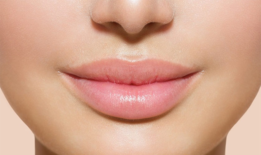
What are lip fillers

Lip augmentation consists of the reshaping and/or enlargement of the visible portion of the lip, the vermilion. Lip augmentation with the use of injectable fillers achieves quick results with minimal downtime. Alteration of the shape of the Cupid’s bow and of the relationship between the vermilion and the skin underlying the nasal columella also falls within the category of lip augmentation. It is also important to consider the relationship between lip height and incisor shown in the anatomic analysis, to evaluate possible maxillary hypoplasia and protrusion, and to consider the patient’s occlusion status.

Lip augmentation with fillers can be performed by injecting the material in any or all of the anatomic parts of the lip, allowing for a very controlled and predictable result. Precautions regarding mode of injection and quantity of the substance injected vary widely with patient profiles. At times, budgetary constraints decide treatments, but that should be avoided, and you need to be told about the optimum filler requirements.

Various fillers, temporary and permanent, have been tried in shaping the lips, with gratifying results. Disasters in the form of granulomas have been reported with both temporary and permanent fillers, more often with the latter.

Hyaluronic acid (HA) and polyacrylamide (PA) are the two main fillers widely available. Hyaluronic acid (HA) is the predominant one, with polyacrylamide (PA) being virtually out of the race because of fears of granuloma. Worldwide usage and published reports clearly confirm the efficacy and safety of HA fillers. They are one of the key components of successful combination treatments of the aging face and lips 1.

Common commercial preparations of hyaluronic acid (HA) that are widely available include the Anteis range (Fortélis® and Esthélis®, Switzerland), Galderma range, USA (Restylane and Perlane), and the Allergan range, Irvine, CA 92612 (Juvéderm Ultra® and Juvéderm Ultra Plus®). Other brands such as Revanesse®, Prollenium Medical Technologies Inc., Canada and Amazing Fill® are also available 2.

Significant ease of use, “off-the-shelf” availability, and widespread acceptance by the public make hyaluronic acid (HA) one of the most commonly used fillers.

Optimizing the aesthetic outcome of lip augmentation with dermal fillers, such as HA, requires skillful application of a suitable injection technique. Moreover, achieving aesthetic goals with minimal risk for adverse events requires knowledge of lip anatomy and function, clinical experience in the use of various injection techniques, and an individualized treatment approach.

How long do lip fillers last?

That depends on the type of lip fillers you are having. For in depth detail discussion see types of dermal lip fillers below.

Here is a table of dermal fillers and their duration

Temporary biodegradable (< 1 year)Semi-permanent biodegradable (1 – 2 years)Permanent non-biodegradable (> 2 years)
Autologous fat transferCalcium hydroxylapatite (CaHA)Collagen implant – porcine
Acellular dermis – cadaverDextran particles (DEAE)Silicone oil (polydimethylsiloxane oil) (LIS)
Collagen – human, bovine, porcineHydroxyethylmethacrylate (HEMA)Polyacrylamide gel (PAAG)
Cultured fibroblasts – humanPolylactic acid (PLA)Polymethylmethacrylate (PMMA)
Fascia autograft – humanPolyvinyl alcohol (PVA)Expanded polytetrafluoroethylene (ePTFE)
Hyaluronic acid (HA) – avian, bacterialPolytetrafluoroethylene (PTFE)
5 Tips for Consumers about Injectable Dermal Fillers
  • ALWAYS work with a licensed health care provider who uses properly labeled, sealed vials for treatments. You also can ask to confirm that you are receiving an FDA-approved filler. And never get injectable fillers from unlicensed providers or in non-medical settings like hotels or private homes.
  • ALWAYS request and read the patient labeling information on U.S. Food and Drug Administration (FDA)-approved injectable wrinkle fillers from your licensed health care provider.
  • ALWAYS know the type of product to be injected and all of its possible side effects. Know where each product used is to be injected. Talk to your licensed health care provider if you have any questions.
  • NEVER buy dermal fillers on the Internet. They may be fake, contaminated, and/or harmful.
  • NEVER get any type of filler or liquid silicone injected for body contouring. This means you should never get breast fillers, “butt” fillers, or fillers for spaces between your muscles. These products, which include certain types of injectable silicone, can be dangerous and can cause serious injury and even death.

Are lip injections safe

These days, people across the country are seeking treatments to smooth smile lines and crow’s feet and to plump up their lips and cheeks. But injectable dermal fillers are not for everyone and may not be indicated for people with certain conditions (such as bleeding disorders or certain allergies) 3. If your health care provider confirms that dermal fillers are an option for you, know that all products have benefits and risks. The U.S. Food and Drug Administration (FDA) advises you to work with a licensed health care provider and to understand all of the risks and benefits before receiving treatment. In May 2015, the FDA issued a warning to healthcare providers and the public about serious complications that can occur if dermal fillers are inadvertently injected into blood vessels in the face. The complications could possibly include vision impairment, blindness, stroke, and damage and/or necrosis of the skin and underlying facial structures. Caution should be used to ensure proper placement of the filler material, and patients should be informed about the potential adverse effects and how to recognize symptoms of impending serious complications.

The US Food and Drug Administration (FDA) is warning healthcare providers and the public about the possibility of rare, but serious, injuries that may occur as a result of unintentional injection of a soft tissue filler into blood vessels in the face. Unintentional injection can block blood vessels and restrict blood supply to tissues and lead to embolization, which could cause vision impairment, blindness, stroke, and damage and/or necrosis of the skin and underlying facial structures, the agency says in a Safety Communication.

Unintentional injections into blood vessels may occur with injection sites anywhere on the face, but the FDA’s review of the literature and submitted adverse event reports pinpoints certain injection locations at which blood vessel blockages have been reported more often. These include the skin between the eyebrows and nose (glabella), in and around the nose, the forehead, and around the eyes (periorbital region), the FDA says.

Two separate but related reports from 2017 and 2018 have reaffirmed the risks, some very serious—including strokes and blindness—that can be associated with dermal fillers 4. A 3-year study (2014-2016) reported swelling, infection, blindness (8 cases), and tissue death as the most common problems. A 10-year analysis (2007-2017) reported nodule formation, infection, inflammation, allergic complications, and vascular complications as the most common problems.

Risks of FDA-approved fillers

Remember to work with a licensed health care provider to ask what you can expect for FDA-approved fillers. Then contact your health care provider if you are concerned about a particular side effect.

The occurrence of adverse reactions relates to both the inherent properties of the product and inappropriate delivery or dilution of the filler, which may lead to harmful sequelae 5.

Underscoring the need for product-specific training, uneven distribution of injected products, due to poor technique, can also lead to postinjection lumps and nodules 6. This is of particular concern with more permanent products because the undesired results are also long-lasting. Overaggressive injection may lead to irregularity or lumpiness, whereas if the product is placed too superficially, beading can occur 7.

The most common side effects include 3:

  • bruising
  • redness
  • swelling
  • pain
  • itching

Additional side effects less commonly reported include 3:

  • infections
  • lumps and bumps
  • discoloration or change in pigmentation

Rare but serious risks include 3:

  • scarring, blurred vision, partial vision loss, and blindness if the dermal filler is inadvertently injected into a blood vessel. It is recommended that health care providers take care to avoid injection into blood vessels (especially around the forehead, nose and eye area) for these reasons.
  • allergic reaction that may lead to a severe reaction (anaphylactic shock) that requires emergency medical help.

Most side effects occur shortly after injection and go away within two weeks. In some cases, side effects may emerge weeks, months, or years later. Talk to your licensed health care provider if you have questions or concerns.
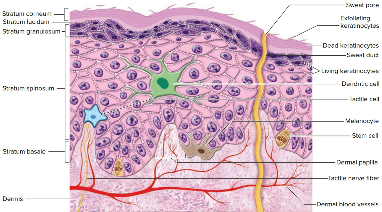
The skin

The skin (integument) is the body’s largest and heaviest organ. In adults, the skin covers an area of 1.5 to 2.0 m2 and accounts for about 15% of the body weight.

The skin consists of two layers:

  1. a stratified squamous epithelium called the Epidermis (Figure 2) and
  2. a deeper connective tissue layer called the Dermis (Figure 1).

The epidermis gives rise to the outer barrier layer of dead cells, the stratum corneum, through terminal differentiation of keratinocytes, which are the predominant cell type. Below the epidermis is the dermis. This layer contains the blood vessels, lymphatics, nerves, and deeper portions of the hair follicles and glands that originate from the epidermis. The dermis is composed largely of extracellular matrix and gives skin its strength and elasticity. Below the dermis is a subcutaneous layer of fatty tissue that gives contour to the skin.

Figure 1. Skin structure

skin structure and layers

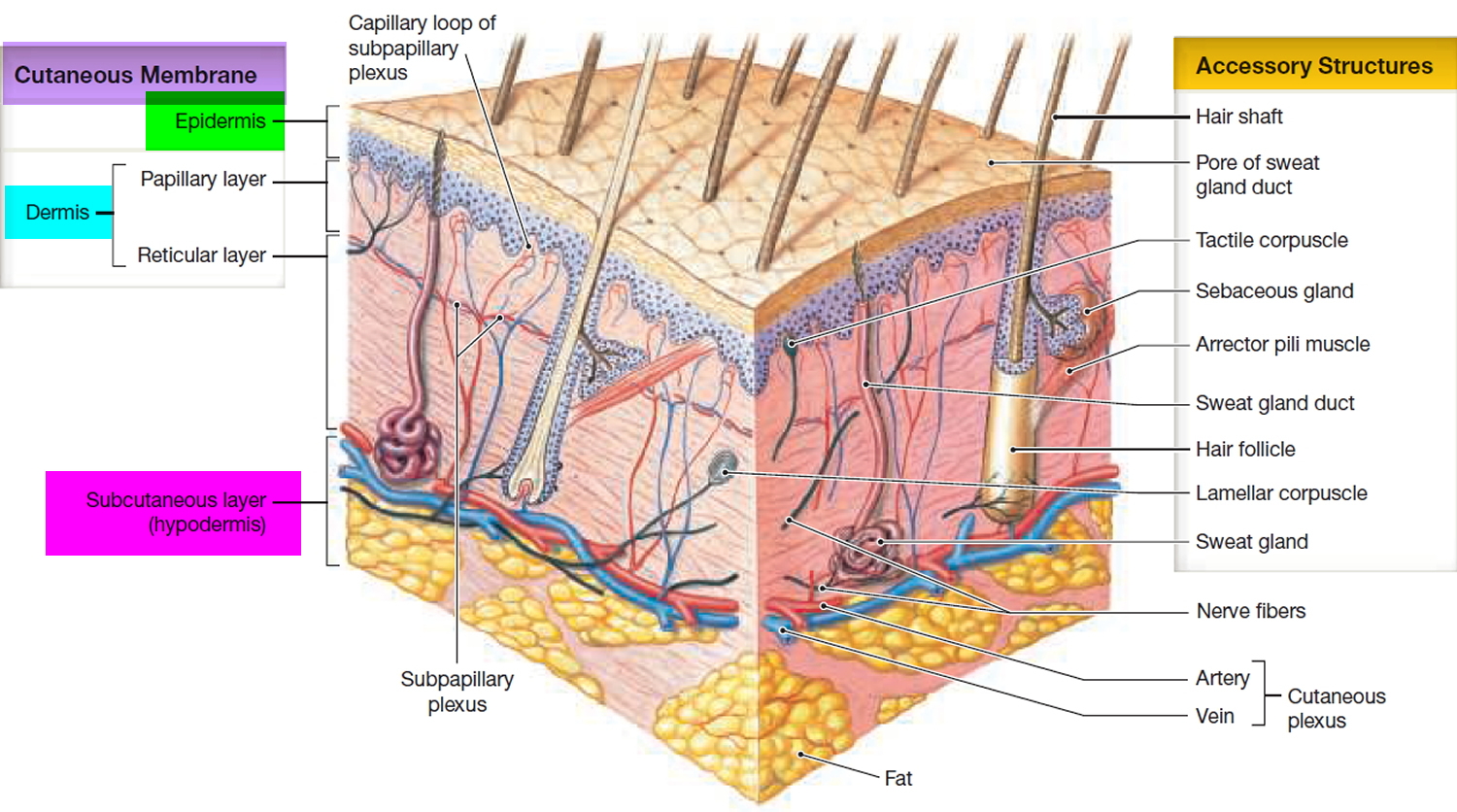
Below the dermis is another connective tissue layer, the hypodermis, which is not part of the skin but is customarily studied in conjunction with it. Most of the skin is 1 to 2 mm thick, but it ranges from less than 0.5 mm on the eyelids to 6 mm between the shoulder blades. The difference is due mainly to variation in thickness of the dermis, although skin is classified as thick or thin based on the relative thickness of the epidermis alone.

Thick skin covers the palms, soles, and corresponding surfaces of the fingers and toes. Its epidermis alone is about 0.5 mm thick, due to a very thick surface layer of dead cells called the stratum corneum (see Figure 2). Thick skin has sweat glands but no hair follicles or sebaceous (oil) glands. The rest of the body is covered with thin skin, which has an epidermis about 0.1 mm thick, with a thin stratum corneum. It possesses hair follicles, sebaceous glands, and sweat glands.

The accessory structures include hair, nails, and a variety of multicellular exocrine glands. These structures are located in the dermis and protrude through the epidermis to the surface.

Figure 2. Structure and skin cells of the Epidermis

structure of epidermis

The ageing process

The process of ageing leads to visible changes in facial contour as there is a loss of subcutaneous volume. Diminished dermal vasculature and blood flow, combined with a reduction of dermal extracellular matrix (including collagen, hyaluronic acid and elastin), lead to progressive thinning of the dermis. This results in a loss of elasticity and underlying atrophy of the natural fat pads. The soft tissue that encircles bony skeleton slowly collapses so that these bony landmarks are more visible 8. This is exacerbated by the effects of gravity, which results in sagging of the soft tissues relative to their substructure 9.
The full fibrous tissue associated with youthful appearance is replaced by less dense, less supple and less supportive tissue 8.

Among the most noticeable changes in the face as it ages, is the development of wrinkles (rhytids) and furrows, and the increased visibility of blood vessels. Critical changes in the perioral area can include vertical rhytids, increased prominence of nasolabial folds (crease that runs from nose to the corner of the mouth), ptosis of the oral commissures (sagging of lines from the corners of the mouth), thinning of the lips, and flattening of the upper lip with less definition of the Cupid’s bow 8. Transverse forehead lines become prominent and can be accompanied by a lowering of the eyebrows.

Lips Anatomy

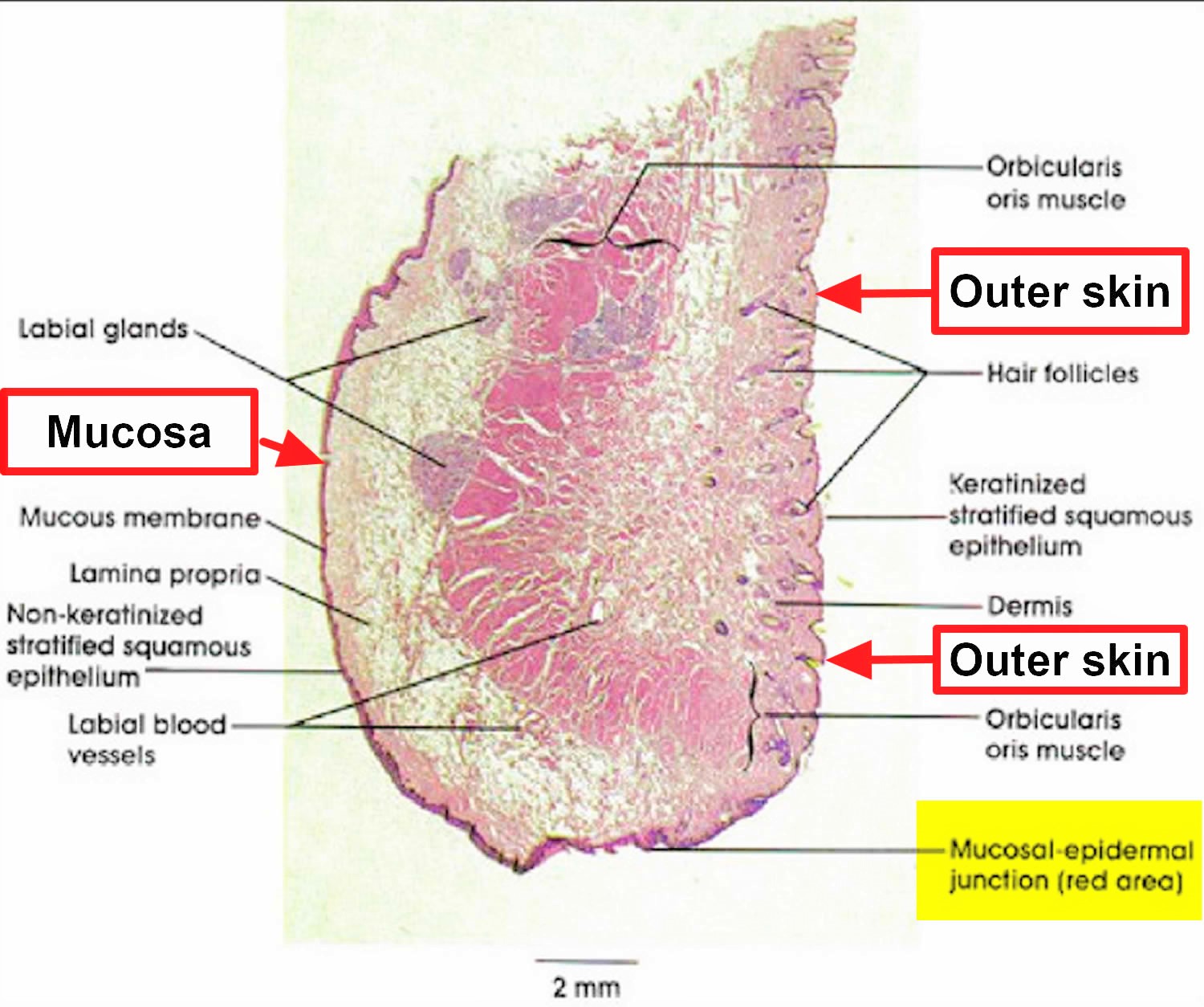
The upper lip extends from the base of the nose superiorly to the nasolabial folds laterally and to the free edge of the vermilion border inferiorly. The lower lip extends from the superior free vermilion edge superiorly, to the commissures laterally, and to the mandible inferiorly. Around the circumferential vermilion/skin border, a fine line of pale skin accentuates the color difference between the vermilion and normal skin. Along the upper vermilion/skin border, two paramedian elevations of the vermilion form the Cupid’s bow. Two raised vertical columns of tissue form a midline depression called the philtrum.

Female lips are, on average, a little fuller than male lips 10. They bulge forward more than male lips – in other words, they are slightly more “pouty.” Female lips are not noticeably bigger when you see them from the front but they do bulge forward more as seen from the side. You need to keep this in mind while receiving treatment for male and female lips. Over-volumization of the male lip can result in feminization of the area.

With the passage of time, photodamage, hereditary factors, and smoking contribute to loss of lip volume, perioral rhytides, and prominence of mentolabial folds. Genetically thin lips and cosmetic asymmetries of the lips are also issues that can be dealt with similarly, that is, by soft tissue augmentations using fillers. Successful rejuvenation of the perioral region requires sophistication in using a combination of technologies and injectables.

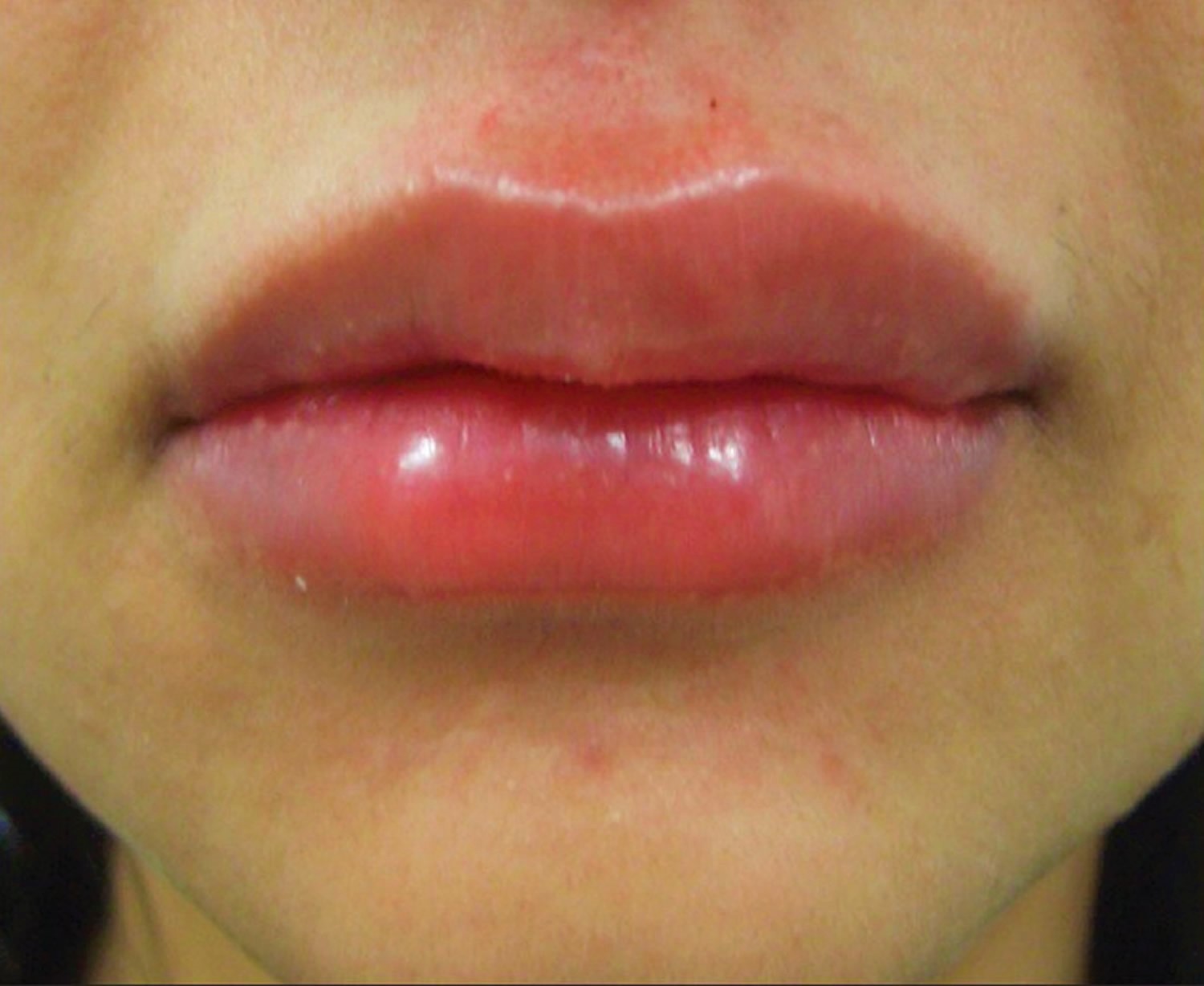
A deflating vermilion (the red part) is the most common complaint, followed by drooping angles of the mouth 10. These two together complete the picture of an aged face. Lips that have good volume can be highlighted by defining them and injecting into the white margins (the vermilion border) (Figures 3). Pouts can be created by injecting the filler below the muscle (Figure 5). Typically, the upper lip is treated more often than the lower lip. The best approach to lip augmentation depends on the nature of the defect and the subject’s aesthetic desires. For genetically thin lips, structural augmentation with a deeper-placed filler followed by volume correction with a superficial filler is ideal. For pure cosmetic enhancement of lips, a superficially placed filler with emphasis on the white roll and expansion of the vermilion is ideal 11.

Figure 3. Lips anatomy for lip dermal filler injections

lips-anatomy-for-dermal-filler

anatomy-of-lips-for-dermal-filler

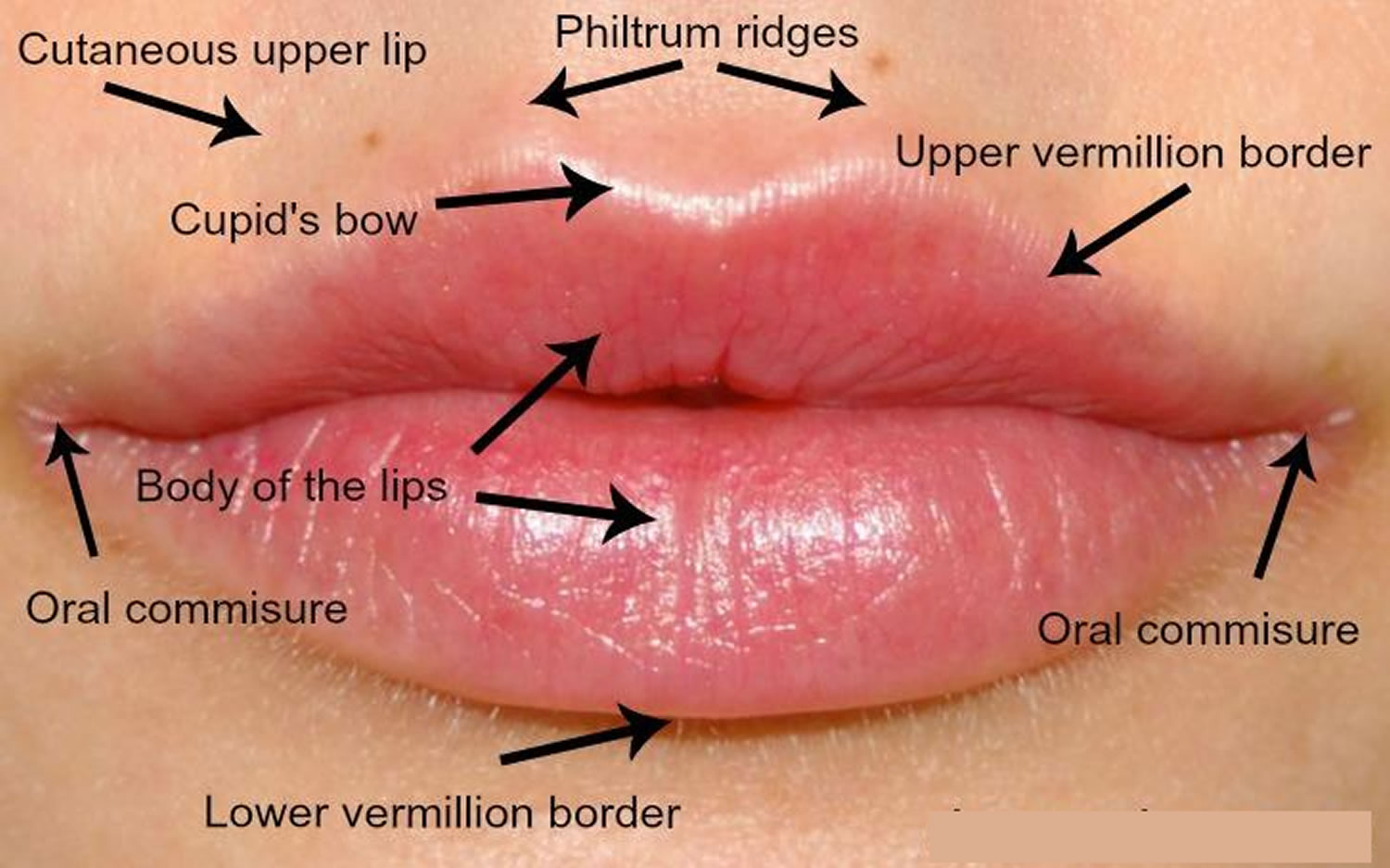
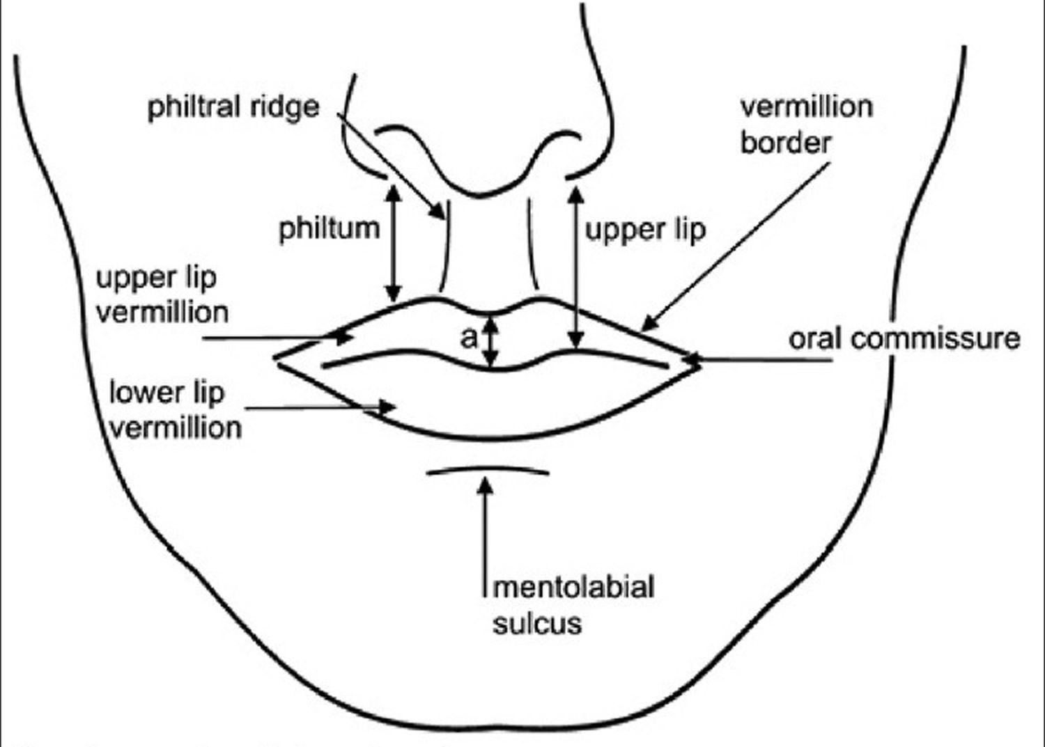
Figure 4. Lips anatomy

lips-anatomy

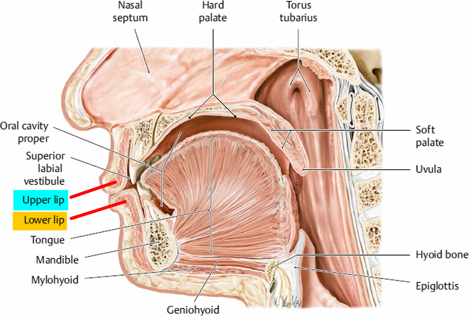
Figure 5. Lips histology

lips-histology

Dermal lip fillers

The perfect dermal filler would be inexpensive, safe, painless to inject, hypoallergenic, and long lasting. In addition, it should have consistent and predictable results, feel natural under the skin, take little time to inject, be ready-to-use, exert no downtime on the patient, and have a low risk of complications. With the increasing desire for people to achieve a more youthful appearance, the aging baby boomer population, and the increased demand for “lunch-time procedures,” the pharmaceutical market has responded by providing the cosmetic surgeon with an increasing number of options to meet the demands of the cosmetic patient. Thus, this segment of cosmetic surgery has been the fastest growing for the past decade.

By definition, a dermal filler is a product that is injected or placed into the dermis. Patients are instructed to not manipulate the treated areas, because the product may shift. The best way to reduce inflammation is to immediately apply a cool pack to the areas that were treated. In current practice, several dermal fillers are available for use in the United States, in addition to subdermal fillers, or those that are placed underneath the dermis in the subcutis.

Injectable dermal fillers and synthetic dermal implants have been developed to address volume loss and contour defects and appear to be useful in the lower third of the face 12. Dermal fillers address volume loss to give the face a fuller, more youthful appearance. Dermal fillers are injected or inserted into the skin to give the face a fuller look, as opposed to the pulled, flat, two-dimensional look that is sometimes associated with facelift surgery (rhytidectomy) 13. Facelifts, which eliminate loose skin folds in the neck and wrinkles (rhytids) in the cheeks by the removing tissue and tightening or ‘lifting’ of the skin, do not augment or ‘fill’ the skin 14.

Dermal fillers have become a popular means of addressing volume loss and contour defects resulting from ageing, photo-damage, disease, trauma and/or scarification 15. Fillers that are biodegradable are resorbed over time by the tissues and require repeat applications. Semi-permanent and permanent dermal fillers have emerged as alternatives, to give patients a longer lasting or permanent effect. However, the potential side effects and complications associated with these fillers have not been well documented, may be difficult to manage, and can last as long as the filler.

Dermal fillers can complement surgical and topical treatments to give a greater and longer lasting aesthetic result 13. A combination of filler materials can be injected or inserted to give a natural contour to the face 16.

Also know that the safety of these products is unknown for use in pregnant or breastfeeding women or in patients under 18 years of age. The safety also is unknown if used with Botox or other wrinkle therapies.

You should discuss the different types of FDA-approved dermal fillers and the results you want to achieve with your licensed health care provider, who can refer you to a licensed dermatologist or plastic surgeon. You may want to contact the American Academy of Dermatology (https://www.aad.org/), the American Society of Plastic Surgeons (https://www.plasticsurgery.org/) or the American Society for Aesthetic Plastic Surgery (https://www.surgery.org/).

Dermal Fillers Approved by the FDA

FDA approval is based on the review of data collected from controlled clinical studies that evaluated the safe and effective use of the wrinkle fillers when injected into specified areas of facial tissue.

Most dermal fillers have a temporary effect, because they contain materials that are absorbed by the body over time. The FDA has approved only one product made from a material that remains in the body and is not absorbed. Some dermal fillers also contain lidocaine, which is intended to decrease pain or discomfort related to the injection.

The materials used in dermal fillers include:

Absorbable (temporary) materials

  • Collagen: Collagen is a type of protein that is a major part of skin and other tissues in the body. Sources of purified collagen used in soft tissue fillers can be from cow (bovine) or human cells. The effects of collagen fillers generally last for 3-4 months. They are the shortest lasting of injectable filler materials.
  • Hyaluronic acid: Hyaluronic acid is a type of sugar (polysaccharide) that is present in body tissues, such as in skin and cartilage. It is able to combine with water and swell when in gel form, causing a smoothing/filling effect. Sources of hyaluronic acid used in dermal fillers can be from bacteria or rooster combs (avian). In some cases, hyaluronic acid used in dermal fillers is chemically modified (crosslinked) to make it last longer in the body. The effects of this material last approximately 6 – 12 months.
  • Calcium hydroxylapatite (CaHA): Calcium hydroxylapatite (CaHA) is a type of mineral that is commonly found in human teeth and bones. For wrinkle filling in the face or for the hand, calcium hydroxylapatite particles are suspended in a gel-like solution and then injected into the wrinkle in the face or under the skin in the back of the hand. The effects of this material last approximately 18 months. While in the body, calcium hydroxylapatite will be visible in x-rays and may obscure underlying features.
  • Polylactic acid (PLA): Polylactic acid (PLA) is a biodegradable, biocompatible man-made polymer. This material has wide uses in absorbable stitches and bone screws. Polylactic acid (PLA) is a long lasting filler material that is given in a series of injections over a period of several months. The effects of polylactic acid (PLA) generally become increasingly apparent over time (over a period of several weeks) and its effects may last up to 2 years.

Non-absorbable (permanent) materials

  • Polymethylmethacrylate beads (PMMA microspheres): Polymethylmethacrylate beads (PMMA microspheres) is a non-biodegradable, biocompatible, man-made polymer. This material is used in other medical devices, such as bone cement and intraocular lenses. Polymethylmethacrylate beads (PMMA microspheres) are tiny, round, smooth particles that are not absorbed by the body. When used as a soft tissue filler, polymethylmethacrylate beads (PMMA microspheres) are suspended in a gel-like solution that contains cow (bovine) collagen and injected into the face.

Table 1. FDA Approved Dermal Fillers

Trade Name (with link to additional information)MaterialApplicantPMA NumberDecision DateApproved For
Restylane, Refyne, Restylane DefyneSodium HyaluronateQ-Med ABP14002912/9/2016Restylane Refyne is indicated for injection into the mid-to-deep dermis for the correction of moderate to severe facial wrinkles and folds (such as nasolabial fold) in patients over the age of 21. Restylane Defyne is indicated for injection into the mid-to-deep dermis for the correction of moderate to severe deep facial wrinkles and folds (such as nasolabial fold) in patients over the age of 21.
JUVEDERM VOLLURE XCHyaluronic AcidAllerganP110033/S0203/17/2017Injection into the mid to deep dermis for correction of moderate to severe facial wrinkles and folds (such as nasolabial folds) in adults over the age of 21.
JUVEDERM VOLBELLA XCHyaluronic
Acid with Lidocaine
AllerganP110033/S0185/31/2016Injection into the lips for lip augmentation and for correction of perioral rhytids in adults over the age of 21.
RESTYLANE LYFT WITH LIDOCAINEHylauronic acid with lidocaineGalderma LaboratoriesP040024 S0737/1/2015Moderate to severe facial folds and wrinkles or in patients over the age of 21 who have age-related volume loss.
RADIESSEHydroxylapatiteBioform Medical, Inc.P050052/S0496/4/2015Subdermal implantation for hand augmentation to correct volume loss in the dorsum of the hands.
RESTYLANE SILKHyaluronic Acid with LidocaineValeant Pharmaceuticals North America LLC/MedicisP040024 S0726/13/2014Indicated for lip augmentation and dermal implantation for correction of perioral rhytids (wrinkles around the lips) in patients over the age of 21.
JUVEDERM VOLUMA XCHyaluronic Acid with LidocaineAllerganP11003310/22/2013Deep (subcutaneous and/or supraperiosteal) injection for cheek augmentation to correct age-related volume deficit in the mid-face in adults over the age of 21.
RESTYLANE-L INJECTABLE GELHyaluronic Acid with LidocaineMedicis Aesthetics Holdings, Inc.P040024 S0568/30/2012Injection into the mid to deep dermis for correction of moderate to severe facial wrinkles/folds (such as nasolabial folds) and for lip augmentation in those over the age of 21 years.
BELOTERO BALANCEHyaluronic AcidMerz PharmaceuticalsP09001611/14/2011Injection into facial tissue to smooth wrinkles and folds, especially around the nose and mouth (nasolabial folds).
RESTYLANE INJECTABLE GELHyaluronic AcidMedicis Aesthetics Holdings, IncP040024 S05110/11/2011Lip augmentation in those over the age of 21 years.
JUVEDERM ULTRA XC AND JUVEDERM ULTRA PLUS XCHyaluronic Acid with LidocaineAllerganP0500471/7/2010The addition of 0.3% Lidocaine into Juvederm Ultra and Juvederm Ultra Plus.
SCULPTRA AESTHETICPoly-L-Lactic Acid (PLLA)Sanofi Aventis U.S.P030050 S0027/28/2009Use in shallow to deep nasolabial fold contour deficiencies and other facial wrinkles.
EVOLENCE COLLAGEN FILLERCollagenColbar Lifescience lP0700136/27/2008The correction of moderate to deep facial wrinkles and folds (such as nasolabial folds).
PREVELLE SILKHyaluronic Acid with LidocaineGenzyme BiosurgeryP030032 S0072/26/2008Injection into the mid to deep dermis for correction of moderate to severe facial wrinkles and folds (such as nasolabial folds).
RADIESSE 1.3CC AND 0.3CCHydroxylapatiteBioform Medical, IncP05003712/22/2006Restoration and/or correction of the signs of facial fat loss (lipoatrophy) in people with HIV.
RADIESSE 1.3CC AND 0.3CCHydroxylapatiteBioform Medical, IncP05005212/22/2006Subdermal implantation for correction of moderate to severe facial wrinkles and folds (such as nasolabial folds).
ELEVESSHyaluronic Acid with LidocaineAnika TherapeuticsP05003312/20/2006Use in mid to deep dermis for correction of moderate to severe facial wrinkles and folds (such as nasolabial folds).
ARTEFILLPolymethylmethacrylate Beads, Collagen and Lidocaine.Suneva Medical, Inc.P02001210/27/2006Use in facial tissue around the mouth (i.e., nasolabial folds).
JUVEDERM 24HV, JUVEDERM 30, and JUVEDERM 30HVHyaluronic AcidAllerganP0500476/2/2006Use in mid to deep dermis for correction of  moderate to severe facial wrinkles and folds (such as nasolabial folds).
RESTYLANE INJECTABLE GELHyaluronic AcidMedicis Aesthetics Holdings, IncP0400243/25/2005Injection into the mid to deep dermis for correction of moderate to severe facial wrinkles and folds (such as nasolabial folds).
CAPTIQUE INJECTABLE GELHyaluronic AcidGenzyme BiosurgeryP030032 S00211/12/2004Injection into the mid to deep dermis for correction of moderate to severe facial wrinkles and folds (such as nasolabial folds).
SCULPTRAPoly-Lactic Acid (PLA)Sanofi Aventis U.S.P0300508/3/2004Restoration and/or correction of the signs of facial fat loss (facial lipoatrophy) in people with Human Immunodeficiency Virus (HIV).
HYLAFORM (HYLAN B GEL)Modified hyaluronic acid derived from a bird (avian) sourceGenzyme BiosurgeryP0300324/22/2004Injection into the mid to deep dermis for correction of moderate to severe facial wrinkles and folds (such as nasolabial folds).
RESTYLANE INJECTABLE GELHyaluronic AcidQ-med AbP02002312/12/2003Injection into the mid to deep dermis for correction of moderate to severe facial wrinkles and folds (such as nasolabial folds).
COSMODERM 1 HUMAN-BASED CCollagenInamed CorporationP800022 S0503/11/2003Injection into the superficial papillary dermis for correction of soft tissue contour deficiencies, such as wrinkles and acne scars.
FIBRELCollagenSerono LaboratoriesP8500532/26/1988The correction of depressed cutaneous scars which are distendable by manual stretching of the scar borders.
ZYPLAST(R)CollagenCollagen Corp.P800022 S0116/24/1985Use in mid to deep dermal tissues for correction of contour deficiencies.
ZYDERM COLLAGEN IMPLANTCollagenAllerganP8000229/18/1981Use in the dermis for correction of contour deficiencies of this soft tissue.
[Source 17]

Hyaluronic Acid

Hyaluronic acid is the most prominent glycosaminoglycan in the skin. Hyaluronic acid potently binds to water and, when injected into the skin, volumizes, softens, and hydrates the skin. In addition to these benefits, it plays a role in cell growth, membrane receptor function, and adhesion.

Hyaluronic acid stabilizes intercellular structures and produces the viscoelastic network for collagen and elastin fibers to bind together. As seen with photoaging, these connections fail, thus resulting in disorganized clumps of collagen and elastin. These benefits make hyaluronic acid an excellent dermal filling agent 18. In February 2003, the FDA approved Restylane, a cross-linked, nonanimal source hyaluronic acid. This dermal filler was quickly found to be relatively long lasting, have minimal adverse effects, was easy to use, was ready to use out of the box, did not require refrigeration, was cost effective, and did not require skin testing prior to treatment.

Because hyaluronic acid is identical in all species, the risk of allergy is remote. Hyaluronic acid has a heparinlike effect, thus resulting in a greater incidence of bruising than is seen with collagen fillers 19. Until 2010, nearly every FDA-approved hyaluronic acid product in the United States did not contain lidocaine, thus significantly increasing the discomfort experienced with injection of these dermal fillers compared with the bovine- and human-derived collagen fillers. Despite these shortcomings, hyaluronic acid fillers still emerged as the leader of dermal filling agents for soft tissue augmentation, owing to their superior cosmetic results.

In the uncommon circumstance when an undesirable outcome occurs with hyaluronic acid, correction is possible with the injection of commercially available hyaluronidase, which breaks down the unwanted hyaluronic acid dermal filler. The use of hyaluronidase for this purpose is not FDA approved and is considered an off-label use. In many cases, 10-30 units of unpreserved hyaluronidase is sufficient to achieve the desired correction. Local site reactions may occur in up to 25% of persons, although they are typically transient and mild. Initial treatment with as little as 5-10 units is commonly recommended and is often effective, although some treat with as much as 75 units with few adverse effects. Additional corrections can be performed, although full correction may take up to 4 weeks to fully appreciate. Some preparations are bovine derived, and skin testing should be considered prior to treatment with these dermal fillers 20.

Hyaluronidase preparations are clear, concentrated liquids that are stored in a refrigerated vial. To reconstitute these dermal fillers, physicians typically add normal saline or lidocaine (with or without epinephrine). When using Amphadase, reconstitution using 3 mL of 1% lidocaine with 1:100,000 epinephrine has been commonly used with great success. After mixing, the vial is gently swirled. Prior to treatment, a skin test can be performed by injecting 3-5 units (0.06-0.1 mL) of the reconstituted solution into the superficial dermis at the antecubital fossa. A positive hypersensitivity reaction consists of a wheal appearing within 5 minutes and lasting 20-30 minutes, accompanied by local itching 21.

Restylane

  • Restylane and Restylane-L

Restylane was approved by the FDA in 2003 for the treatment of nasolabial folds. This dermal filler is produced by fermentation in bacterial cultures of equine streptococci. This dermal filler contains approximately 100,000 particles per mL (20 mg/mL). These particles are approximately 300 mm and are highly cross-linked (single cross-linked) using an ether bond, making these dermal fillers one of the stiffest hyaluronic acid fillers. This dermal filler has been used for correction of the nasolabial folds, marionette lines, tear troughs, and glabellar frown lines, in addition to lip enhancement and cheek augmentation. Other clinical uses include correction of the jowls and nasal deformities. In general, most patients can expect 6 months of correction, if not longer 22. In 2010, Restylane-L became available, which contains lidocaine to reduce pain upon injection.

  • Restylane Lyft with lidocaine

This dermal filler was originally marketed as Perlane-L, but had a name change in 2015. Restylane Lyft is identical to Restylane except that it consists of larger gel particles. This dermal filler is suitable for the correction of deeper folds, such as the nasolabial folds, and works well for cheek enhancement. Additionally, some experienced physicians prefer this dermal filler over Restylane for lip augmentation. Most patients can expect 6-12 months of correction with Perlane. In 2010, Perlane-L became available, which contains lidocaine to reduce pain upon injection.

  • Restylane Silk

This dermal filler was FDA approved in 2014 for submucosal implantation for lip augmentation and dermal implantation for correction of perioral rhytids in patients older than 21 years. It has the same concentration of hyaluronic acid and a of pH 7.0 as the other Restylane products, but the molecules are smaller. This product is intended to be used for fine perioral lines, although other areas on the face can be treated as well.

  • Precautions

Use of products that contain lidocaine (Restylane-L, Restylane Lyft, and Restylane Silk) reduces pain upon injection and is recommended, unless contraindications to the use of lidocaine are present. Erythema and edema are common after treatment and typically last a few days. When injected too superficially, a bluish Tyndall effect can be seen, which represents visible hyaluronic acid seen through the translucent epidermis. Fortunately, these bluish cysts are easily corrected by nicking the skin with a small-gauge needle (30 gauge) or No. 11 blade and expressing the superficial, unwanted dermal filler 23.

Occasionally, palpable nodules can be felt under the skin, which often occurs when the multiple-puncture technique is used or depot injections are performed to place the dermal filler. For this reason, linear threading is now commonly used, which minimizes the risk of nodules. Some prefer using a small (30-gauge) blunt-tipped cannula to inject instead of the 30-gauge needle that comes with the product. As with other fillers, patients should avoid all blood thinners for 10 days prior to treatment 24.

Juvederm (Juvederm Ultra/Juvederm Ultra Plus)

In 2006, the FDA approved Juvederm, which is also a nonanimal stabilized hyaluronic dermal filler. In the United States, three types of Juvederm dermal fillers are FDA approved. Both Juvederm Ultra and Juvederm Ultra Plus contain 24 mg/mL of hyaluronic acid, but Juvederm Ultra Plus has a higher proportion of cross-linking than Juvederm Ultra. Juvederm is a homologous gel with the highest degree of cross-linking of any of the hyaluronic acid fillers and thus has a smooth consistency 25. Juvederm Ultra and Juvederm Ultra Plus have indications similar to those of Restylane and Perlane, respectively, and also do not require refrigeration or skin tests prior to use. In 2010, both Juvederm Ultra and Juvederm Ultra Plus became available with lidocaine, to reduce pain with injection, both labeled as Juvederm (R).

  • Precautions

The adverse effects of Juvederm are similar to those seen with Restylane and Perlane 26. Like all of the hyaluronic acid filling agents that are FDA approved, Juvederm does not contain lidocaine. Therefore, in addition to discomfort with injection, one may see erythema, swelling, and bruising. If injected too superficially, a bluish Tyndall effect and nodules may appear 27.

Hylaform/Hylaform Plus

A sterile, nonpyrogenic, viscoelastic, clear gel implant composed of cross-linked molecules of hyaluronic acid derived from an avian (bird) source. Hylaform contains 5.5 mg/mL of medium-sized particles of hylan B. Hylaform Plus contains larger molecules of the same concentration of Hylaform. The indications for Hylaform and Hylaform Plus are similar to Juvederm Ultra and Juvederm Ultra Plus, respectively 24.

  • Precautions

These dermal fillers have precautions similar to those of Restylane and Perlane. Additionally, because this dermal filler is derived from an avian (bird) source, persons with a known allergy to avian proteins should not be treated with these dermal fillers. The Hylaform dermal fillers are stored at room temperature and no skin test is required before use.

Captique

This dermal filler is identical to Hylaform except that it is derived from a bacterial source through fermentation. This dermal filler is slightly stiffer than Hylaform 25. Captique is stored at room temperature and no skin test is required before use.

Puragen

This dermal filler contains 20 mg/mL of bacterially derived hyaluronic acid. The particle size is approximately 200 mm, which is slightly smaller than Restylane, and is double–cross-linked with both ether and ester bonds. Ester bond linkages make this dermal filler more stable, protecting the ether bonds, and they are hydrophobic, thus making the product less susceptible to breakdown by hyaluronidase 28. Pilot studies have shown that this dermal filler may be degraded more slowly than Restylane, although larger studies are needed to confirm this. This dermal filler has indications and precautions similar to those of Restylane.

Prevelle Silk

This dermal filler was FDA approved for use in the United States in 2008 and was the first hyaluronic acid dermal filler to contain lidocaine, which reduces pain upon injection. Prevelle Silk is indicated for the treatment of moderate-to-severe facial wrinkles, although it may be better suited for fine lines. Injection into the mid-to-deep dermis is recommended by Mentor, the manufacturer. Similar to other hyaluronic dermal fillers, the most common adverse effects include temporary injection site reactions such as swelling, pain, tenderness, redness, lumps, and bumps.

Polymethylmethacrylate (PMMA) With Bovine Collagen

Artefill (now called Bellafill) was FDA approved for use in the United States in 2007. This dermal filler is composed of nonresorbable polymethylmethacrylate (PMMA) microspheres, which are 30-50 μ m in diameter, suspended in a water-based carrier gel composed of 3.5% bovine collagen, 92.6% buffered isotonic water, 0.3% lidocaine, 2.7% phosphate buffer, and 0.9% sodium chloride 29. Because this dermal filler contains lidocaine, the injection is less painful compared with other dermal fillers that do not contain lidocaine. This dermal filler is indicated for the correction of the nasolabial folds, although it has been used for acne scars and forehead furrows. Using a 26-gauge needle, Bellafill should be injected directly beneath the skin fold into the deep dermis and not into the subcutis. Most practitioners prefer a threading injection technique. Unlike the other dermal fillers, this dermal filler should be considered a permanent dermal filler 30.

  • Precautions

Because this product contains bovine collagen, skin testing must be performed prior to treatment, as would be indicated with Zyderm or Zyplast (see Collagen). Because results are permanent, it is often best not to try to achieve full correction in one session, but to accomplish the desired result over several treatment sessions. In addition to allergy, other adverse effects may include lumpiness, persistent swelling or redness, and increased sensitivity at the injection site 31. The product must be refrigerated.

Poly-L-Lactic Acid (PLA)

This novel product (Sculptra) differs from all other agents in several aspects. Poly-L-lactic acid is a synthetic, biodegradable, biocompatible, immunologically inert peptide polymer that is believed to stimulate fibroblasts to produce more collagen, thus increasing facial volume. In the United States, poly-L-lactic acid is only FDA approved for the treatment of HIV-associated lipoatrophy; and it is used for the correction of skin folds (ie, nasolabial folds) and other facial wrinkles in immune-competent persons 32.

Although poly-L-lactic acid is nearly always injected subdermally, dermal neocollagenesis occurs, thus it is a dermal stimulating agent, not a true dermal filling agent. Several limitations have prevented poly-L-lactic acid from becoming as popular as other products. Poly-L-lactic acid must be premixed prior to use, making immediate treatment impossible. Unlike dermal fillers, results are not appreciated for 4 or more weeks. Lastly, most patients require 2-3 treatment sessions that are at least 4-6 weeks apart 33.

  • Precautions

Because poly-lactic acid is not a true filler, but relies on neocollagenesis to achieve clinical improvement, the clinical results from this agent are less predictable than the true dermal fillers. Patients should be properly educated that results take 4-6 weeks to be appreciated. Dermal nodules have been reported after treatment and often take 7 months or much longer to develop 34. When treating the face, these nodules can often be felt, but not seen. Reconstitution of the product with 6 or more milliliters of sterile water, in addition to vigorous posttreatment massage, is believed to reduce the incidence of nodule formation.

Use of this product on the hands has increased; however, unfortunately, the incidence of nodules in this location may be as high as 10% and the nodules are often visible, unsightly, and difficult to treat. In this location, treatment with Sculptra reconstituted with as much as 10 mL of sterile water has still resulted in nodule formation, which may take 1-3 years to appear after treatment 35. Additional long-term studies are needed to fully assess the safety of poly-L-lactic acid for the treatment of the hands in an immune-competent individual.

No skin test is required prior to treatment. The product is stored at room temperature, although it must be reconstituted prior to treatment.

Calcium Hydroxylapatite (CaHA)

This novel filler, Radiesse, was FDA approved in December 2006 for the correction of facial wrinkles and folds and for the correction of HIV-associated facial atrophy. In 2009, it received FDA approval for cosmetic use in non-HIV patients as well. The subdermal filler is composed of 30% calcium hydroxylapatite and 70% carrier gel 36. The clinical results may last as long as 12 months or longer, although the carrier gel lasts no longer than 6 months, thus often resulting in a slight decrease in correction by that time.

Radiesse is nearly always injected subdermally at the dermal-subcutaneous junction or just above the periosteum; thus, this product is not a true dermal filler. Studies from 2008 suggest that calcium hydroxylapatite may induce neocollagenesis, although further research is needed 37. Most commonly, Radiesse is used for the correction of nasolabial folds, atrophic cheeks, and temporal wasting.

Because of the pain associated with injection, some practitioners added lidocaine to the syringe, without a clinical appreciable decrease in effect. Mariano Busso 38, had treated numerous patients by adding 1 drop of 10% lidocaine to the previously available 1.3-mL syringe of Radiesse to decrease the discomfort associated with injection. In 2010, Radiesse with lidocaine was released to reduce pain upon injection and no premixing is needed.

Treatment of the hands has been accomplished with the addition of 0.15-0.23 mL of 2% lidocaine per 1.3-mL syringe of Radiesse. This can be accomplished using a nose-to-nose (female-to-female) Luer-lock connector to connect the syringe of Radiesse to a 3-mL syringe containing the lidocaine. At least 10 passes of the product back and forth between the two syringes is recommended to achieve adequate and even distribution of the lidocaine. This makes the consistency of the Radiesse slightly thinner, thus making it easier to spread when using a bolus injection technique. Similarly, this mixture is often preferred for the treatment of the temples.

  • Precautions

Injection into the dermis may result in nodule formation and should be avoided. Extreme care must be taken to avoid injection while withdrawing the needle out of the skin, which will result in the deposition of material into the dermis. Treatment of the lips has resulted in cyst formation containing the carrier gel 29. For this reason, most cosmetic surgeons avoid treating the lips with Radiesse.

No skin test is required prior to treatment, and the product is stored at room temperature.

Collagen replacement therapy

Collagen is a natural substance that is found in skin, muscle, tendons and bones and provides structural support. In the dermis (the mid-layer of skin), collagen is made by fibroblast cells. It forms a fibrous network on which new cells can grow. Through the natural processes of aging, collagen in the dermis is gradually lost and contributes to the formation facial lines.

Injectable bovine collagen is made of sterile, purified collagen from cow skin. Human collagen implants are highly purified and isolated from human skin grown in a laboratory. The cells have been grown for the last ten years or so primarily to manufacture living skin-equivalents to treat burns and ulcers.

When injected into the body’s skin both forms of collagen are accepted as if they were the body’s own collagen, forming a network of collagen fibres.

Zyderm® and Zyplast® are popular brands of injectable bovine collagen. Others include Resoplast®. Human collagen brands include Dermalogen®, Cymetra™, CosmoDerm® and CosmoPlast®.

Zyderm® and Cosmoderm® are used for fine lines and Zyplast® and CosmoDerm® are used for deeper furrows.

There are also products derived from porcine collagen, including Evolence™, and Fibrel®. Fibrel is injected with the patient’s own serum. The injections are said to be rather painful and may cause allergic reactions.

A person’s own skin may be used to produce fibroblast cultures (Isolagen®) or a suspension of collagen (Autologen®). It can also be mixed with synthetic PMMA beads (Artecoll®).

How long does the collagen implant last?

Collagen implants are not permanent. Because collagen is a natural protein it slowly breaks down into amino acids that are then absorbed by the body. In most cases, implants last anywhere between one to six months, although in some people one implant may be sufficient for up to two years. Repeat treatments will be necessary to maintain the results.

The longevity of the implant will depend on its location and individual response. Muscular activity such as smiling and frowning will reduce how long it lasts. Conversely, it may last longer than expected in scars because these are not caused by facial muscle movement.

Precautions

People with severe allergies (anaphylaxis) or allergy to injected local anaesthetics should not be treated with bovine, porcine or human collagen.

Since injectable bovine collagen is derived from cows it may cause allergic reactions. Approximately 3% of the population is allergic to bovine collagen; these individuals should not receive these implants. Human collagen is a better choice in the following circumstances:

  • Allergies to foods, especially meat products
  • Family or personal history of severe allergies (including asthma, hay fever and atopic dermatitis)
  • Any previous reaction to a test dose of collagen.

To see if you are eligible for bovine collagen replacement therapy you will require one or two skin tests. No skin test is necessary for human collagen as allergic reactions are very unlikely.

Procedure for collagen skin test

  1. Test dose of bovine collagen injected just below the skin’s surface on forearm
  2. Observe the site closely for at least 4 weeks for any of the following signs:
    • redness
    • swelling
    • hardness
    • itching
    • tenderness
  3. Most reactions will occur within the first 3 days, but can happen at anytime within this timeframe.
  4. If it appears that you have a very mild reaction, the doctor may need you to have a second skin test on the other arm to confirm sensitivity.
  5. Report all reactions to your doctor. After the 4-week observation period (or 8 weeks if second test is required) your doctor will advise whether or not you can proceed with bovine collagen replacement therapy.

Collagen replacement therapy side effects

At the time of treatment most patients report minor discomfort. This is minimized by the addition of lignocaine (lidocaine) to the collagen preparation. This is local anaesthetic to numb the treatmnet area, and is known as ‘lidocaine’ in America.

Immediately after treatment the area may be red, swollen and tender; this usually improves over the following days. Temporary bruising and discolouration may also occur. The collagen is sometimes visible for a while in the form of small white bumps at the treatment site. This generally smoothes out within a few weeks.

Very rarely, if a blood vessel is accidentally blocked by the collagen injection, a small area of skin may die resulting in an ulcer that scabs. This reaction may leave a permanent scar. It has most often been reported in the glabellar area.

Injections through the skin carry a risk of bacterial infection (impetigo). This is more likely if inflammation is present in the treated area such as acne or rosacea spots. Injections in and around the lips may also provoke cold sores in those prone to them.

Allergic reaction to bovine collagen is usually recognized after one or two test doses. In rare cases, allergic reactions may occur during the course of treatment even though there was no reaction to the test dose. They may occur rapidly after treatment or arise weeks to months afterwards. The reaction may clear up in a few days or persist for months. See your doctor immediately if you have an allergic reaction.

Allergic reactions may include:

  • Shortness of breath, low blood pressure, and chest pain.
  • Urticaria (hives)
  • Red, itchy or sore lumps at injection sites
  • Scarring when the lumps have resolved
  • Shorter lasting effect of collagen injections

People with connective tissue diseases such as rheumatoid arthritis, systemic lupus erythematosus, systemic sclerosis and dermatomyositis may be more likely to experience an allergic reaction to bovine collagen, and the effect of the treatment might not last as long. There have also been reports of connective tissue disease arising for the first time after bovine collagen injections. However a connection between the injections and the connective tissue disease has not been established.

The safety of collagen implants in pregnancy or in children has not been established.

Autologous Cell Therapy

Azficel-T (laViv) is an autologous aesthetic cell therapy indicated to improve the appearance of moderate-to-severe nasolabial fold wrinkles in adults. The product is available from Fibrocell Science, Inc, a company focused on developing personalized cell therapies for aesthetic, medical, and scientific applications.

According to the company, creating azficel-T involves a patented technology whereby fibroblasts are extracted from behind the patient’s ear and sent to the Fibrocell Science laboratory, where they are multiplied for about 3 months and are then frozen until needed. Only physicians who complete a Fibrocell-approved training program will be able to administer it.

This is a biological that works over time that provides gradual and natural results.

The FDA approval was based largely on two identical phase 3 multicenter, randomized, double-blind, placebo-controlled studies involving 421 patients who underwent 3 treatment sessions approximately 5 weeks apart 39.

On the basis of investigators’ and patients’ assessments, a significantly greater proportion of patients demonstrated a positive response to treatment with azficel-T than with placebo. The treatment improved the appearance of nasolabial fold wrinkles for the 6 months of patient follow-up after the last treatment. The company said studies are ongoing, looking at how long beyond 6 months after the last treatment the effect may last.

In clinical trials, the most common adverse events were mild-to-moderate injection-site reactions that usually resolved within 1 week.

Unapproved Dermal Fillers

The FDA is aware that unapproved versions of Juvederm, such as Juvederm Ultra 2, 3, and 4 are being sold and distributed in the U.S., including by online retailers 17. Juvederm is a prescription device that should only be injected and sold by or on the prescription of a licensed health care provider. The FDA is warning health care providers and patients not to use any Juvederm Ultra 2, 3 or 4, because these products are not approved for use in the U.S. As such, the safety and effectiveness of these products cannot be assured. A list of approved products and indications for use in the U.S. can be found in the table below.

The FDA has NOT approved liquid silicone or silicone gel for injection to fill wrinkles or augment tissues anywhere in the body.

Types of lip fillers

Dermal fillers can be temporary, semi-permanent or permanent and can be classed on the basis of biodegradability or their mechanism of action (Table 2). Permanence of fillers refers to a lack of degradation of the in vivo material over time rather than to a permanent cosmetic result. Permanent aesthetic results are seldom possible because of continued tissue volume loss and other factors associated with the ageing face.

The longevity of the effects of dermal filler products vary and are generally based on their composition and level of biodegradability within the tissues. Permanent and semi-permanent dermal fillers were developed as an alternative to biodegradable dermal fillers to increase the durability of the aesthetic effect, with a lower risk, less expense and a faster recovery time 40.

Fillers that are placed superficially can be injected without anaesthesia, whereas deeper implants, such as those used for lip augmentation, usually require nerve blockade 12. Autologous fat transfer requires local or general anaesthesia or deep sedation depending on the volume of fat harvested and the harvesting location. Harvesting is done using cannulae and syringes under negative pressure. Other materials, such as expanded polytetrafluoroethylene (ePTFE), are inserted in the tissues through small incisions under local anaesthesia.

Table 2. Classification of dermal fillers by composition

Temporary biodegradable (< 1 year)Semi-permanent biodegradable (1 – 2 years)Permanent non-biodegradable (> 2 years)
Autologous fat transferCalcium hydroxylapatite (CaHA)Collagen implant – porcine
Acellular dermis – cadaverDextran particles (DEAE)Silicone oil (polydimethylsiloxane oil) (LIS)
Collagen – human, bovine, porcineHydroxyethylmethacrylate (HEMA)Polyacrylamide gel (PAAG)
Cultured fibroblasts – humanPolylactic acid (PLA)Polymethylmethacrylate (PMMA)
Fascia autograft – humanPolyvinyl alcohol (PVA)Expanded polytetrafluoroethylene (ePTFE)
Hyaluronic acid (HA) – avian, bacterialPolytetrafluoroethylene (PTFE)

Table 3. Summary and taxonomy of the semi-permanent and permanent dermal fillers available in the US.

Type of FillerProduct NameMode of ActionLongevity

SUSPENSION OF INSOLUBLE POLYMER FRAGMENTS OR MICROSPHERES AND A RESORBABLE LIQUID

SILICONE PARTICLES

100 – 600 μm irregular shaped, solid silicone particles suspended in a polyvinylpyrrolidone (PVP) carrier

Bioplastique™

Bioplasty Inc, St Paul, MN, USA

Injected into subcutaneous tissue (below dermis).

Collagen encapsulates and localises the particles. Particles remain in tissue and produce a local foreign body host response, ending in fibrosis, which contributes to the filling effect.

Not for fine wrinkles.

No allergy test needed.

Permanent
POLYMETHYLMETHACRYLATE (PMMA) MICROSPHERES

30-42 μm smooth particles of PMMA suspended in bovine collagen

Arte-Fill®

Artes Medical, Inc, San Diego, CA, USA.

Superceeded products: Artecoll & Arteplast

Injected into sub-dermis (at junction of dermis and subcutaneous fat).

Collagen encapsulates and localises the particles. Collagen degraded in 1-3 months and replaced by body’s own connective tissue. Particles remain in tissue and produce a local foreign body host response.

Final result takes up to 1 – 2 years.

Not for fine wrinkles.

Allergy test recommended.

Permanent
POLYVINYLHYDROXIDE (PVOH) PARTICLES IN POLYACRYLAMIDE GEL (PAAG)

5 – 80 μm porous PVOH microspheres suspended in PAAG

Evolution™

ProCytech Labs, Bordeaux, France

Gel is phagocytosed. Microparticles surrounded by fibrous capsule. After 9 months implant is totally infiltrated by macrophages, fibroblasts and giant cells.

No allergy test needed.

Permanent
HYDROXYETHYLMETHACRYLATE (HEMA) AND ETHYLMETHACRYLATE (EMA) PARTICLES IN HYALURONIC ACID (HA) GEL

Smooth, non-spherical acrylic hydrogel (HA) particles (copolymer of hydroxyethylmethacrylate (HEMA) and ethylmethacrylate (EMA)) and crosslinked hyaluronic acid

DermaLive® 45 – 65 μm

DermaDeep® 80 – 110 μm

Dermatech, Paris, France

DermaLive injected into deeper layers of dermis (junction of dermis and hypodermis).

DermaDeep injected into subperiosteal layer or hypodermis.

Hyaluronic acid is completely broken down over months. Particles produce a local foreign body host response.

No skin test required.

Semi-permanent

SUSPENSION OF SLOWY DEGRADABLE POLYMER MICROSPHERES AND A RESORBABLE LIQUID

POLYLACTIC ACID (PLA) MICROSPHERES

1 – 50 μm PLA microspheres in mannitol/ carbomethoxycellulose gel

Sculptra®

Dermik Laboratories, Berwyn, PA

(Also known as New-Fill™)

Injected into deep dermis or subcutaneous space.

Local foreign body host response.

Polylactic acid is completely broken down within months. New collagen growth can last for up to 2 years or longer.

No skin test necessary.

Semi-permanent
CALCIUM HYDROXYLAPATITE (CaHA) PARTICLES

25 – 45 μm calcium hydroxylapatite (CaHA) microspheres suspended in cellulose gel.

Radiesse®

BioForm Medical, Inc. Franksville, Wisconsin

(formerly Radiance FN)

Injected subcutaneously (sub-dermis).

Product incorporated by tissues and then replaced by collagen.

CaHA broken down into calcium and phosphate.

Good for deeper folds.

No skin test necessary.

Semi-permanent
DEXTRAN PARTICLES IN HA
40 – 60 μm dextran micro beads suspended HA gelReviderm® Intra

Rofil Medical International, Breda, The Netherlands

After injection, there is in an initial macrophage response followed by fibroblast proliferation and new collagen formation.Semi-permanent
80-120 μm micro particles with a positively charged surface suspended in cross linked hyaluronic acidMatridex®

BioPolymer GmbH & Co. KG

Stimulates new collagen production.Semi-permanent
Biodegradable cross-linked HA gel with positively charged dextranomer beadsReDexis® (formerly HylaDex)

Prollenium Medical Technologies Inc,

ON, Canada

Positively charged dextranomer beads attract body’s own collagen and elastin to injection site. Results in filling through cross-linked HA and collagen regeneration. HA broken down, but microspheres remain permanently.Semi-permanent

HOMOGENOUSLY BUILT POLYMER GEL (SILICONE GEL , POLYACRYLAMIDE HYDROGEL – NO PARTICLES/SPHERES)

LIQUID SILICONE

Injectable liquid silicone -poly dimethylsiloxane

Silikon™ 1000, Alcon Laboratories Inc, Fort Worth, TX, USA

PMS 350®, Vikomed, Meinerzhagen,Germany

VitreSil® 1000 & SilSkin® 1000, RJ Development Corp. Peabody, MA, USA

Adatosil® 5000, Bausch & Lomb Inc

Injected into sub-dermis.

Each micro-droplet induces its own fibroblastic response resulting in augmentation by forming a fibrous capsule around each silicone particle.

No skin test needed.

Permanent
POLYACRYLAMIDE GEL (PAAG)

Polyacrylamide gel: 95% sterile water and 5% hydrophilic, cross-linked polyacrylamide polymer

Aquamid® Contura, Soeberg, Denmark

(aka Formacryl (Russia), Interfall (Ukraine), Royamid (Sweden),

BioFormacryl, Argiform, Kosmogel)

Phigel®

Outline™, Original (2 years), Ultra (5 years) & Fine Line (1 year), ProCytech Labs, Bordeaux, France

Beautical 2® (2 years) & Beautical 5® (5 years), Rofil Medical International, Breda, The Netherlands

Eutrophill®, Mediform

Aquamid injected subcutaneously.

Permanent filler where the volume of the implant is given by water not from a solid product. Body accepts the gel readily and forms a thin membrane around the implant which helps to keep it in place; as the gel is very elastic it moves with all facial expressions.

Aquamid marketed under different names in different countries.

Similar products available with different polyacrylamide/water ratio.

Permanent
Polyalkylimide gel (based on polyacrylamide gel): 96% sterile water and 4% hydrophilic, cross-linked polymer (poly-Alkyl-Imide)Bio-Alcamid®

Polymekon, Biotech Industrie, Milan, Italy

Injected into subcutaneous tissue.

Product becomes covered by thin collagen capsule which completely surrounds the gel, isolating it from the host tissues.

Does not become integrated into surrounding tissues and can be removed. Volume of the implant given by water not from a solid product.

Permanent
Based on polyacrylamide gel: 92% sterile water and 8% polyvinyl alcoholBioinblue®

Polymekon, Biotech Industrie, Milan, Italy

As for polyacrylamide gelSemi-permanent

Temporary fillers

Most biological materials such as collagen, hyaluronic acid (HA) gels and humanderived products last temporarily, persisting generally for less than 12 months before being resorbed into the tissues 41. To maintain the visible filling effect produced by these materials, patients must receive regular reapplications. Temporary fillers can be appealing to patients who want to experiment with soft tissue augmentation, but do not necessarily want to commit to their new look.

Semi-permanent fillers

To achieve a longer visible filling effect, slowly resorbable substances such as polylactic acid (PLA), calcium hydroxylapatite (CaHA), hydroxyethylmethacrylate
(HEMA) and dextran (DEAE) have been used 42. These substances slowly biodegrade and typically persist for one to two years in the tissues. In some cases, multiple injections may initially be required to get the desired effect. Over time, this may need to be repeated because the visible effect deteriorates as the filler breaks down.

Permanent fillers

To address the issue of materials being resorbed over time, non-biodegradable materials have emerged as dermal filler materials. Liquid injectable silicone (LIS),
certain polyacrylamide gels (PAAG), polymethylmethacrylate (PMMA) and polytetrafluoroethylene (PTFE) are non-resorbable and remain in the tissues permanently 41. Used correctly, permanent fillers offer certain advantages over non-permanent ones, particularly as they do not require touch-up sessions once the expected result has been achieved 43. However, permanent dermal fillers are much more technique sensitive than temporary fillers.

How do lip injections work

Dermal fillers can be classed by their mechanism of action as either volumisers (fillers) or stimulators (sculptors) 44. When injected into the skin, volumizers increase facial volume and fill out the skin directly. Volumizers are normally of minimal viscosity and are injected into the upper layers of the dermis to fill superficial lines 41. They do not initiate a chronic immune response and hence cause little foreign body  reaction in the tissues. Volumizers include silicone, collagen, certain polyacrylamide gels (PAAGs) and hyaluronic acid (HA).

Stimulators can also directly create volume, but primarily, they stimulate the tissues to create a foreign body reaction over a limited time. This stimulates long-term or permanent collagen deposition. Stimulators include polylactic acid (PLA), dextran particles (DEAE), calcium hydroxylapatite (CaHA), hydroxyethylmethacrylate (HEMA) and polymethylmethacrylate (PMMA). Fillers such as polylactic acid (PLA) and dextran particles (DEAE) cause a foreign body reaction for a limited time before they are absorbed, whereas polymethylmethacrylate (PMMA) is non-biodegradable and stimulates collagen deposition indefinitely 41. Stimulators are intended for deeper lines and furrows, and are generally injected into the deep dermis or upper subcutaneous tissue 44. Some fillers contain both volumizing and stimulating components and fall into both categories.

The majority of stimulators consist of microparticles suspended in a carrier gel. The carrier gel provides some distance between the particles and holds them in place so they do not move during the initial phases of wound healing. According to their chemical composition, size, shape, surface structure and surface charge, different microparticles demonstrate different biocompatibility 45. According to the chemical composition and surface charge of the microparticle, the tissue reacts either with protein attachment and consequent encapsulation with fibrous tissue, or with an attempt to phagocytose the particle 42. Microparticle size determines whether it is phagocytosable, and hence biodegradable. Small particles (< 60 μm) are phagocytosed, whereas larger particles remain in the tissue 46. Macrophages containing phagocytosed particles may migrate toward distant organs such as the spleen, the lymph nodes or liver 42.

Smooth-walled microparticles become enveloped by a fibrous capsule that holds them in place. A monolayer of macrophages then surrounds the surface of the  microparticle. Fibroplasia is initiated and results in fibroblast adhesion and collagen synthesis, resulting in augmentation 47. There is no chronic inflammation, and the microparticle becomes embedded in the ‘new’ collagen.

Microparticles with rough surfaces and irregular shape encourage tissue ingrowth and create a foreign body reaction 48. Activated macrophages and tissue monocytes release cytokines, such as angiogenic growth factors, to induce the invasion of capillaries into the granulation tissue. Collagen is then synthesized, which forms a scaffold for cellular reconstruction and remodelling in this tissue.

Lip augmentation surgery patient selection and outcomes

Careful patient selection, history, and a detailed consultation outlining the benefits, limitations, and adverse events of lip reshaping go a long way toward providing the desired results. Discussing the immediate aftermath of a lip augmentation, that is, swelling and bruising, is a key component to counselling as often patients tend to be secretive about these procedures and do not wish to disclose any treatment taken. The existence of downtime, varying from 2 days to 2 weeks, needs to be emphasized; the recent use of cannulae instead of needles has helped to reduce it. What the patient may desire may not be realistically possible and it is crucial to align them on what to expect (Figure 6).

Patient alignment checklist

  • Synchronization of patient and physician expectations
  • Information on immediate and late postprocedure outcomes
  • Number of syringes and cost
  • Longevity of product used
  • Repetitive and temporary nature of treatment
  • Adverse events
  • Postprocedure care

Figure 6. Swelling of the lips immediately after HA injection with needles

Swelling of the lips immediately after HA injection with needles

Lip augmentation treatment techniques

Anesthesia is first administered with either a eutectic mixture of lidocaine and prilocaine or regional blocks (infraorbital for the upper lip and mental for the lower lip). It is critical not to distort the shape of the lips. Using premixed hyaluronic acid (HA) with lidocaine as an add-on reduces the pain of the injection. Some patients may experience anxiety over the amount of swelling and bruising, and may require ice soaks, nonsteroidal anti-inflammatory drugs (NSAIDs; not in the first 6 hours as that might mask any signs of vascular compromise), and even prednisolone 11.

Medium-depth fillers such as Restylane®, Juvéderm Ultra®, and Esthélis Basic® are preferred, using either a 30-G needle or a 27-G cannula.

Expected postprocedure outcomes of lip augmentation include edema, bruising, and ecchymosis. Complications are extremely rare and include nodules and lumps, which can be massaged in or dissolved with hyaluronidase injections. Intravascular injections may result in immediate blanching, but the collateral circulation of the lips is highly forgiving. Warm compresses, use of hyaluronidase, and topical nitroglycerin help. Herpetic reactivation can be prevented with oral antivirals (acyclovir, famciclovir or valaciclovir).

Overcorrection is not indicated. Simultaneous vermilion and vermilion border augmentations result in a complete effect.

The techniques for injection of HA for lip augmentation have included serial puncture and linear threading, which may be antegrade or retrograde. The choice of one technique over another or a combination of techniques may be influenced by aesthetic goals and patient factors.

The use of cannulae has cut down the downtime involved in this procedure and increased the percentage of patients returning for repeat augmentations [Figure 7]. Only one point each on either side at the oral commissure is utilized to reach both the upper and the lower lip. This technique needs getting accustomed to and is difficult for a beginner. The author advises using needles initially but ultimately moving to a cannula–it pays off in the long run with better results and less downtime [Figure 8] 10.

Figure 7. Lip augmentation with a cannula

Lip augmentation with a cannula

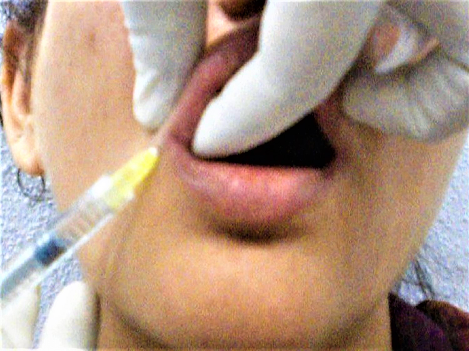
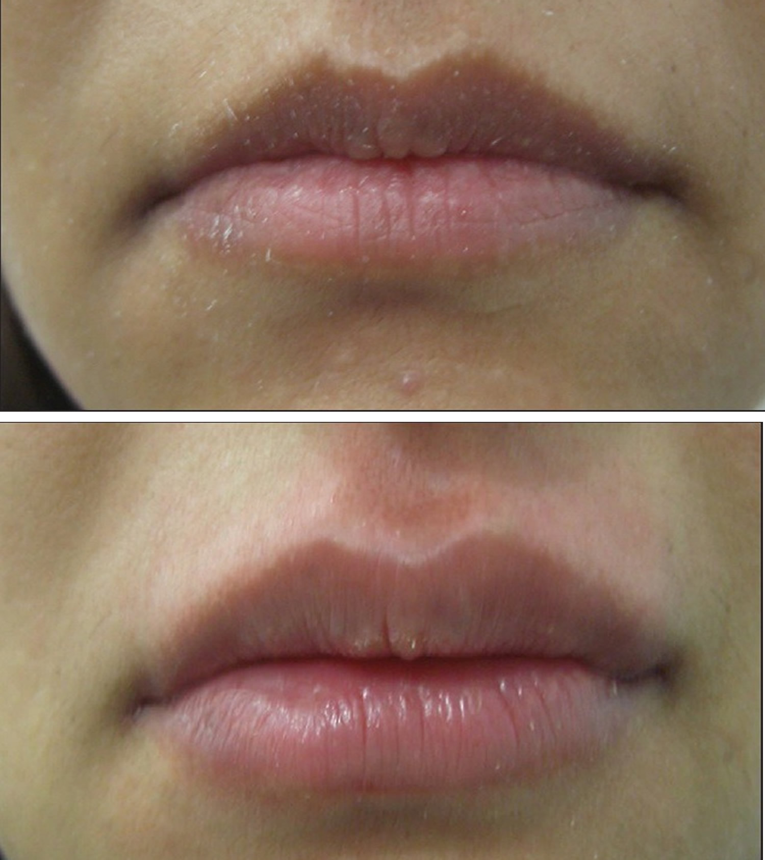
Figure 8. Lip augmentation before and after – shaping of upper and lower lips with 1 mL of HA

Lip augmentation before and after

References
  1. Lupo PM. Hyaluronic acid fillers in facial rejuvenation. Semin Cutan Med Surg. 2006;25:122–6. https://www.ncbi.nlm.nih.gov/pubmed/17055390
  2. Luthra A, Kumar V. Fillers and autologus fat. In: Khunger M, editor. Step by Step Treatment of Acne Scars. India: Jaypee Brothers; 2014. pp. 153–71.
  3. Filling in Wrinkles Safely. https://www.fda.gov/forconsumers/consumerupdates/ucm049349.htm
  4. Doheny K. Dermal Fillers: The Risks to Eliminating Wrinkles. Medscape News & Perspectives. https://www.medscape.com/viewarticle/894549#vp_1
  5. Lowe NJ. Temporary dermal fillers — European experiences. In: Lowe NJ, editor. Textbook of Facial Rejuvenation. London: Martin Dunitz/Taylor and Francis; 2002. pp. 177–88.
  6. Rudolph CM, Soyer HP, Sculler-Petrovic S, Kerl H. Foreign body granulomas due to injectable aesthetic microimplants. Am J Surg Pathol. 1999;23:113–7. https://www.ncbi.nlm.nih.gov/pubmed/9888711
  7. Lemperle G, Romano JJ, Busso M. Soft tissue augmentation with Artecoll: 10-year history, indications, techniques, and complications. Dermatol Surg. 2003;29:573–87. https://www.ncbi.nlm.nih.gov/pubmed/12786699
  8. Chisholm BB. Facial implants: Facial augmentation and volume restoration. Oral & Maxillofacial Surgery Clinics of North America 2005; 17(1): 77-84.
  9. Mandeville JT and Rubin PA. Injectable Agents for Facial Rejuvenation: Botulinum Toxin and Dermal Filling Agents. International Ophthalmology Clinics 2004; 44(1):189-212.
  10. Luthra A. Shaping Lips with Fillers. Journal of Cutaneous and Aesthetic Surgery. 2015;8(3):139-142. doi:10.4103/0974-2077.167269. https://www.ncbi.nlm.nih.gov/pmc/articles/PMC4645142/
  11. Carruthers J, Narurkar VA. Management of the lips and mouth corners. In: Carruthers J, Carruthers A, editors. Soft Tissue Augmentation. Philadelphia: Saunders; 2006. pp. 109–19.
  12. Narins RS & Bowman PH. Injectable skin fillers. Clinics in Plastic Surgery 2005; 32(2):151-162.
  13. de Maio M. The minimal approach: an innovation in facial cosmetic procedures. Aesthetic Plastic Surgery 2004; 28(5): 295–300.
  14. Dermal Fillers. https://emedicine.medscape.com/article/1125066-overview
  15. Dermal fillers. https://emedicine.medscape.com/article/1125066-overview
  16. Godin MS, Majmundar MV, Chrzanowski DS, Dodson KM. Use of Radiesse in combination with Restylane for facial augmentation. Archives of Facial Plastic Surgery 2006; 8(2): 92–97.
  17. Dermal Fillers Approved by the Center for Devices and Radiological Health. https://www.fda.gov/MedicalDevices/ProductsandMedicalProcedures/CosmeticDevices/WrinkleFillers/ucm227749.htm
  18. Sundaram H, Cassuto D. Biophysical Characteristics of Hyaluronic Acid Soft-Tissue Fillers and Their Relevance to Aesthetic Applications. Plast Reconstr Surg. 2013 Oct. 132(4S-2 Clinical Introduction to the Hyaluronic Acid Dermal Filler Using Cohesive Polydensified Matrix Technology):5S-21S.
  19. Homicz MR, Watson D. Review of injectable materials for soft tissue augmentation. Facial Plast Surg. 2004 Feb. 20(1):21-9.
  20. Cassuto D, Sundaram H. A Problem-Oriented Approach to Nodular Complications from Hyaluronic Acid and Calcium Hydroxylapatite Fillers: Classification and Recommendations for Treatment. Plast Reconstr Surg. 2013 Oct. 132(4S-2 Clinical Introduction to the Hyaluronic Acid Dermal Filler Using Cohesive Polydensified Matrix Technology):48S-58S.
  21. Lambros V. The use of hyaluronidase to reverse the effects of hyaluronic acid filler. Plast Reconstr Surg. 2004 Jul. 114(1):277
  22. Dover JS, Carruthers A, Carruthers J, Alam M. Clinical Use of RESTYLANE. Skin Therapy Lett. 2005 Feb. 10(1):5-7.
  23. Hirsch RJ, Narurkar V, Carruthers J. Management of injected hyaluronic acid induced Tyndall effects. Lasers Surg Med. 2006 Mar. 38(3):202-4.
  24. Rao J, Chi GC, Goldman MP. Clinical comparison between two hyaluronic acid-derived fillers in the treatment of nasolabial folds: hylaform versus restylane. Dermatol Surg. 2005 Nov. 31(11 Pt 2):1587-90.
  25. Monheit GD, Prather CL. Juvéderm: a hyaluronic acid dermal filler. J Drugs Dermatol. 2007 Nov. 6(11):1091-5.
  26. Flynn TC, Thompson DH, Hyun SH. Molecular Weight Analyses and Enzymatic Degradation Profiles of the Soft-Tissue Fillers Belotero Balance, Restylane, and Juvéderm Ultra. Plast Reconstr Surg. 2013 Oct. 132(4S-2 Clinical Introduction to the Hyaluronic Acid Dermal Filler Using Cohesive Polydensified Matrix Technology):22S-32S.
  27. Ballin AC, Cazzaniga A, Brandt FS. Long-term efficacy, safety and durability of Juvéderm® XC. Clin Cosmet Investig Dermatol. 2013. 6:183-9.
  28. Kono T, Kinney BM, Groff WF, Chan HH, Ercocen AR, Nozaki M. Randomized, evaluator-blind, split-face comparison study of single cross-linked versus double cross-linked hyaluronic acid in the treatment of glabellar lines. Dermatol Surg. 2008 Jun. 34 Suppl 1:S25-30.
  29. Alam M, Gladstone H, Kramer EM, Murphy JP Jr, Nouri K, Neuhaus IM, et al. ASDS guidelines of care: injectable fillers. Dermatol Surg. 2008 Jun. 34 Suppl 1:S115-48.
  30. ArteFill – Instructions for Use [package insert]. San Diego, Calif: Artes Medical, Inc. 2008.
  31. Lemperle G, Romano JJ, Busso M. Soft tissue augmentation with artecoll: 10-year history, indications, techniques, and complications. Dermatol Surg. 2003 Jun. 29(6):573-87; discussion 587.
  32. Butterwick K. Understanding injectable poly-L-lactic acid. Cosmet Dermatol. 2007. 20:388-92.
  33. Vleggaar D. Soft-tissue augmentation and the role of poly-L-lactic acid. Plast Reconstr Surg. 2006 Sep. 118(3 Suppl):46S-54S.
  34. Narins RS, Brandt FS, Lorenc ZP, Maas CS, Monheit GD, Smith SR. Twelve-month persistency of a novel ribose-cross-linked collagen dermal filler. Dermatol Surg. 2008 Jun. 34 Suppl 1:S31-9.
  35. Cohen JL. Understanding, avoiding, and managing dermal filler complications. Dermatol Surg. 2008 Jun. 34 Suppl 1:S92-9.
  36. Coleman KM, Voigts R, DeVore DP, Termin P, Coleman WP 3rd. Neocollagenesis after injection of calcium hydroxylapatite composition in a canine model. Dermatol Surg. 2008 Jun. 34 Suppl 1:S53-5.
  37. Berlin AL, Hussain M, Goldberg DJ. Calcium hydroxylapatite filler for facial rejuvenation: a histologic and immunohistochemical analysis. Dermatol Surg. 2008 Jun. 34 Suppl 1:S64-7.
  38. Busso M, Voigts R. An investigation of changes in physical properties of injectable calcium hydroxylapatite in a carrier gel when mixed with lidocaine and with lidocaine/epinephrine. Dermatol Surg. 2008 Jun. 34 Suppl 1:S16-23; discussion S24.
  39. azficel-T (laViv) [package insert]. Fibrocell Science, Inc.
  40. Dermal Fillers https://emedicine.medscape.com/article/1125066-overview
  41. Carruthers J. Review of long-lasting dermal fillers. Aesthetic Buyers Guide Supplement 2006; 2(1): 1-28.
  42. Morhenn VB, Lemperle G, Gallo RL. Phagocytosis of different particulate dermal filler substances by human macrophages and skin cells. Dermatologic Surgery 2002; 28(6): 484-490.
  43. Haneke E. Polymethyl methacrylate microspheres in collagen. Seminars in Cutaneous Medicine & Surgery 2004; 23(4): 227-232.
  44. Thioly-Bensoussan D. A new option for volumetric restoration: Poly-l-lactic acid. Journal of the European Academy of Dermatology & Venereology 2006; 20(Suppl 1):12-16.
  45. Marler JJ, Guha A, Rowley T, Koka R, Mooney D, Vacanti JP. Soft-tissue augmentation with injectable alginate and syngeneic fibroblasts. Plastic & Reconstructive Surgery 2000; 105(6): 2059-2058.
  46. Ersek RA & Beisang AA, III. Bioplastique: a new textured copolymer microparticle promises permanence in soft tissue augmentation. Plastic & Reconstructive Surgery 1991; 87(4): 693-702.
  47. Laeschke K. Biocompatibility of microparticles into soft tissue fillers. Seminars in Cutaneous Medicine & Surgery 2004; 23(4): 214-217.
  48. Lemperle G, Morhenn VB, Pestonjamasp V, Gallo RL. Migration studies and histology of injectable microspheres of different sizes in mice. Plastic & Reconstructive Surgery 2004; 113(5): 1380-1390.
read more
Procedures

MCH blood test

mch blood test

What is MCH

MCH is short for mean corpuscular hemoglobin is also called mean cell hemoglobin. MCH (mean corpuscular hemoglobin) is a calculation of the average amount of hemoglobin inside a single red blood cell. MCH blood test shows hemoglobin amount of red blood cells in circulation. Normal levels of MCH (mean corpuscular hemoglobin) are between 27 and 31 picograms (pg) of hemoglobin per red blood cell. To calculate MCH, the hemoglobin is divided by red blood cell, yielding an average amount of hemoglobin per red blood cell. In contrast to MCH (mean corpuscular hemoglobin), MCHC (mean corpuscular hemoglobin concentration) indicates the amount of hemoglobin per unit volume and MCHC correlates the hemoglobin content with the volume of the cell. It is expressed as g/dl of red blood cells or as a percentage value. The normal values for MCHC (mean corpuscular hemoglobin concentration) are 34 ± 2 g/dl.

Figure 1. Mean corpuscular hemoglobin formula

Hemoglobin is the iron-containing protein found in all red blood cells (RBCs) that gives the cells their characteristic red color. Hemoglobin enables red blood cells to bind to oxygen in the lungs and carry it to tissues and organs throughout the body. Hemoglobin also helps transport a small portion of carbon dioxide, a product of cell metabolism, from tissues and organs to the lungs, where it is exhaled.

The MCH blood test is done as a component of a blood test called a Complete Blood Count (CBC), which evaluates the composition of the blood, checking hematocrit, white blood cells (WBC) and platelets as well as hemoglobin and red blood cells (red blood cell). It is usually ordered to get an overview of general health. MCH is not measured directly, but calculated based on the hemoglobin value (Hb), which is the total measure of hemoglobin in the blood, and the red blood cell, which is the number of red blood cells in the blood.

A Complete Blood Count (CBC) is commonly included in bloodwork ordered by your doctor during an annual checkup. Also included in this standard test bundling is a comprehensive metabolic panel, urinalysis, hemoglobin A1c, and lipid panel.

The hemoglobin test measures the amount of hemoglobin in a person’s sample of blood. A hemoglobin level can be performed alone or with a hematocrit (Hct), a test that measures the proportion of blood that is made up of red blood cells, to quickly evaluate an individual’s red blood cells. Red blood cells, which make up about 40% (ranging 37-49%) of the blood’s volume, are produced in the bone marrow and are released into the bloodstream when they are, or nearly are, mature. The typical lifespan of an red blood cell is 120 days, and the bone marrow must continually produce new red blood cells to replace those that age and degrade or are lost through bleeding.

Several diseases and conditions can affect red blood cells and consequently the level of hemoglobin in the blood. In general, the hemoglobin level and hematocrit rise when the number of red blood cells increases. The hemoglobin level and hematocrit fall to less than normal when there is a drop in production of red blood cells by the bone marrow, an increase in the destruction of red blood cells, or if blood is lost due to bleeding. A drop in the red blood cell count, hemoglobin and hematocrit can result in anemia, a condition in which tissues and organs in the body do not get enough oxygen, causing fatigue and weakness. If too many red blood cells are produced, polycythemia results and the blood can become thickened, causing sluggish blood flow and related problems.

Abnormally high or low levels of MCH, as determined by blood testing, can be an indication of a number of problems within the body, ranging from nutrient deficiencies to chronic diseases.

Several diseases and conditions can affect red blood cells and consequently the level of hemoglobin in the blood. In general, the hemoglobin level and hematocrit rise when the number of red blood cells increases. The hemoglobin level and hematocrit fall to less than normal when there is a drop in production of red blood cells by the bone marrow, an increase in the destruction of red blood cells, or if blood is lost due to bleeding. A drop in the red blood cell count, hemoglobin and hematocrit can result in anemia, a condition in which tissues and organs in the body do not get enough oxygen, causing fatigue and weakness. If too many red blood cells are produced, polycythemia (erythrocytosis) results and the blood can become thickened, causing sluggish blood flow and related problems.

The best way to evaluate your MCH result is to look the all parameters of complete blood count (CBC) together and diagnose the anemia correctly. Complete blood count laboratory test is a simple and quick test but with it’s lots of sub parameters provides a lot of information about your general health condition and also diseases.

MCH levels

MCH normal range

Normal range of MCH for adults is 27 to 31 picograms/red blood cell (pg/cell)

This range may change according to the laboratory and the analyzer that is used.

  • 1-3 days old baby: 31-37 pg
  • 1 week-1 month old baby: 28-40 pg
  • 3-6 months baby: 25-35 pg
  • 6 months- 2 years old baby: 23-31 pg
  • 2-6 years old child: 24-30 pg
  • 6-12 years old child: 25-33 pg
  • 12-18 years old teen: 25-35 pg

MCH low

MCH levels below 27 pg (hypochromia) are considered abnormally low. Common causes of Low MCH results include blood loss, iron deficiency and microcytic anemia, which is a condition in which red blood cells are abnormally small, carrying less hemoglobin. Other potential causes of a low MCH test include hemoglobinopathy, which is a group of disorders that cause changes in the structure of hemoglobin, and iron-deficiency anemia.

MCH high

MCH levels over 31 pg (hyperchromia) are generally considered abnormally high. The most common reason for high MCH is macrocytic anemia or megaloblastic anemia, which is a blood disorder in which the body fails to produce enough red blood cells. In macrocytic anemia, red blood cells that are produced are larger than usual, each carrying more hemoglobin than normal-sized cells would. This condition can be caused by deficient levels of vitamin B-12 or folic acid in the body; nutrients found in foods like fish, liver, green leafy vegetables and fortified cereals. These contribute to efficient red blood cell production and may be lacking in your diet, you may not be able to absorb them or there may be other reasons your body cannot process them. Other possible causes of megaloblastic anemia, hypothyroidism, liver disease, and primary bone marrow dysplasias (including myelodysplasia and myeloproliferative disorders) are some of the more common causes.

Symptoms of macrocytic anemia can include:

  • Unexplained fatigue
  • Heart palpitations
  • Pallor
  • Heart complications. Since this can interfere with heart function, early diagnosis of high MCH (macrocytic) anemia is important.
  • Other possible reasons for a high MCH test include several other forms of anemia, thyroid dysfunction, chemotherapy, certain infections, over use of estrogen-containing medications, some forms of leukemia and hereditary spherocytosis; a condition that causes a shortage of red blood cells.

What are red blood cells

Blood transports a variety of materials between interior body cells and those that exchange substances with the external environment. In this way, blood helps maintain stable internal environmental conditions. Blood is composed of formed elements suspended in a fluid extracellular matrix called blood plasma. The “formed elements” include red blood cells, white blood cells, and cell fragments called platelets. Most blood cells form in red marrow within the hollow parts of certain long bones.

Most blood samples are roughly 37% to 49% red blood cells by volume – adult females is 38–46% (average = 42%) and for adult males, it is 40–54% (average = 47). This percentage is called the hematocrit. The white blood cells and platelets account for less than 1% of blood volume. The remaining blood sample, about 55%, is the plasma, a clear, straw-colored liquid. Blood plasma is a complex mixture of water, gases, amino acids, proteins, carbohydrates, lipids, vitamins, hormones, electrolytes, and cellular wastes (see Figure 1).

Blood volume varies with body size, percent adipose tissue, and changes in fluid and electrolyte concentrations. An average-size adult has a blood volume of about 5 liters (5.3 quarts), 4–5 liters in a female and 5–6 liters in a male.

Red blood cell (also called erythrocyte) is biconcave disc without a nucleus. This biconcave shape is an adaptation for transporting the gases oxygen and carbon dioxide. It increases the surface area through which oxygen and carbon dioxide can diffuse into and out of the cell. The characteristic shape of a red blood cell also places the cell membrane closer to oxygen-carrying hemoglobin molecules in the cell reducing the distance for diffusion.

Each red blood cell is about one-third hemoglobin by volume. This protein imparts the color of blood. When hemoglobin binds oxygen, the resulting oxyhemoglobin is bright red, and when oxygen is released, the resulting deoxyhemoglobin is darker.

Prolonged oxygen deficiency (hypoxia) causes cyanosis, in which the skin and mucous membranes appear bluish due to an abnormally high blood concentration of deoxyhemoglobin in the superficial blood vessels. Exposure to low temperature may also result in cyanosis by constricting superficial blood vessels. This response to environmental change slows skin blood flow. As a result, more oxygen than usual is removed from the blood flowing through the vessels, increasing the concentration of deoxyhemoglobin.

Note: Blood is a complex mixture of formed elements in a liquid extracellular matrix, called blood plasma. Note that water and proteins account for 99% of the blood plasma.

Figure 1. Blood composition

blood composition

blood compositionblood-composition

Note: Blood consists of a liquid portion called plasma and a solid portion (the formed elements) that includes red blood cells, white blood cells, and platelets. When blood components are separated by centrifugation, the white blood cells and platelets form a thin layer, called the “buffy coat,” between the plasma and the red blood cells, which accounts for about 1% of the total blood volume. Blood cells and platelets can be seen under a light microscope when a blood sample is smeared onto a glass slide.

Blood Cell Formation

The process of blood cell formation, called hematopoiesis, begins in the yolk sac, which lies outside the human embryo. Later in the fetal development, red blood cells are manufactured (erythropoiesis) in the liver and spleen, and still later they form in bone marrow. After birth, these cells are produced in the red bone marrow.

Bone marrow is a soft, netlike mass of connective tissue within the medullary cavities of long bones, in the irregular spaces of spongy bone, and in the larger central canals of compact bone tissue. It is of two kinds: red and yellow. Red bone marrow functions in the formation of red blood cells (erythrocytes), white blood cells (leukocytes), and blood platelets. The color comes from the oxygen-carrying pigment hemoglobin in the red blood cells.

In an infant, red marrow occupies the cavities of most bones. As a person ages, yellow bone marrow, which stores fat, replaces much of the red marrow. Yellow marrow is not active in blood cell production. In an adult, red marrow is primarily found in the spongy bone of the skull, ribs, breastbone (sternum), collarbones (clavicles), backbones (vertebrae), and hip bones. If the supply of blood cells is deficient, some yellow marrow may become red marrow, which then reverts to yellow marrow when the deficiency is corrected.

Figure 3 illustrates the stages in the formation of red blood cells from hematopoietic stem cells (blood-forming cells), which are also called hemocytoblasts.

Red blood cells have nuclei during their early stages of development but lose their nuclei as the cells mature. Losing the nuclei provides more space for hemoglobin. Because mature red blood cells do not have nuclei, they cannot divide. They use none of the oxygen they carry because they do not have mitochondria. Mature red blood cells produce ATP through glycolysis only.

The average life span of a red blood cell is 120 days. Many of these cells are removed from the circulation each day, and yet the number of cells in the circulating blood remains relatively stable. This observation suggests a homeostatic control of the rate of red blood cell production.

The hormone erythropoietin (EPO) controls the rate of red blood cell formation through negative feedback. The kidneys, and to a lesser extent the liver, release erythropoietin in response to prolonged oxygen deficiency (Figure 6). At high altitudes, for example, where the amount of oxygen in the air is reduced, the blood oxygen level initially decreases. This drop in the blood oxygen level triggers the release of erythropoietin, which travels via the blood to the red bone marrow and stimulates red blood cell production.

After a few days of exposure to high altitudes, many newly formed red blood cells appear in the circulating blood. The increased rate of production continues until the number of erythrocytes in the circulation is sufficient to supply tissues with oxygen. When the availability of oxygen returns to normal, erythropoietin release decreases, and the rate of red blood cell production returns to normal as well. An excessive increase in red blood cells is called polycythemia. This condition increases blood viscosity, slowing blood flow and impairing circulation.

Figure 2. Bone marrow anatomy

bone marrow anatomy

Anatomy of the bone. The bone is made up of compact bone, spongy bone, and bone marrow. Compact bone makes up the outer layer of the bone. Spongy bone is found mostly at the ends of bones and contains red marrow. Bone marrow is found in the center of most bones and has many blood vessels. There are two types of bone marrow: red and yellow. Red marrow contains blood stem cells that can become red blood cells, white blood cells, or platelets. Yellow marrow is made mostly of fat.

Dietary Factors Affecting Red Blood Cell Production

Availability of B-complex vitamins—vitamin B12 and folic acid—significantly influences red blood cell production. Because these vitamins are required for DNA synthesis, they are necessary for the growth and division of cells. Cell division is frequent in blood-forming (hematopoietic) tissue, so this tissue is especially vulnerable to a deficiency of either of these vitamins.

Hemoglobin synthesis and normal red blood cell production also require iron. The small intestine absorbs iron slowly from food. The body reuses much of the iron released by the decomposition of hemoglobin from damaged red blood cells. Nonetheless, insufficient dietary iron can reduce hemoglobin synthesis.

A deficiency of red blood cells or a reduction in the amount of hemoglobin they contain results in a condition called anemia. This reduces the oxygen-carrying capacity of the blood, and the affected person may appear pale and lack energy. A pregnant woman may have a normal number of red blood cells, but she develops a relative anemia because her plasma volume increases due to fluid retention. This shows up as a decreased hematocrit.

In contrast to anemia, the inherited disorder called hemochromatosis results in the absorption of iron in the small intestine at ten times the normal rate. Iron builds up in organs, to toxic levels. Treatment is periodic blood removal, as often as every week.

Figure 3. Blood cell development. A blood stem cell goes through several steps to become a red blood cell, platelet, or white blood cell

blood cell development

Figure 4. Blood cells

blood cells

Note: Blood tissue consists of red blood cells, white blood cells, and platelets suspended in plasma. (a) Idealized representation of a sample of blood. (b) Micrograph of a sample of blood (1,000x).

Figure 5. Red blood cells

red blood cells

What is the function of red blood cells

Red blood cells function is to transports oxygen and carbon dioxide. to body tissues by blood flow via circulatory system. Red blood cells take oxygen from the lungs and release it into the cells or tissues. Lipids and proteins make up the cell membrane of red blood cells. Hemoglobin, an iron containing biomolecule, is the rich component of the cytoplasm of red blood cells mainly responsible for the oxygen binding and red color of the erythrocytes (see Figure 6).

Figure 6. Red blood cell formation

red blood cell formation

Note: Low blood oxygen causes the kidneys and to a lesser degree, the liver to release erythropoietin. Erythropoietin stimulates target cells in the red bone marrow to increase the production of red blood cells, which carry oxygen to tissues.

Destruction of Red Blood Cells

The average life span of red blood cells is about four months (120 days) after which it breaks down. Red blood cells are elastic and flexible, and they readily bend as they pass through small blood vessels. As the cells near the end of their four-month life span, however, they become more fragile. The cells may sustain damage simply passing through capillaries, particularly those in active muscles that must withstand strong forces. Macrophages phagocytize and destroy damaged red blood cells, primarily in the liver and spleen. Macrophages are large, phagocytic, wandering cells. During phagocytosis, the iron from the hemoglobin is retained in the liver and spleen cells and is again used in the formation of red blood cells in the body. About 2-10 million red blood cells are formed and destroyed each second in a normal person.

Hemoglobin molecules liberated from red blood cells break down into their four component polypeptide “globin” chains, each surrounding a heme group. The heme further decomposes into iron and a greenish pigment called biliverdin. The blood may transport the iron, combined with a protein, to the hematopoietic tissue in red bone marrow to be reused in synthesizing new hemoglobin. About 80% of the iron is stored in the liver in the form of an iron-protein complex. Biliverdin eventually is converted to an orange-yellow pigment called bilirubin. Biliverdin and bilirubin are secreted in the bile as bile pigments. Figure 7 summarizes the life cycle of a red blood cell.

In jaundice (yellow discoloration of the skin and the whites of the eyes), accumulation of bilirubin turns the skin and eyes yellowish. Newborns can develop physiologic jaundice a few days after birth. This condition may be the result of immature liver cells that ineffectively secrete bilirubin into the bile. Treatment includes exposure to fluorescent light, which breaks down bilirubin in the tissues, and feedings that promote bowel movements. In hospital nurseries, babies being treated for physiological jaundice lie under “bili lights,” clad only in diapers and protective goggles.

Figure 7. Red blood cell hemoglobin

Hemoglobin

Figure 8. Lifecycle of a red blood cell

lifecycle of red blood cell

What is anemia?

Anemia is a condition that occurs when the amount of hemoglobin in a person’s blood drops below normal. A decrease in hemoglobin is often associated with a decrease in the number of red blood cells (red blood cells) and hematocrit. Hemoglobin is contained within red blood cells and is necessary to transport and deliver oxygen from the lungs to the rest of the body. Without a sufficient supply of oxygen, many tissues and organs throughout the body can be adversely affected. People with anemia may experience fatigue and weakness and may lack energy.

Anemia is a fairly common condition, affecting both men and women of all ages, races, and ethnic groups. However, certain people have increased risk of developing anemia. These include people with diets poor in iron and vitamins, chronic diseases such as kidney disease, diabetes, cancer, inflammatory bowel disease, a family history of inherited anemia, chronic infections such as tuberculosis or HIV, and those who have had significant blood loss from injury or surgery. Anemia can be mild, moderate, or severe depending on how much the red blood cell count and/or hemoglobin levels are decreased.

In general, the main causes of anemia include:

  • Impaired or decreased production of red blood cells by the bone marrow due to nutritional deficiency (e.g., iron deficiency, B vitamin deficiencies), bone marrow failure (e.g., aplastic anemia, myelodysplastic syndrome), or diseases that involve the bone marrow (e.g., infection, lymphoma, solid tumor)
  • Loss of red blood cells due to bleeding or to increased destruction of red blood cells as in hemolytic anemia

Anemia may be acute or chronic. Chronic anemia may develop slowly over a period of time with long-term illnesses such as diabetes, chronic kidney disease, or cancer. In these situations, the anemia-related symptoms may not be apparent because the underlying disease masks its symptoms and/or the body adapts to anemia when it develops over a period of time. The presence of anemia in chronic conditions may often go undetected for a period of time and sometimes may only be discovered during tests or examinations for other conditions.

Anemia may also occur in acute episodes such as with substantial blood loss (extensive injury or invasive surgery) or with certain anemias in which a significant number of red blood cells are destroyed known as hemolytic anemia. Signs and symptoms may become apparent very quickly, and the cause can be determined from a combination of physical examination, medical history, and testing.

Types of anemia

Anemias can also be described based on the red blood cell size and concentration of hemoglobin in them. If cell size is much smaller than normal, it is known as microcytic anemia. If it is much bigger than normal (high MCV), then it is macrocytic anemia. Likewise, if the concentration of hemoglobin is much lower than normal, it is hypochromic anemia (low MCH); if the concentration is much higher than normal, the red blood cells are called hyperchromic (high MCH).

Within the two broad categories of general causes of anemia, there are several types with different specific causes. Some of the most common types are summarized in the table below. See the sections below to read more about each one.

Type of AnemiaDescriptionExamples of Causes
Iron DeficiencyDeficiency of iron leads to decreased production of hemoglobin; low levels of hemoglobin in turn leads to production of smaller and hypochromic red blood cells (low MCV and low MCH).Blood loss; diet low in iron; poor absorption of iron
Pernicious Anemia and B Vitamin DeficiencyRed blood cells do not develop as they normally would because of a lack of B vitamins (B12 and folate); leads to decreased production of red blood cells.Lack of intrinsic factor (needed for B12 absorption); diet low in B vitamins; decreased absorption of B vitamins
AplasticBone marrow is unable to produce enough blood cells; a life-threatening condition.Cancer therapy, exposure to toxins, autoimmune disease, viral infections
HemolyticRed blood cells are destroyed faster than the bone marrow can replace them.Inherited causes such as sickle cell disease and hereditary spherocytosis; other causes include transfusion of incompatible blood, autoimmune disease, certain drugs (penicillin)
Anemia of Chronic DiseasesVarious conditions over the long term can cause decreased production of red blood cells.Kidney disease, rheumatoid arthritis, diabetes, tuberculosis, HIV, Crohn disease, cancer, and others

There are many other conditions that can, for various reasons, result in some level of anemia, such as:

Bleeding—significant bleeding resulting from, for example, trauma or surgery (acute) or from gastrointestinal bleeding (ulcers) occurring over time (chronic)
Leukemia (acute or chronic)
Lymphoma
Myelodysplastic syndrome
Multiple myeloma
Myeloproliferative neoplasms (e.g., myelofibrosis)
Infections (e.g., HIV)

Anemia signs and symptoms

Though different types of anemia have different causes, the signs and symptoms can be very similar. Mild or moderate forms of anemia may cause few, if any, symptoms. Some of the most common symptoms are:

  • General feeling of tiredness or weakness (fatigue)
  • Lack of energy
  • Pale skin (pallor)
  • Dizziness
  • Headaches

Other signs and symptoms that may develop as the anemia becomes more severe include a feeling of cold or numbness in hands and/or feet, shortness of breath, fast or irregular heartbeat, and chest pain.

Tests for anemia

Several routine laboratory tests may be used to help diagnose anemia as well as help to determine the underlying cause. These are listed below. Depending on the results of these, the medical history of the person, and signs and symptoms, other tests may be done as follow up to help diagnose the cause of anemia and to help guide treatment. (Click on the links for the different types of anemia at the top of this page to read about these specific tests.)

Complete Blood Count (CBC)

Anemia may first be detected when a routine test that counts the number and relative proportion of each of the different types of cells in the blood stream, called a complete blood count (CBC), is done during a health exam or as part of testing for other conditions. A complete blood count (CBC) is often ordered as part of a yearly physical exam and helps to evaluate overall health and to screen for a wide variety of disorders.

With anemia, some of the components of the complete blood count (CBC) that may show abnormal results include:

  • Red blood cell count—typically low
  • Hemoglobin—low
  • Hematocrit—low
  • Red blood cell indices—these include mean corpuscular volume (MCV), mean corpuscular hemoglobin (MCH), and mean corpuscular hemoglobin concentration (MCHC). They give a healthcare practitioner information about the size of the red blood cells and the amount and concentration of hemoglobin in red blood cells present in someone’s blood at that moment. For example, the size and hemoglobin concentration of red blood cells can help with diagnosing anemia because those characteristics can vary for different kinds of anemia.

Blood Smear and Differential

If results of the CBC indicate anemia, it may be followed up with an examination of a blood smear or a differential, which counts white blood cells. The smear review can provide additional information, such as the shape of red blood cells and the presence of abnormal cells, which can help diagnose and classify anemia.

Reticulocyte Count

This test provides information on the number of relatively immature red blood cells in a person’s blood sample. When someone has anemia (low red blood cell count, hemoglobin, and hematocrit), the results of this test can help determine the cause and/or help classify the type of anemia. For example, for a person with anemia, an inappropriately low reticulocyte count often indicates decrease in red blood cell production in the bone marrow.

Results from these tests may give clues as to the cause. Several other tests may be run to help determine the cause of the anemia and to guide treatment. See the individual discussions of the different types of anemia for more on these.

High MCV and MCH

Macrocytosis, defined as a MCV (mean corpuscular volume) greater than 100 fL (femtoliter or 1015 liter), occurs in approximately 3 percent of the general population 1. Debate persists about the upper limit of normal values of MCV (mean corpuscular volume). In a recent study, 7 percent of patients had a mean corpuscular volume greater than 96 fL, and 1.7 percent had a mean corpuscular volume greater than 100 fL 2. With the advent of automated complete blood cell (CBC) counters and the increased use of certain medications, elevations in the MCV (mean corpuscular volume) may be more commonly encountered.

Causes of high MCV and their prevalence in different populations are shown in Table 1 3. Although high MCV is associated with anemia, hypothyroidism is a more common cause in older persons than in other age groups. Results of a study in New York City indicated that medications for treating human immunodeficiency virus (HIV) infection have become a more prominent cause of high MCV (macrocytosis). Alcoholism is the cause in as many as 80 percent of patients in some populations 4. Bone marrow biopsy was only performed in these studies when another cause could not be determined, and resulted in diagnosis in approximately one fourth of these remaining patients 1. In recent years, an association has been found between Helicobacter pylori infection and vitamin B12 deficiency 5.

Table 1. Major causes of macrocytosis

EtiologyStudy population
Hospitalized patients in New York City (%)Outpatients in Finland (%)Finnish persons older than 75 years (%)Finnish and American patients (%)

Alcohol

26

65

15

36

B12 and/or folate deficiency

6

9

28

21

Medications

37*

3

2

11

Hypothyroidism

1

12

5

Bone marrow dysplasias

6

1

5

5

Liver disease (nonalcoholic)

6

2

6

Reticulocytosis

8

7

Miscellaneous

3

21

13

7

Not established

7

22

12


Footnote: Causes listed from most to least common.

*— 13 percent from zidovudine (Retrovir).

[Source 3]

The causes of high MCV (macrocytosis) can be broadly classified as megaloblastic and nonmegaloblastic (Table 2). Megaloblastic processes are characterized on the peripheral smear by macroovalocytes and hypersegmented neutrophils, which are absent in nonmegaloblastic macrocytic processes (Figure 9). Nonmegaloblastic processes have round macrocytes or macroreticulocytes (Figure 10). Because the mechanisms producing macrocytosis are not completely understood, the separation between megaloblastic and nonmegaloblastic causes is somewhat artificial. However, this concept remains useful for identifying the most predominant etiology for high MCV (macrocytosis). For example, whereas the effect of alcohol is thought to be primarily a nonmegaloblastic process, in chronic alcoholism there may be concomitant vitamin B12 or folate deficiency 6.

Figure 9. Megaloblastic anemia

Megaloblastic anemiaFigure 10. Microangiopathic hemolytic anemia (nonmegaloblastic), with polychromatophilic macrocytes (black arrow) and schistocytes (red arrows) typical of this particular disorder are also present. Schistocytes (red arrows) have an irregular shape and lack the area of central pallor usually seen in red blood cells (erythrocytes). 

Microangiopathic hemolytic anemia

Table 2. Differential diagnosis of high MCV (macrocytosis)

Megaloblastic (involving vitamin B12 and/or folate deficiencies)

Atrophic gastritis

Enteral malabsorption

Human immunodeficiency virus treatments

Anticonvulsants (some cause folate depletion)

Primary bone marrow disorders

Nitrous oxide abuse

Inherited disorders

Nonmegaloblastic

Alcohol abuse

Medication side effects (see Table 3)

Myelodysplasia

Hypothyroidism

Liver disease

Hemolysis

Hemorrhage

Chronic obstructive pulmonary disease

Splenectomy

False elevations

Cold agglutinins

Hyperglycemia

Marked leukocytosis


Footnote: Diagnoses listed from most to least common.

[Source 3]

In megaloblastic processes, erythrogenic precursors are larger than mature red blood cells (red blood cells) because folate and vitamin B12 deficiencies result in defective RNA and DNA syntheses. Serum elevations in homocysteine and methylmalonic acid result from defective biochemical processes in folate and B12 deficiencies, and could be used to clarify the cause of megaloblastic anemia, although this is not yet standard clinical practice 7.

Nonmegaloblastic processes develop from multiple mechanisms and have not been fully outlined. Macrocytosis can occur when there is increased red blood cell production secondary to peripheral blood cell destruction (i.e., hemolysis) or loss (i.e., hemorrhage), leading to a reticulocytosis. Reticulocytes are incompletely processed red blood cells and, therefore, are slightly larger than the average red blood cell.

Table 3. Medications that may cause high MCV (macrocytosis)

Treatments for human immunodeficiency virus: reverse transcriptase inhibitors (e.g., stavudine [Zerit], lamivudine [Epivir], zidovudine [Retrovir])

Anticonvulsants (e.g., valproic acid [Depakote], phenytoin [Dilantin])

Folate antagonists (e.g., methotrexate)

Chemotherapeutics (e.g., alkylating agents, pyrimidine, purine inhibitors)

Trimethoprim/sulfamethoxazole (Bactrim, Septra)

Biguanides (e.g., metformin [Glucophage]), cholestyramine (Questran)

[Source 3]

High MCV (Macrocytosis) Specific Causes

Vitamin B12 deficiency

Vitamin B12 is absorbed by the ileum when it is bound by intrinsic factor, which is produced by the parietal cells of the gastric mucosa. In pernicious anemia, the loss of parietal cells leads to insufficient absorption of vitamin B12, which then leads to vitamin B12 deficiency over time. Pernicious anemia is most commonly caused by auto-immune atrophic gastritis, in which autoantibodies are directed against parietal cells and intrinsic factor. Less commonly, pernicious anemia can be caused by nonautoimmune gastritis secondary to Helicobacter pylori (H. pylori) infections and Zollinger-Ellison syndrome.

Patients with vitamin B12 deficiency may describe paresthesias related to peripheral neuropathy, poor or strict vegan diet, lack of socioeconomic resources, bowel-related symptoms (including diarrhea), or a history of bowel surgery for weight loss. Findings on physical examination may include neurologic signs such as ataxia, decreased proprioception, and vibratory sensation. Patients may also have poor dentition or nonspecific oral stomatitis or glossitis.

Because pregnant women take folic acid routinely in prenatal vitamins, macrocytic anemia is much less common during pregnancy. Consider nitrous oxide abuse in at-risk populations, because nitrous oxide inactivates vitamin B12 through oxidation 8. Other uncommon causes include Diphyllobothrium latum (i.e., fish tapeworm) infection or inherited disorders of cobalamin metabolism, including Imerslund syndrome (a congenital vitamin B12 malabsorption associated with proteinuria) 9. Only 10 percent of persons with vitamin B12 deficiency are actually anemic 10.

The normal range for serum measures of vitamin B12 varies among laboratories. If the vitamin B12 level s borderline low (i.e., 100 to 400 pg per mL [74 to 295 pmol per L]), methylmalonic acid and homocysteine levels should be ordered and, if elevated, may provide evidence of B12 deficiency 7. The Schilling test (i.e., measuring enteral absorption of vitamin B12) is not widely available at this time.

Oral therapy appears to be as effective as intramuscular therapy for the treatment of vitamin B12 deficiency. Relapse of pernicious anemia occurs at a mean interval of 65 months after cessation of treatment. It is important for patients to adhere to long-term therapy because the deficiency will recur if treatment is stopped, unless a reversible cause is identified 11.

Folate deficiency

The history of folate deficiency may mimic the history of vitamin B12 deficiency in regard to poor nutritional intake or absorption. In addition, 35 percent of patients with alcoholism and macrocytic anemia are folate deficient, which can be caused by poor nutritional intake, malabsorption, hepatobiliary dysfunction, and possibly increased folate catabolism 12. Some medications that are used to treat seizure disorders, cancer, and autoimmune diseases can lead to folate deficiency. For example, methotrexate directly inhibits dihydrofolate reductase, which leads to a functional folate deficiency 13. Other medications that affect folate metabolism include 5-fluorouracil (Adrucil), hydroxyurea (Hydrea), pyrimethamine (Daraprim), trimethoprim/sulfamethoxazole (Bactrim, Septra), pentamidine (Pentam), and phenytoin (Dilantin) 14. Medications can also affect folate absorption, including metformin (Glucophage) and cholestyramine (Questran). Supplementing with folate may be necessary when treating a patient with such medications 15.

Serum folate levels are not useful because they fluctuate rapidly with dietary intake and are not cost effective 16. Red blood cell folate levels more accurately correlate with folate stores and should be performed if folate deficiency is suspected 17. In differentiating the cause of megaloblastic anemia, a methylmalonic acid level that is within normal range also points toward a diagnosis of folate deficiency, especially if the serum vitamin B12 level is within the normal range. Note that homocysteine levels will be elevated with vitamin B12 and folate deficiencies.

HIV medications

Treatment of HIV with reverse transcriptase inhibitors (e.g., stavudine [Zerit], lamivudine [Epivir], zidovudine [Retrovir]) will cause macrocytosis because they interfere with DNA production, which may lead to megaloblastic changes. Most patients with HIV who are being treated with reverse transcriptase inhibitors will display macrocytosis without anemia. This indicates medication compliance by the patient, and no treatment is necessary 18. Table 3 lists other medications that may cause macrocytosis 19.

Reticulocytosis (hemolysis or hemorrhage)

For the patient’s history, questions that are pertinent to evidence of blood loss, whether chronic or acute, can point to reticulocytosis. As part of the family history, physicians should ask about the presence of certain hematologic syndromes, including sickle cell disease, hereditary spherocytosis, and glucose-6-phosphate dehydrogenase deficiency (G6PD). In persons who do distance running, hemolysis from constant foot pounding has infrequently been shown to cause macrocytosis 20. The physical examination may reveal hepatosplenomegaly in hemolysis or other physical manifestations of blood loss (e.g., conjunctiva, mucosal pallor). In further diagnostic testing, the peripheral smear will reveal evidence of hemolyzed red blood cells (e.g., bite cells, helmet cells). Macrocytosis results from the marrow’s response to increased cell destruction or blood loss, with release of reticulocytes into the peripheral circulation. Treatment should be focused on the underlying cause of hemolysis or hemorrhage.

Alcohol abuse

The Michigan Alcoholism Screening test and obtaining γ-glutamyltransferase levels were found to be the two most sensitive tests for detecting alcohol abuse in patients with macrocytosis 4. Physical findings consistent with alcoholism include gynecomastia, caput medusae, and jaundice. Alcohol use more commonly causes macrocytosis through its toxic effect than through folate deficiency secondary to alcoholism. The mean corpuscular volume is generally less than 110 fL with chronic alcohol use. Abstinence from alcohol rapidly corrects the elevated mean corpuscular volume 6.

Bone marrow dysfunction

As noted above, myeloproliferative disorders (sometimes called refractory anemia) are a more common cause of macrocytosis and anemia among older persons than in younger populations. Although the peripheral smear may be suggestive, bone marrow biopsy is required to establish this diagnosis. Referral will likely be necessary for the work-up and management.

Other causes

Medications, hypothyroidism, liver and renal disease, and chronic obstructive pulmonary disease are associated with less dramatic elevations in the mean corpuscular volume. Nonalcoholic liver disease and hypothyroidism account for a substantial portion of macrocytosis. Patient history and physical examination should include a history of these medical diseases. Splenectomy may cause macrocytosis because cells are not processed as thoroughly when the spleen is absent. Down syndrome also may be associated with reticulocytosis or myeloproliferative disorders 21. Treatment, if needed, can be directed at the underlying disorder.

High MCV diagnosis

Once macrocytosis is identified, the history and physical examination help narrow the differential diagnosis. The presence of anemia, the degree of elevation of the mean corpuscular volume, and the patient’s overall health guide how aggressively the work-up progresses. At least some amount of investigation is warranted if the diagnosis is not readily apparent or if the patient is anemic (defined by the World Health Organization as a hemoglobin level less than 13 g per dL [130 g per L] in men and less than 12 g per dL [120 g per L] in women) 22.

The algorithm outlined in Figure 11 suggests a work-up for macrocytosis. Physicians should begin by ordering a peripheral smear, a reticulocyte count, and a vitamin B12 serum level for all patients with macrocytosis. It may be necessary to specifically order a reticulocyte index in some laboratories, which assesses if there is an adequate bone marrow response. Hemorrhage or hemolysis is the most likely cause if the reticulocyte count is elevated, but anemia recovery also causes an elevation in the reticulocyte count. Measures of vitamin B12 are a useful part of the initial work-up, because if vitamin B12 deficiency is present but undiagnosed, folate repletion will correct the megaloblastic anemia, but not the possible neuropathic changes that occur with B12 deficiency 10.

Although uncommon, consider the possibility of spurious macrocytosis. This may be caused by cold agglutinins, hyperglycemia, or leukocytosis. Cold agglutinins cause the RBCs to clump, making them appear larger to the automatic counter 23. Hyperglycemic blood is more concentrated, and when it is diluted to measure the mean corpuscular volume, the cells swell more than usual, causing a false elevation 24. Increased turbidity of a sample with marked leukocytosis also can cause the machine to overestimate the cell size 25.

When the history and physical examination, peripheral smear, B12 level, and reticulocyte count have not lead to an obvious diagnosis, consider a comprehensive metabolic panel to look for liver and kidney disease, thyroid-stimulating hormone for thyroid disorders, and methylmalonic acid and homocysteine levels to assess for vitamin B12 deficiency, despite a normal vitamin B12 level. If the cause remains elusive, consider again whether the degree of anemia or the patient’s overall health warrants referral to a hematologist for bone marrow biopsy, or search for rarer causes, keeping in mind that the most extensive work-up will result in a diagnosis in approximately 90 percent of patients.

Figure 11. Evaluation of Macrocytic Anemia – RBC = red blood cell; MMA = methylmalonic acid

Evaluation of Macrocytic Anemia

Low MCV and MCH

If MCH is found lower than 27 pg this means red blood cells hemoglobin is decreased too. Blood loss and iron deficiency are common causes of low MCH levels and this is called microcytic anemia (low MCV anemia). Red blood cell size is smaller than normal in microcytic anemia so red blood cells can’t carry enough hemoglobin and oxygen to the tissues.

Microcytosis (low MCV) is usually encountered incidentally when a complete blood count (CBC) is performed for various reasons. The condition is defined as a mean corpuscular volume of less than 80 μm³ (80 fL) in adults and is often associated with anemia 26. Normal mean corpuscular volume and hemoglobin levels vary during childhood, and normal hemoglobin levels can vary based on factors such as ethnicity, tobacco use, and altitude (Table 4) 27.

Table 4. Variations in Hemoglobin Level and Mean Red Blood Cell Volume (MCV)

AgeHemoglobin level in g per dL (g per L)Mean corpuscular volume in μm3 (fL)
Mean*Diagnostic of anemiaMeanDiagnostic of microcytosis

3 to 6 months

11.5 (115)

9.5 (95)

91 (91)

74 (74)

6 months to 2 years

12.0 (120)

10.5 (105)

78 (78)

70 (70)

2 to 6 years

12.5 (125)

11.5 (115)

81 (81)

75 (75)

6 to 12 years

13.5 (135)

11.5 (115)

86 (86)

77 (77)

12 to 18 years (female)

14.0 (140)

12.0 (120)

90 (90)

78 (78)

12 to 18 years (male)

14.5 (145)

13.0 (130)

88 (88)

78

20 to 59 years (white men)

13.7 (137)

90

80 (80)

60 years and older (white men)

13.2 (132)

90

80

20 to 59 years (white women)

12.2 (122)

90

80

60 years and older (white women)

12.2 (122)

90

80

20 to 59 years (black men)

12.9 (129)

90

80

60 years and older (black men)

12.7 (127)

90

80

20 to 59 years (black women)

11.5 (115)

90

80

60 years and older (black women)

11.5 (115)

90

80

—No data available for some age groups.

[Source 28]

Low MCV causes

In the United States, the most common cause of microcytosis is iron deficiency anemia. Other causes include anemia of chronic disease, lead toxicity, sideroblastic anemia, and thalassemia trait 29. Table 5 presents the differential diagnosis of microcytosis 30.

Table 5. Differential Diagnosis of Low MCV (Microcytosis)

Children and adolescents

Iron deficiency anemia
Thalassemia trait
Other hemoglobinopathies
Lead toxicity
Chronic inflammation
Sideroblastic anemia

Menstruating women

Iron deficiency anemia
Thalassemia trait
Pregnancy
Anemia of chronic disease
Sideroblastic anemia

Men and nonmenstruating women

Iron deficiency anemia
Anemia of chronic disease
Unexplained anemia
Thalassemia trait

Footnote: Listed in descending order of frequency

[Source 28]

Low MCV Diagnosis

History and Physical Examination

Once microcytosis is diagnosed, the history can sometimes provide clues to the underlying etiology. Important history information includes nutritional intake (especially whole milk intake in children) 27; pica or cravings for ice, which are symptoms of iron deficiency anemia; occupational or residential exposure to toxins, such as lead; family history of anemia or ethnicity suggestive of anemia due to an underlying hemoglobinopathy; and systemic symptoms of an underlying chronic infectious or inflammatory process 27. A review of gastrointestinal symptoms, including abdominal discomfort, hematochezia, and bright red rectal bleeding, is warranted in adults, and a menstrual history should be obtained in menstruating women. Finally, depending on the severity and acuity of the anemia, the patient may have varying levels of fatigue or dyspnea.

Most patients with microcytosis are asymptomatic, and physical examination findings are often limited. As the severity of anemia increases, physical findings may include a systolic murmur and pallor of the mucous membranes, nail beds, and palmar creases 31.

Laboratory Evaluation

Laboratory tests that may help in differentiating the cause of microcytosis include red blood cell distribution width using the complete blood count (CBC), serum iron levels, serum ferritin levels, total iron-binding capacity (TIBC), transferrin saturation, hemoglobin electrophoresis, and occasionally reticulocyte blood count and peripheral blood smears. Data do not support the routine use of other complete blood count (CBC) parameters in the evaluation of microcytosis 32. Table 6 provides a summary of laboratory results that suggest causes of microcytosis 26.

Table 6. Laboratory Tests in the Differential Diagnosis of Low MCV (Microcytosis)

TestSuggested diagnosis
Iron deficiency anemiaThalassemiaAnemia of chronic diseaseSideroblastic anemia

Serum ferritin level

Decreased

Increased

Normal to increased

Normal to increased

Red blood cell distribution width

Increased

Normal to increased

Normal

Increased

Serum iron level

Decreased

Normal to increased

Normal to decreased

Normal to increased

Total iron-binding capacity

Increased

Normal

Slightly decreased

Normal

Transferrin saturation

Decreased

Normal to increased

Normal to slightly decreased

Normal to increased

[Source 26]

Red blood cell distribution width (RDW) measures the variation in red blood cell size and is often increased in persons with iron deficiency, but normal in those with anemia of chronic disease 26. However, this measurement is not sensitive or specific enough to differentiate iron deficiency and beta-thalassemia trait 33. The red blood cell count can help differentiate the two causes because it is often in the high to normal range with beta-thalassemia trait 34.

Serum iron levels are decreased in iron deficiency anemia and, to a lesser extent, in anemia of chronic disease. Serum iron levels can have diurnal variations with higher concentrations later in the day. Transient increases in serum iron levels may occur with meat ingestion or iron supplementation, but do not represent an increase in true iron stores 35.

Ferritin and transferrin saturation levels and total iron-binding capacity (TIBC) are useful in differentiating iron deficiency anemia and anemia of chronic disease when the serum iron level is decreased. Total iron-binding capacity (TIBC) refers to the ability of unsaturated transferrin, the transport protein for iron, to bind to iron. This measure is usually increased in iron deficiency, decreased in anemia of chronic disease, and normal in the less severe thalassemias. Similar to iron, total iron-binding capacity (TIBC) is affected by diurnal variations.

Transferrin saturation is a percentage calculated as serum iron concentration/TIBC × 100. Levels indicate the number of free binding sites on transferrin. A value less than 16 percent is often indicative of iron deficiency anemia 36.

Ferritin is a complex of iron and the binding protein apoferritin 36. Ferritin reflects true iron stores and is not susceptible to the short-term variations that occur with serum iron levels and total iron-binding capacity (TIBC). However, ferritin is also an acute phase reactant and can be elevated with liver disease, malignancy, and chronic renal disease.18 Iron deficiency anemia is likely if the ferritin level is less than 15 ng per mL (15 mcg per L) in an otherwise healthy person, or less than 50 ng mL (50 mcg per L) in a person with an underlying source of chronic inflammation. Iron deficiency can usually be excluded when the ferritin level is greater than 100 ng per mL (100 mcg per L) 37.

Low MCV diagnostic algorithm

Figure 12 is a suggested algorithm for diagnosing the cause of microcytosis in adults 26. After confirmation of microcytosis on complete blood count, physicians should first order a serum ferritin level 37. If the ferritin level is consistent with iron deficiency anemia, identifying the underlying cause of the anemia is the priority. It is critical to exclude gastrointestinal malignancy in men and nonmenstruating women 38. Gastrointestinal sources of blood loss should also be considered in menstruating women and adolescent girls when the anemia is refractory to iron treatment or when gastrointestinal symptoms are present 39.

If the serum ferritin level is not consistent with iron deficiency anemia, the next stage of the evaluation should include measurement of serum iron level, total iron-binding capacity (TIBC), and transferrin saturation. Iron deficiency anemia is still probable if the serum iron level and transferrin saturation are decreased and total iron-binding capacity (TIBC) is increased. On the other hand, if the serum iron level is decreased and the total iron-binding capacity (TIBC) and transferrin saturation are decreased or normal, anemia of chronic disease is most likely 40.

If the diagnosis remains unclear, hemoglobin electrophoresis can identify beta-thalassemia trait and less common inherited causes of microcytosis. Hemoglobin electrophoresis may be considered earlier in the evaluation of children and young adults, in whom beta-thalassemia trait is more common 41. A bone marrow biopsy can help identify sideroblastic anemia (a group of disorders in which iron is deposited in bone marrow erythrocytes). A serum lead test can detect lead toxicity, and the Centers for Disease Control and Prevention provide guidelines on which high-risk groups of children to screen 42. Even in the presence of lead toxicity, microcytic anemia can be caused by coexisting iron deficiency anemia 43.

Figure 12. Diagnosing the cause of low MCV (microcytosis) algorithm

diagnosing-causes-of-low-MCV-algorithm

Low MCV (Microcytosis) Specific Causes

Iron deficiency anemia

Iron deficiency anemia is a common type of anemia and it has many causes. Iron deficiency anemia occurs when the absorption of iron through dietary intake does not match the needs of the body. The mismatch occurs from inadequate dietary intake or increased needs, which usually cause only mild anemia, or from blood loss or malabsorption, which can lead to more significant anemia. Young children, women of childbearing age, and pregnant women have the highest prevalence of the condition 36. Complications include developmental delays and behavior disturbances in children and preterm delivery in pregnant women 44. Heavy menstrual losses leading to significant anemia warrant additional evaluation for clotting disorders (e.g., von Willebrand disease).

Symptoms are related to the overall decrease in the number of red blood cells (red blood cells) and the level of hemoglobin. If the iron deficiency anemia is mild to moderate, there may be no signs or symptoms. In addition to the most common signs and symptoms, there are some that are more unique to iron deficiency and may appear as iron stores in the body are chronically depleted. These may include:

  • Brittle or spoon-shaped nails
  • Swollen or sore tongue
  • Cracks or ulcers at the corners of the mouth
  • Difficulty in swallowing
  • Craving to eat unusual non-food substances such as ice or dirt (also known as “pica”)

Iron is an essential trace element and is necessary for the production of healthy red blood cells. It is one component of heme, a part of hemoglobin, which is the protein in red blood cells that binds to oxygen and allows red blood cells to transport oxygen throughout the body. If not enough iron is taken in compared to what the body needs, then iron stored in the body begins to be used up. As iron stores are depleted, the body makes fewer red blood cells with decreased amounts of hemoglobin in them, resulting in anemia.

Some of the causes of iron deficiency include:

  • Chronic bleeding—if bleeding is excessive over a period of the time (chronic), the body’s stored iron is gradually depleted and, as a result, the body cannot produce enough hemoglobin and red blood cells. In women, iron deficiency may be due to heavy menstrual periods or bleeding fibroids. In older women and in men, the bleeding is usually from disease of the intestines such as ulcers and cancer.
  • Dietary deficiency—iron deficiency may be due to a diet poor in iron. Meat, poultry, fish, and iron-fortified foods or dark leafy greens and certain beans are good sources of iron. Children and pregnant or nursing women especially need more iron due to increased requirements. In pregnant women, lack of iron can lead to low birth weight babies and premature delivery. Women who are pregnant or planning to become pregnant routinely take iron supplements to prevent these complications. Newborns who are nursing from deficient mothers tend to have iron deficiency anemia as well.
  • Absorption problem—certain conditions affect the absorption of iron from food in the gastrointestinal (GI) tract and over time can result in anemia. These include celiac disease, Crohn disease, intestinal surgery such as gastric bypass, and reduced stomach acid from taking prescription medications.

Iron deficiency anemia laboratory tests

Initial blood tests typically include a complete blood count (CBC). Results may show:

  • Hemoglobin (Hb)—may be normal early in the disease but will decrease as anemia worsens
  • Red blood cell indices—early on, the red blood cells may be a normal size and color (normocytic, normochromic) but as the anemia progresses, the red blood cells become smaller (microcytic) and paler (hypochromic) than normal.
  • Average size of red blood cells (mean corpuscular volume, MCV)—decreased
  • Average amount of hemoglobin in red blood cells (mean corpuscular hemoglobin, MCH)—decreased
  • Hemoglobin concentration (mean corpuscular hemoglobin concentration, MCHC)—decreased
  • Increased variation in the size of red blood cells (red cell distribution width, RDW)

A blood smear may reveal red blood cells that are smaller and paler than normal as well as red blood cells that vary in size (anisocytosis) and shape (poikilocytosis).

If a healthcare provider suspects that someone’s anemia is due to iron deficiency, several follow-up tests may be run to confirm the iron deficiency. These may include:

  • Serum iron—the level of iron in someone’s blood, which is usually decreased
  • Ferritin—a protein used to store iron; the small quantity of ferritin that is released into the blood is a reflection of the amount of stored iron in the body and is usually low with iron deficiency anemia. It is considered to be the most specific test for identifying iron deficiency anemia, unless infection or inflammation are present.
  • Transferrin and total iron-binding capacity (TIBC)—transferrin is a protein that binds to and carries iron through the blood; TIBC is a reflection of how much transferrin is available to bind to iron. In iron deficiency anemia, the transferrin level and TIBC are high.
  • Reticulocyte count—reticulocytes are young, immature red blood cells; the number of reticulocytes in iron deficiency anemia is low because there is insufficient iron to produce new red blood cells.
  • Soluble transferrin receptor (sTfR)—this test is primarily ordered to help distinguish between anemia that is caused by iron deficiency and anemia that is caused by inflammation or a chronic illness. It may be ordered as an alternative to ferritin when a person has a chronic illness and/or inflammation is present or suspected. It will be high in iron deficiency.

If the iron deficiency is thought to be due to chronic blood loss, such as GI (gastrointestinal) tract bleeding, then other tests and procedures may be performed. Laboratory tests that may be able to detect GI bleeding are the fecal occult blood test (FOBT) or fecal immunochemical test (FIT).

A test for Helicobacter pylori may detect a bacterium that can cause ulcers in the GI (gastrointestinal) tract that may be a cause of chronic bleeding. If any of these tests are positive or if it is strongly suspected that a GI (gastrointestinal) bleed exists, then procedures such as endoscopy or colonoscopy may be done to find the location of the bleeding so that it can be treated.

Iron deficiency anemia treatment

Treatment of iron deficiency typically involves iron supplements and/or a change in diet. Vitamin C also helps with iron absorption. However, if iron-deficiency is suspected to result from abnormal blood loss, further testing is often required to determine the reason for the bleeding. People with severe iron deficiency may require a transfusion of blood cells or iron therapy through intravenous (IV) or injections. When the underlying cause is found and treated, then the anemia usually resolves.

Thalassemia

Thalassemia is a group of inherited blood disorders that can be passed from parents to their children and affect the amount and type of hemoglobin the body produces.

Hemoglobin (Hb or Hgb) is a substance present in all red blood cells (RBCs). It is important for proper red blood cell function because it carries the oxygen that RBCs deliver around the body. One portion of hemoglobin called heme is the molecule with iron at the center. Another portion is made of up four protein chains called globins. Each of the four globin chains holds a heme group containing one iron atom. Depending on their structure, the globin chains are designated as alpha, beta, gamma, or delta.

Not all hemoglobin is the same. Different types of hemoglobin are classified according to the type of globin chains they contain. The type of globin chains present is important in hemoglobin’s ability to transport oxygen.

Normal hemoglobin types include:

  • Hemoglobin A – this is the predominant type of Hb in adults (about 95-98%); Hb A contains two alpha (α) protein chains and two beta (ß) protein chains.
  • Hb A2 – makes up about 2-3.5% of Hb found in adults; it has two alpha (α) and two delta (δ) protein chains.
  • Hb F – makes up to 2% of Hb found in adults; it has two alpha (α) and two gamma (γ) protein chains. Hb F is the primary hemoglobin produced by a developing baby (fetus) during pregnancy. Its production usually falls to a low level within a year after birth.

People with thalassemia have one or more genetic mutations that they have inherited and that result in a decreased production of normal hemoglobin. When the body doesn’t make enough normal hemoglobin, red blood cells do not function properly and oxygen delivery suffers. This can lead to anemia with signs and symptoms that can range from mild to severe, depending on the type of thalassemia that a person has. Examples of signs and symptoms include weakness, fatigue, and pale skin (pallor). See the Classifications section for more about the signs, symptoms, and complications of the different types of thalassemia.

For hemoglobin, there are four genes in our DNA that code for the alpha globin chains and two genes (each) for the beta, delta, and gamma globin chains. Since everyone inherits a set of chromosomes from each parent, each person inherits two alpha globulin genes and one beta globulin gene from each parent.

With thalassemias, mutations in one or more of the globin genes cause a reduction in the amount of the particular globin chain produced. This can upset the balance of alpha to beta chains, resulting in unusual forms of hemoglobin or an increase in the amount of normally minor hemoglobin, such as Hb A2 or Hb F. The thalassemias are usually classified by the type of globin chain whose synthesis is decreased. For example, the most common alpha chain-related condition is called alpha thalassemia. The severity of this condition depends on the number of genes affected.

Beta-thalassemia is an autosomal recessive genetic condition in which the normal beta globin chains that make up hemoglobin are underproduced. Beta-thalassemia trait is the heterozygous form of the disease. Beta- thalassemia major (also known as Cooley anemia) is the homozygous form. Both are more common in black persons, and in persons of Southeast Asian, Greek, Italian, or Mediterranean descent 26.

Most patients with beta-thalassemia trait have mild anemia (hemoglobin level is rarely less than 9.3 g per dL [93 g per L]). In addition, the mean corpuscular volume can sometimes reach much lower levels than with iron deficiency anemia alone. Ultimately, the diagnosis of beta-thalassemia trait is made when hemoglobin electrophoresis shows a slight increase in hemoglobin A2 45. Coexisting iron deficiency anemia can lower hemoglobin A2 levels; therefore, iron deficiency anemia must be corrected before hemoglobin electrophoresis results can be appropriately evaluated 46.

Alpha-thalassemia is caused by an underproduction of alpha globin chains, and is most prevalent in persons of African or Southeast Asian descent. The production of alpha globin chains is controlled by four genes on two chromosomes. One gene deletion results in a silent carrier status with normal hematologic findings. Two gene deletions result in alpha-thalassemia trait, which usually leads to microcytosis without anemia. Hemoglobin electrophoresis is often normal in patients with silent carrier status or with alpha-thalassemia trait. The diagnosis is usually one of exclusion 47.

Thalassemia Tests and Diagnosis

Several laboratory tests may be used to help detect and diagnose thalassemia:

Complete blood count (CBC). The CBC is an evaluation of the cells in the blood. Among other things, the CBC determines the number of red blood cells present and how much hemoglobin is in them. It evaluates the size and shape of the red blood cells present, reported as the red cell indices. These include the mean corpuscular volume (MCV), a measurement of the size of the red blood cells. A low MCV is often the first indication of thalassemia. If the MCV is low and iron deficiency has been ruled out as a cause, thalassemia should be considered.

Blood smear (also called peripheral smear and manual differential). In this test, a trained laboratory professional examines a thin layer of blood that is treated with a special stain, on a slide, under a microscope. The number and type of white blood cells, red blood cells, and platelets are evaluated to see if they are normal and mature. With thalassemia, the red blood cells often appear smaller than normal (microcytic, low MCV). Red cells may also:

  • Be paler than normal (hypochromic or low MCH)
  • Vary in size and shape (anisocytosis and poikilocytosis)
  • Be nucleated (normal, mature red blood cells do not have a nucleus)
  • Have uneven hemoglobin distribution (producing “target cells” that look like a bull’s-eye under the microscope)

The greater the percentage of abnormal-looking red blood cells, the greater the likelihood of an underlying disorder and decreased ability of the red blood cells to cary oxygen.

Iron studies. These may include: iron, ferritin, unsaturated iron binding capacity (UIBC), total iron binding capacity (TIBC), and percent saturation of transferrin. These tests measure different aspects of the body’s iron storage and usage. The tests are ordered to help determine whether an iron deficiency is the cause of a person’s anemia. One or more of them may also be ordered to help monitor the degree of iron overload in an individual with thalassemia.

  • Alpha thalassemia is sometimes confused with iron deficiency anemia because both disorders have smaller than usual (microcytic) red blood cells. If someone has thalassemia, his or her iron levels are not expected to be low. Iron therapy will not help people with alpha thalassemia and may lead to iron overload, which can cause organ damage over time.
  • Erythrocyte porphyrin tests may be used to distinguish an unclear beta thalassemia minor diagnosis from iron deficiency or lead poisoning. Individuals with beta thalassemia will have normal porphyrin levels, but those with the latter conditions will have elevated porphyrin.

Hemoglobinopathy (Hb) evaluation (hemoglobin electrophoresis). This test assess the type and relative amounts of hemoglobin present in red blood cells. Hemoglobin A (Hb A), composed of both alpha and beta globin, is the type of hemoglobin that normally makes up 95% to 98% of hemoglobin in adults. Hemoglobin A2 (HbA2) is usually 2% to 3% of hemoglobin in adults, while hemoglobin F usually makes up less than 2%.

Beta thalassemia upsets the balance of beta and alpha hemoglobin chain formation and causes an increase in those minor hemoglobin components. So individuals with the beta thalassemia major usually have larger percentages of Hb F. Those with beta thalassemia minor usually have elevated fraction of Hb A2. Hb H is a less common form of hemoglobin that may be seen in some cases of alpha thalassemia. Hb S is the hemoglobin more common in people with sickle cell disease.

Hemoglobinopathy (Hb) evaluations are used for state-mandated newborn hemoglobin screening and prenatal screening when parents are at high risk for hemoglobin abnormalities.

DNA analysis. These tests are used to help confirm mutations in the alpha and beta globin-producing genes. DNA testing is not routinely done but can be used to help diagnose thalassemia and to determine carrier status, if indicated.

  • For beta thalassemia, the hemoglobin beta gene, HBB, may be analyzed or sequenced to confirm the presence of thalassemia-causing mutations. Genetic tests may also be given for other HBB mutations such as Hb S mutation, which is associated with sickle cell disease. More than 250 mutations have been associated with beta thalassemia, though some cause no signs or symptoms. However, others decrease the amount of beta globin production and some prevent it completely. The presence of one of those mutations confirms a diagnosis of beta thalassemia.
  • The primary molecular test available for alpha thalassemia detects common mutations (e.g., deletions) in the two alpha genes HBA1 and HBA2. Each person has two copies of each of these genes, called alleles, in their cells, one from their mother and one from their father. These alleles govern alpha globin production and if mutations lead to functional loss of one or more of alpha genes, alpha thalassemia occurs.

Since having relatives who carry mutations for thalassemia increases a person’s risk of carrying the same mutant gene, family studies may be done to evaluate carrier status and the types of mutations present in other family members if deemed necessary by a healthcare practitioner.

Genetic testing of amniotic fluid is used in the rare instances a fetus is at increased risk for thalassemia. This is especially important if both parents likely carry a mutation because that increases the risk that their child may inherit a combination of abnormal genes, causing a more severe form of thalassemia.

Thalassemia treatment

Most individuals with mild thalassemia traits require no treatment. They may want to consider genetic counseling, however, because they may pass the mutant gene on to their children.

People with hemoglobin H disease or beta thalassemia intermedia will experience variable amounts of anemia throughout their life. They can live relatively normal lives but will require regular monitoring and may occasionally need blood transfusion. Folic acid supplementation is often given, but iron supplementation is not recommended.

Those with beta thalassemia major will usually require regular blood transfusions, as frequently as every few weeks, and chelation therapy to remove iron throughout their life. These transfusions help maintain hemoglobin at a high enough level to provide oxygen to the body and prevent growth abnormalities and organ damage. Frequent transfusions, however, can raise body iron to toxic levels, resulting in deposits of iron in the liver, heart, and other organs. Regular iron chelation therapy is used to help decrease iron in the body.

Bone marrow transplant known as hematopoietic stem cell transplantation can also be used for treatment of beta thalassemia major.

Fetuses with alpha thalassemia major are usually miscarried, stillborn, or die shortly after birth. Experimental treatments, such as fetal blood transfusions and even fetal marrow transplant, have been successful in a very few cases in bringing a baby to term.

Anemia of chronic disease

Some chronic (long-term) illnesses can cause anemia. Anemia of chronic disease can be caused by chronic infections or inflammatory processes. Increased levels of cytokines cause a decrease in erythropoietin production, a decreased response to erythropoietin, and interference with iron metabolism 48. Although anemia of chronic disease is usually normocytic, about one fourth to one third of cases are mildly microcytic. The anemia is usually mild and not progressive. Additionally, although serum iron levels are decreased in anemia of chronic disease (similar to iron deficiency anemia), ferritin levels are increased because ferritin is an acute phase reactant 26.

Often, anemia caused by chronic diseases goes undetected until a routine test such as a complete blood count (CBC) reveals abnormal results. Several follow-up tests may be used to determine the underlying cause. There are many chronic conditions and diseases that can result in anemia. Some examples of these include:

  • Kidney disease—red blood cells are produced by the bone marrow in response to a hormone called erythropoietin, made primarily by the kidneys. Chronic kidney disease can cause anemia resulting from too little production of this hormone; the anemia can be treated by giving erythropoietin injections.
  • Anemia of chronic disease—whenever there are chronic diseases that stimulate the body’s inflammatory response, the ability of the bone marrow to respond to erythropoietin is decreased, mainly due to impairment in body iron regulation. For example, rheumatoid arthritis (a severe form of joint disease caused by the body attacking its own joints, called an autoimmune disease) can cause anemia by this mechanism. Other diseases that can produce anemia in the same way include chronic infections such as HIV or tuberculosis (TB).

Chronic disease anemia laboratory tests

A number of tests may be used as follow up to abnormal results of initial tests such as a CBC and blood smear to determine the underlying cause of chronic anemia. Some of these may include:

  • Reticulocyte count—will typically be low
  • Comprehensive metabolic panel—used to detect evidence of chronic disorders

Tests for anemia of chronic disease may include:

  • Tests for inflammation such as C-Reactive Protein (CRP)
  • Erythropoietin—is typically mildly increased
  • Tests for infections such as HIV and TB
  • Iron and transferrin (TIBC)—are typically both low
  • Soluble transferrin receptor (sTfR)—is typically normal or low

Chronic disease anemia treatment

Treatment of anemia due to chronic conditions usually involves determining and/or resolving the underlying disease. Blood transfusions may be used to treat the condition in the short term.

References
  1. Seppä K, Heinilä K, Sillanaukee P, Saarni M. Evaluation of macrocytosis by general practitioners. J Stud Alcohol. 1996;57(1):97–100.
  2. Rumsey SE, Hokin B, Magin PJ, Pond D. Macrocytosis—an Australian general practice perspective. Aust Fam Physician. 2007;36(7):571–572.
  3. Evaluation of Macrocytosis. Am Fam Physician. 2009 Feb 1;79(3):203-208. https://www.aafp.org/afp/2009/0201/p203.html
  4. Seppä K, Laippala P, Saarni M. Macrocytosis as a consequence of alcohol abuse among patients in general practice. Alcohol Clin Exp Res. 1991;15(5):871–876.
  5. Kaptan K, Beyan C, Ural AU, et al. Helicobacter pylori–is it a novel causative agent in Vitamin B12 deficiency? Arch Intern Med. 2000;160(9):1349–1353.
  6. Maruyama S, Hirayama C, Yamamoto S, et al. Red blood cell status in alcoholic and non-alcoholic liver disease. J Lab Clin Med. 2001;138(5):332–337.
  7. Savage DG, Lindenbaum J, Stabler SP, Allen RH. Sensitivity of serum methylmalonic acid and total homocysteine determinations for diagnosing cobalamin and folate deficiencies. Am J Med. 1994;96(3):239–246.
  8. Miller MA, Martinez V, McCarthy R, Patel MM. Nitrous oxide “whippit” abuse presenting as clinical B12 deficiency and ataxia. Am J Emerg Med. 2004;22(2):124.
  9. Zimran A, Hershko C. The changing pattern of megaloblastic anemia: megaloblastic anemia in Israel. Am J Clin Nutr. 1983;37(5):855–861.
  10. Chui CH, Lau FY, Wong R, et al. Vitamin B12 deficiency—need for a new guideline. Nutrition. 2001;17(11–12):917–920.
  11. Savage D, Lindenbaum J. Relapses after interruption of cyanocobalamin therapy in patients with pernicious anemia. Am J Med. 1983;74(5):765–772.
  12. Suh JR, Herbig AK, Stover PJ. New perspectives on folate catabolism. Annu Rev Nutr. 2001;21:255–282.
  13. Waxman S, Metz J, Herbert V. Defective DNA synthesis in human megaloblastic bone marrow: effects of homocysteine and methionine. J Clin Invest. 1969;48(2):284–289.
  14. Moran RG, Keyomarsi K. Biochemical rationale for the synergism of 5-fluorouracil and folinic acid. NCI Monogr. 1987;(5):159–163.
  15. Ortiz Z, Shea B, Suarez Almazor M, Moher D, Wells G, Tugwell P. Folic acid and folinic acid for reducing side effects in patients receiving methotrexate for rheumatoid arthritis. Cochrane Database Syst Rev. 2000;(2):CD000951.
  16. Robinson AR, Mladenovic J. Lack of clinical utility of folate levels in the evaluation of macrocytosis or anemia. Am J Med. 2001;110(2):88–90.
  17. Phekoo K, Williams Y, Schey SA, Andrews VE, Dudley JM, Hoff-brand AV. Folate assays: serum or red cell? J R Coll Physicians Lond. 1997;31(3):291–295.
  18. Geené D, Sudre P, Anwar D, Goehring C, Saaïdia A, Hirschel B. Causes of macrocytosis in HIV-infected patients not treated with zidovudine. Swiss HIV Cohort Study. J Infect. 2000;40(2):160–163.
  19. DRUGDEX System, PDR Electronic Library. Thompson Micromedex. Greenwood Village, Co.; 1974–2007.
  20. Dang CV. Runner’s anemia. JAMA. 2001;286(6):714–716.
  21. Wachtel TJ, Pueschel SM. Macrocytosis in Down Syndrome. Am J Ment Retard. 1991;95(4):417–420.
  22. World Health Organization. Nutritional Anaemias: Report of a WHO Scientific Group. Geneva: World Health Organization; 1968.
  23. Gloster ES, Strauss RA. More on spurious red blood cell parameters. Am J Clin Pathol. 1985;84(6):775–776.
  24. van Duijnhoven HL, Treskes M. Marked interference of hyperglycemia in measurements of mean (red) cell volume by Technicon H analyzers. Clin Chem. 1996;42(1):76–80.
  25. Francis DA, Francis JL, Roath OS. Improved assessment of haemoglobin and red cell indices in blood samples with high white cell counts. Med Lab Sci. 1985;42(3):285–286.
  26. Hematologic diseases. In: Wallach J. Interpretation of Diagnostic Tests. 8th ed. Boston, Mass.: Little Brown and Company; 2006:385–419.
  27. Richardson M. Microcytic anemia [published corrections appear in Pediatr Rev. 2007;28(7):275, Pediatr Rev. 2009;30(5):181, and Pediatr Rev. 2007;28(4):151]. Pediatr Rev. 2007;28(1):5–14.
  28. Evaluation of Microcytosis. Am Fam Physician. 2010 Nov 1;82(9):1117-1122. https://www.aafp.org/afp/2010/1101/p1117.html
  29. Moreno Chulilla JA, Romero Colás MS, Gutiérrez Martín M. Classification of anemia for gastroenterologists. World J Gastroenterol. 2009;15(37):4627–4637.
  30. Guralnik JM, Eisenstaedt RS, Ferrucci L, Klein HG, Woodman RC. Prevalence of anemia in persons 65 years and older in the United States: evidence for a high rate of unexplained anemia. Blood. 2004;104(8):2263–2268.
  31. Muhe L, Oljira B, Degefu H, Jaffar S, Weber MW. Evaluation of clinical pallor in the identification and treatment of children with moderate and severe anaemia [published correction appears in Trop Med Int Health. 2001;6(4):326]. Trop Med Int Health. 2000;5(11):805–810.
  32. AlFadhli SM, Al-Awadhi AM, AlKhaldi D. Validity assessment of nine discriminant functions used for the differentiation between iron deficiency anemia and thalassemia minor. J Trop Pediatr. 2007;53(2):93–97.
  33. Ntaios G, Chatzinikolaou A, Saouli Z, et al. Discrimination indices as screening tests for beta-thalassemic trait. Ann Hematol. 2007;86(7):487–491.
  34. Demir A, Yarali N, Fisgin T, Duru F, Kara A. Most reliable indices in differentiation between thalassemia trait and iron deficiency anemia. Pediatr Int. 2002;44(6):612–616.
  35. Brugnara C. Iron deficiency and erythropoiesis: new diagnostic approaches. Clin Chem. 2003;49(10):1573–1578.
  36. Recommendations to prevent and control iron deficiency in the United States. Centers for Disease Control and Prevention. MMWR Recomm Rep. 1998;47(RR-3):1–29.
  37. Knovich MA, Storey JA, Coffman LG, Torti SV, Torti FM. Ferritin for the clinician. Blood Rev. 2009;23(3):95–104.
  38. Hershko C, Skikne B. Pathogenesis and management of iron deficiency anemia: emerging role of celiac disease, helicobacter pylori, and autoimmune gastritis. Semin Hematol. 2009;46(4):339–350.
  39. Vannella L, Aloe Spiriti MA, Cozza G, et al. Benefit of concomitant gastrointestinal and gynaecological evaluation in premenopausal women with iron deficiency anaemia. Aliment Pharmacol Ther. 2008;28(4):422–430
  40. Thomas C, Thomas L. Anemia of chronic disease: pathophysiology and laboratory diagnosis. Lab Hematol. 2005;11(1):14–23.
  41. Jain S, Kamat D. Evaluation of microcytic anemia. Clin Pediatr (Phila). 2009;48(1):7–13.
  42. Centers for Disease Control and Prevention. Screening for lead at the domestic refugee medical examination. https://www.cdc.gov/immigrantrefugeehealth/pdf/lead.pdf
  43. Clark M, Royal J, Seeler R. Interaction of iron deficiency and lead and the hematologic findings in children with severe lead poisoning. Pediatrics. 1988;81(2):247–254.
  44. U.S. Preventive Services Task Force. Screening for iron deficiency anemia. https://www.uspreventiveservicestaskforce.org/Page/Document/UpdateSummaryFinal/iron-deficiency-anemia-in-pregnant-women-screening-and-supplementation
  45. Henderson S, Timbs A, McCarthy J, et al. Incidence of haemoglobinopathies in various populations-the impact of immigration. Clin Biochem. 2009;42(18):1745–1756.
  46. El-Agouza A, Abu Shahla A, Sirdah M. The effect of iron deficiency anaemia on the levels of haemoglobin subtypes: possible consequences for clinical diagnosis. Clin Lab Haemotol. 2002;24(5):285–289.
  47. Muncie HL Jr, Campbell J. Alpha and beta thalassemia. Am Fam Physician. 2009;80(4):339–344.
  48. Thomas C, Kirschbaum A, Boehm D, Thomas L. The diagnostic plot: a concept for identifying different states of iron deficiency and monitoring the response to epoetin therapy. Med Oncol. 2006;23(1):23–36.
read more
Procedures

Pacemaker

pacemaker

What is a pacemaker

A pacemaker is a small medical device that gives off electrical impulses to your heart, that’s placed in your chest or abdomen to correct certain heart problems and to help control abnormal heart rhythms. A pacemaker monitors your heart rhythm and triggers an electrical impulse if your heart is beating too slowly (bradycardia, heart rate <60 beats per minute) or too fast (tachycardia, heart rate >100 beats per minute) to make your heart beat at a normal rate. Newer pacemakers can monitor your blood temperature, breathing, and other factors. They also can adjust your heart rate to changes in your activity. A pacemaker consists of a lithium battery, a tiny computer and a pulse generator in a small titanium box, along with one, two, or three leads (wires) that are inserted into the heart thus connecting the pacemaker to your heart. Your doctor or heart specialist may suggest you have a pacemaker inserted if you have an irregular heartbeat, heart block or some other heart conditions.

Pacemakers are used to treat arrhythmias. Arrhythmias are problems with the rate or rhythm of the heartbeat. During an arrhythmia, the heart can beat too fast, too slow, or with an irregular rhythm.

A heartbeat that’s too fast is called tachycardia (heart rate >100 beats per minute). A heartbeat that’s too slow is called bradycardia (heart rate <60 beats per minute).

During an arrhythmia, the heart may not be able to pump enough blood to the body. This can cause symptoms such as fatigue (tiredness), shortness of breath, or fainting. Severe arrhythmias can damage the body’s vital organs and may even cause loss of consciousness or death.

Pacemakers monitor your heartbeat and, if it’s too slow, the pacemaker will speed up your heart rate by sending electrical signals to your heart. In addition, most pacemakers have sensors that detect body motion or breathing rate, which signals the pacemaker to increase your heart rate during exercise to meet your body’s increased need for blood and oxygen.

A pacemaker can relieve some arrhythmia symptoms, such as fatigue and fainting. A pacemaker also can help a person who has abnormal heart rhythms resume a more active lifestyle.

There are different types of pacemakers and your doctor will discuss which one is best for you. Pacemaker pulse generators are checked two to three times a year and must be replaced every five to ten years.

To have a pacemaker inserted, you will have minor surgery under local anesthesia to numb the area of any incisions with IV medication to help you relax. Pacemaker implantation takes about one to three hours in the Electrophysiology Lab with special X-ray equipment.

During surgery, your doctor or cardiologist will insert a wire into a large vein under your collarbone and thread it through to your heart. The doctor will check it is in the right place with an X-ray. The small box containing the battery and pulse generator is inserted in a little cut just under the skin of your chest or abdomen. It is attached to the wire from your heart. Sometimes a second wire is needed.

Once your pacemaker is implanted, the battery should last 5 to 15 years, which is the average battery life. When a pacemaker’s battery wears out, the pacemaker’s pulse generator is replaced. The leads of your pacemaker can be left in place — though they may need to be replaced eventually — and the procedure to change your pacemaker’s battery is often quicker and requires less recovery time than the procedure to first implant your pacemaker.

Pacemakers are a standard treatment for many conditions affecting your heart’s electrical system. By preventing a slow heart rate, pacemakers can treat symptoms, such as fatigue, lightheadedness and fainting. Because most of today’s pacemakers automatically adjust your heart rate to match your level of physical activity, they can allow you to resume a more active lifestyle.

Pacemaker indications

Pacemakers are implanted to help control your heartbeat. Your doctor may suggest you have a temporary pacemaker while your heart recovers from a heart attack, heart surgery or drug overdose.

Pacemakers can also be implanted permanently to correct a slow heartbeat (bradycardia) or, in some cases, to help treat heart failure.

A permanent artificial pacemaker may be recommended if you have:

  • Arrhythmias including heart block
  • Heart disease
  • Other conditions that affect your heart rate
  • Atrial fibrillation (AF): A common heart rhythm disorder in which the heart beats too fast and chaotically. Sometimes, people with AFib can also have slow rhythms. Medications used to control atrial fibrillation may result in slow rhythms, which are treated by pacemakers.
  • Heart failure: A condition in which the heartbeat is not strong enough to carry a normal amount of blood and oxygen to the brain and other parts of the body. A special pacemaker can be programmed to increase the force of heart muscle contractions. This is called “biventricular pacing” or “resynchronization” therapy.
  • Syncope: A condition best known as “Fainting,” usually not serious. Some patients faint when their heart rate becomes too slow. A pacemaker prevents slow heart rates and can cure syncope in some patients.

Doctors recommend pacemakers for many reasons, however the most common reasons for a permanent pacemaker are bradycardia and heart block. Bradycardia is a heartbeat that is slower than normal (less than 60 beats per minute) causing symptoms such as fatigue, dizziness or Fainting spells. Heart block is a disorder that occurs if an electrical signal is slowed or disrupted as it moves through the heart. Heart block can happen as a result of aging, damage to the heart from a heart attack, or other conditions that disrupt the heart’s electrical activity. Some nerve and muscle disorders also can cause heart block, including muscular dystrophy.

People who have arrhythmias find their heart beats slower or faster than normal or has an irregular rhythm. Their heart may then not be able to pump enough blood to your body. This can make you feel tired, shortness of breath, dizzy or faint. It can be dangerous.

There are other treatments for arrhythmia. Your doctor will discuss with you whether a pacemaker is the best treatment for you and your problem.

Your doctor also may recommend a pacemaker if:

  • Aging or heart disease damages your sinus node’s ability to set the correct pace for your heartbeat. Such damage can cause slower than normal heartbeats or long pauses between heartbeats. The damage also can cause your heart to switch between slow and fast rhythms. This condition is called sick sinus syndrome.
  • You’ve had a medical procedure to treat an arrhythmia called atrial fibrillation. A pacemaker can help regulate your heartbeat after the procedure.
  • You need to take certain heart medicines, such as beta blockers. These medicines can slow your heartbeat too much.
  • You faint or have other symptoms of a slow heartbeat. For example, this may happen if the main artery in your neck that supplies your brain with blood is sensitive to pressure. Just quickly turning your neck can cause your heart to beat slower than normal. As a result, your brain might not get enough blood flow, causing you to feel faint or collapse.
  • You have heart muscle problems that cause electrical signals to travel too slowly through your heart muscle. Your pacemaker may provide cardiac resynchronization therapy for this problem. Cardiac resynchronization therapy devices coordinate electrical signaling between the heart’s lower chambers.
  • You have long QT syndrome, which puts you at risk for dangerous arrhythmias.

Doctors also may recommend pacemakers for people who have certain types of congenital heart disease or for people who have had heart transplants. Children, teens, and adults can use pacemakers.

Before recommending a pacemaker, your doctor will consider any arrhythmia symptoms you have, such as dizziness, unexplained fainting, or shortness of breath. He or she also will consider whether you have a history of heart disease, what medicines you’re currently taking, and the results of heart tests.

Figure 1. Pacemaker

pacemaker

Normal pacemaker of the heart

To understand the causes of heart rate or rhythm problems such arrhythmias, it helps to understand how the heart’s internal electrical system works.

Your heart is made up of four chambers — two upper chambers (atria) and two lower chambers (ventricles). The rhythm of your heart is normally controlled by a natural pacemaker called the sinoatrial (SA) node — or sinus node — an area of specialized cells in the right atrium. The sinoatrial (SA) node produces electrical impulses that normally start each heartbeat. This natural pacemaker produces the electrical impulses that trigger the normal heartbeat. From the sinus node, electrical impulses travel across the atria to the ventricles, causing them to contract and pump blood to your lungs and body.

From the sinus node, electrical impulses travel across the atria, causing the atrial muscles to contract and pump blood into the ventricles.

The electrical impulses then arrive at a cluster of cells called the atrioventricular (AV) node — usually the only pathway for signals to travel from the atria to the ventricles.

The atrioventricular (AV) node slows down the electrical signal before sending it to the ventricles. This slight delay allows the ventricles to fill with blood. When electrical impulses reach the muscles of the ventricles, they contract, causing them to pump blood either to the lungs or to the rest of the body.

When anything disrupts this complex system, it can cause the heart to beat too fast (tachycardia), too slow (bradycardia) or with an irregular rhythm.

Figure 2. The anatomy of the heart

Heart-Anatomy
Figure 3. The anatomy of the heart chambers

Heart-Anatomy-Cut-SectionFigure 4. The heart’s electrical system

electrical system of the heartheart-electrical-system

What does a pacemaker do

Faulty electrical signaling in the heart causes arrhythmias. Pacemakers use low-energy electrical pulses to overcome this faulty electrical signaling. Pacemakers can:

  • Speed up a slow heart rhythm.
  • Help control an abnormal or fast heart rhythm.
  • Make sure the ventricles contract normally if the atria are quivering instead of beating with a normal rhythm (a condition called atrial fibrillation).
  • Coordinate electrical signaling between the upper and lower chambers of the heart.
  • Coordinate electrical signaling between the ventricles. Pacemakers that do this are called cardiac resynchronization therapy (biventricular pacemaker) devices. Biventricular pacemakers are used to treat heart failure.
  • Prevent dangerous arrhythmias caused by a disorder called long QT syndrome.

Pacemakers also can monitor and record your heart’s electrical activity and heart rhythm. Newer pacemakers can monitor your blood temperature, breathing rate, and other factors. They also can adjust your heart rate to changes in your activity.

Pacemakers can be temporary or permanent. Temporary pacemakers are used to treat short-term heart problems, such as a slow heartbeat that’s caused by a heart attack, heart surgery, or an overdose of medicine.

Temporary pacemakers also are used during emergencies. They might be used until your doctor can implant a permanent pacemaker or until a temporary condition goes away. If you have a temporary pacemaker, you’ll stay in a hospital as long as the device is in place.

Permanent pacemakers are used to control long-term heart rhythm problems. This article mainly discusses permanent pacemakers, unless stated otherwise.

Doctors also treat arrhythmias with another device called an implantable cardioverter defibrillator (ICD). An ICD is similar to a pacemaker. However, besides using low-energy electrical pulses, an ICD also can use high-energy pulses to treat life-threatening arrhythmias.

Pacemaker types

Single chamber pacemaker

This type of pacemaker usually carries electrical impulses from the pulse generator to the right ventricle of your heart.

Dual chamber pacemaker

A dual chamber pacemaker carries electrical impulses from the pulse generator to both the right ventricle and the right atrium of your heart. The impulses help control the timing of contractions between the two chambers.

Rate responsive pacemaker

Rate Responsive Pacemakers adjust the heart rate to a patient’s level of activity. They pace faster when a patient is exercising and slower when a patient is resting.

Newer and smaller leadless cardiac pacemaker

Smaller pacemakers about the size of a pill have been developed and are currently undergoing clinical trials. This new, leadless cardiac pacemaker can be implanted directly into the heart, where it emits an electrical impulse to control the heartbeat. Because a lead isn’t required, this pill size leadless pacemaker can minimize the risk of infection and speed recovery time.

Biventricular pacemaker

Cardiac resynchronization therapy is a procedure to implant a biventricular pacemaker in your chest to make your heart’s chambers contract in a more organized and efficient way.

Cardiac resynchronization therapy is used to treat the delay in heart ventricle contractions that occur in some people with advanced heart failure. Heart failure means your heart’s pumping power is weaker than normal and may not be able to pump out enough blood to support your body. This can be worsened if your heart’s chambers aren’t in sync with each other. With heart failure, blood moves through your heart and body at a slower rate, and pressure in the heart increases. A delay between the contraction of the right and left ventricles often occurs with heart failure, so the walls of the left ventricle are unable to contract at the same time.

Cardiac resynchronization therapy uses a biventricular pacemaker that delivers electrical signals to both of the lower chambers of your heart (ventricles). The signals tell your ventricles to contract at the same time, maximizing the amount of blood that’s pumped out of your heart. Cardiac resynchronization therapy may reduce your symptoms of heart failure and reduce your risk of heart failure complications, including death.

A biventricular pacemaker is an electronic, battery-powered device that is surgically implanted under the skin. A biventricular pacemaker has 2 or 3 leads (wires) that are positioned in the heart to help the heart beat in a more balanced way. The leads are implanted through a vein in the right atrium and right ventricle and into the coronary sinus vein to pace the left ventricle.

Figure 5. Biventricular pacemaker

biventricular pacemaker

How biventricular pacemaker works

When your heart rate drops below the set rate (programmed by your doctor), the biventricular pacemaker generates (fires) small electrical impulses that pass through the leads to the heart muscle. These impulses make the lower chambers (ventricles) of the heart muscle contract, causing the right and left ventricles to pump together. The end result is improved cardiac function.

Sometimes the biventricular pacemaker also contains an implantable cardioverter-defibrillator (ICD), which can deliver stronger electrical shocks if your heart rhythm becomes dangerously erratic.

Who is eligible to receive a biventricular pacemaker?

People with heart failure who have a poor ejection fraction (<35%) are at risk for fast, irregular and sometimes life- threatening heart rhythms. Ejection fraction is the measurement of how much blood is being pumped out of the left ventricle of the heart. Biventricular pacemaker may be appropriate for people who:

  • Have severe or moderately severe heart failure symptoms
  • Are taking medications to treat heart failure
  • Have delayed electrical activation of the heart (such as intraventricular conduction delay or bundle branch block)
  • Have a history of cardiac arrest or are at risk for cardiac arrest

Together, you and your doctor will determine if this treatment is right for you. You will receive an instruction sheet that describes how to prepare for the procedure.

Should I take my medications?

If you take Coumadin, the results of your INR test (a blood test to evaluate the blood clotting) must be within a suitable range before the implant procedure can be performed. Usually you will be instructed to stop taking anticoagulant medications, including aspirin or Coumadin (warfarin), a few days before the procedure.

Your doctor may also ask you to stop taking other medications, such as those that control your heart rate. Do not discontinue any of your medications without first talking to your health care provider. Ask your doctor which medications you should stop taking and when to stop taking them.

If you have diabetes, ask the nurse how to adjust your diabetes medications or insulin.

Can I eat?

Eat a normal meal the evening before your procedure. However, DO NOT eat, drink or chew anything after 12 midnight before your procedure. This includes gum, mints, water, etc. If you must take medications, only take them with small sips of water. When brushing your teeth, do not swallow any water.

What should I wear?

Remove all makeup and nail polish. Wear comfortable clothes when you come to the hospital. You will change into a hospital gown for the procedure. Please leave all jewelry (including wedding rings), watches and valuables at home. The clothing you are wearing that morning will be returned to the person who accompanies you.

What should I bring?

You will not need a robe or toiletries when you first arrive. You may pack these items – your family member will need to keep your bag until after the procedure. Bring a one-day supply of your prescription medications. Do not take these medications without first talking with the doctor or nurse. You may bring guided imagery tapes or music and the appropriate player.

What happens before the procedure?

Before the procedure begins, a nurse will help you get ready. You will lie on a bed and the nurse will start an IV (intravenous line) in a vein in your arm or hand. The IV is used to deliver medications and fluids during the procedure.To prevent infection and to keep the pacemaker insertion site sterile:

  • An antibiotic will be given through the IV at the beginning of the procedure
  • For men: The left or right side of your chest will be shaved
  • A special soap will be used to cleanse the area
  • Sterile drapes are used to cover you from your neck to your feet
  • A soft strap will be placed across your waist and arms to prevent your hands from touching the sterile area

Will I be awake?

A medication will be given through your IV to relax you and make you feel drowsy, but you will not be asleep during the procedure (with the endocardial or transvenous approach).

Will I be monitored?

The nurse will connect you to several monitors that allow the health care team to check your heart rhythm and blood pressure during the procedure. The nurse continually monitors you during the procedure.

Monitors During the Procedure

  • Defibrillator/pacemaker/cardioverter: Attached to one sticky patch placed on the center of your back and one on your chest. This allows the doctor and nurse to pace your heart rate if it is too slow, or deliver energy to your heart if the rate is too fast.
  • Electrocardiogram or EKG: Attached to several sticky electrode patches placed on your chest, as well as inside your heart. Provides a picture on the monitors of the electrical impulses traveling through the heart.
  • Blood pressure monitor: Connected to a blood pressure cuff on your arm. Checks your blood pressure throughout the procedure.
  • Oximeter monitor: Attached to a small clip placed on your finger. Checks the oxygen level of your blood.
  • Fluoroscopy: A large X- ray machine will be positioned above you to help the doctors see the leads on an X-ray screen during the procedure.

Biventricular pacemaker implant procedure

Cardiac resynchronization therapy requires a minor surgical procedure to implant a biventricular pacemaker in your chest.

You’ll likely be awake during the procedure, though the area where the biventricular pacemaker is implanted is numbed and you’ll receive medication to help you relax (conscious sedation). The procedure typically takes a few hours.

During surgery, insulated wires (leads, or electrodes) are inserted into a major vein under or near your collarbone and guided to your heart with the help of X-ray images. One end of each wire is secured to the appropriate position in your heart, while the other end is attached to a pulse generator, which is usually implanted under the skin beneath your collarbone.

Types of cardiac resynchronization therapy devices include:

  • Cardiac resynchronization therapy with a biventricular pacemaker. The device used for cardiac resynchronization therapy has 3 leads that connect the pacemaker to the right upper chamber of your heart (right atria) and both lower chambers (ventricles).
  • Cardiac resynchronization therapy with a biventricular pacemaker and an implantable cardioverter-defibrillator (ICD). People with heart failure who also have a risk of sudden cardiac death may benefit from an implantable cardioverter-defibrillator (ICD) that can detect dangerous heart rhythms and deliver a stronger correcting shock of energy than a pacemaker can deliver. In these cases, a cardiac resynchronization therapy device that works as both a pacemaker and an implantable cardioverter-defibrillator (ICD) may be recommended.

You’ll usually stay overnight in the hospital after cardiac resynchronization therapy. Your doctor will test your device to make sure it’s programmed correctly before you leave the hospital. Most people can return to their usual activities after a few days.

The transvenous (endocardial) technique is technically challenging. In some cases, this technique may not be successful due to the size, shape or location of the vein(s). If the transvenous (endocardial) approach cannot be used or is unsuccessful, the epicardial approach will be used.

The epicardial approach may also be used to place the cardiac resynchronization therapy if you are already having surgery to treat another heart condition. With the epicardial (surgical) approach, general anesthesia is given to put you to sleep during the procedure. The leads are guided to the heart with the aid of the fluoroscopy machine. Two leads are guided to the right atrium and right ventricle, while the third lead is guided through the coronary sinus to the left ventricle. The lead tips are attached to the heart muscle, while the other ends of the leads are attached to the pulse generator. The generator is placed in a pocket created under the skin in the lower abdomen. The hospital recovery time is generally 3 to 5 days. Although recovery with the epicardial (surgical) approach is longer than that of the transvenous approach, minimally invasive techniques enable a shorter hospital stay and quicker recovery time. Your doctor will determine the best implant procedure approach for you, depending on your condition.

How long does the procedure last?

The device implant procedure may last from 2 to 5 hours.

Will I have to stay in the hospital?

Yes, you will be admitted to the hospital overnight. Usually you will be able to go home the day after your device was implanted, unless the epicardial approach was used during the procedure.

What should I expect during the recovery?

In your hospital room, a special monitor, called a telemetry monitor, will continually monitor your heart rhythm. The telemetry monitor consists of a small box connected by wires to your chest with sticky electrode patches. The box displays your heart rhythm on several monitors in the nursing unit. The nurses will be able to observe your heart rate and rhythm.

You will also have a holter monitor a small recorder attached to your chest with sticky electrode patches. The holter monitor records your heart rhythm for 12 hours to ensure that the pacemaker is functioning properly.

What tests will be done after the procedure?

A chest X-ray will be done after the device implant to check your lungs and the position of the device and leads. Before you are discharged, the holter monitor will be removed, and the results will be given to your doctor. You will then go to the Device Clinic.

What happens at the Device Clinic?

  • You will sit in a reclining chair. Small sticky patches (electrodes) will be placed on your chest and connected via wires to a computer. A nurse will place a small device called a programmer directly over the cardiac resynchronization therapy device. The programmer allows the nurse to change the device settings and to check the device and lead function. You may feel your heart beat faster or slower. Although this is normal, please tell the nurse what symptoms you are experiencing. The results of the device check are reported to your doctor, who then determines the appropriate settings for the device. The holter monitor results also are reviewed.
  • Home-going instructions including incision care, activity guidelines and follow-up schedule also are reviewed.
  • An echocardiogram may be performed as part of the Device Clinic evaluation or at your next follow-up appointment.
  • If an echocardiogram (echo) is performed at your pacemaker check, the pacemaker nurse will be there during your echo and will change your pacemaker at least 3 times. The echo will be repeated with each change to evaluate heart function. The pacemaker will keep the settings that demonstrated your best heart function.

How will I feel?

You may feel discomfort at the pacemaker implant site during the first 48 hours after the procedure. The doctor will tell you what medications you can take for pain relief. Please tell your doctor or nurse if your symptoms are prolonged or severe.

This information is about procedures and may include instructions specific to Cleveland Clinic. Please consult your physician for information pertaining to your specific procedure.

How does a pacemaker work

An implanted electronic pacemaker mimics the action of your natural pacemaker. A pacemaker consists of a battery, a computerized generator, and wires with sensors at their tips. (The sensors are called electrodes.) The battery powers the generator, and both are surrounded by a thin metal box. The wires connect the generator to the heart.

An implanted pacemaker consists of two parts:

  1. The pulse generator. This small metal container houses a battery and the electrical circuitry that regulates the rate of electrical pulses sent to your heart.
  2. Leads (electrodes). One to three flexible, insulated wires are each placed in a chamber, or chambers, of your heart and deliver the electrical pulses to adjust your heart rate.

A pacemaker helps monitor and control your heartbeat. The electrodes detect your heart’s electrical activity and send data through the wires to the computer in the generator.

If your heart rhythm is abnormal, the computer will direct the generator to send electrical pulses to your heart. The pulses travel through the wires to reach your heart.

Newer pacemakers can monitor your blood temperature, breathing, and other factors. They also can adjust your heart rate to changes in your activity.

The pacemaker’s computer also records your heart’s electrical activity and heart rhythm. Your doctor will use these recordings to adjust your pacemaker so it works better for you.

Your doctor can program the pacemaker’s computer with an external device. He or she doesn’t have to use needles or have direct contact with the pacemaker.

Pacemakers have one to three wires that are each placed in different chambers of the heart:

  • The wires in a single-chamber pacemaker usually carry pulses from the generator to the right ventricle (the lower right chamber of your heart).
  • The wires in a dual-chamber pacemaker carry pulses from the generator to the right atrium (the upper right chamber of your heart) and the right ventricle. The pulses help coordinate the timing of these two chambers’ contractions.
  • The wires in a biventricular pacemaker carry pulses from the generator to an atrium and both ventricles. The pulses help coordinate electrical signaling between the two ventricles. This type of pacemaker also is called a cardiac resynchronization therapy device.

Pacemaker programming

The two main types of programming for pacemakers are demand pacing and rate-responsive pacing.

A demand pacemaker monitors your heart rhythm. It only sends electrical pulses to your heart if your heart is beating too slow or if it misses a beat.

A rate-responsive pacemaker will speed up or slow down your heart rate depending on how active you are. To do this, the device monitors your sinus node rate, breathing, blood temperature, and other factors to determine your activity level.

Your doctor will work with you to decide which type of pacemaker is best for you.

Pacemaker surgery preparation

Before your doctor decides if you need a pacemaker, you’ll have several tests done to find out the cause of your irregular heartbeat. These could include:

  • Electrocardiogram. In this noninvasive test, sensor pads with wires attached, called electrodes, are placed on your chest and sometimes your limbs to measure your heart’s electrical impulses. Your heart’s beating pattern can offer clues to the type of irregular heartbeat you have.
  • Holter monitoring. Also known as an ambulatory monitor, a Holter monitor records your heart rhythms for an entire 24-hour period. Wires from electrodes on your chest go to a battery-operated recording device carried in your pocket or worn on a belt or shoulder strap. While you’re wearing the monitor, you’ll keep a diary of your activities and symptoms. Your doctor will compare the diary with the electrical recordings to try to figure out the cause of your symptoms.
  • Echocardiogram. This noninvasive test uses harmless sound waves that allow your doctor to see your heart without making an incision. During the procedure, a small instrument called a transducer is placed on your chest. It collects reflected sound waves (echoes) from your heart and transmits them to a machine that uses the sound wave patterns to compose images of your beating heart on a monitor. These images show how well your heart is functioning, and recorded pictures allow your doctor to measure the size and thickness of your heart muscle.
  • Stress test. Some heart problems occur only during exercise. For a stress test, an electrocardiogram is taken before and immediately after walking on a treadmill or riding a stationary bike. In some cases, an echocardiogram or nuclear imaging may be done. Other types of treadmill exercise tests also can be done to evaluate your heart, including an oxygen consumption test that measures how much oxygen your body is using.

Pacemaker surgery

Placing a pacemaker requires minor surgery. The surgery usually is done in a hospital or special heart treatment laboratory.

Before the surgery, an intravenous (IV) line will be inserted into one of your veins. You will receive medicine through the IV line to help you relax. The medicine also might make you sleepy.

Your chest is cleaned with an antibacterial soap, and an IV line is placed in your arm on the same side as the pacemaker.

Your doctor will numb the area where he or she will put the pacemaker so you don’t feel any pain. Your doctor also may give you antibiotics to prevent infection.

First, your doctor will insert a needle into a large vein, usually near the shoulder opposite your dominant hand. Your doctor will then use the needle to thread the pacemaker wires into the vein and to correctly place them in your heart.

An x-ray “movie” of the wires as they pass through your vein and into your heart will help your doctor place them. Once the wires are in place, your doctor will make a small cut into the skin of your chest or abdomen.

He or she will slip the pacemaker’s small metal box through the cut, place it just under your skin, and connect it to the wires that lead to your heart. The box contains the pacemaker’s battery and generator.

Once the pacemaker is in place, your doctor will test it to make sure it works properly. He or she will then sew up the cut. The entire surgery takes a few hours.

After the pacemaker procedure

You’ll usually stay in the hospital for one day after having a pacemaker implanted. Before you leave, your pacemaker is programmed to fit your particular pacing needs. A return visit is often scheduled to make sure your pacemaker’s settings are correct.

After that, most pacemakers can be checked remotely using wireless technology. Using your cellphone or radiofrequency signals, your pacemaker transmits and receives information between you and your doctor’s office, where your doctor can access the data — including your heart rate and rhythm, how your pacemaker is functioning, and remaining battery life.

Remote transmissions can be made at scheduled intervals or at unscheduled times if your pacemaker sends an alert, or you can send a transmission if you have a concern. Remote technology means fewer trips to the doctor’s office, but you’ll still need to be seen by your doctor in person for scheduled checkups.

After your procedure to implant your pacemaker, your doctor may recommend that you avoid vigorous exercise or heavy lifting for about a month. You may have some aches and pains near the area where your pacemaker was implanted. These pains can be relieved with over-the-counter medicines, such as acetaminophen (Tylenol, others) or ibuprofen (Advil, Motrin IB, others), but talk to your doctor before taking any pain relievers.

Ongoing Care

Your doctor will want to check your pacemaker regularly (about every 3 months). Over time, a pacemaker can stop working properly because:

  • Its wires get dislodged or broken
  • Its battery gets weak or fails
  • Your heart disease progresses
  • Other devices have disrupted its electrical signaling

To check your pacemaker, your doctor may ask you to come in for an office visit several times a year. Some pacemaker functions can be checked remotely using a phone or the Internet.

Your doctor also may ask you to have an EKG (electrocardiogram) to check for changes in your heart’s electrical activity.

Battery Replacement

Pacemaker batteries last between 5 and 15 years (average 6 to 7 years), depending on how active the pacemaker is. Your doctor will replace the generator along with the battery before the battery starts to run down.

Replacing the generator and battery is less-involved surgery than the original surgery to implant the pacemaker. Your pacemaker wires also may need to be replaced eventually.

Your doctor can tell you whether your pacemaker or its wires need to be replaced when you see him or her for followup visits.

Pacemaker precautions

Most people with pacemakers can play sport, swim, have sex and keep up other physical activities. They should avoid contact sport though such as football. You’ll need regular check-ups by your specialist to make sure the pacemaker is working properly.

Occasionally electrical devices with a strong magnetic field, including mobile phones, microwave ovens and high tension wires, can interfere with a pacemaker. Talk to your doctor if you are experiencing any problems.

Your doctor will give you a medical ID bracelet or a card to keep in your wallet to let people know you have a pacemaker. You’ll also get instructions about being careful with some electrical equipment and medical procedures.

Pacemaker special precautions

It’s unlikely that your pacemaker would stop working properly because of electrical interference. Still, you’ll need to take a few precautions:

  • Cellphones and MP3 players (for example, iPods). It’s safe to talk on a cellphone, but avoid placing your cellphone directly over your pacemaker implantation site when the phone is turned on. Although unlikely, your pacemaker could misinterpret the cellphone signal as a heartbeat and withhold pacing, producing symptoms, such as sudden fatigue. To be safe, some experts recommend not putting your cell phone or MP3 player in a shirt pocket over your pacemaker (if the devices are turned on). You may want to hold your cell phone up to the ear that’s opposite the site where your pacemaker is implanted. If you strap your MP3 player to your arm while listening to it, put it on the arm that’s farther from your pacemaker.
  • Security systems. Passing through an airport metal detector won’t interfere with your pacemaker, although the metal in it may sound the alarm. But avoid lingering near or leaning against a metal-detection system. If security personnel insist on using a hand-held metal detector, ask them not to hold the device near your pacemaker any longer than necessary or ask for an alternative form of personal search. To avoid potential problems, carry an ID card stating that you have a pacemaker.
  • Medical equipment. If a doctor is considering any medical procedure that involves intensive exposure to electromagnetic energy, tell him or her that you have a pacemaker. Such procedures include magnetic resonance imaging (MRI scan), therapeutic radiation for cancer treatment (radiotherapy) and shock wave lithotripsy (ESWL), which uses shock waves to break up large kidney stones or gallstones. If you’re having surgery, a procedure to control bleeding (electrocautery) also can interfere with pacemaker function.
  • Power-generating equipment. Stand at least 2 feet (60 centimeters) from welding equipment, high-voltage transformers or motor-generator systems. If you work around such equipment, your doctor can arrange a test in your workplace to determine whether it affects your pacemaker.

Devices that are unlikely to interfere with your pacemaker include microwave ovens, televisions and remote controls, radios, toasters, electric blankets, electric shavers, and electric drills.

Pacemakers and end-of-life issues

If you have a pacemaker and become terminally ill with a condition unrelated to your heart, such as cancer, it’s possible that your pacemaker could prolong the process of dying. Doctors and researchers have varied opinions on turning off a pacemaker in end-of-life situations.

Talk to your doctor if you have a pacemaker and are concerned about turning it off. You may also want to talk to family members or another person designated to make medical decisions for you about what you’d like to do in end-of-life care situations.

Pacemaker risks

Pacemaker surgery is usually safe and your body won’t reject a pacemaker. Complications from having surgery to implant your pacemaker are uncommon, but could include:

  • Infection where the pacemaker was implanted
  • Allergic reaction to the dye or anesthesia used during your surgery
  • Swelling, bruising or bleeding at the generator site, especially if you are taking blood thinners
  • Damage to your blood vessels or nerves near the pacemaker
  • Collapsed lung
  • Swelling, bruising or bleeding
  • Blood vessel or nerve damage

Life-threatening complications of pacemaker implantation are rare.

Talk to your doctor about the risks of pacemaker implant surgery.

read more
Procedures

Laminectomy

laminectomy

What is a laminectomy

A laminectomy is a surgery on your back or neck to remove the lamina (see Figures 1 to 6 below), the back part of a vertebra that covers your spinal canal. Laminectomy opens up your spinal canal so your spinal nerves have more room. Laminectomy may be done along with a diskectomy, foraminotomy, and spinal fusion. Laminectomy also known as decompression surgery, laminectomy enlarges your spinal canal to relieve pressure on the spinal cord or nerves. A laminectomy is done if your spinal cord or the nerves of the spine are compressed. The pressure might be due to problems with your bones, your joints or the discs in your spine, which can occur in people who have arthritis in their spines. These overgrowths are sometimes referred to as bone spurs, but they’re a normal side effect of the aging process in some people. The pressure may also be due to other problems such as an injury, infection or tumor. The pressure might cause numbness, tingling or pain in the arms or legs. Laminectomy is designed to stop pressure on your spinal cord or on the spinal nerves in your neck or back. Laminectomy may also be done to remove bone spurs in your spine.

Laminectomy is generally used only when more-conservative treatments — such as medication, physical therapy or injections have failed to relieve symptoms. Laminectomy may also be recommended if symptoms are severe or worsening dramatically.

Before a laminectomy is done, your doctor will first look into the reason for the spinal cord or the spinal nerves pressure and will carry out a scan, such as an MRI or CT scan. The doctor will also examine your spine, muscle strength, reflexes and sense of feeling.

Pressure on the spinal cord or nerves can cause problems such as:

  • numbness, cramping or pain in your back, neck, arms or legs
  • weakness in the arm or leg
  • difficulty or poor balance when walking
  • problems with bowel movement or urinating

If you have these problems there may be other treatments, such as physiotherapy and medication, that might be tried first before the surgeon considers that a laminectomy would be suitable for you.

Lumbar laminectomy

Lumbar laminectomy is a surgical procedure to relieve discomfort, cramps, pain, tingling and numbness in the buttocks or legs caused by pressure on the spinal cord, the cauda equina or spinal nerve roots.

The aim of surgery is to remove the pressure by opening the spinal canal and widening it from the back. The surgeon removes bone and other tissue pressing on the affected nerves, providing more space for the nerves and reducing irritation and inflammation.

Laminectomies are typically performed to treat lumbar spinal stenosis. This is a narrowing of the spinal canal that contains the spinal cord and the spinal nerves that arise from the spinal cord, as shown in Figure 2 below. At the lumbar level of L1, the spinal cord becomes a nerve bundle called the cauda equina.

Other treatment for spinal problems

Even with signs of spinal nerve pressure, such as sciatica, recovery without any treatment may occur. Alternative treatment to surgery isn’t always possible, but generally should be tried first.

Other treatment options may include:

  • stabilization exercises
  • stretching and strengthening exercises
  • cortisone injections
  • non-steroidal anti-inflammatory drugs (NSAIDS) or a course of oral steroids
  • training on how to safely use your back (such as proper lifting techniques)
  • physiotherapy
  • switching to ergonomic furniture.

Discectomy (removal of the disc) without laminectomy may also be an option, and this can sometimes be done as day surgery as a lumbar microdiscectomy.

Key points

  • A laminectomy is a surgical incision into the backbone to obtain access to the spinal cord.
  • One of the most common reasons for laminectomy is a prolapsed or herniated intervertebral disc.
  • Occasionally, the operation doesn’t work and the original symptoms remain.
  • At other times, the operation isn’t expected to relieve symptoms, but is performed to prevent the area from deteriorating further.

Why is laminectomy done

Bony overgrowths within the spinal canal can narrow the space available for your spinal cord and nerves. This pressure can cause pain, weakness or numbness that can radiate down your arms or legs.

Because the laminectomy restores spinal canal space but does not cure you of arthritis, it more reliably relieves radiating symptoms from compressed nerves than it does back pain from spinal joints.

Your doctor may recommend laminectomy if:

  • Conservative treatment, such as medication or physical therapy, fails to improve your symptoms
  • You have muscle weakness or numbness that makes standing or walking difficult
  • You experience loss of bowel or bladder control

In some situations, laminectomy may be necessary as part of surgery to treat a herniated spinal disk. Your surgeon may need to remove part of the lamina to gain access to the damaged disk.

Figure 1. Vertebral lamina

vertebral-lamina

Figure 2. Lumbar laminectomy – a lumbar laminectomy involves the removal of the back portion of a vertebra in your lower back to create more room within the spinal canal.

lumbar-laminectomy

Figure 3. Spinal cord

spinal cord

Figure 4. Spinal cord anatomy

spinal cord anatomy

Figure 5. Vertebral column

vertebral column

Laminectomy complications

Laminectomy is generally a safe procedure. But as with any surgery, complications may occur. Potential complications include:

  • Damage to a spinal nerve, causing pain, weakness, numbness or loss of feeling
  • Spinal fluid leak, which can be repaired
  • Paralysis, paraplegia or quadriplegia (depending on the site and severity of the spinal cord injury), which is very uncommon
  • Bleeding – superficial or deep
  • Infection or breakdown of the wound
  • Blood clots in the legs
  • Nerve injury
  • Chest infection
  • Damage to the dura (which covers the spinal cord) leading to leakage of spinal fluid
  • Delayed instability.

The greatest risk of the surgery is that it might not work. People who have surgery for back pain don’t always feel better afterwards.

Laminectomy surgery

How you prepare

You’ll need to avoid eating and drinking for a certain amount of time before surgery. Your doctor can give you specific instructions about the types of medications you should and shouldn’t take before your surgery.

What happens during laminectomy

You will have a general anaesthetic, which means you will be unconscious and won’t feel any pain during the procedure.

Your surgical team will monitor your heart rate, blood pressure and blood oxygen levels throughout the procedure. After you’re unconscious and can’t feel any pain:

  • The surgeon makes an incision in your neck or your back over the affected vertebrae and moves the muscles away from your spine as needed. Small instruments are used to remove the appropriate lamina. The size of the incision may vary depending on your condition and body size. Minimally invasive surgeries typically use smaller incisions than those used for open procedures.
  • If laminectomy is being performed as part of surgical treatment for a herniated disk, the surgeon also removes the herniated portion of the disk and any pieces that have broken loose (diskectomy).
  • If one of your vertebrae has slipped over another or if you have curvature of the spine, spinal fusion (join two or more vertebrae together) may be necessary to stabilize your spine. During spinal fusion, the surgeon permanently connects two or more of your vertebrae together using bone grafts and, if necessary, metal rods and screws to strengthen the spine after a laminectomy.
  • Depending on your condition and individual needs, the surgeon may use a smaller (minimally invasive) incision and a special surgical microscope to perform the operation.
  • Sometimes a small plastic tube will be put in to drain excess fluid. The tube will be removed a few days after surgery.

You should discuss all these possibilities beforehand.

Laminectomy recovery

After surgery, you’re moved to a recovery room where the health care team watches for complications from the surgery and anesthesia. You may also be asked to move your arms and legs. Your doctor may prescribe medication to relieve pain at the incision site.

You might go home the same day as the surgery, although some people may need few days hospital stay. Your doctor may recommend physical therapy, exercises or rehabilitation after a laminectomy to improve your strength and flexibility.

Depending on the amount of lifting, walking and sitting your job involves, you may be able to return to work within a few weeks. If you also have spinal fusion, your recovery time will be longer. Your doctor might also suggest that you protect your back from further stress by limiting lifting or heavy work.

Self-care after a laminectomy

Be guided by your doctor, but general suggestions include:

  • Continue to take your medications as ordered, especially the full course of antibiotics.
  • Avoid activities that strain the spine – such as sitting or standing for too long, flexing your spine, bending at the waist, climbing too many stairs or going for long trips in the car.
  • Avoid wearing high-heeled shoes.
  • Sleep on a firm mattress.
  • Continue with any exercises you were shown in hospital. Gradually increase them, although if you develop pain, stop the activity.
  • Beware of lifting, bending or twisting.
  • Be guided by your physiotherapist and doctor as to what specific activities you can and can’t do, including walking and driving.
  • Report any signs of infection to your doctor, such as wound redness or seeping, elevated temperature or persistent headaches.

Laminectomy results

Most people report measurable improvement in their symptoms after laminectomy, particularly a decrease in pain that radiates down the leg or arm. But this benefit may lessen over time if you have a particularly aggressive form of arthritis. Laminectomy is less likely to improve pain in the back itself.

Occasionally, laminectomy doesn’t work and the original symptoms remain. At other times, laminectomy isn’t expected to relieve symptoms, but is performed to prevent the area from deteriorating further. In this case, your original symptoms will probably remain, but not get any worse. Make sure to discuss your concerns and expectations with your doctor.

A regular exercise program following surgery is most important to increase your spinal muscle strength and flexibility, and to protect against future injury. This is usually started in hospital and continued as an outpatient.

Cervical laminectomy

Cervical laminectomy is a surgical procedures to treat symptoms of cervical nerve root or spinal cord compression. Cervical laminectomy is usually performed under general anaesthesia. Patients with cervical spinal cord compression will generally have these symptoms: neck stiffness; unilateral or bilateral deep, aching neck, arm and shoulder pain; and possibly stiffness or clumsiness while walking. Cervical spinal cord compression usually develops insidiously. In the early stages of cervical spinal cord compression, complaints of neck stiffness are common because of the presence of advanced cervical spondylosis 1. Other common complaints include crepitus in the neck with movement; brachialgia, which is characterized as a stabbing pain in the pre- or postaxial border of the arm, elbow, wrist or fingers; a dull “achy” feeling in the arm; and numbness or tingling in the hands. Cervical laminectomy can treat spinal canal stenosis, a narrowing of the spinal canal. Cervical laminectomy surgery can remove the pressure on nerves and spinal cord by opening the spinal canal and widening it. The surgeon creates more space around the spinal cord and the nerve roots. This may reduce inflammation and irritation.

In patients with a disc herniation between the sixth and seventh vertebrae, pain radiates into the shoulder, upper arm, elbow, and index and middle fingers. It is typically unilateral. Numbness and weakness follow the same distribution.

The hallmark symptom of cervical spinal cord compression is weakness or stiffness in the legs 1, 2. Patients with cervical spinal cord compression may also present with unsteadiness of gait. Weakness or clumsiness of the hands in conjunction with the legs is also characteristic of cervical spinal cord compression. Symptoms may be asymmetric particularly in the legs. Loss of sphincter control or frank incontinence is rare; however, some patients may complain of slight hesitancy on urination.

Common symptoms:

  • Clumsy or weak hands
  • Leg weakness or stiffness
  • Neck stiffness
  • Pain in shoulders or arms
  • Unsteady gait

Common signs:

  • Atrophy of the hand musculature
  • Hyperreflexia
  • Lhermitte’s sign (intermittent electric shock-like sensation down the center of the back following flexion of the neck)
  • Sensory loss.

Spinal stenosis occurs mainly in older patients due to age-related changes such as:

  • osteoarthritis of the spine and degenerative changes in cervical vertebrae
  • enlargement of facet joints
  • thickening of facet-joint tissue
  • formation of bone spurs on a vertebra
  • thickening, hardening and calcification of spinal ligaments
  • thinning of intervertebral discs
  • herniated or bulging discs
  • forward displacement or “slippage” of one vertebra over another.

These conditions cause a decrease in space around the spinal nerves or spinal cord, increasing the risk of compression.

Other conditions that can lead to spinal stenosis include:

  • rheumatoid arthritis of the spine
  • epidural abscess
  • spinal cord tumour
  • Paget’s disease
  • acute vertebral trauma
  • congenital spinal stenosis
  • scoliosis (curvature of the spine)
  • achondroplasia (a hereditary condition).

Figure 6. Cervical laminectomy – a cervical laminectomy involves the removal of the back portion of a vertebra in your neck to create more room within the spinal canal.

cervical laminectomy

Cervical spinal cord compression diagnosis

The physical and neurologic examination is used to confirm the presence of spinal cord dysfunction. Flexion of the neck may cause a generalized “electric shock-like” sensation down the center of the back, referred to as Lhermitte’s sign. Atrophy of the hands, particularly the intrinsic musculature, may be present.

Sensory abnormalities have a variable pattern on examination. Loss of vibratory sense or proprioception in the extremities (especially the feet) can occur. Superficial sensory loss may be asymmetric and persons are variably affected. The sensory examination may be confounded by the presence of diabetes mellitus and a concurrent peripheral neuropathy.

A characteristic physical finding of cervical spinal cord compression is hyperreflexia. The biceps and supinator reflexes (C5 and C6) may be absent, with a brisk triceps reflex (C7). This pattern is almost pathognomonic of cord compression because of cervical spondylosis at the C5-C6 interspace 3. Ankle clonus and Babinski’s sign (pathologic extension of the great toe elicited by stroking the foot) in the feet may also be revealed. Hoffmann’s sign (a reflex contraction of the thumb and index finger after nipping the middle finger) is a subtle indicator of spinal cord dysfunction. A stiff or spastic gait is also characteristic of cervical spinal cord compression in its later stages.

When cervical spondylosis is isolated to the C6-7, C7-T1 spinal levels, the arm reflexes may be normal. A hyperactive pectoralis muscle reflex elicited by tapping the pectoralis tendon in the deltopectoral groove causing adduction and internal rotation of the shoulder is a sign of cord compression in the upper cervical spine (C2-3, C3-4 spinal levels) 4. The “dynamic” Hoffmann’s sign (when a typical Hoffmann’s sign is elicited after having the patient flex and extend the neck multiple times) may be an indicator of early cervical spinal cord compression 5. Hyperreflexia may be absent in cervical spinal cord compression patients who have concurrent diabetes, causing a peripheral neuropathy.

Imaging and Diagnostic Studies

Magnetic resonance imaging (MRI) of the cervical spine is the procedure of choice during the initial screening process of patients with suspected cervical spinal cord compression 6. MRI is noninvasive and provides images of the spine and spinal cord in several planes (Figure 3). In addition to giving an assessment of the degree of spinal canal stenosis, an MRI can identify intrinsic spinal cord lesions that can also present with myelopathy (e.g., tumors). High signal changes seen in the spinal cord of patients with cervical spinal cord compression may indicate myelomalacia or permanent spinal cord damage.

Computed tomography (CT) is complementary to MRI. CT scan may give a more accurate assessment of the amount of canal compromise because it is superior to MRI in evaluating bone (osteophytes) 7. Myelography or the intrathecal injection of a contrast agent is used in conjunction with CT scan. Since the advent of MRI, the use of myelography has decreased; however, it still provides useful information in some instances for surgical planning. Plain radiographs alone are of little use as an initial diagnostic procedure.

Treatment Options

Evaluating the efficacy of any particular treatment strategy for cervical spinal cord compression is difficult because reports show that as many as 18 percent of patients with cervical spinal cord compression will improve spontaneously, 40 percent will stabilize and approximately 40 percent will deteriorate if no treatment is given 8. Unfortunately, the current understanding of cervical spinal cord compression does not allow physicians to predict the course of a patient. Also, the literature regarding various treatment strategies (surgical and nonsurgical) for cervical spinal cord compression is flawed because of a lack of prospective controlled studies.

A decision whether to have laminectomy

As you make the decision whether to have surgery, make sure that you understand its risks, benefits and limitations. If you do not have surgery to relieve compression of a spinal nerve, further damage may occur, with more pain, numbness, paralysis or loss of bladder or bowel control.

Only you can decide if surgery is right for you. If you have any questions, ask your surgeon.

Nonsurgical Treatment

In patients who are mildly affected by cervical spinal cord compression, a “careful watching” approach can be taken. A variety of nonsurgical strategies have been used with variable success for the treatment of cervical spinal cord compression. These include cervical traction, cervical immobilization (collar or neck brace), skull traction and physical therapy. Cervical immobilization is the most commonly used treatment in the United States. Some studies demonstrate the benefits of wearing a brace, while other studies show that immobilization does not improve the patient’s condition 9. It has also been reported that symptomatic patients may deteriorate neurologically during bracing, causing many to advocate earlier surgical intervention 10. A nonsurgical approach is usually inadvisable.

Surgical Treatment

Once frank myelopathy occurs, surgical intervention is necessary. The primary goal of surgery is to decompress the spinal cord, thus giving the neural elements more room. Traditionally, cervical laminectomy, a posterior approach, has been used for surgical treatment of cervical spinal cord compression. However, over the past 20 years, it has been increasingly recognized that laminectomy is not appropriate for all patients. Further neurologic deterioration after laminectomy is attributed to a development of latent instability of the spine with development of kyphotic spinal deformities and to the inability of posterior approaches to directly address anterior vector compression secondary to osteophytic overgrowth.

For this reason, anterior approaches to the spine have been increasingly used 11. Through an anterior cervical approach, one can directly address and remove osteophytes and disc material for decompression of the spinal cord. Also, with the addition of interposition bone grafts and, in some cases, cervical plates (instrumentation) to promote spinal fusion, the development of instability of the neck can be prevented. A variety of factors must be considered when deciding whether to use an anterior or posterior approach, but the primary goal of both approaches is to provide adequate space for the spinal cord.

Many surgical series show “improvement,” or at least stabilization of symptoms with posterior and anterior approaches. After reviewing the surgical literature, one investigator found that the rate of successful outcome after surgery was at best 50 percent with the potential for significant postsurgical morbidity 11. The older surgical literature has been criticized because of the uncertainty of whether nonspondylotic myelopathy conditions (e.g., multiple sclerosis, ALS) had been sufficiently excluded before surgery 11.

A variety of factors determine success after surgery. Factors that may portend a less than satisfactory surgical outcome include severe preoperative neurologic deficits, abnormal signal changes within the spinal cord and/or spinal cord atrophy seen on MRI, and severity of cord compression seen on radiographic studies 12, 13, 14.

Laminectomy discectomy

Discectomy is a surgical procedure to remove the damaged portion of a herniated disk in your spine. A herniated disk occurs when some of the softer material inside the disk pushes out through a crack in the tougher exterior. A herniated disk can irritate or compress nearby nerves and cause pain, numbness or weakness. Discectomy is most effective for treating pain that radiates down your arms or legs.

The discectomy procedure is less helpful for treating actual back pain or neck pain. Most people who have back pain or neck pain find relief with more-conservative treatments, such as physical therapy.

Your doctor may suggest discectomy if conservative, nonsurgical treatments haven’t worked or if your symptoms worsen. There are several ways to perform a discectomy. Many surgeons now prefer minimally invasive discectomy, which uses small incisions and a tiny video camera for viewing the procedure.

Figure 7. Discectomy

DiscectomyWhy discectomy is done

A discectomy is performed to relieve the pressure a herniated disk (also called a slipped, ruptured or bulging disk or disk prolapse) places on a spinal nerve. A herniated disk occurs when some of the softer material inside the disk pushes out through a crack in the tougher exterior.

Your doctor may recommend discectomy if:

  • You have trouble standing or walking because of nerve weakness
  • Conservative treatment, such as physical therapy or steroid injections, fails to improve your symptoms after six to 12 weeks
  • Pain radiating into your buttocks, legs, arms or chest becomes too much to manage

Discectomy risks and possible complications

Discectomy is considered a safe procedure. But as with any surgery, discectomy carries a risk of complications. Potential complications include:

  • Bleeding
  • Infection
  • Leaking spinal fluid
  • Injury to blood vessels or nerves in and around the spine

How you prepare for a discectomy surgery

You’ll likely need to avoid eating and drinking for a certain amount of time before surgery. If you take blood-thinning medications, you may need to adjust your dosing schedule before surgery. Your doctor will give you specific instructions.

During discectomy procedure

Surgeons usually perform discectomy using general anesthesia, so you’re unconscious during the procedure. Small amounts of spinal bone and ligament may be removed to gain access to the herniated disk.

Ideally, just the fragment of disk that is pinching the nerve is removed, relieving the pressure but leaving most of the disk intact.

If the entire disk must be taken out, your surgeon may need to fill the space with a piece of bone — taken from a deceased donor or from your own pelvis — or a synthetic bone substitute. The adjoining vertebrae are then fused together with metal hardware.

Discectomy recovery

After surgery, you’re moved to a recovery room where the health care team watches for complications from the surgery and anesthesia. You may be healthy enough to go home the same day you have surgery, although a short hospital stay may be necessary — particularly if you have any serious pre-existing medical conditions.

Depending on the amount of lifting, walking and sitting your job involves, you may be able to return to work in two to six weeks. If you have a job that includes heavy lifting or operating heavy machinery, your doctor may advise you to wait six to eight weeks before returning to work.

Discectomy results

Discectomy reduces herniated disk symptoms in most people who have clear signs of nerve compression, such as radiating pain. However, discectomy may not be a permanent cure, because it doesn’t do anything to reverse the process that allowed the disk to become herniated in the first place.

To avoid re-injuring your spine, your doctor may recommend weight loss, prescribe a low-impact exercise program, and ask that you limit some activities that involve extensive or repetitive bending, twisting or lifting.

Laminectomy and spinal fusion

Spinal fusion is a surgical procedure used to correct problems with the small bones in the spine (vertebrae). It is essentially a “welding” process. The basic idea is to fuse together two or more vertebrae so that they heal into a single, solid bone. This is done to eliminate painful motion or to restore stability to the spine.

Spine surgery is usually recommended only when your doctor can pinpoint the source of your pain. To do this, your doctor may use imaging tests, such as x-rays, computerized tomography (CT) scans, and magnetic resonance imaging (MRI) scans.

Spinal fusion may help relieve symptoms of many back problems, including:

  • Degenerative disk disease
  • Spondylolisthesis
  • Spinal stenosis
  • Scoliosis
  • Fractured vertebra
  • Infection
  • Herniated disk
  • Tumor

Spinal fusion eliminates motion between vertebrae. It also prevents the stretching of nerves and surrounding ligaments and muscles. It is an option when motion is the source of pain, such as movement that occurs in a part of the spine that is arthritic or unstable due to injury, disease, or the normal aging process. The theory is if the painful vertebrae do not move, they should not hurt.

If you have leg pain or arm pain in addition to back pain, your surgeon may also perform a decompression (laminectomy). This procedure involves removing bone and diseased tissues that are putting pressure on spinal nerves.

Fusion will take away some spinal flexibility, but most spinal fusions involve only small segments of the spine and do not limit motion very much. The majority of patients will not notice a decrease in range of motion. Your surgeon will talk with you about whether your specific procedure may impact flexibility or range of motion in your spine.

Figure 8. Spinal fusion

Spinal fusion

Lumbar and cervical spinal fusion procedure

Lumbar and cervical spinal fusion have been performed for decades. There are several different techniques that may be used to fuse the spine. There are also different “approaches” your surgeon can take to reach your spine.

  • Your surgeon may approach your spine from the front. This is called an anterior approach and requires an incision in the lower abdomen for a lumbar fusion or in the front of the neck for a cervical fusion.
  • A posterior approach is done from the back.
  • In a lateral approach, your surgeon approaches your spine from the side.

Minimally invasive techniques have also been developed. These allow fusions to be performed with smaller incisions.

The right procedure for you will depend on the nature and location of your disease.

Bone Grafting

All spinal fusions use some type of bone material, called a bone graft, to help promote the fusion. Generally, small pieces of bone are placed into the space between the vertebrae to be fused.

A bone graft is primarily used to stimulate bone healing. It increases bone production and helps the vertebrae heal together into a solid bone. Sometimes larger, solid pieces are used to provide immediate structural support to the vertebrae.

In the past, a bone graft harvested from the patient’s pelvis was the only option for increasing the material needed for fusing the vertebrae. This type of graft is called an autograft. Harvesting a bone graft requires an additional incision during the operation. It lengthens surgery and can cause increased pain after the operation.

If you are having a laminectomy procedure, the surgeon may harvest your bone from the site of the laminectomy decompression and use it as the graft. This type of graft is called a local autograft. The bone is essentially recycled; it is moved from where it is compressing your nerves to the area the surgeon wants to fuse.

One alternative to harvesting a bone graft is an allograft, which is cadaver bone. An allograft is typically acquired through a bone bank.

Today, several artificial bone graft materials have also been developed:

  • Demineralized bone matrices. Calcium is removed from cadaver bone to create demineralized bone matrices. Without the mineral, the bone can be changed into a putty or gel-like consistency. Demineralized bone matrices are usually combined with other grafts, and may contain proteins that help in bone healing.
  • Bone morphogenetic proteins. These very powerful synthetic bone-forming proteins promote a solid fusion. They are approved by the U.S. Food and Drug Administration for use in the spine in certain situations. Autografts may not be needed when bone morphogenetic proteins are used.
  • Synthetic bone. Synthetic bone grafts are made from calcium/phosphate materials and are often called “ceramics.” They are similar in shape and consistency to autograft bone.

Your surgeon will discuss with you the type of bone graft material that will work best for your condition and procedure.

Immobilization

After bone grafting, the vertebrae need to be held together to help the fusion progress. Your surgeon may suggest that you wear a brace.

In many cases, surgeons will use plates, screws, and rods to help hold the spine still. This is called internal fixation, and may increase the rate of successful healing. With the added stability from internal fixation, most patients are able to move earlier after surgery.

Laminectomy and spinal fusion complications

As with any surgery, there are risks associated with spinal fusion. Your doctor will discuss each of the risks with you before your procedure and will take specific measures to help avoid potential complications. Potential risks and complication of spinal fusion include:

  • Infection. Antibiotics are regularly given to the patient before, during, and often after surgery to lessen the risk of infections.
  • Bleeding. A certain amount of bleeding is expected, but this is not typically significant. It is not usually necessary to donate blood before spinal fusion.
  • Pain at graft site. A small percentage of patients will experience persistent pain at the bone graft site.
  • Recurring symptoms. Some patients may experience a recurrence of their original symptoms. There are various causes for this. If your original symptoms recur, inform your doctor so that he or she can determine what is causing your symptoms.
  • Pseudarthrosis. This is a condition in which there is not enough bone formation. Patients who smoke are more likely to develop a pseudarthrosis. Other causes include diabetes and older age. Moving too soon—before the bone is able to start fusing—may also result in a pseudarthrosis. If this occurs, a second surgery may be needed in order to obtain a solid fusion.
  • Nerve damage. It is possible that nerves or blood vessels may be injured during these operations. These complications are very rare.
  • Blood clots. Another uncommon complication is the formation of blood clots in the legs. These pose significant danger if they break off and travel to the lungs.

Warning Signs

It is important that you carefully follow any instructions from your doctor relating to the warning signs of blood clots and infection. These complications are most likely to occur during the first few weeks after surgery.

Blood clots. Warning signs of a possible blood clot include:

  • Swelling in the calf, ankle or foot
  • Tenderness or redness, which may extend above or below the knee
  • Pain in the calf

Occasionally, a blood clot will travel through the bloodstream and may settle in the lungs. If this happens, you may experience sudden chest pain and shortness of breath or coughing. If you experience any of these symptoms, you should notify your doctor immediately. If you cannot reach your doctor, have someone take you to the hospital emergency room or call your local emergency services number.

Infection following spine surgery occurs very rarely. Warning signs of infection include:

  • Redness, tenderness, and swelling around the wound edges
  • Drainage from the wound
  • Pain or tenderness
  • Shaking chills
  • Elevated temperature, usually above 101°F if taken with an oral thermometer

If any of these symptoms occur, you should contact your doctor or go to the nearest emergency room immediately.

Laminectomy and spinal fusion recovery

Pain Management

After surgery, you will feel some pain. This is a natural part of the healing process. Your doctor and nurses will work to reduce your pain, which can help you recover from surgery faster.

Medications are often prescribed for short-term pain relief after surgery. Many types of medicines are available to help manage pain, including opioids, nonsteroidal anti-inflammatory drugs (NSAIDs), and local anesthetics. Your doctor may use a combination of these medications to improve pain relief, as well as minimize the need for opioids.

Be aware that although opioids help relieve pain after surgery, they are a narcotic and can be addictive. Opioid dependency and overdose has become a critical public health issue in the U.S. It is important to use opioids only as directed by your doctor. As soon as your pain begins to improve, stop taking opioids. Talk to your doctor if your pain has not begun to improve within a few days of your surgery.

Rehabilitation

The fusion process takes time. It may be several months before the bone is solid, although your comfort level will often improve much faster. During this healing time, the fused spine must be kept in proper alignment. You will be taught how to move properly, reposition, sit, stand, and walk.

Your symptoms will gradually improve, as will your activity level. Right after your operation, your doctor may recommend only light activity, like walking. As you regain strength, you will be able to slowly increase your activity level. Physical therapy is typically started from 6 weeks to 3 months after surgery. Your surgeon will talk with you about whether physical therapy is needed in your situation.

Maintaining a healthy lifestyle and following your doctor’s instructions will greatly increase your chances for a successful outcome.

Post laminectomy syndrome

Post-laminectomy syndrome or “Failed Back Surgery Syndrome” is defined by the International Association for the Study of Pain as back pain, with or without referred or radiating pain, that is located mainly in the lower limbs, is of unknown origin and persists or begins after surgical procedures are performed to treat lumbar disc herniations 15. Although not well known, post laminectomy syndrome is an important cause of chronic back pain. Low back pain is a common complaint, with a reported peak prevalence in the adult population of 37 % and a lifetime prevalence between 60 and 85 %. In addition to the suffering and disability that post laminectomy syndrome may inflict on patients, its impact on society is considerable 16.

Compared with other models of chronic pain, post laminectomy syndrome patients with neuropathic pain experience intense levels of pain, lower quality of life, greater disability and higher rates of unemployment 16. As many as one-third of the patients undergoing surgery for the correction of lumbar disc conditions experience recurrent postoperative symptoms 17. Walker 18 states that 20–40 % of the patients undergoing lumbar surgery will not experience benefits from the procedure and that the condition will worsen in 1–10 %.

The main causes of post laminectomy syndrome are foraminal stenosis, internal disc extrusion, pseudoarthrosis and neuropathic pain, which in combination account for more than 70 % of the cases 19.

Several authors 20 have suggested that the misinterpretation that a herniated disc is causing low back pain is the most common reason behind the spinal surgeries that result in post-laminectomy chronic pain syndrome beginning immediately after the procedures. This misinterpretation may be partially caused by an overestimation of the anatomical findings that are revealed during the imaging evaluations but are not related to the lumbago and usually do not explain the pain or justify surgical intervention. The overvaluation of complementary exams by contemporary medicine might be responsible for the high prevalence of post laminectomy syndrome. Hasty diagnoses using imaging methods rather than clinical observations can lead to unnecessary treatments (including surgery) that, in turn, cause iatrogenic conditions.

It should also be noted that this subject is controversial. Some investigations have shown beneficial results from laminectomy surgery. A prospective non-randomized observational study 21 suggests that patients who suffer from low back pain caused by lumbar disc herniations and who undergo surgery exhibit positive responses to treatment, as do those patients who are treated conservatively. The same investigation has also demonstrated that patients who opt for interventional treatment report significantly greater improvements than those who elect nonsurgical treatments; however, the study was non-randomized and supported only by subjective reports from the patients, warranting a careful interpretation of the results. However, a controlled and randomized cohort study 22 has demonstrated that patients with disc herniations who are treated surgically experience significantly greater improvements in their pain, function, satisfaction and self-assessments of their progress over 4 years of follow-up compared with patients who are treated conservatively, despite worse results for the motor activity in the former group after surgery.

The pain stems from the involvement of various anatomical structures and manifests itself in various and unique ways in each patient 23. Chronic pain is debilitating and often resistant to pharmacological, rehabilitational, psychotherapeutic and surgical treatments 24. Chronic pain evokes emotions that may be equally disabling or even more disabling than the actual underlying condition that caused it. The pain can generate ideas about hypothetical losses and physical disability, which together can affect the emotional, social, cultural and family performance of the patient. Many patients’ lives begin to be solely centered on their pain. Their physical, mental and social deterioration—as well as the immobility caused by fear of exacerbating the pain—may result in a worsening of the associated mental illnesses and difficulties associated with their treatment. The difficulties that the patients encounter in understanding their problems and finding a cure, combined with a lack of coherent explanations from the professionals who assist them, destabilize and worsen their mental states 25.

A detailed psychiatric evaluation revealed that most of the post laminectomy syndrome patients had defined psychiatric diagnoses, were suffering from personality disorders, had normal pre-pain personalities or suffered from reactive depression 26. These results confirm the influence of chronic pain on the process of mental health, which requires psychological and/or psychiatric counseling for the management of these individuals to obtain satisfactory results in their treatment.

References
  1. Adams RD, Victor M. Diseases of the spinal cord, peripheral nerve and muscle. In: Adams RD, Victor M, eds. Principles of neurology. 5th ed. New York:McGraw-Hill, Health Professions Division, 1993:1100–1.
  2. Brain NR, Northfield D, Wilkinson M. The neurological manifestations of cervical spondylosis. Brain. 1952;75:187–225.
  3. Patten J. Neurological differential diagnosis. 2d ed. New York: Springer, 1996.
  4. Watson JC, Broaddus WC, Smith MM, Kubal WS. Hyperactive pectoralis reflex as an indicator of upper cervical spinal cord compression. Report of 15 cases. J Neurosurg. 1997;86:159–61.
  5. Denno JJ, Meadows GR. Early diagnosis of cervical spondylotic myelopathy. A useful clinical sign. Spine. 1991;16:1353–5.
  6. Al-Mefty O, Harkey LH, Middleton TH, Smith RR, Fox JL. Myelopathic cervical spondylotic lesions demonstrated by magnetic resonance imaging. J Neurosurg. 1988;68:217–22.
  7. Freeman TB, Martinez CR. Radiological evaluation of cervical spondylotic disease: limitation of magnetic resonance imaging for diagnosis and preoperative assessment. Perspect Neurol Surg. 1992;3:34–6.
  8. Kumar VG, Rea GL, Mervis LJ, McGregor JM. Cervical spondylotic myelopathy: functional and radiographic long-term outcome after laminectomy and posterior fusion. Neurosurgery. 1999;44:771–8.
  9. Roberts AH. Myelopathy due to cervical spondylosis treated by collar immobilization. Neurology. 1966;16:951–4.
  10. Zeidman SM, Ducker TB. Cervical disk diseases: part 1. Treatment options and outcomes. Nuerosurgy Quarterly. 1992;2:116–43.
  11. Saunders RL. Corpectomy for cervical spondylotic myelopathy. In: Menezes AH, Sonntag VK, eds. Principles of spinal surgery. New York: McGraw-Hill Companies, Health Professions Division, 1996: 559–69.
  12. Naderi S, Ozgen S, Pamir MN, Ozek MM, Erzen C. Cervical spondylotic myelopathy: surgical results and factors affecting prognosis. Neurosurgery. 1998;43:43–50.
  13. Bucciero A, Vizioli L, Tedeschi G. Cord diameters and their significance in prognostication and decisions about management of cervical spondylotic myelopathy. J Neurosurg Sci. 1993;37:223–8.
  14. Chiles BW 3d, Leonard MA, Choudhri HF, Cooper PR. Cervical spondylotic myelopathy: patterns of neurological deficit and recovery after anterior cervical decompression. Neurosurgery. 1999;44:762–70.
  15. Merskey H, Bogduk N. Classification of chronic pain—descriptions of chronic pain syndromes and definitions of pain terms. Seatle: IASP Press; 2004.
  16. Chan CW, Peng P. Failed back surgery syndrome. Pain Med. 2011;12:577–606. doi: 10.1111/j.1526-4637.2011.01089.x https://www.ncbi.nlm.nih.gov/pubmed/21463472
  17. McKinstry CS, Bell KE. The failed back syndrome: the diagnostic contribution of computed tomography. Ulster Med J. 1990;59:122–130 https://www.ncbi.nlm.nih.gov/pmc/articles/PMC2448294/pdf/ulstermedj00079-0025.pdf
  18. Walker BF. Failed back surgery syndrome. COMSIG Rev. 1992;1:3–6 https://www.ncbi.nlm.nih.gov/pmc/articles/PMC2050006/pdf/cr011-003b.pdf
  19. Rodrigues FF, Dozza DC, de Oliveira CR, de Castro RG. Failed back surgery syndrome: casuistic and etiology. Arq Neuropsiquiatr. 2006;64:757–761. doi: 10.1590/S0004-282X2006000500011 https://www.ncbi.nlm.nih.gov/pubmed/17057881
  20. Teixeira MJ, Yeng LT, Garcia OG, Fonoff ET, Paiva WS, Araujo JO. Failed back surgery pain syndrome: therapeutic approach descriptive study in 56 patients. Rev Assoc Med Bras. 2011;57:282–287. doi: 10.1016/S0104-4230(11)70060-4 https://www.ncbi.nlm.nih.gov/pubmed/21691691
  21. An analysis of reasons for failed back surgery syndrome and partial results after different types of surgical lumbar nerve root decompression. Bokov A, Isrelov A, Skorodumov A, Aleynik A, Simonov A, Mlyavykh S. Pain Physician. 2011 Nov-Dec; 14(6):545-57. https://www.ncbi.nlm.nih.gov/pubmed/22086096/
  22. Surgical vs nonoperative treatment for lumbar disk herniation: the Spine Patient Outcomes Research Trial (SPORT) observational cohort. Weinstein JN, Lurie JD, Tosteson TD, Skinner JS, Hanscom B, Tosteson AN, Herkowitz H, Fischgrund J, Cammisa FP, Albert T, Deyo RA. JAMA. 2006 Nov 22; 296(20):2451-9. https://www.ncbi.nlm.nih.gov/pmc/articles/PMC2562254/
  23. Hedtmann A. The so-called post-discotomy syndrome—failure of intervertebral disk surgery? Z Orthop Ihre Grenzgeb. 1992;130:456–466. doi: 10.1055/s-2008-1039653 https://www.ncbi.nlm.nih.gov/pubmed/1492448
  24. Fiume D, Sherkat S, Callovini GM, Parziale G, Gazzeri G. Treatment of the failed back surgery syndrome due to lumbo-sacral epidural fibrosis. Acta Neurochir Suppl. 1995;64:116–118. doi: 10.1007/978-3-7091-9419-5_25. https://www.ncbi.nlm.nih.gov/pubmed/8748597
  25. Garcia JBS, Rodrigues DP, Leite DRB, do Nascimento Câmara S, da Silva Martins K, de Moraes ÉB. Clinical evaluation of the post-laminectomy syndrome in public hospitals in the city of São Luís, Brazil. BMC Research Notes. 2015;8:451. doi:10.1186/s13104-015-1400-9 https://www.ncbi.nlm.nih.gov/pmc/articles/PMC4574019/
  26. Clinical features of the failed-back syndrome. Long DM, Filtzer DL, BenDebba M, Hendler NH. J Neurosurg. 1988 Jul; 69(1):61-71. https://www.ncbi.nlm.nih.gov/pubmed/2967891/
read more
Procedures

Cancer immunotherapy

immunotherapy

What is cancer immunotherapy

Cancer immunotherapy is treatment that uses your immune system to fight cancer. Immunotherapy for cancer works by helping certain parts of your immune system to recognize and attack cancer cells. Some types of immunotherapy are also called targeted treatments or biological therapies (biotherapies). You might have immunotherapy on its own or with other cancer treatments.

Immunotherapy is a treatment for some types of cancer, for example melanoma that has spread. Immunotherapy uses natural body substances, or drugs made from natural body substances, to treat cancer.

Cancer immunotherapies are helpful in cancer treatment because cancer cells are different from normal cells. And the immune system can recognize and kill abnormal cells.

Cancer immunotherapy can be done in a couple of ways:

  • Stimulating your own immune system to work harder or smarter to attack cancer cells
  • Giving you immune system components, such as man-made immune system proteins

In the last few decades immunotherapy has become an important part of treating some types of cancer. Newer types of immune treatments are now being studied, and they’ll impact how doctors treat cancer in the future.

Immunotherapy includes treatments that work in different ways. Some boost your body’s immune system in a very general way. Others help train the immune system to attack cancer cells specifically.

Immunotherapy works better for some types of cancer than for others. Immunotherapy is used by itself for some of these cancers, but for others it seems to work better when used with other types of treatment.

Scientists can produce, in the laboratory, different chemicals that are part of the immune response. So, they can make different types of immunotherapy such as:

  • monoclonal antibodies (MABs), which recognize and attack certain proteins on the surface of cancer cells
  • vaccines to help the immune system to recognize and attack cancer
  • cytokines to help to boost the immune system
  • adoptive cell transfer to change the genes in a person’s white blood cells

Immunotherapy side effects

Immunotherapy can cause side effects, which affect people in different ways. The side effects you may have and how they make you feel will depend on how healthy you are before treatment, your type of cancer, how advanced it is, the type of therapy you are getting, and the dose. Doctors and nurses cannot know for certain how you will feel during treatment.

The most common side effects are skin reactions at the needle site. These side effects include:

  • Pain
  • Swelling
  • Soreness
  • Redness
  • Itchiness
  • Rash

You may have flu-like symptoms, which include:

  • Fever
  • Chills
  • Weakness
  • Dizziness
  • Nausea or vomiting
  • Muscle or joint aches
  • Fatigue
  • Headache
  • Trouble breathing
  • Low or high blood pressure

Other side effects might include:

  • Swelling and weight gain from retaining fluid
  • Heart palpitations
  • Sinus congestion
  • Diarrhea
  • Risk of infection

Immunotherapies may also cause severe or even fatal allergic reactions. However, these reactions are rare.

Potentially serious side effects of specific immunotherapies include:

Immune Checkpoint Inhibitors

Organ-damaging immune-mediated reactions involving the digestive system, liver, skin, nervous system, and heart and in the hormone-producing glands. These reactions can cause immune-mediated pneumonitis, colitis, hepatitis, nephritis and renal (kidney) dysfunction, myocarditis (inflammation of the heart muscle), and hypothyroidism and hyperthyroidism.

Immune Cell Therapy

  • Cytokine release syndrome (CAR T-cell therapy)
  • Capillary leak syndrome (TIL therapy)

Therapeutic Antibodies and Other Immune System Molecules

  • Cytokine release syndrome (blinatumomab)
  • Infusion reactions, capillary leak syndrome, and loss of visual acuity (denileukin diftitox)

Therapeutic Vaccines

  • Flu-like symptoms
  • Severe allergic reaction
  • Stroke (Sipuleucel-T)
  • Tumor lysis syndrome, herpes virus infection (T-VEC)

Immune System Modulators

  • Flu-like symptoms, severe allergic reaction, lowered blood counts, changes in blood chemistry, organ damage (cytokines)
  • Flu-like symptoms, severe allergic reaction, urinary side effects (BCG)
  • Severe birth defects if taken during pregnancy, blood clots/venous embolism, neuropathy (thalidomide, lenalidomide, pomalidomide)
  • Skin reactions (imiquimod)

How Immunotherapy is given

Different forms of immunotherapy may be given in different ways. These include:

Intravenous (IV)

The immunotherapy goes directly into a vein.

Oral

The immunotherapy comes in pills or capsules that you swallow.

Topical

The immunotherapy comes in a cream that you rub onto your skin. This type of immunotherapy can be used for very early skin cancer.

Intravesical

The immunotherapy goes directly into the bladder.

Your immune system

Your immune system is a collection of organs, special cells, and substances that help protect you from infections caused by bacteria, viruses, fungi or parasites, illnesses and some other diseases. Immune cells and the substances they make travel through your body to protect it from germs that cause infections. Your immune system also help protect you from the development of cancer in some ways.

Your immune system includes the lymph glands, spleen and white blood cells. Normally, it can spot and destroy faulty cells in the body, stopping cancer developing. But a cancer might develop when:

  • your immune system recognizes cancer cells but it is not strong enough to kill the cancer cells
  • the cancer cells produce signals that stop your immune system from attacking it
  • the cancer cells hide or escape from your immune system

Your immune system is important to cancer patients in many ways because:

  • cancer can weaken your immune system
  • cancer treatments may weaken your immune system
  • your immune system may help to fight cancer

Your immune system keeps track of all of the substances normally found in your body. Any new substance that the immune system doesn’t recognize raises an alarm, causing the immune system to attack it. For example, germs contain substances such as certain proteins that are not normally found in the human body. The immune system sees these as “foreign” and attacks them. The immune response can destroy anything containing the foreign substance, such as germs or cancer cells.

Your immune system has a tougher time targeting cancer cells, though. This is because cancer starts when cells become altered and start to grow out of control. The immune system doesn’t always recognize cancer cells as foreign.

Clearly there are limits on the immune system’s ability to fight cancer on its own, because many people with healthy immune systems still develop cancer. Sometimes the immune system doesn’t see the cancer cells as foreign because the cells aren’t different enough from normal cells. Sometimes the immune system recognizes the cancer cells, but the response might not be strong enough to destroy the cancer. Cancer cells themselves can also give off substances that keep the immune system in check.

To overcome this, researchers have found ways to help the immune system recognize cancer cells and strengthen its response so that it will destroy them.

Cancer and treatments may weaken immunity

Cancer can weaken your immune system by spreading into the bone marrow. The bone marrow makes blood cells that help to fight infection. This happens most often in leukaemia or lymphoma, but it can happen with other cancers too. The cancer can stop the bone marrow from making so many blood cells.

Certain cancer treatments can temporarily weaken your immune system. This is because they can cause a drop in the number of white blood cells made in the bone marrow. Cancer treatments that are more likely to weaken the immune system are:

  • chemotherapy
  • targeted cancer drugs
  • radiotherapy
  • high dose of steroids

Your immune system can help to fight cancer

Some cells of the immune system can recognize cancer cells as abnormal and kill them. Unfortunately, this may not be enough to get rid of a cancer altogether. But some new treatments aim to use the immune system to fight cancer.

There are 2 main parts of the immune system:

  • the protection you have from birth (in built immune protection)
  • the protection you develop after having certain diseases (acquired immunity)

In built immune protection

This is also called innate immunity. These mechanisms are always ready and prepared to defend your body from infection. They can act immediately (or very quickly). This in built protection comes from:

  • a barrier formed by the skin around the body
  • the inner linings of the gut and lungs, which produce mucus and trap invading bacteria
  • hairs that move the mucus and trapped bacteria out of the lungs
  • stomach acid, which kills bacteria
  • helpful bacteria growing in the bowel, which prevent other bacteria from taking over
  • urine flow, which flushes bacteria out of the bladder and urethra
  • white blood cells called neutrophils, which can find and kill bacteria

Different things can overcome and damage these natural protection mechanisms. For example:

  • something may break the skin barrier, such as having a drip in your arm or a wound from surgery
  • a catheter into your bladder can become a route for bacteria to get inside the bladder and cause infection
  • antacid medicines for heartburn may neutralize the stomach acid that kills bacteria

Certain cancer treatments can also overcome these protection mechanisms. Chemotherapy can temporarily reduce the number of neutrophils in the body, making it harder for you to fight infections. Radiotherapy to the lung can damage the hairs and mucus producing cells that help to remove bacteria.

Neutrophils

These white blood cells are very important for fighting infection. They can:

  • move to areas of infection in the body
  • stick to the invading bacteria, viruses or fungi
  • swallow up the bacteria, viruses or fungi and kill them with chemicals

Your normal neutrophil count is between 2,000 and 7,500 per cubic millimeter of blood. When you don’t have enough neutrophils in your blood, doctors may say that you are neutropenic.

Chemotherapy, targeted cancer drugs and some radiotherapy treatments can lower the neutrophil count. So, you may be more likely to get bacterial or fungal infections after these treatments.

It is important for you to know the following when having cancer treatment:

  • infections can become serious very quickly in people with low neutrophil counts
  • antibiotics could save your life, so if you get a fever or feel ill, phone your cancer center or go to hospital straight away
  • you may need to take antibiotics to help prevent severe infection if your blood counts are low

You are more likely to become ill from bugs you carry around with you, than from catching someone else’s. This means that you usually don’t have to avoid contact with your family, friends or children after treatment.

You can ask your doctor or nurse what precautions you should take against infection.

Acquired immunity

This is immune protection that the body learns after having certain diseases. The body learns to recognize each different kind of bacteria, fungus or virus it meets for the first time. So, the next time the same bug invades the body, the immune system is ready for it and able to fight it off more easily. This is why you usually only get some infectious diseases, such as measles or chicken pox, once.

Vaccination works by using this type of immunity. A vaccine contains a small amount of protein from a disease. This is not harmful, but it allows the immune system to recognize the disease if it meets it again. The immune response can then stop you getting the disease.

Some vaccines use small amounts of the live bacteria or virus. These are live attenuated vaccines. It means that scientists have changed the virus or bacteria so that it stimulates the immune system to make antibodies. A live vaccine won’t cause an infection.

Other types of vaccine use killed bacteria or viruses, or parts of proteins produced by bacteria and viruses.

B cells and T cells

Lymphocytes are a type of white blood cells involved in the acquired immune response. There are 2 main types of lymphocytes:

  • B cells (B lymphocytes)
  • T cells (T lymphocytes)

The bone marrow produces all blood cells, including B and T lymphocytes. Like the other blood cells, they have to fully mature before they can help in the immune response.

B cells mature in the bone marrow. But T cells mature in the thymus gland. Once they are fully mature, the B and T cells travel to the spleen and lymph nodes ready to fight infection.

What B cells do

B cells react against invading bacteria or viruses by making proteins called antibodies. The antibody made is different for each different type of germ (bug). The antibody locks onto the surface of the invading bacteria or virus. The invader is then marked with the antibody so that the body knows it is dangerous and needs to be killed. Antibodies can also detect and kill damaged cells.

The B cells are part of the memory of the immune system. The next time the same germ tries to invade, the B cells that make the right antibody are ready for it. They are able to make their antibody very quickly.

How antibodies work

Antibodies have 2 ends. One end sticks to proteins on the outside of white blood cells. The other end sticks to the germ or damaged cell and helps to kill it. The end of the antibody that sticks to the white blood cell is always the same. Scientists call this the constant end (constant domain).

The end of the antibody that recognizes germs and damaged cells varies, depending on the cell it needs to recognize. So it is called the variable end (constant domain). Each B cell makes antibodies with a different variable end from other B cells.

Cancer cells are not normal cells. So some antibodies with variable ends recognize cancer cells and stick to them.

Figure 1. Antibody
Antibody

What T cells do

There are different kinds of T cells called:

  • helper T cells
  • killer T cells

The helper T cells stimulate the B cells to make antibodies and help killer cells develop.

Killer T cells kill the body’s own cells that have been invaded by the viruses or bacteria. This prevents the germ from reproducing in the cell and then infecting other cells.

Types of cancer immunotherapy

The main types of immunotherapy now being used to treat cancer include:

  • Monoclonal antibodies (MAb): These are man-made versions of immune system proteins. Antibodies can be very useful in treating cancer because they can be designed to attack a very specific part of a cancer cell.
  • Immune checkpoint inhibitors: These drugs basically take the ‘brakes’ off the immune system, which helps it recognize and attack cancer cells.
  • Cancer vaccines: Vaccines are substances put into the body to start an immune response against certain diseases. We usually think of them as being given to healthy people to help prevent infections. But some vaccines can help prevent or treat cancer.
  • Adoptive cell transfer, which is a treatment that attempts to boost the natural ability of your T cells to fight cancer. In this treatment, T cells are taken from your tumor. Then those that are most active against your cancer are grown in large batches in the lab. The process of growing your T cells in the lab can take 2 to 8 weeks. During this time, you may have treatments such as chemotherapy and radiation therapy to reduce your immune cells. After these treatments, the T cells that were grown in the lab will be given back to you via a needle in your vein. For more information about a specific type of adoptive cell transfer see CAR T-Cell Therapies: Chimeric antigen receptor (CAR) T-cell therapy and Tumor-infiltrating lymphocytes (or TILs).
  • Other, non-specific immunotherapies: These treatments boost the immune system in a general way, but this can still help the immune system attack cancer cells.

Immunotherapy drugs are now used to treat many different types of cancer. For more information about immunotherapy as a treatment for a specific cancer, please see our information on that type of cancer.

Many newer types of immunotherapy are now being studied for use against cancer.

Monoclonal antibodies to treat cancer

One way the immune system attacks foreign substances in the body is by making large numbers of antibodies. An antibody is a protein that sticks to a specific protein called an antigen. Antibodies circulate throughout the body until they find and attach to the antigen. Once attached, they can recruit other parts of the immune system to destroy the cells containing the antigen.

Researchers can design antibodies that specifically target a certain antigen, such as one found on cancer cells. They can then make many copies of that antibody in the lab. These are known as monoclonal antibodies (mAbs).

Monoclonal antibodies are used to treat many diseases, including some types of cancer. To make a monoclonal antibody, researchers first have to identify the right antigen to attack. For cancer, this is not always easy, and so far mAbs have proven to be more useful against some cancers than others.

Over the past couple of decades, the US Food and Drug Administration (FDA) has approved more than a dozen monoclonal antibodies to treat certain cancers. As researchers have found more antigens linked to cancer, they have been able to make monoclonal antibodies (mAbs) against more and more cancers. Clinical trials of newer monoclonal antibodies are now being done on many types of cancer.

Figure 2. How monoclonal antibodies treat cancer

how monoclonal antibodies treat cancer

Types of monoclonal antibodies

Different types of monoclonal antibodies are used in cancer treatment.

Naked monoclonal antibodies

Naked monoclonal antibodies (mAbs) are antibodies that work by themselves. There is no drug or radioactive material attached to them. These are the most common type of monoclonal antibodies used to treat cancer.

Most naked monoclonal antibodies attach to antigens on cancer cells, but some work by binding to antigens on other, non-cancerous cells, or even free-floating proteins.

Naked monoclonal antibodies can work in different ways.

  • Some boost a person’s immune response against cancer cells by attaching to them and acting as a marker for the body’s immune system to destroy them. An example is alemtuzumab (Campath®), which is used to treat some patients with chronic lymphocytic leukemia (CLL). Alemtuzumab binds to the CD52 antigen, which is found on cells called lymphocytes (which include the leukemia cells). Once attached, the antibody attracts immune cells to destroy these cells.
  • Some naked monoclonal antibodies boost the immune response by targeting immune system checkpoints. (See Immune checkpoint inhibitors to treat cancer.)
  • Other naked monoclonal antibodies work mainly by attaching to and blocking antigens on cancer cells (or other nearby cells) that help cancer cells grow or spread. For example, trastuzumab (Herceptin®) is an antibody against the HER2 protein. Breast and stomach cancer cells sometimes have large amounts of this protein on their surface. When HER2 is activated, it helps these cells grow. Trastuzumab binds to these proteins and stops them from becoming active.

Conjugated monoclonal antibodies

Monoclonal antibodies (mAbs) joined to a chemotherapy drug or to a radioactive particle are called conjugated monoclonal antibodies. The monoclonal antibodies is used as a homing device to take one of these substances directly to the cancer cells. The monoclonal antibodies circulates throughout the body until it can find and hook onto the target antigen. It then delivers the toxic substance where it is needed most. This lessens the damage to normal cells in other parts of the body.

Conjugated monoclonal antibodies are also sometimes referred to as tagged, labeled, or loaded antibodies.

Radiolabeled antibodies: Radiolabeled antibodies have small radioactive particles attached to them. Ibritumomab tiuxetan (Zevalin®) is an example of a radiolabeled monoclonal antibodies. This is an antibody against the CD20 antigen, which is found on lymphocytes called B cells. The antibody delivers radioactivity directly to cancerous B cells and can be used to treat some types of non-Hodgkin lymphoma.

Treatment with this type of antibody is sometimes known as radioimmunotherapy.

Chemolabeled antibodies: These monoclonal antibodies have powerful chemotherapy (or other) drugs attached to them. They are also known as antibody-drug conjugates. (The drug is often too powerful to be used on its own – it would cause too many side effects if not attached to an antibody.)

Chemolabeled antibodies used to treat cancer include:

  • Brentuximab vedotin (Adcetris®), an antibody that targets the CD30 antigen (found on lymphocytes), attached to a chemo drug called MMAE. This drug is used to treat Hodgkin lymphoma and anaplastic large cell lymphoma.
  • Ado-trastuzumab emtansine (Kadcyla®, also called TDM-1), an antibody that targets the HER2 protein, attached to a chemo drug called DM1. It’s used to treat some breast cancer patients whose cancer cells have too much HER2.

A related drug known as denileukin diftitox (Ontak®) is an immune system protein known as interleukin-2 (IL-2) attached to a toxin from the germ that causes diphtheria. Although it’s not an antibody, IL-2 normally attaches to certain cells in the body that contain the CD25 antigen, which makes it useful for delivering the toxin to these cells. Denileukin diftitox is used to treat lymphoma of the skin (also known as cutaneous T-cell lymphoma). It’s also being studied for use against a number of other cancers.

Bispecific monoclonal antibodies

These drugs are made up of parts of 2 different mAbs, meaning they can attach to 2 different proteins at the same time. An example is blinatumomab (Blincyto), which is used to treat some types of acute lymphocytic leukemia (ALL). One part of blinatumomab attaches to the CD19 protein, which is found on some leukemia and lymphoma cells. Another part attaches to CD3, a protein found on immune cells called T cells. By binding to both of these proteins, this drug brings the cancer cells and immune cells together, which is thought to cause the immune system to attack the cancer cells.

Monoclonal antibodies side effects

Monoclonal antibodies are given intravenously (injected into a vein). The antibodies themselves are proteins, so giving them can sometimes cause something like an allergic reaction. This is more common while the drug is first being given. Possible side effects can include:

  • Fever
  • Chills
  • Weakness
  • Headache
  • Nausea
  • Vomiting
  • Diarrhea
  • Low blood pressure
  • Rashes

Compared with chemotherapy drugs, naked monoclonal antibodies (mAbs) tend to have fewer serious side effects. But they can still cause problems in some people. Some monoclonal antibodies (mAbs) can have side effects that are related to the antigens they target. For example:

  • Bevacizumab (Avastin®) is an mAb that targets a protein called VEGF that affects tumor blood vessel growth. It can cause side effects such as high blood pressure, bleeding, poor wound healing, blood clots, and kidney damage.
  • Cetuximab (Erbitux®) is an antibody that targets a cell protein called EGFR, which is found on normal skin cells (as well as some types of cancer cells). This drug can cause serious rashes in some people.

Conjugated antibodies can be more powerful than naked monoclonal antibodies, but they can also cause more side effects. The side effects depend on which type of substance they’re attached to.

Immune checkpoint inhibitors to treat cancer

An important part of your immune system is its ability to tell between normal cells in the body and those it sees as “foreign.” This lets the immune system attack the foreign cells while leaving the normal cells alone. To do this, it uses “checkpoints” – molecules on certain immune cells that need to be activated (or inactivated) to start an immune response.

Cancer cells sometimes find ways to use these checkpoints to avoid being attacked by the immune system. But drugs called checkpoint inhibitors block proteins that stop the immune system attacking cancer cells. Checkpoint inhibitors are also described as a type of monoclonal antibody (mAb) or targeted treatment.

Checkpoint inhibitors block different proteins, including PD-1 and PD-L1 (programmed death ligand 1). So you might also hear some of these drugs called PD-1 inhibitors or PD-L1 inhibitors. Checkpoint inhibitors are used to treat cancers such as melanoma skin cancer and lung cancer. Researchers are also looking at them in clinical trials for other types of cancer.

How checkpoint inhibitors work

Our immune system protects us from disease, killing bacteria and viruses. One main type of immune cell that does this is called a T cell.

T cells have proteins on them that turn on an immune response and other proteins that turn it off. These are called checkpoints.

Some checkpoint proteins help tell T cells to become active, for example when an infection is present. But if T cells are active for too long, or react to things they shouldn’t, they can start to destroy healthy cells and tissues. So other checkpoints help tell T cells to switch off.

Some cancer cells make high levels of proteins. These can switch off T cells, when they should really be attacking the cancer cells. So the cancer cells are pushing a stop button on the immune system. And the T cells can no longer recognize and kill cancer cells.

Drugs that block checkpoint proteins are called checkpoint inhibitors. They stop the proteins on the cancer cells from pushing the stop button. This turns the immune system back on and the T cells are able to find and attack the cancer cells.

Types of checkpoint inhibitors

These drugs block different checkpoint proteins including:

  • CTLA-4 (cytotoxic T lymphocyte associated protein 4)
  • PD-1 (programmed cell death protein 1)
  • PD-L1 (programmed death ligand 1)

CTLA-4 and PD-1 are found on T cells. PD-L1 are on cancer cells.

Drugs that target PD-1 or PD-L1

PD-1 (programmed cell death protein 1) is a checkpoint protein on immune cells called T cells. It normally acts as a type of “off switch” that helps keep the T cells from attacking other cells in the body. It does this when it attaches to PD-L1 (programmed death ligand 1), a protein on some normal (and cancer) cells. When PD-1 binds to PD-L1, it basically tells the T cell to leave the other cell alone. Some cancer cells have large amounts of PD-L1, which helps them evade immune attack.

Monoclonal antibodies that target either PD-1 or PD-L1 can block this binding and boost the immune response against cancer cells. These drugs have shown a great deal of promise in treating certain cancers.

PD-1 inhibitors: Examples of drugs that target PD-1 (programmed cell death protein 1) include:

  • Pembrolizumab (Keytruda)
  • Nivolumab (Opdivo)

Nivolumab and pembrolizumab are treatments for some people with:

  • melanoma skin cancer
  • Hodgkin lymphoma
  • non small cell lung cancer
  • head and neck cancers
  • cancer of the urinary tract (urothelial cancer) – kidney cancer and bladder cancer

The urinary tract includes the:

  • center of the kidney (renal pelvis)
  • tubes that take urine from the kidneys to the bladder (ureters)
  • bladder
  • tube that drains urine from the bladder and out of the body (urethra)

They are also being studied for use against many other types of cancer.

PD-L1 inhibitors: Examples of drugs that target PD-L1 (programmed death ligand 1) include:

  • Atezolizumab (Tecentriq) (also known as MPDL3280A)
  • Avelumab (Bavencio)
  • Durvalumab (Imfinzi)

These drugs have also been shown to be helpful in treating different types of cancer, including bladder cancer, non-small cell lung cancer, and Merkel cell skin cancer (Merkel cell carcinoma). They are also being studied for use against other types of cancer.

Atezolizumab is a treatment for some people with lung cancer and urothelial cancers. It is also used in clinical trials for other cancers including breast cancer.

One concern with all of these drugs is that they can allow the immune system to attack some normal organs in the body, which can lead to serious side effects in some people. Common side effects of these drugs can include fatigue, cough, nausea, loss of appetite, skin rash, and itching. Less often they can cause more serious problems in the lungs, intestines, liver, kidneys, hormone-making glands, or other organs.

Many other drugs that target either PD-1 or PD-L1 are now being tested in clinical trials as well, both alone and combined with other drugs.

Drugs that target CTLA-4

CTLA-4 (cytotoxic T lymphocyte associated protein 4) is another protein on some T cells that acts as a type of “off switch” to keep the immune system in check.

Ipilimumab (Yervoy) is a monoclonal antibody that attaches to CTLA-4 (cytotoxic T lymphocyte associated protein 4) and stops it from working. This can boost the body’s immune response against cancer cells.

Ipilimumab (Yervoy) is used as a treatment for advanced melanoma of the skin. It is also being studied for use against other cancers.

Because ipilimumab affects the immune system, it can sometimes cause serious or even life-threatening side effects. In fact, compared to drugs that target PD-1 (programmed cell death protein 1) or PD-L1 (programmed death ligand 1), serious side effects seem to be more likely with ipilimumab.

When might you have immune checkpoint inhibitors drugs?

Ask your doctor if these immune checkpoint inhibitors drugs are suitable for you. Whether you can have this treatment depends on your type of cancer. It might also depend on:

  • the stage of your cancer
  • whether you have already had certain treatments

To find out if you can have pembrolizumab (Keytruda) for non small cell lung cancer, you need to have your cancer cells tested. To have this PD-1 inhibitor for lung cancer, you need to have large amounts of the PD-L1 protein on your cancer cells. This is called PL-L1 positive cancer.

This testing does not apply to all checkpoint inhibitors. Your doctor or specialist nurse can tell you if this applies to you.

You might be offered checkpoint inhibitors as part of a clinical trial. Or you could ask your doctor whether there are any trials that you could take part in.

How you have immune checkpoint inhibitors treatment

You usually have these drugs as a treatment through a drip into your bloodstream.

Immune checkpoint inhibitors side effects

These drugs boost all the immune cells, not just the ones that target cancer. So the overactive T cells can cause possible side effects. These might include:

  • tiredness (fatigue)
  • feeling or being sick
  • dry, itchy skin, skin rash
  • loss of appetite
  • diarrhea
  • breathlessness and a dry cough, caused by inflammation of the lungs

These drugs can also disrupt the normal working of the liver, kidneys and hormone making glands (such as the thyroid). You have regular blood tests to check for this.

Some of these side effects can be serious. Your medical team will talk through the possible side effects so you know what to look out for. Tell your doctor or nurse if you have any side effects so they can treat them as soon as possible.

Side effects, such as diarrhea, can be serious. And you are also likely to have worse side effects if you have ipilimumab (Yervoy) and nivolumab (Opdivo) together. You might have these 2 drugs if you have advanced melanoma.

Cancer vaccines

You probably know about vaccines given to healthy people to help prevent infections, such as measles and chicken pox. These vaccines use weakened or killed germs like viruses or bacteria to start an immune response in the body. Getting the immune system ready to defend against these germs helps keep people from getting infections.

Most cancer vaccines work the same way, cancer vaccines stimulate your immune system into action to attack cancer cells. The immune system makes antibodies that can recognize and attack the harmless versions of the disease. Once the body has made these antibodies it can recognize the disease if you come into contact with it again. The goal is to help treat cancer or to help keep it from coming back after other treatments. But there are also some vaccines that may actually help prevent certain cancers.

Vaccines to prevent cancer

Many people might not realize it, but some cancers are caused by viruses. Vaccines that help protect against infections with these viruses might also help prevent some of these cancers.

  • Some strains of the human papilloma virus (HPV) have been linked to cervical, anal, throat, and some other cancers. Vaccines against HPV may help protect against some of these cancers.
  • People who have chronic (long-term) infections with the hepatitis B virus (HBV) are at higher risk for liver cancer. Getting the vaccine to help prevent HBV infection may therefore lower some people’s risk of getting liver cancer.

These are traditional vaccines that target the viruses that can cause certain cancers. They may help protect against some cancers, but they don’t target cancer cells directly.

These types of vaccines are only useful for cancers known to be caused by infections. But most cancers, such as colorectal, lung, prostate, and breast cancers, are not thought to be caused by infections. Doctors are not yet sure if it’s possible to make vaccines to prevent these other cancers. Some researchers are now trying, but this research is still in very early stages. Even if such vaccines prove to be possible, it will be many years before they become available.

Vaccines to treat cancer

Cancer treatment vaccines are different from the vaccines that work against viruses. These vaccines try to get the immune system to mount an attack against cancer cells in the body. Instead of preventing disease, they are meant to get the immune system to attack a disease that already exists.

Some cancer treatment vaccines are made up of cancer cells, parts of cells, or pure antigens. Sometimes a patient’s own immune cells are removed and exposed to these substances in the lab to create the vaccine. Once the vaccine is ready, it’s injected into the body to increase the immune response against cancer cells.

Vaccines are often combined with other substances or cells called adjuvants that help boost the immune response even further.

Cancer vaccines cause the immune system to attack cells with one or more specific antigens. Because the immune system has special cells for memory, it’s hoped that the vaccine might continue to work long after it’s given.

Cancer vaccines might help to:

  • stop further growth of a cancer
  • prevent a cancer from coming back
  • destroy any cancer cells left behind after other treatments

Sipuleucel-T (Provenge®)

This is the only vaccine approved in the US to treat cancer so far. It’s used to treat advanced prostate cancer that is no longer being helped by hormone therapy.

For this vaccine, immune system cells are removed from the patient’s blood and sent to a lab. There they are exposed to chemicals that turn them into special immune cells called dendritic cells. They are also exposed to a protein called prostatic acid phosphatase (PAP), which should produce an immune response against prostate cancer cells.

The dendritic cells are then given back to the patient by infusion into a vein (IV). This process is repeated twice more, 2 weeks apart, so that the patient gets 3 doses of cells. Back in the body, the dendritic cells help other immune system cells attack the prostate cancer.

Although the vaccine doesn’t cure prostate cancer, it has been shown to help extend patients’ lives by several months on average. Studies to see if this vaccine can help men with less advanced prostate cancer are now being done.

Side effects are usually mild and can include fever, chills, fatigue, back and joint pain, nausea, and headache. A few men may have more severe symptoms, including problems breathing and high blood pressure.

Other vaccines

Scientists are studying many different types of cancer vaccines and how they work in different ways. More research is needed before they have a full picture of how well this type of treatment works and which cancers it may treat.

Many different types of cancer vaccines have shown some promise in clinical trials, but they are not yet approved in the United States to treat cancer.

The following types of cancer vaccines are most commonly under investigation throughout the world:

  • Antigen vaccines

These vaccines are made from special proteins (antigens) in cancer cells. They aim to stimulate your immune system to attack the cancer. Scientists have worked out the genetic codes of many cancer cell proteins, so they can make them in the lab in large quantities.

  • Whole cell vaccines

A whole cell vaccine uses the whole cancer cell, not just a specific cell protein (antigen), to make the vaccine. Scientists make the vaccine from your own cancer cells, another person’s cancer cells or cancer cells that were grown in the laboratory.

  • Dendritic cell vaccines

Dendritic cells help the immune system recognize and attack abnormal cells, such as cancer cells. To make the vaccine, scientists grow dendritic cells alongside cancer cells in the lab. The vaccine then stimulates your immune system to attack the cancer.

  • DNA vaccines

These vaccines are made with bits of DNA from cancer cells. They can be injected into the body to make the cells of the immune system better at responding to and destroying cancer cells.

  • Anti idiotype vaccines

This vaccine stimulates the body to make antibodies against cancer cells.

Tumor-infiltrating lymphocytes

This approach uses T cells that are naturally found in a patient’s tumor, called tumor-infiltrating lymphocytes (TILs). TILs that best recognize the patient’s tumor cells in laboratory tests are selected, and these cells are grown to large numbers in the laboratory. The cells are then activated by treatment with immune system signaling proteins called cytokines and infused into the patient’s bloodstream.

The idea behind this approach is that the TILs have already shown the ability to target tumor cells, but there may not be enough of them in the tumor microenvironment to kill the tumor or to overcome the immune suppressive signals that the tumor is releasing. Introducing massive amounts of activated TILs can help to overcome these barriers.

CAR T-cell therapy

Chimeric antigen receptor (CAR) T-cell therapy is still quite new and researchers are looking into how well it works as a treatment for cancer. You might have it as part of a clinical trial.

To understand CAR T-cell therapy more, it helps to understand what T cells do.

White blood cells called lymphocytes play an important part in fighting infection and diseases, including cancer. There are different types of lymphocytes. T cells are one type.

T cells move around the body to find and destroy defective cells. When you come into contact with a new infection or disease, the body makes T cells to fight that specific infection or disease. It then keeps some in reserve so that if you come across the infection again your body can recognise it and attack it immediately.

T cells are good at fighting infection. But it can be difficult for them to tell the difference between a cancer cell and a normal cell. So the cancer cells can hide away and not be recognised.

Your immune system has many different kinds of cells that work together to destroy foreign substances. First, the immune system has to recognize that these substances do not belong in the body. It does this by finding proteins on the surface of those cells, called antigens. Some immune cells, like T-cells, have their own proteins (called receptors) that attach to foreign antigens and help trigger other parts of the immune system to destroy the foreign substance. The relationship between antigens and immune receptors is like a lock and key. Just as every lock can only be opened with the right key, each foreign antigen has a unique immune receptor that is able to bind to it. Cancer cells also have antigens, but the immune system has a tougher time knowing cancer cells are foreign. If your immune cells do not have the right receptor (protein) to find a cancer cell’s antigen, they cannot attach to it and help destroy the cancer cell.

Scientists are trying to find ways to get T cells to recognise cancer cells. One possible way to do this might be CAR T-cell therapy.

What happens with CAR T-cell therapy

With this treatment, you have a sample of T cells taken from your blood. Your medical team do this through a process called apheresis or leukapheresis.

First you have a tube put into a vein in each arm. One tube removes the blood and passes it into an apheresis machine. The machine separates the different parts of the blood. For CAR T-cell therapy, the machine takes out your T cells. The rest of your blood cells and normal blood fluid go back into your body through the tube in your other arm.

In the lab, scientists change the T cells. You might hear this called genetically engineering the T cell by adding the specific chimeric antigen receptor (CAR). The T cell is now a CAR T-cell. CAR stands for chimeric antigen receptor. These CAR T-cells are designed to recognise and target a specific protein on the cancer cells.

These changed T cells grow and multiply in the lab. It can take a few weeks to finish making CAR T-cells because a very large number of CAR T-cells are needed for this therapy. Once there are enough CAR T-cells, they will be given back to the patient  into your bloodstream to launch a precise attack against the cancer cells. The aim is for the CAR T-cells to then recognise and attack the cancer cells.

The changes they make in the lab mean that they can stay in your body for long periods of time, recognising and attacking the specific cancer cells. Researchers are still looking into how long they might stay in the body.

Which cancer types are currently treated with CAR T-cell therapy

Currently, there are three CAR T-cell therapies approved for use in the United States. Tisagenlecleucel (Kymriah™) is for advanced or recurrent acute lymphoblastic leukemia (ALL) in children and young adults. Axicabtagene ciloleucel (Yescarta™) is approved for patients with certain types of B-cell non-Hodgkin lymphoma who have not responded to or who have relapsed after at least two other kinds of treatment. The other two are for certain types of advanced or recurrent large B-cell lymphoma. This type of lymphoma is one of several types of non-Hodgkin’s lymphoma. This technique has shown very encouraging results in clinical trials against these cancers. In many patients the cancer could not be found after treatment, although it’s not yet clear if these therapies will result in a long-term cure. In some patients the CAR T-cells seem to go away after the cancer has been in remission for a while and researchers are studying whether those patients have a higher risk of their cancer coming back. Researchers are also studying long-term side effects of this kind of treatment. Other CAR T-cell therapies to treat different types of cancer are being studied and are currently only available in clinical trials.

Researchers have produced CAR T-cells for these cancers to target a protein on the surface of the cell called CD19. This protein is on the surface of nearly all B cells.

There are trials looking at CAR T-cell therapy for different types of cancer, including:

  • leukemia
  • myeloma – a cancer of the plasma cells in the bone marrow
  • neuroblastoma – a rare cancer that develops from particular types of nerve cells called neuroblasts, it mostly affects children under the age of 5 years old.

Side effects of CAR T-cell therapy

Some people have had serious side effects from this treatment, especially as the CAR T-cells multiply in the body to fight the cancer. Serious side effects can include very high fevers and dangerously low blood pressure in the days after it’s given.

This is a new treatment, so doctors might not know about all the possible side effects yet. Two known side effects include:

  • cytokine-release syndrome
  • no B cells or fewer B cells after treatment with CAR T-cells that target CD19

Cytokine release syndrome

Cytokines are group of proteins in the body that play an important part in boosting the immune system. CAR T-cell therapy stimulates the immune system to make large amounts of cytokines. It causes symptoms such as:

  • fever (high temperature)
  • dizziness due to low blood pressure
  • difficulty breathing

This syndrome might happen in the first week of treatment. You can have treatment to reverse the syndrome.

Fewer B cells in some types of CAR T-cell therapy

In treatment for some leukemias, CAR T-cells are designed to recognise a protein called CD 19. CD 19 is found on the surface of most B cells. B cells are a type of white blood cell and, like T cells, they play an important part in fighting infection.

CAR T-cell therapy that targets the CD 19 protein also destroys the B cells. It kills normal B cells as well as cancerous B cells. This either reduces the number of B cells or destroys them all. This makes it difficult for you to fight infections.

You might need treatment for this side effect. This treatment is called immunoglobulin therapy. It contains antibodies to help you fight infection.

Research is still trying to find out if this is a temporary or long term side effect.

Cytokines

Cytokines are a group of proteins in the body that play an important part in boosting your immune system. Interferon and interleukin are types of cytokines found in the body. Scientists have developed man made versions of these to treat cancer.

The man made version of interleukin is called aldesleukin.

Cytokines are injected, either under the skin, into a muscle, or into a vein.

How interferon and aldesleukin work

Interferon and aldesleukin work in several ways, including:

interfering with the way cancer cells grow and multiply
stimulating the immune system and encouraging killer T cells and other cells to attack cancer cells
encouraging cancer cells to produce chemicals that attract immune system cells to them

Interferon

Interferon is also called interferon alfa or Intron A.

Doctors use interferon for several different types of cancer including:

kidney cancer (renal cell cancer)
melanoma
multiple myeloma
some types of leukaemia

You are more likely to have interferon as an injection just under the skin (subcutaneously). Or you might have it into the bloodstream through a drip (infusion).

How often you have it depends on which type of cancer you are having treatment for. Most people have interferon 3 times a week. Or you might have it as a daily injection.

Interleukins

Interleukins are a group of cytokines that act as chemical signals between white blood cells.

Interleukin-2 (IL-2) helps immune system cells grow and divide more quickly. Interleukin-2 (IL-2) activates certain white blood cells in the body called lymphocytes, which fight diseases and infections. A man-made version of Interleukin 2 (IL-2) called Aldesleukin or Proleukin is approved to treat advanced kidney cancer and metastatic melanoma.

Interleukin-2 (IL-2) can be used as a single drug treatment for these cancers, or it can be combined with chemotherapy or with other cytokines such as interferon-alfa. Using interleukin-2 (IL-2) with these treatments might help make them more effective against some cancers, but the side effects of the combined treatment are also increased.

Side effects of interleukin-2 (IL-2) can include flu-like symptoms such as chills, fever, fatigue, and confusion. Most people gain weight. Some have nausea, vomiting, or diarrhea. Many people develop low blood pressure, which can be treated with other medicines. Rare but potentially serious side effects include an abnormal heartbeat, chest pain, and other heart problems. Because of these possible side effects, if interleukin-2 (IL-2) is given in high doses, it must be done in a hospital.

Other interleukins, such as IL-7, IL-12, and IL-21, are now being studied for use against cancer too, both as adjuvants and as stand-alone agents.

Aldesleukin

Aldesleukin (IL-2, Proleukin or interleukin 2) is a man made protein. It is very similar to a protein produced by the body called interleukin-2. Interleukin-2 is part of the immune system.

Doctors use it most often to treat kidney cancer. It is also in clinical trials for some other types of cancer.

You are most likely to have it as an injection just under the skin (subcutaneously). But you may have it into a vein, either as an injection or through a drip.

How often you have this drug depends on which cancer you are being treated for.

Aldesleukin works in a number of ways. It:

  • works directly on cancer cells by interfering with how the cells grow and multiply
  • stimulates the immune system by encouraging the growth of killer T cells and other cells that attack cancer cells
  • encourages cancer cells to send out chemicals that attract immune system cells

How you have it

You usually have adesleukin as an injection just under the skin. You can also have it into your bloodstream.

You have blood tests before starting treatment and during your treatment. They check your levels of blood cells and other substances in the blood. They also check how well your liver and kidneys are working.

  • Injection just under the skin

You usually have injections under the skin (subcutaneous injection) into the stomach, thigh or top of your arm.

You might have stinging or a dull ache for a short time after this type of injection but they don’t usually hurt much. The skin in the area may go red and itchy for a while.

You usually have a daily injection under the skin for 5 days then 2 days rest. You have it like this for 4 weeks and then have a week without treatment. Then this 5 week cycle is repeated. The number of cycles of treatment you need depends on how well your cancer responds.

  • Drugs into your bloodstream

You have the treatment through a drip into your arm. A nurse puts a small tube (a cannula) into one of your veins and connects the drip to it.

You might need a central line. This is a long plastic tube that gives the drugs into a large vein, either in your chest or through a vein in your arm. It stays in while you’re having treatment, which may be for a few months.

You might have aldesleukin into a vein over several days. This means you will need to stay in hospital. The team caring for you can keep a close eye on how you are coping with it.

Aldesleukin side effects

Each of these effects happens in more than 1 in 10 people (10%). You might have one or more of them.

  • Increased risk of infection: Signs of an infection include headaches, aching muscles, a cough, a sore throat, pain passing urine, or feeling cold and shivery.
  • Breathlessness and looking pale: Cancer treatment can cause the level of red blood cells to fall (anemia). This makes you breathless and look pale.
  • Bruising and bleeding. This is due to a drop in the number of platelets that help clot your blood. If your platelets get very low you may have lots of tiny red spots or bruises on your arms or legs called petechiae. You might notice you:
    • bruise more easily
    • have nosebleeds
    • have bleeding gums when you brush your teeth
  • Tiredness (fatigue): You might feel very tired during your treatment.
  • Flu like symptoms: You may have headaches, muscle aches (myalgia), a high temperature and shivering.
  • Confusion, depression or sleepiness: You might have some mood changes while having this drug.
  • Fluid leaking out of small blood vessels: Fluid may leak out of the small blood vessels in your body (vascular leak syndrome) if you have aldesleukin through a drip.
  • Inflammation of the lung tissue: This drug can cause inflammation of the lungs (pneumonitis).
  • Skin rash: A rash can also be itchy. Tell your doctor or nurse if you have a skin rash. They can prescribe medicine to stop the itching and soothe your skin.
  • Feeling or being sick
  • Diarrhea
  • Loss of appetite
  • Difficulty sleeping
  • Anxiety: You might feel more worried or panicky than usual.
  • Sore mouth: Your mouth might get sore.
  • Pain at the injection site
  • Low blood pressure.

Interferons

Interferons are chemicals that help the body resist virus infections and cancers. The types of interferon (IFN) are named after the first 3 letters of the Greek alphabet:

  • IFN-alfa
  • IFN-beta
  • IFN-gamma

Only interferon-alfa is used to treat cancer. It boosts the ability of certain immune cells to attack cancer cells. It may also slow the growth of cancer cells directly, as well as the blood vessels that tumors need to grow.

Interferon-alfa can be used to treat these cancers:

  • Hairy cell leukemia
  • Chronic myelogenous leukemia (CML)
  • Follicular non-Hodgkin lymphoma
  • Cutaneous (skin) T-cell lymphoma
  • Kidney cancer
  • Melanoma
  • Kaposi sarcoma

Side effects of interferons can include:

  • Flu-like symptoms (chills, fever, headache, fatigue, loss of appetite, nausea, vomiting)
  • Low white blood cell counts (which increase the risk of infection)
  • Skin rashes
  • Thinning hair

These side effects can be severe and can make treatment with interferon hard for many people to tolerate. Most side effects don’t last long after the treatment stops, but fatigue can last longer. Other rare long-term effects include damage to nerves, including those in the brain and spinal cord.

Other drugs that boost the immune system

Some other drugs boost the immune system in a non-specific way, similar to cytokines. But unlike cytokines, these drugs are not naturally found in the body.

Thalidomide, lenalidomide, and pomalidomide

Thalidomide (Thalomid®), lenalidomide (Revlimid®), and pomalidomide (Pomalyst®) are known as immunomodulating drugs (or IMiDs). They are thought to work in a general way by boosting the immune system, although it’s not exactly clear how they do this. These drugs are used to treat multiple myeloma and some other cancers.

The drugs can cause side effects such as drowsiness, fatigue, constipation, low blood cell counts, and neuropathy (painful nerve damage). There is also an increased risk of serious blood clots (that start in the leg and can travel to the lungs). These tend to be more likely with thalidomide than with the other drugs.

These drugs can also cause severe birth defects if taken during pregnancy.

Bacille Calmette-Guérin

Bacille Calmette-Guérin (BCG) is a germ that doesn’t cause serious disease in humans, but it does infect human tissues and helps activate the immune system. This makes BCG useful as a form of cancer immunotherapy. BCG was one of the earliest immunotherapies used against cancer and is still being used today.

BCG is used to treat early stage bladder cancer. It is a liquid put into the bladder through a catheter. BCG attracts the body’s immune system cells to the bladder, where they can attack the bladder cancer cells. Treatment with BCG can cause symptoms that are like having the flu, such as fever, chills, and fatigue. It can also cause a burning feeling in the bladder.

BCG can also be used to treat some melanoma skin cancers by injecting it directly into the tumors.

Imiquimod

Imiquimod (Zyclara®) is a drug that is applied to the skin as a cream. It stimulates a local immune response against skin cancer cells. It is used to treat some very early stage skin cancers (or pre-cancers), especially if they are in sensitive areas such as on the face.

The cream is applied anywhere from once a day to twice a week for several months. Some people have serious skin reactions to this drug.

Immunotherapy vs Chemotherapy

Chemotherapy also called chemo is a cancer treatment where powerful chemicals, medicines or drugs is used to kill fast-growing cancer cells in your body. Chemotherapy is most often used to treat cancer, since cancer cells grow and multiply much more quickly than most cells in the body. Surgery and radiation therapy remove, kill, or damage cancer cells in a certain area, but chemotherapy can work throughout the whole body. This means chemotherapy can kill cancer cells that have spread (metastasized) to parts of the body far away from the original (primary) tumor.

Chemotherapy may be used to:

  • Keep the cancer from spreading
  • Make the cancer grow slower
  • Kill cancer cells that may have spread to other parts of the body (metastasized)
  • Make side effects from cancer better, like pain or blockages
  • Cure cancer

There are many different types of chemotherapy medication, but they all work in a similar way. Chemotherapy stop cancer cells reproducing by targeting cells at different phases of the process of forming new cells, called the cell cycle, which prevents them from growing and spreading in the body. Chemotherapy drugs can be used alone or in combination to treat a wide variety of cancers.

Though chemotherapy is an effective way to treat many types of cancer, chemotherapy treatment also carries a risk of side effects. Some chemotherapy side effects are mild and treatable, while others can cause serious complications.

Sometimes chemotherapy is the only treatment you need. More often, chemotherapy is used with surgery or radiation therapy or both. Here’s why:

  • Chemotherapy may be used to shrink a tumor before surgery or radiation therapy. Chemotherapy used in this way is called neoadjuvant chemotherapy.
  • Chemotherapy may be used after surgery or radiation therapy to help kill any remaining cancer cells. Chemotherapy used in this way is called adjuvant chemotherapy.
  • Chemotherapy may be used with other treatments if your cancer comes back.

When chemotherapy is used

Chemotherapy may be used if cancer has spread or there’s a risk it will.

Chemotherapy can be used to:

  • try to cure the cancer completely (curative chemotherapy)
  • make other treatments more effective – for example, it can be combined with radiotherapy (chemoradiation) or used before surgery (neo-adjuvant chemotherapy)
  • reduce the risk of the cancer coming back after radiotherapy or surgery (adjuvant chemotherapy)
  • relieve symptoms if a cure isn’t possible (palliative chemotherapy)

The effectiveness of chemotherapy varies significantly. Ask your doctors about the chances of treatment being successful for you.

Goals of chemotherapy

If your doctor has recommended chemotherapy to treat your cancer, it’s important to understand the goals of chemotherapy treatment when making treatment decisions. There are three main goals for chemotherapy (chemo) in cancer treatment:

  1. Cure
  2. Control
  3. Palliation

Cure

If possible, chemotherapy is used to cure cancer, meaning that the cancer is destroyed – it goes away and doesn’t come back.

Most doctors don’t use the word “cure” except as a possibility or intention. So, when giving treatment that has a chance of curing a person’s cancer, the doctor may describe it as treatment with curative intent.

There are no guarantees, and though cure may be the goal, it doesn’t always work out that way. It often takes many years to know if a person’s cancer is really cured.

Control

If cure is not possible, the goal may be to control the disease. Chemo is used to shrink tumors and/or stop the cancer from growing and spreading. This can help the person with cancer feel better and live longer.

In many cases, the cancer doesn’t completely go away, but is controlled and managed as a chronic disease, much like heart disease or diabetes. In other cases, the cancer may even seem to have gone away for a while, but it’s expected to come back. Then chemo can be given again.

Palliation

Chemo can also be used to ease symptoms caused by the cancer. This is called palliative chemotherapy or palliation.

When the cancer is at an advanced stage, meaning it’s not under control and has spread from where it started to other parts of the body, the goal may be to improve the quality of life or help the person feel better. For instance, chemo may be used to help shrink a tumor that’s causing pain or pressure.

It’s important to know that any treatment that’s used to reduce symptoms or improve comfort is called palliative care. For example, anti-nausea treatments or pain medicines are palliative, and can be used at all stages of treatment. It can be confusing when chemo is used as a palliative treatment, because it’s most often used to try to cure or control the cancer. But when it’s used with the goal of comfort, chemo becomes palliative care.

Chemotherapy types

Chemotherapy can be given in several ways. Your doctors will recommend the best type for you.

The most common types are:

  • chemotherapy given into a vein (intravenous chemotherapy) – this is usually done in hospital and involves medicine being given through a tube in a vein in your hand, arm or chest
  • chemotherapy tablets (oral chemotherapy) – this usually involves taking a course of medication at home, with regular check-ups in hospital

You may be treated with one type of chemotherapy medicine or a combination of different types.

You’ll usually have several treatment sessions, which will typically be spread over the course of a few months.

How chemotherapy kills cancer cells

Chemotherapy circulates throughout your body in the bloodstream. So it can treat cancer cells almost anywhere in the body. This is known as systemic treatment.

Chemotherapy kills cells that are in the process of splitting into 2 new cells.

Body tissues are made of billions of individual cells. Once we are fully grown, most of the body’s cells don’t divide and multiply much. They only divide if they need to repair damage.

When cells divide, they split into 2 identical new cells. So where there was 1 cell, there are now 2. Then these divide to make 4, then 8 and so on.

In cancer, the cells keep on dividing until there is a mass of cells. This mass of cells becomes a lump, called a tumor.

Because cancer cells divide much more often than most normal cells, chemotherapy is much more likely to kill them.

Some drugs kill dividing cells by damaging the part of the cell’s control center that makes it divide. Other drugs interrupt the chemical processes involved in cell division.

The effects on dividing cells

Chemotherapy damages cells as they divide.

In the center of each living cell is a dark blob, called the nucleus. The nucleus is the control center of the cell. It contains chromosomes, which are made up of genes.

These genes have to be copied exactly each time a cell divides into 2 to make new cells.

Chemotherapy damages the genes inside the nucleus of cells.

Some drugs damage cells at the point of splitting. Some damage the cells while they’re making copies of all their genes before they split. Chemotherapy is much less likely to damage cells that are at rest, such as most normal cells.

You might have a combination of different chemotherapy drugs. This will include drugs that damage cells at different stages in the process of cell division. This means there’s more chance of killing more cells.

Why chemotherapy causes side effects

The fact that chemotherapy drugs kill dividing cells helps to explain why chemotherapy causes side effects. It affects healthy body tissues where the cells are constantly growing and dividing, such as:

  • your hair, which is always growing
  • your bone marrow, which is constantly producing blood cells
  • your skin and the lining of your digestive system, which are constantly renewing themselves

Because these tissues have dividing cells, chemotherapy can damage them. But normal cells can replace or repair the healthy cells that are damaged by chemotherapy.

So the damage to healthy cells doesn’t usually last. Most side effects disappear once your treatment is over. Some side effects such as sickness or diarrhea might only happen during the days you are actually having the drugs.

Immunotherapy for bladder cancer

Bladder cancer begins when cells in the urinary bladder start to grow uncontrollably. As more cancer cells develop, they can form a tumor and spread to other areas of the body.

Depending on the stage of the bladder cancer and other factors, treatment options for people with bladder cancer can include:

  • Surgery
  • Intravesical therapy
  • Chemotherapy
  • Radiation therapy
  • Immunotherapy

Immunotherapy for Bladder Cancer

Intravesical BCG (for some early-stage bladder cancers)

Bacille Calmette-Guérin (BCG) is a type of bacteria related to the one that causes tuberculosis (TB). While BCG doesn’t usually cause a person to get sick, it can help trigger an immune response. Bacille Calmette-Guérin (BCG) can be put directly into the bladder (as a liquid) through a catheter. This activates immune system cells in the bladder, which in turn can attack bladder cancer cells.

For some early-stage cancers, Bacille Calmette-Guérin (BCG) can be used after transurethral resection of bladder tumor to help keep the cancer from coming back. Treatment is usually started a few weeks after a transurethral resection of bladder tumor and is given once a week for 6 weeks. Sometimes long-term maintenance BCG therapy is given.

Treatment with Bacille Calmette-Guérin (BCG) can cause symptoms that feel like having the flu, such as fever, chills, and fatigue. Bacille Calmette-Guérin (BCG) can also cause a burning feeling in the bladder. Rarely, BCG can spread through the body, leading to a serious infection. One sign of this can be a high fever that isn’t helped by aspirin or similar medicines. If this happens, call your doctor right away.

Immune checkpoint inhibitors (for advanced bladder cancers)

An important part of the immune system is its ability to keep itself from attacking normal cells in the body. To do this, it uses “checkpoints” – molecules on immune cells that need to be turned on (or off) to start an immune response. Cancer cells sometimes use these checkpoints to avoid being attacked by the immune system. But newer drugs that target these checkpoints hold a lot of promise as cancer treatments.

Atezolizumab (Tecentriq), durvalumab (Imfinzi), and avelumab (Bavencio) are drugs that target PD-L1, a protein on cells (including some cancer cells) that helps keep the immune system from attacking them. By blocking PD-L1, these drugs boost the immune response against the cancer cells. This can shrink some tumors or slow their growth.

Nivolumab (Opdivo) and pembrolizumab (Keytruda) target PD-1, another protein that normally helps keep the immune system in check. Blocking PD-1 can help the immune system attack the cancer cells.

Any of these drugs can be used in people with advanced bladder that starts growing again after chemotherapy. Atezolizumab and pembrolizumab can also be used in people who can’t get the chemo drug cisplatin for some reason.

These drugs are given as intravenous (IV) infusions, typically every 2 or 3 weeks.

Possible side effects

Common side effects of these drugs include fatigue, nausea, loss of appetite, fever, urinary tract infections, rash, diarrhea, and constipation.

Less often, more serious side effects can occur. These drugs work by basically removing the brakes on the body’s immune system. Sometimes the immune system starts attacking other parts of the body, which can cause serious or even life-threatening problems in the lungs, intestines, liver, hormone-making glands, or other organs.

It’s very important to report any new side effects to your health care team promptly. If serious side effects do occur, treatment may need to be stopped and you may get high doses of corticosteroids to suppress your immune system.

Immunotherapy for breast cancer

Most women with breast cancer will have some type of surgery to remove the tumor. Depending on the type of breast cancer and how advanced it is, you might need other types of treatment as well, either before or after surgery, or sometimes both.

Your treatment plan will depend on other factors as well, including your overall health and personal preferences.

There are several ways to treat breast cancer, depending on its type and stage.

Local treatments: Some treatments are local, meaning they treat the tumor without affecting the rest of the body. Types of local therapy used for breast cancer include:

  • Surgery
  • Radiation therapy

Systemic treatments: Drugs used to treat breast cancer are considered systemic therapies because they can reach cancer cells almost anywhere in the body. They can be given by mouth or put directly into the bloodstream. Depending on the type of breast cancer, different types of drug treatment might be used, including:

  • Chemotherapy
  • Hormone therapy
  • Targeted therapy

Many women get more than one type of treatment for their cancer.

Targeted therapy for HER2-positive breast cancer

For about 1 in 5 women with breast cancer, the cancer cells have too much of a growth-promoting protein known as HER2/neu (or just HER2) on their surface. These cancers, known as HER2-positive breast cancers, tend to grow and spread more aggressively. A number of drugs have been developed that target this protein:

  • Trastuzumab (Herceptin): This is a monoclonal antibody, which is a man-made version of a very specific immune system protein. It is often given along with chemo, but it might also be used alone (especially if chemo alone has already been tried). Trastuzumab can be used to treat both early- and late-stage breast cancer. When started before or after surgery to treat early breast cancer, this drug is usually given for a total of 6 months to a year. For advanced breast cancer, treatment is often given for as long as the drug is helpful. This drug is given into a vein (IV).
  • Pertuzumab (Perjeta): This monoclonal antibody can be given with trastuzumab and chemo, either before surgery to treat early-stage breast cancer, or to treat advanced breast cancer. This drug is given into a vein (IV).
  • Ado-trastuzumab emtansine (Kadcyla, also known as TDM-1): This is a monoclonal antibody attached to a chemotherapy drug. It is used by itself to treat advanced breast cancer in women who have already been treated with trastuzumab and chemo. This drug is also given in a vein (IV).
  • Lapatinib (Tykerb): This is a kinase inhibitor. It is a pill taken daily. Lapatinib is used to treat advanced breast cancer, and might be used along with certain chemotherapy drugs, trastuzumab, or hormone therapy drugs.
  • Neratinib (Nerlynx): This is another kinase inhibitor. It is a pill that is taken daily. Neratinib is used to treat early-stage breast cancer after a woman has completed one year of trastuzumab and is usually given for one year. Some clinical trials show that it may also be effective in advanced breast cancer, as well.

Side effects of targeted therapy for HER2-positive breast cancer

The side effects of these drugs are often mild, but some can be serious. Discuss what you can expect with your doctor.

Some women develop heart damage during or after treatment with trastuzumab, pertuzumab, or ado-trastuzumab emtansine. This can lead to congestive heart failure. For most (but not all) women, this effect lasts a short time and gets better when the drug is stopped. The risk of heart problems is higher when these drugs are given with certain chemo drugs that also can cause heart damage, such as doxorubicin (Adriamycin) and epirubicin (Ellence). Because these drugs can cause heart damage, doctors often check your heart function (with an echocardiogram or a MUGA scan) before treatment, and again while you are taking the drug. Let your doctor know if you develop symptoms such as shortness of breath, leg swelling, and severe fatigue.

Lapatinib and neratinib can cause severe diarrhea, so it’s very important to let your health care team know about any changes in bowel habits as soon as they happen. Lapatinib can also cause hand-foot syndrome, in which the hands and feet become sore and red, and may blister and peel. Pertuzumab can also cause diarrhea.

If you are pregnant, you should not take these drugs. They can harm and even cause death to the fetus. If you could become pregnant, talk to your doctor about using effective birth control while taking these drugs.

Targeted therapy for hormone receptor-positive breast cancer

About 2 of 3 breast cancers are hormone receptor-positive (ER-positive or PR-positive). For women with these cancers, treatment with hormone therapy is often helpful. Certain targeted therapy drugs can make hormone therapy even more effective, although these targeted drugs might also add to the side effects.

CDK4/6 inhibitors

Palbociclib (Ibrance), ribociclib (Kisqali), and abemaciclib (Verzenio) are drugs that block proteins in the cell called cyclin-dependent kinases (CDKs), particularly CDK4 and CDK6. Blocking these proteins in hormone receptor-positive breast cancer cells helps stop the cells from dividing. This can slow cancer growth.

These drugs are approved for women who have gone through menopause and have advanced hormone receptor-positive, HER2-negative breast cancer. They are used along with certain hormone therapy drugs such as fulvestrant or an aromatase inhibitor (such as letrozole). These drugs are taken as pills, typically once or twice a day.

Abemaciclib can also be used by itself in women who have previously been treated with hormone therapy and chemotherapy.

Side effects of these drugs tend to be mild. The most common side effects are low blood cell counts and fatigue. Nausea and vomiting, mouth sores, hair loss, diarrhea, and headache are less common side effects. Very low white blood cell counts can increase the risk of serious infection.

Everolimus (Afinitor)

Everolimus is used for women who have gone through menopause and have advanced hormone receptor-positive, HER2-negative breast cancer. It is used along with the aromatase inhibitor exemestane (Aromasin) for women whose cancers have grown while being treated with either letrozole or anastrozole (or if the cancer started growing shortly after treatment with these drugs was stopped).

This targeted therapy drug blocks mTOR, a protein in cells that normally helps them grow and divide. Everolimus may also stop tumors from developing new blood vessels, which can help limit their growth. In treating breast cancer, this drug seems to help hormone therapy drugs work better. Everolimus is a pill that is taken once a day.

Common side effects of everolimus include mouth sores, diarrhea, nausea, feeling weak or tired, low blood counts, shortness of breath, and cough. Everolimus can also increase blood lipids (cholesterol and triglycerides) and blood sugars, so your doctor will check your blood work periodically while you are taking this drug. It can also increase your risk of serious infections, so your doctor will watch you closely for infection.

Everolimus is also being studied for use in earlier-stage breast cancer, with other hormone therapy drugs, and in combination with other treatments.

Targeted therapy for women with BRCA gene mutations

Olaparib (Lynparza) is a type of drug known as a PARP inhibitor. PARP proteins normally help repair damaged DNA inside cells. The BRCA genes (BRCA1 and BRCA2) also help repair DNA (in a slightly different way), but mutations in one of those genes can stop this from happening. PARP inhibitors work by blocking the PARP proteins. Because tumor cells with a mutated BRCA gene already have trouble repairing damaged DNA, blocking the PARP proteins often leads to the death of these cells.

Olaparib can be used to treat metastatic, HER2-negative breast cancer in women with a BRCA mutation who have already gotten chemotherapy (and hormone therapy if the cancer is hormone receptor-positive). Only a small portion of women with breast cancer have a mutated BRCA gene. If you are not known to have a BRCA mutation, your doctor will test your blood to be sure you have one before starting treatment with this drug.

This drug comes in pills that are taken once a day.

Side effects can include nausea, vomiting, diarrhea, fatigue, loss of appetite, taste changes, low red blood cell counts (anemia), belly pain, and muscle and joint pain. Rarely, some people treated with a PARP inhibitor have developed a blood cancer, such as myelodysplastic syndrome or acute myeloid leukemia (AML).

Immunotherapy for colon cancer

Treatment for colon cancer is based largely on the stage (extent) of the cancer, but other factors can also be important.

People with colon cancers that have not spread to distant sites usually have surgery as the main or first treatment. Chemotherapy may also be used after surgery (called adjuvant treatment). Most adjuvant treatment is given for about 6 months.

Stage IV colon cancers have spread from the colon to distant organs and tissues. Colon cancer most often spreads to the liver, but it can also spread to other places like the lungs, brain, peritoneum (the lining of the abdominal cavity), or to distant lymph nodes. If the cancer has spread too much to try to cure it with surgery, chemotherapy is the main treatment. Most people with stage IV cancer will get chemotherapy and/or targeted therapies to control the cancer. If one of these regimens is no longer working, another may be tried. For people with certain gene changes in their cancer cells, another option after initial chemotherapy might be treatment with an immunotherapy drugs such as pembrolizumab (Keytruda) and nivolumab (Opdivo).

Immune checkpoint inhibitors

An important part of the immune system is its ability to keep itself from attacking the body’s normal cells. To do this, it uses “checkpoint” proteins on immune cells, which act like switches that need to be turned on (or off) to start an immune response. Cancer cells sometimes use these checkpoints to keep the immune system from attacking them. But drugs that target these checkpoints hold a lot of promise as cancer treatments.

Pembrolizumab (Keytruda) and Nivolumab (Opdivo) are drugs that target PD-1 (programmed cell death protein 1). PD-1 is a protein found on immune system cells called T cells. It normally helps keep these cells from attacking “good” cells in the body.

Pembrolizumab and nivolumab block the cancer cells’ ability to attach to PD-1, so, the immune system can then “see” the cells as “bad.” This boosts the immune response against the cancer cells and can shrink some tumors or slow their growth.

These drugs can be used for people whose colorectal cancer cells have tested positive for specific gene changes, such as a high level of microsatellite instability (MSI-H), or changes in one of the mismatch repair (MMR) genes. The drugs are used for people whose cancer is still growing after treatment with chemotherapy. They might also be used to treat people whose cancer can’t be removed with surgery, has come back (recurred) after treatment, or has spread to other parts of the body (metastasized).

Pembrolizumab (Keytruda) is given as an intravenous (IV) infusion. Treatment takes about 30 minutes and is given every 3 weeks.

Nivolumab (Opdivo) is given as an (IV infusion) that takes 1 hour. It’s given every 2 weeks.

Possible side effects

Side effects can include:

  • Fatigue
  • Fever
  • Cough
  • Shortness of breath
  • Itching
  • Skin rash
  • Nausea
  • Loss of appetite
  • Diarrhea
  • Constipation
  • Muscle and/or joint pain

Other, more serious side effects occur less often. Checkpoint inhibitors work by basically removing the brakes on the body’s immune system. Sometimes the immune system starts attacking other parts of the body, which can cause serious or even life-threatening problems in the lungs, intestines, liver, hormone-making glands, kidneys, or other organs.

It’s very important to tell your cancer care team about new side effects right away. Let them know about any changes you notice. If serious side effects do occur, treatment may need to be stopped and you may get high doses of steroids to suppress your immune system.

Immunotherapy for lung cancer

If you’ve been diagnosed with non-small cell lung cancer, your cancer care team will discuss your treatment options with you.

Depending on the stage of the cancer and other factors, treatment options for people with non-small cell lung cancer can include:

  • Surgery
  • Radiofrequency ablation
  • Radiation therapy
  • Chemotherapy
  • Targeted therapies
  • Immunotherapy

Palliative treatments can also be used to help with symptoms.

In many cases, more than one of type of treatment is used.

Immunotherapy for Non-Small Cell Lung Cancer

Immune checkpoint inhibitors

An important part of the immune system is its ability to keep itself from attacking normal cells in the body. To do this, it uses “checkpoints” – molecules on immune cells that need to be turned on (or off) to start an immune response. Cancer cells sometimes use these checkpoints to avoid being attacked by the immune system. But newer drugs that target these checkpoints hold a lot of promise as cancer treatments.

  • Nivolumab (Opdivo) and pembrolizumab (Keytruda) target PD-1, a protein on immune system cells called T cells that normally helps keep these cells from attacking other cells in the body. By blocking PD-1, these drugs boost the immune response against cancer cells. This can shrink some tumors or slow their growth.
  • Atezolizumab (Tecentriq) targets PD-L1, a protein related to PD-1 that is found on some tumor cells and immune cells. Blocking this protein can also help boost the immune response against cancer cells.

Nivolumab, pembrolizumab and atezolizumab can be used in people with certain types of non-small cell lung cancer whose cancer starts growing again after chemotherapy or other drug treatments. Pembrolizumab can also be used as the first treatment in some people, either along with or instead of chemo.

  • Durvalumab (Imfinzi) also targets the PD-L1 protein. This drug is used a little differently than the other immunotherapy drugs. It is used in people with certain types of non-small cell lung cancer whose cancer has not gotten worse after they have already received chemotherapy along with radiation (chemoradiation). The goal of treatment with this drug is to keep the cancer from getting worse for as long as possible.

These immunotherapy drugs are given as an intravenous (IV) infusion every 2 or 3 weeks.

Possible side effects

Side effects of these drugs can include fatigue, cough, nausea, itching, skin rash, loss of appetite, constipation, joint pain, and diarrhea.

Other, more serious side effects occur less often. These drugs work by basically removing the brakes on the body’s immune system. Sometimes the immune system starts attacking other parts of the body, which can cause serious or even life-threatening problems in the lungs, intestines, liver, hormone-making glands, kidneys, or other organs.

It’s very important to report any new side effects to your health care team promptly. If serious side effects do occur, treatment may need to be stopped and you may get high doses of corticosteroids to suppress your immune system.

Immunotherapy for melanoma

Once melanoma has been diagnosed and staged, your cancer care team will discuss your treatment options with you. Based on the stage of the melanoma cancer and other factors, your treatment options might include:

  • Surgery
  • Immunotherapy
  • Targeted therapy
  • Chemotherapy
  • Radiation therapy

Early-stage melanomas can often be treated with surgery alone, but more advanced cancers often require other treatments. Sometimes more than one type of treatment is used.

Immune checkpoint inhibitors for melanoma skin cancer

These newer drugs have shown a lot of promise in treating advanced melanomas. An important part of the immune system is its ability to keep itself from attacking normal cells in the body. To do this, it uses “checkpoints”, which are proteins on immune cells that need to be turned on (or off) to start an immune response. Melanoma cells sometimes use these checkpoints to avoid being attacked by the immune system. But these drugs target the checkpoint proteins, helping to restore the immune response against melanoma cells.

PD-1 inhibitors

Pembrolizumab (Keytruda) and nivolumab (Opdivo) are drugs that target PD-1, a protein on immune system cells called T cells that normally help keep these cells from attacking other cells in the body. By blocking PD-1, these drugs boost the immune response against melanoma cells. This can often shrink tumors and help people live longer (although it’s not yet clear if these drugs can cure melanoma).

These drugs are given as an intravenous (IV) infusion every 2 or 3 weeks.

Side effects of these drugs can include fatigue, cough, nausea, itching, skin rash, decreased appetite, constipation, joint pain, and diarrhea.

Other, more serious side effects occur less often. These drugs work by basically removing the brakes from the body’s immune system. Sometimes the immune system starts attacking other parts of the body, which can cause serious or even life-threatening problems in the lungs, intestines, liver, hormone-making glands, kidneys, or other organs.

It’s very important to report any new side effects to your health care team promptly. If serious side effects do occur, treatment may need to be stopped and you may get high doses of corticosteroids to suppress your immune system.

CTLA-4 inhibitor

Ipilimumab (Yervoy) is another drug that boosts the immune response, but it has a different target. It blocks CTLA-4, another protein on T cells that normally helps keep them in check.

This drug is given as an intravenous (IV) infusion, usually once every 3 weeks for 4 treatments. In patients with melanomas that can’t be removed by surgery or that have spread to other parts of the body, this drug has been shown to help people live longer, although it’s not clear if it can cure the melanoma.

The most common side effects from this drug include fatigue, diarrhea, skin rash, and itching.

Serious side effects seem to happen more often with this drug than with the PD-1 inhibitors. Like the PD-1 inhibitors, this drug can cause the immune system to attack other parts of the body, which can lead to serious problems in the intestines, liver, hormone-making glands, nerves, skin, eyes, or other organs. In some people these side effects can be life threatening.

It’s very important to report any new side effects during or after treatment to your health care team promptly. If serious side effects do occur, you may need to stop treatment and take high doses of corticosteroids to suppress your immune system.

Cytokines (interferon-alfa and interleukin-2)

Cytokines are proteins in the body that boost the immune system in a general way. Man-made versions of cytokines, such as interferon-alfa and interleukin-2 (IL-2), are sometimes used in patients with melanoma. They are given as intravenous (IV) infusions, at least at first. Some patients or caregivers may be able to learn how to give injections under the skin at home.

For advanced melanomas: Both interferon-alfa and IL-2 can shrink advanced melanomas in about 10% to 20% of patients when used alone. These drugs may also be given along with chemotherapy drugs (known as biochemotherapy) for stage IV melanoma.

Side effects can include flu-like symptoms such as fever, chills, aches, severe tiredness, drowsiness, and low blood cell counts. Interleukin-2, particularly in high doses, can cause fluid to build up in the body so that the person swells up and can feel quite sick. Because of this and other possible serious side effects, high-dose IL-2 is given only in the hospital, in centers that have experience with this type of treatment.

After surgery for some earlier-stage melanomas: Thicker melanomas are more likely than thinner melanomas to come back in another part of the body after surgery, even if all of the cancer is thought to have been removed. Interferon-alfa can sometimes be used as an added (adjuvant) therapy after surgery to try to prevent this. This may delay the recurrence of melanoma, but it’s not yet clear if it improves survival.

High doses must be used for the interferon to be effective, but many patients can’t take the side effects of high-dose therapy. These can include fever, chills, aches, depression, feeling very tired, and effects on the heart and liver. Patients getting this drug need to be watched closely by a doctor who is experienced with this treatment.

When deciding whether to use adjuvant interferon therapy, patients and their doctors need to take into account the potential benefits and side effects of this treatment.

Oncolytic virus therapy

Viruses are a type of germ that can infect and kill cells. Some viruses can be altered in the lab so that they infect and kill mainly cancer cells. These are known as oncolytic viruses. Along with killing the cells directly, the viruses can also alert the immune system to attack the cancer cells.

Talimogene laherparepvec (Imlygic), also known as T-VEC, is an oncolytic virus that can be used to treat melanomas in the skin or lymph nodes that can’t be removed with surgery. The virus is injected directly into the tumors, typically every 2 weeks. This treatment can sometimes shrink these tumors, but it hasn’t been shown to shrink tumors in other parts of the body. It’s also not clear if this treatment can help people live longer. Side effects can include flu-like symptoms and pain at the injection site.

Bacille Calmette-Guerin (BCG) vaccine

BCG is a germ related to the one that causes tuberculosis. BCG doesn’t cause serious disease in humans, but it does activate the immune system. The BCG vaccine is sometimes used to help treat stage III melanomas by injecting it directly into tumors.

Imiquimod cream

Imiquimod (Zyclara) is a drug that is put on the skin as a cream. It stimulates a local immune response against skin cancer cells. For very early (stage 0) melanomas in sensitive areas on the face, some doctors may use imiquimod if surgery might be disfiguring. It can also be used for some melanomas that have spread along the skin. Still, not all doctors agree it should be used for melanoma.

The cream is usually applied 2 to 5 times a week for around 3 months. Some people have serious skin reactions to this drug. Imiquimod is not used for more advanced melanomas.

Immunotherapy for pancreatic cancer

If you’ve been diagnosed with pancreatic cancer, your cancer care team will discuss your treatment options with you.

Depending on the type and stage of the cancer and other factors, treatment options for people with pancreatic cancer can include:

  • Surgery
  • Ablation or embolization treatments
  • Radiation therapy
  • Chemotherapy and other drugs

Pain control is also an important part of treatment for many patients.

Sometimes, the best option might include more than one type of treatment.

The cell surface molecule CD40 is a member of the tumor necrosis factor receptor superfamily and is broadly expressed by immune cells, in particular B cells, dendritic cells and monocytes, as well as other normal cells and some malignant cells 1. CD40 agonist antibodies (αCD40) promote antigen-presenting cell maturation and enhance macrophage tumoricidal activity 2 Beatty et al. 3 reported on a small cohort of pancreatic ductal adenocarcinoma patients treated with gemcitabine chemotherapy plus anti-CD40 agonist antibodies and observed tumor regressions in a CD40-dependent mechanism by targeting tumor stroma. More recent studies suggested that CD40 agonists can mediate both T-cell-independent and T-cell-dependent immune mechanisms of tumor regression in pancreatic cancer. An early clinical trial testing agonist CD40 monoclonal antibody in combination of gemcitabine was well-tolerated and associated with increased antitumor activity in patients with pancreatic ductal adenocarcinoma 4.

Immunotherapy for prostate cancer

Depending on each case, treatment options for men with prostate cancer might include:

  • Watchful waiting or active surveillance
  • Surgery
  • Radiation therapy
  • Cryotherapy (cryosurgery)
  • Hormone therapy
  • Chemotherapy
  • Vaccine treatment
  • Bone-directed treatment

These treatments are generally used one at a time, although in some cases they may be combined.

Cancer vaccine treatment for prostate cancer

Sipuleucel-T (Provenge) is a cancer vaccine. Unlike traditional vaccines, which boost the body’s immune system to help prevent infections, this vaccine boosts the immune system to help it attack prostate cancer cells.

The vaccine is used to treat advanced prostate cancer that’s no longer responding to hormone therapy but is causing few or no symptoms.

This vaccine is made specifically for each man. To make it, white blood cells (cells of the immune system) are removed from your blood over a few hours while you are hooked up to a special machine. The cells are then sent to a lab, where they are exposed to a protein from prostate cancer cells called prostatic acid phosphatase (PAP). The cells are then sent back to the doctor’s office or hospital, where they are given back to you by infusion into a vein (IV). This process is repeated 2 more times, 2 weeks apart, so that you get 3 doses of cells. The cells help your other immune system cells attack the prostate cancer.

The vaccine hasn’t been shown to stop prostate cancer from growing, but it seems to help men live an average of several months longer. As with hormone therapy and chemotherapy, this type of treatment has not been shown to cure prostate cancer.

Studies are now being done to see if this vaccine can help men with less advanced prostate cancer.

Possible side effects of vaccine treatment

Side effects from the vaccine tend to be milder than those from hormone therapy or chemotherapy. Common side effects can include fever, chills, fatigue, back and joint pain, nausea, and headache. These most often start during the cell infusions and last no more than a couple of days. A few men may have more severe symptoms, including problems breathing and high blood pressure, which usually get better after treatment.

References
  1. Prospect of targeting the CD40 pathway for cancer therapy. Vonderheide RH. Clin Cancer Res. 2007 Feb 15; 13(4):1083-8. http://clincancerres.aacrjournals.org/content/13/4/1083.long
  2. Molecular mechanism and function of CD40/CD40L engagement in the immune system. Elgueta R, Benson MJ, de Vries VC, Wasiuk A, Guo Y, Noelle RJ. Immunol Rev. 2009 May; 229(1):152-72. https://www.ncbi.nlm.nih.gov/pmc/articles/PMC3826168/
  3. Vonderheide R.H., Bajor D.L., Winograd R., Evans R.A., Bayne L.J., Beatty G.L. CD40 Agonists Alter Tumor Stroma and Show Efficacy Against Pancreatic Carcinoma in Mice and Humans. Science. 2011;331:1612–1616 https://www.ncbi.nlm.nih.gov/pmc/articles/PMC3406187/
  4. Beatty G.L., Torigian D.A., Chiorean E.G., Saboury B., Brothers A., Alavi A., Troxel A.B., Sun W., Teitelbaum U.R., Vonderheide R.H., et al. A phase I study of an agonist CD40 monoclonal antibody (CP-870,893) in combination with gemcitabine in patients with advanced pancreatic ductal adenocarcinoma. Clin. Cancer Res. 2013;19:6286–6295. doi: 10.1158/1078-0432.CCR-13-1320 https://www.ncbi.nlm.nih.gov/pmc/articles/PMC3834036/
read more
Procedures

Abstinence birth control

abstinence birth control

What is abstinence

Sexual abstinence simply means no sexual contact or not having sex and refraining from sexual intercourse. A person who decides to practice abstinence has decided not to have sex or any type of intimate sexual contact. Because you’re not having any sexual contact, abstinence is 100 percent effective without any side effects. Some sexually transmitted diseases (STDs) spread through oral–genital sex, anal sex, or even intimate skin-to-skin contact without actual penetration (genital warts and herpes can be spread this way). So only avoiding all types of intimate genital contact — including anal sex and oral sex — can prevent sexually transmitted diseases (STDs). This is called complete abstinence. Abstinence is easy to do as no action is required, but practicing abstinence does take willpower. You may want to have a back-up birth control method, such as condoms. To be effective, any method of contraception must be used consistently and correctly.

The cons of abstinence:

Requires willpower and discipline

How does abstinence work?

Abstinence is the most effective form of birth control. Abstinence prevents pregnancy because sexual intercourse does not take place. Abstinence involves refraining from any activity that leads to an exchange of body fluids. Periodic abstinence is often used by couples who are practicing the fertility awareness method of birth control as a means of preventing pregnancy during the fertile period of a woman’s cycle.

Effects of abstinence on females

If a man and a woman don’t have sex, then sperm can’t fertilize an egg and there’s no possibility of a pregnancy. Some forms of birth control depend on barriers that prevent the sperm from reaching the egg (such as condoms or diaphragms). Others interfere with the menstrual cycle (as birth control pills do). With abstinence, no barriers or pills are necessary because the person is not being sexually intimate with others.

You don’t have to be a virgin to practice abstinence. Sometimes people who have been having sex decide not to continue having sex. Even someone who has been having sex can still choose abstinence to prevent pregnancy and sexually transmitted diseases (STDs) in the future.

Effects of abstinence on males

None. For most people, abstinence is the absence of sexual contact altogether. It is the healthiest way to avoid pregnancy and more importantly, the best way to avoid contracting a sexually transmitted infection (STI).

Benefits of abstinence

Abstinence means you don’t have sex at all. The advantages of abstinence are:

  • 0 percent chance of getting pregnant
  • Free
  • Protects you from sexually transmitted diseases (STDs) including HIV
  • Has no side effects or health risks
  • You don’t have to go to a doctor or clinic
  • You don’t need to buy devices or remember to take anything
  • Abstinence allows your relationship to grow without the pressures having sex can bring
  • Reduces emotional and psychological challenges related to relationships that involve sexual activity

Abstinence facts

Research suggests that 95% of Americans have participated in premarital sex 1. A recent study was conducted with teens who made a public pledge to abstain until marriage. The study followed up with the youth again six years after they made their pledge and found that over 60% had broken their vow to remain abstinent until marriage. The study also found that while the teens who took virginity pledges begin engaging in vaginal intercourse later than non-pledging teens, those same pledgers were more likely to engage in oral or anal sex than non-pledging teens. Most importantly, these pledgers were less likely to use condoms once they became sexually active. The study found that pledgers were less likely than non-pledgers to use contraception the first time they had sex and also were less likely than other teens to have undergone sexually transmitted disease (STD) testing and to know their sexually transmitted disease (STD) status. As a result, the sexually transmitted disease (STD) rates between pledgers and non-pledgers were statistically similar 2. These studies help conclude that abstinence from sex until marriage is an unrealistic expectation for the youth of America.

  • About three out of 10 young women become pregnant at least once before they reach the age of 17, approximately 750,000 per year.
  • 25% of teen females and 18% of teen males did not use contraception the first time they had intercourse.
  • The interval between the time an adolescent female starts sexual activity and seeks health care services is approximately a year.
  • About 20% of adolescent pregnancies occur within one month of the beginning of sexual activity, and 50% occur within six months.
  • An estimated half of all new HIV infections occur in people under age 25.
  • Even though they only represent 25 percent of the sexually active population, 15 to 24 year olds acquire nearly one-half of all new sexually transmitted diseases (STDs) 3.

There has been a decrease in adolescent pregnancy that can be almost entirely attributed to improved contraceptive use. There has been an 84% decline in teen pregnancy since 1995 and the rates are lower than they have been in 65 years. Even still, United States leads all developed nations in teenage birth rate 4.

How can I be successful at abstinence?

Abstinence is most successful when you are diligent and use planning within your relationships.

To make it easier, try some of the following ideas:

  • Do things with friends or in groups
  • Go on double dates
  • Minimize physical affection that could lead to passion and desire, making it harder to abstain from sexual intercourse
  • Avoid situations where you are alone

What about teenagers and abstinence?

Relationships that involve sexual intercourse are filled with physical, emotional, and psychological risks. Abstinence provides teenagers the opportunity to avoid those risks. Individuals who abstain from sexual intercourse during their teenage years tend to have fewer sexual partners in their future.

Remaining abstinent as a teenager means that you will be less likely to:

  • Contract a sexually transmitted disease, which may also lead to infertility
  • Develop cancer of the cervix
  • Experience an unplanned pregnancy

How can my partner get to know me?

Sexual intercourse is not the only way two people can get to know each other. Too often, people open this door for drawing closer to one another only to regret the decision later, because they did not really know each other at all.

Intimacy can be developed through a variety of means such as:

  • Talking and listening
  • Sharing joys, hurts, dreams, goals, wishes and other aspects of life
  • Honesty and respect for one another
  • Having fun and playing together

Why should someone choose abstinence?

Abstinence is chosen by women and men for a number of reasons. If you are a teenager, it is the best way to avoid being a pregnant teen or getting an STD.

Some of the reasons people choose abstinence are noted below:

  • Honor of personal, moral, or religious beliefs
  • Wait until they are married and in a monogamous and committed relationship
  • Pursue school, career, and other activities
  • To avoid pregnancy and sexually transmitted diseases

How can I express my affection?

Intimacy and affection can be expressed in a number of ways other than sexual intercourse. Kissing, hugging, massaging, and holding hands are some of the ways that couples express their affection in a physical manner. Intimacy and affection can also be expressed in other ways such as:

  • Conversations
  • Cards, letters, and love notes
  • Support in your partner’s activities
  • Creative and fun dating
  • The caution with any physical affection is that it can lead to passion and a desire for something more.

How well does abstinence work?

Abstinence is 100% effective in preventing pregnancy if used correctly and consistently. Abstinence prevents the transmission of sexually transmitted infections 100% of the time when practiced appropriately and consistently. Although many birth control methods can have high rates of success if used properly, they can fail occasionally. Practicing abstinence ensures that a girl won’t become pregnant because there’s no opportunity for sperm to fertilize an egg.

Table 1. Types of birth control comparison

Method Number of pregnancies per 100 women within their first year of typical use1Side effects and risks*

*These are not all of the possible side effects and risks. Talk to your doctor or nurse for more information.

How often you have to take or use
Abstinence (no sexual contact)Unknown

(0 for perfect use)

No medical side effectsNo action required, but it does take willpower. You may want to have a back-up birth control method, such as condoms.
Permanent sterilization surgery for women (tubal ligation, “getting your tubes tied”)Less than 1
  • Possible pain during recovery (up to 2 weeks)
  • Bleeding or other complications from surgery
  • Less common risk includes ectopic (tubal) pregnancy
No action required after surgery
Permanent sterilization implant for women (Essure®)Less than 1
  • Pain during the insertion of Essure; some pain during recovery
  • Cramping, vaginal bleeding, back pain during recovery
  • Implant may move out of place
  • Less common but serious risk includes ectopic (tubal) pregnancy
No action required after surgery
Permanent sterilization surgery for men (vasectomy)Less than 1
  • Pain during recovery
  • Complications from surgery
No action required after surgery
Implantable rod (Implanon®, Nexplanon®)Less than 1
  • Headache
  • Irregular periods
  • Weight gain
  • Sore breasts
  • Less common risk includes difficulty in removing the implant
No action required for up to 3 years before removing or replacing
Copper intrauterine device (IUD) (ParaGard®)Less than 1
  • Cramps for a few days after insertion
  • Missed periods, bleeding between periods, heavier periods
  • Less common but serious risks include pelvic inflammatory disease and the IUD being expelled from the uterus or going through the wall of the uterus.
No action required for up to 10 years before removing or replacing
Hormonal intrauterine devices (IUDs) (Liletta, Mirena®, and Skyla®)Less than 1
  • Irregular periods, lighter or missed periods
  • Ovarian cysts
  • Less common but serious risks include pelvic inflammatory disease and the IUD being expelled from the uterus or going through the wall of the uterus.
No action required for 3 to 5 years, depending on the brand, before removing or replacing
Shot/injection (Depo-Provera®)6
  • Bleeding between periods, missed periods
  • Weight gain
  • Changes in mood
  • Sore breasts
  • Headaches
  • Bone loss with long-term use (bone loss may be reversible once you stop using this type of birth control)
Get a new shot every 3 months
Oral contraceptives, combination hormones (“the pill”)9
  • Headache
  • Upset stomach
  • Sore breasts
  • Changes in your period
  • Changes in mood
  • Weight gain
  • High blood pressure
  • Less common but serious risks include blood clots, stroke and heart attack; the risk is higher in smokers and women older than 35
Take at the same time every day
Oral contraceptives, progestin-only pill (“mini-pill”)9
  • Spotting or bleeding between periods
  • Weight gain
  • Sore breasts
  • Headache
  • Nausea
Take at the same time every day
Skin patch
(Xulane®)
9

May be less effective in women weighing 198 pounds or more2

  • Skin irritation
  • Upset stomach
  • Changes in your period
  • Changes in mood
  • Sore breasts
  • Headache
  • Weight gain
  • High blood pressure
  • Less common but serious risks include blood clots, stroke and heart attack; the risk is higher in smokers and women older than 35
Wear for 21 days, remove for 7 days, replace with a new patch
Vaginal ring (NuvaRing®)9
  • Headache
  • Upset stomach
  • Sore breasts
  • Vaginal irritation and discharge
  • Changes in your period
  • High blood pressure
  • Less common but serious risks include blood clots, stroke and heart attack; the risk is higher in smokers and women older than 35
Wear for 21 days, remove for 7 days, replace with a new ring
Diaphragm with spermicide (Koromex®, Ortho-Diaphragm®)12

If you gain or lose than 15 pounds, or have a baby, have your doctor check you to make sure the diaphragm still fits.

  • Irritation
  • Allergic reactions
  • Urinary tract infection (UTI)
  • Vaginal infections
  • Rarely, toxic shock if left in for more than 24 hours
  • Using a spermicide often might increase your risk of getting HIV
Insert each time you have sex
Sponge with spermicide (Today Sponge®)12

(among women who have never given birth before)

or

24

(among women who have given birth)3

  • Irritation
  • Allergic reactions
  • Rarely, toxic shock if left in for more than 24 hours
  • Using a spermicide often might increase your risk of getting HIV
Insert each time you have sex
Cervical cap with spermicide (FemCap®)23
  • Vaginal irritation or odor
  • Urinary tract infections (UTIs)
  • Allergic reactions
  • Rarely, toxic shock if left in for more than 48 hours
  • Using a spermicide often might increase your risk of getting HIV
Insert each time you have sex
Male condom18
  • Irritation
  • Condom may tear, break or slip off
  • Allergic reactions to latex condoms
Use each time you have sex
Female condom21
  • Irritation
  • Condom may tear or slip out
  • Allergic reaction
Use each time you have sex
Withdrawal — when a man takes his penis out of a woman’s vagina (or “pulls out”) before he ejaculates (has an orgasm or “comes”)22
  • Sperm can be released before the man pulls out, putting you at risk for pregnancy
Use each time you have sex
Natural family planning (rhythm method)24
  • Can be hard to know the days you are most fertile (when you need to avoid having sex or use back-up birth control)
Depending on method used, takes planning each month
Spermicide alone28

Works best if used along with a barrier method, such as a diaphragm

  • Irritation
  • Allergic reactions
  • Urinary tract infection
  • Frequent use of a spermicide might increase your risk of getting HIV
Use each time you have sex

Protection Against STDs

Abstinence protects people against STDs. Some sexually transmitted diseases (STDs) spread through oral–genital sex, anal sex, or even intimate skin-to-skin contact without actual penetration (genital warts and herpes can be spread this way). So only avoiding all types of intimate genital contact — including anal sex and oral sex — can prevent STDs. This is called complete abstinence.

Consistent abstinence means that someone practices abstinence all the time. Having sex even once means that the person risks getting an infection.

Only complete and consistent abstinence can totally prevent pregnancy and protect against sexually transmitted diseases (STDs). Because a person does not have any type of intimate sexual contact when he or she practices complete and consistent abstinence, there is no risk of passing on a sexually transmitted infection.

Abstinence does not prevent AIDS, hepatitis B, and hepatitis C infections that come from nonsexual activities, like using contaminated needles for doing drugs, tattooing, or injecting steroids.

Practicing abstinence

Not having sex may seem easy because it’s not doing anything. But peer pressure and things you see on TV and in the movies can make the decision to practice abstinence more difficult.

If it seems like everybody else is having sex, some people may feel they have to do it, too, just to be accepted. Don’t let kidding or pressure from friends, a girlfriend, a boyfriend, or even the media push you into something that’s not right for you. The truth is that most teens are not having sex.

A couple can still have a relationship without having sex. If you’ve made a decision not to have sex, it’s an important personal choice and the people who care about you should respect that.

You may have questions about making this choice or about other methods of birth control. Your doctor or nurse — or an adult you trust, such as a parent, teacher, or counselor — can help provide some answers.

Abstinence side effects

There are no side effects or health risks related to abstinence.

References
  1. Finer L. Trends in premarital sex in the United States, 1954-2003. Public Health Reports, 2007; 23: 73.
  2. Bearman PS, Brückner H. Promising the future: virginity pledges and first intercourse. American Journal of Sociology 2001; 106:859-912.
  3. Guttmacher Institute. U.S. Teenage Pregnancy Statistics: National and State Trends and Trends by Race and Ethnicity. New York: Author, 2006
  4. Hamilton BE et al. Births, preliminary data for 2005. National Vital Statistics Report 2007; 55.
read more
Procedures

Red blood cell count

red blood cell count

What is red blood cell count

Red blood cell count is the number of red blood cells in a microliter (μL or mcL or 1 mm3) of blood. Red blood cell count number varies from time to time even in healthy individuals. However, the typical red blood cell count range for adult males is 4,700,000 to 6,100,000 cells per microliter (μL), and that for adult females is 4,200,000 to 5,400,000 cells per microliter (μL).

Normal red blood cell count:

  • Male – 4.7 to 6.1 million cells per microlitre (cells/mcL)
  • Female – 4.2 to 5.4 million cells/mcL

The absolute numbers for red blood cell, white blood cell, and platelet counts can vary depending on how they are measured and the instruments used to measure them. For this reason, different sources may present different, but very similar, ranges of normal red blood cell count values.

An increase in the number of circulating red blood cells increases the blood’s oxygen-carrying capacity, much as a decrease in the number of circulating red blood cells decreases the blood’s oxygen-carrying capacity. Changes in this number may affect health. For this reason, red blood cell counts are routinely consulted to help diagnose and evaluate the courses of certain diseases.

A red blood cell count is typically ordered as part of a complete blood count (CBC) and may be used as part of a health checkup to screen for a variety of conditions. Red blood cell count test may also be used to help diagnose and/or monitor a number of diseases that affect the production or lifespan of red blood cells (RBCs).

Red blood cells (RBCs) circulate in the blood and carry oxygen throughout the body. They are produced in the bone marrow and then released into the bloodstream as they mature. Red blood cells (RBCs) have a typical lifespan of about 120 days and are continuously renewed and replaced as they age and degrade or are lost through bleeding. A relatively stable number of red blood cells is maintained in the circulation by increasing or decreasing the rate of production by the bone marrow.

Some conditions affect red blood cell production and may cause an increase or decrease in the number of mature red blood cells released into the blood circulation. Other conditions may affect the lifespan of red blood cells in circulation, especially if the red blood cells are deformed due to an inherited or acquired defect or abnormality. If red blood cells are lost or destroyed faster than they can be replaced, if bone marrow production is disrupted, or if the red blood cells produced do not function normally, then a person will become anemic, which affects the amount of oxygen reaching tissues.

If too many red blood cells are produced and released, then a person can develop polycythemia. This can cause decreased blood flow and related problems.

While a red blood cell count can be used to detect a problem with red blood cell production and/or lifespan, it cannot determine the underlying cause. In addition to the full complete blood count (CBC), some other tests may be performed at the same time or as follow up to help establish a diagnosis. Examples include:

  • Blood smear
  • Reticulocyte count
  • Iron studies
  • Vitamin B12 and folate levels
  • In more severe conditions, a bone marrow examination

When is red blood cell count ordered?

A red blood cell count count is ordered as a part of the complete blood count (CBC), often as part of a routine physical or as part of a pre-surgical workup. When someone has signs and symptoms suggesting a disease that might affect red blood cell production, a complete blood count (CBC) that includes the red blood cell count may help make a diagnosis.

Some common signs and symptoms associated with low red blood cell count (anemia) that generally lead to a health practitioner ordering a CBC are:

  • Weakness or fatigue
  • Lack of energy
  • Paleness

Some signs and symptoms that may appear with a high red blood cell count count (polycythemia) include:

  • Disturbed vision
  • Headache, dizziness
  • Flushing
  • Enlarged spleen

Red blood cell count test may also be performed on a regular basis to monitor people who have been diagnosed with conditions such as blood disorders, kidney disease, bleeding problems, chronic anemia, and polycythemia. Chemotherapy or radiation therapy often decreases bone marrow production of all the blood cells. Thus, a complete blood count (CBC) is typically ordered at regular intervals when monitoring people who are undergoing treatment for cancer.

What does abnormal red blood cell count test result mean?

Since a red blood cell count count is performed as part of a complete blood count (CBC), results from other components are taken into consideration. A rise or drop in the red blood cell count must be interpreted in conjunction with other parameters, such as hemoglobin (Hb), hematocrit (Hct), reticulocyte count, and/or red blood cell indices (e.g., MCV, MCHC, RDW).

Blood or red cell loss that occurs suddenly or over time and diseases and conditions that decrease red blood cell production in the bone marrow will result in a low red blood cell count.

A recent blood transfusion can affect results of an red blood cell count.

Alteration of the number of red blood cells is often temporary and can be easily corrected and/or returned to normal levels by treating and resolving the underlying condition.

During pregnancy, body fluids tend to accumulate, thus decreasing the red blood cell count in relation to fluid volume.

Living at high altitudes causes an increase in red blood cell count; this is the body’s response to the decreased oxygen available at these heights.

Women tend to have slightly lower red blood cell counts than men.

Some causes of a low red blood cell count (anemia) include:

  • Trauma
  • Red blood cell destruction, for example hemolytic anemia caused by autoimmunity or defects in the red cell itself; the defects could be a hemoglobinopathy (e.g., sickle cell anemia), thalassemia, an abnormality in the red blood cell membrane (e.g., hereditary spherocytosis), or enzyme defect (e.g., G6PD deficiency).
  • Sudden (acute) or chronic bleeding from the digestive tract (e.g., ulcers, polyps, colon cancer) or other sites, such as the bladder or uterus (in women, heavy menstrual bleeding, for example)
  • Nutritional deficiency such as iron deficiency or vitamin B12 or folate deficiency
  • Bone marrow damage (e.g., toxin, radiation or chemotherapy, infection, drugs)
  • Bone marrow disorders such as leukemia, multiple myeloma, myelodysplasia, or lymphoma or other cancers that spread to the marrow
  • Chronic inflammatory disease or condition
  • Kidney failure—severe and chronic kidney diseases lead to decreased production of erythropoietin, a hormone produced by the kidneys that stimulates red blood cell production by the bone marrow.

Some causes of a high red blood cell count (polycythemia) include:

  • Dehydration—as the volume of fluid in the blood drops, the count of red blood cells per volume of fluid artificially rises.
  • Lung (pulmonary) disease—if someone is unable to breathe in and absorb sufficient oxygen, the body tries to compensate by producing more red blood cells.
  • Pulmonary fibrosis – a lung condition that causes scarring of the lungs
  • Congenital heart disease—with this condition, the heart is not able to pump blood efficiently, resulting in a decreased amount of oxygen getting to tissues.
  • The body tries to compensate by producing more red blood cells.
  • Kidney tumor that produces excess erythropoietin
  • Smoking
  • Genetic causes (altered oxygen sensing, abnormality in hemoglobin oxygen release)
  • Polycythemia vera—a rare disease in which the body produces excess red blood cells inappropriately.

Do diet and nutrition help keep red blood cells at healthy levels?

Yes, to the extent that if you eat a well-balanced diet, you can prevent anemia due to a lack of iron, vitamin B12, or folate in the foods you eat. Sometimes use of a supplement is recommended if you are at risk of a vitamin deficiency. However, the most common cause of vitamin B12 deficiency is malabsorption, and the most common cause of iron deficiency is bleeding. These conditions and other red blood cell problems that are caused by diseases other than nutritional deficiencies will not be corrected by diet.

Are there symptoms I should recognize if my red blood cells are dangerously low or high?

Fatigue and weakness may indicate a low or high red blood cell count. Fainting, pallor, shortness of breath, dizziness, and/or altered mental status can also indicate a low red blood cell count. Disturbed vision, headache, and flushing may be present with increased numbers of red blood cells.

What are red blood cells

Blood transports a variety of materials between interior body cells and those that exchange substances with the external environment. In this way, blood helps maintain stable internal environmental conditions. Blood is composed of formed elements suspended in a fluid extracellular matrix called blood plasma. The “formed elements” include red blood cells, white blood cells, and cell fragments called platelets. Most blood cells form in red marrow within the hollow parts of certain long bones.

Most blood samples are roughly 37% to 49% red blood cells by volume – adult females is 38–46% (average = 42%) and for adult males, it is 40–54% (average = 47). This percentage is called the hematocrit. The white blood cells and platelets account for less than 1% of blood volume. The remaining blood sample, about 55%, is the plasma, a clear, straw-colored liquid. Blood plasma is a complex mixture of water, gases, amino acids, proteins, carbohydrates, lipids, vitamins, hormones, electrolytes, and cellular wastes (see Figure 1).

Blood volume varies with body size, percent adipose tissue, and changes in fluid and electrolyte concentrations. An average-size adult has a blood volume of about 5 liters (5.3 quarts), 4–5 liters in a female and 5–6 liters in a male.

Red blood cell (also called erythrocyte) is biconcave disc without a nucleus. This biconcave shape is an adaptation for transporting the gases oxygen and carbon dioxide. It increases the surface area through which oxygen and carbon dioxide can diffuse into and out of the cell. The characteristic shape of a red blood cell also places the cell membrane closer to oxygen-carrying hemoglobin molecules in the cell reducing the distance for diffusion.

Each red blood cell is about one-third hemoglobin by volume. This protein imparts the color of blood. When hemoglobin binds oxygen, the resulting oxyhemoglobin is bright red, and when oxygen is released, the resulting deoxyhemoglobin is darker.

Prolonged oxygen deficiency (hypoxia) causes cyanosis, in which the skin and mucous membranes appear bluish due to an abnormally high blood concentration of deoxyhemoglobin in the superficial blood vessels. Exposure to low temperature may also result in cyanosis by constricting superficial blood vessels. This response to environmental change slows skin blood flow. As a result, more oxygen than usual is removed from the blood flowing through the vessels, increasing the concentration of deoxyhemoglobin.

Note: Blood is a complex mixture of formed elements in a liquid extracellular matrix, called blood plasma. Note that water and proteins account for 99% of the blood plasma.

Figure 1. Blood composition

blood composition

blood compositionblood-composition

Note: Blood consists of a liquid portion called plasma and a solid portion (the formed elements) that includes red blood cells, white blood cells, and platelets. When blood components are separated by centrifugation, the white blood cells and platelets form a thin layer, called the “buffy coat,” between the plasma and the red blood cells, which accounts for about 1% of the total blood volume. Blood cells and platelets can be seen under a light microscope when a blood sample is smeared onto a glass slide.

Blood Cell Formation

The process of blood cell formation, called hematopoiesis, begins in the yolk sac, which lies outside the human embryo. Later in the fetal development, red blood cells are manufactured (erythropoiesis) in the liver and spleen, and still later they form in bone marrow. After birth, these cells are produced in the red bone marrow.

Bone marrow is a soft, netlike mass of connective tissue within the medullary cavities of long bones, in the irregular spaces of spongy bone, and in the larger central canals of compact bone tissue. It is of two kinds: red and yellow. Red bone marrow functions in the formation of red blood cells (erythrocytes), white blood cells (leukocytes), and blood platelets. The color comes from the oxygen-carrying pigment hemoglobin in the red blood cells.

In an infant, red marrow occupies the cavities of most bones. As a person ages, yellow bone marrow, which stores fat, replaces much of the red marrow. Yellow marrow is not active in blood cell production. In an adult, red marrow is primarily found in the spongy bone of the skull, ribs, breastbone (sternum), collarbones (clavicles), backbones (vertebrae), and hip bones. If the supply of blood cells is deficient, some yellow marrow may become red marrow, which then reverts to yellow marrow when the deficiency is corrected.

Figure 3 illustrates the stages in the formation of red blood cells from hematopoietic stem cells (blood-forming cells), which are also called hemocytoblasts.

Red blood cells have nuclei during their early stages of development but lose their nuclei as the cells mature. Losing the nuclei provides more space for hemoglobin. Because mature red blood cells do not have nuclei, they cannot divide. They use none of the oxygen they carry because they do not have mitochondria. Mature red blood cells produce ATP through glycolysis only.

The average life span of a red blood cell is 120 days. Many of these cells are removed from the circulation each day, and yet the number of cells in the circulating blood remains relatively stable. This observation suggests a homeostatic control of the rate of red blood cell production.

The hormone erythropoietin (EPO) controls the rate of red blood cell formation through negative feedback. The kidneys, and to a lesser extent the liver, release erythropoietin in response to prolonged oxygen deficiency (Figure 6). At high altitudes, for example, where the amount of oxygen in the air is reduced, the blood oxygen level initially decreases. This drop in the blood oxygen level triggers the release of erythropoietin, which travels via the blood to the red bone marrow and stimulates red blood cell production.

After a few days of exposure to high altitudes, many newly formed red blood cells appear in the circulating blood. The increased rate of production continues until the number of erythrocytes in the circulation is sufficient to supply tissues with oxygen. When the availability of oxygen returns to normal, erythropoietin release decreases, and the rate of red blood cell production returns to normal as well. An excessive increase in red blood cells is called polycythemia. This condition increases blood viscosity, slowing blood flow and impairing circulation.

Figure 2. Bone marrow anatomy

bone marrow anatomy

Anatomy of the bone. The bone is made up of compact bone, spongy bone, and bone marrow. Compact bone makes up the outer layer of the bone. Spongy bone is found mostly at the ends of bones and contains red marrow. Bone marrow is found in the center of most bones and has many blood vessels. There are two types of bone marrow: red and yellow. Red marrow contains blood stem cells that can become red blood cells, white blood cells, or platelets. Yellow marrow is made mostly of fat.

Dietary Factors Affecting Red Blood Cell Production

Availability of B-complex vitamins—vitamin B12 and folic acid—significantly influences red blood cell production. Because these vitamins are required for DNA synthesis, they are necessary for the growth and division of cells. Cell division is frequent in blood-forming (hematopoietic) tissue, so this tissue is especially vulnerable to a deficiency of either of these vitamins.

Hemoglobin synthesis and normal red blood cell production also require iron. The small intestine absorbs iron slowly from food. The body reuses much of the iron released by the decomposition of hemoglobin from damaged red blood cells. Nonetheless, insufficient dietary iron can reduce hemoglobin synthesis.

A deficiency of red blood cells or a reduction in the amount of hemoglobin they contain results in a condition called anemia. This reduces the oxygen-carrying capacity of the blood, and the affected person may appear pale and lack energy. A pregnant woman may have a normal number of red blood cells, but she develops a relative anemia because her plasma volume increases due to fluid retention. This shows up as a decreased hematocrit.

In contrast to anemia, the inherited disorder called hemochromatosis results in the absorption of iron in the small intestine at ten times the normal rate. Iron builds up in organs, to toxic levels. Treatment is periodic blood removal, as often as every week.

Figure 3. Blood cell development. A blood stem cell goes through several steps to become a red blood cell, platelet, or white blood cell

blood cell development

Figure 4. Blood cells

blood cells

Note: Blood tissue consists of red blood cells, white blood cells, and platelets suspended in plasma. (a) Idealized representation of a sample of blood. (b) Micrograph of a sample of blood (1,000x).

Figure 5. Red blood cells

red blood cells

What is the function of red blood cells

Red blood cells function is to transports oxygen and carbon dioxide. to body tissues by blood flow via circulatory system. Red blood cells take oxygen from the lungs and release it into the cells or tissues. Lipids and proteins make up the cell membrane of red blood cells. Hemoglobin, an iron containing biomolecule, is the rich component of the cytoplasm of red blood cells mainly responsible for the oxygen binding and red color of the erythrocytes (see Figure 6).

Figure 6. Red blood cell formation

red blood cell formation

Note: Low blood oxygen causes the kidneys and to a lesser degree, the liver to release erythropoietin. Erythropoietin stimulates target cells in the red bone marrow to increase the production of red blood cells, which carry oxygen to tissues.

Destruction of Red Blood Cells

The average life span of red blood cells is about four months (120 days) after which it breaks down. Red blood cells are elastic and flexible, and they readily bend as they pass through small blood vessels. As the cells near the end of their four-month life span, however, they become more fragile. The cells may sustain damage simply passing through capillaries, particularly those in active muscles that must withstand strong forces. Macrophages phagocytize and destroy damaged red blood cells, primarily in the liver and spleen. Macrophages are large, phagocytic, wandering cells. During phagocytosis, the iron from the hemoglobin is retained in the liver and spleen cells and is again used in the formation of red blood cells in the body. About 2-10 million red blood cells are formed and destroyed each second in a normal person.

Hemoglobin molecules liberated from red blood cells break down into their four component polypeptide “globin” chains, each surrounding a heme group. The heme further decomposes into iron and a greenish pigment called biliverdin. The blood may transport the iron, combined with a protein, to the hematopoietic tissue in red bone marrow to be reused in synthesizing new hemoglobin. About 80% of the iron is stored in the liver in the form of an iron-protein complex. Biliverdin eventually is converted to an orange-yellow pigment called bilirubin. Biliverdin and bilirubin are secreted in the bile as bile pigments. Figure 7 summarizes the life cycle of a red blood cell.

In jaundice (yellow discoloration of the skin and the whites of the eyes), accumulation of bilirubin turns the skin and eyes yellowish. Newborns can develop physiologic jaundice a few days after birth. This condition may be the result of immature liver cells that ineffectively secrete bilirubin into the bile. Treatment includes exposure to fluorescent light, which breaks down bilirubin in the tissues, and feedings that promote bowel movements. In hospital nurseries, babies being treated for physiological jaundice lie under “bili lights,” clad only in diapers and protective goggles.

Figure 7. Red blood cell hemoglobin

Hemoglobin

Figure 8. Lifecycle of a red blood cell

lifecycle of red blood cell

High red blood cell count

Polycythemia, or erythrocytosis, means having a high concentration of red blood cells in your blood. This makes the blood thicker and less able to travel through blood vessels and organs. Many of the symptoms of polycythaemia are caused by this sluggish flow of blood.

The definition of a high red blood cell count varies from one medical practice to another. A normal range in adults is generally considered to be 700,000 to 5.2 million red blood cells per microliter (mcL) of blood for men and 500,000 to 4.6 million red blood cells per mcL of blood for women. In children, the threshold for high red blood cell count varies with age and sex.

A high red blood cell count is usually found when your doctor has ordered tests to help diagnose a condition you have. Talk to your doctor about what your test results mean. A high red blood cell count and results from other tests may indicate the cause of your illness. Or your doctor may suggest additional tests to monitor your condition.

High red blood cell count causes

High red blood cell count (polycythemia) can be divided into several different types, depending on the underlying cause of the condition. In some cases, an underlying cause can’t be identified.

Apparent polycythemia

“Apparent polycythemia” is where your red cell count is normal, but you have a reduced amount of a fluid called plasma in your blood, making it thicker.

The condition is often caused by being overweight, smoking, drinking excessive amounts of alcohol or taking certain medications – such as diuretics. A similar condition that’s sometimes called “relative polycythemia” can also occur as a result of dehydration.

Apparent polycythemia may improve if the underlying cause is identified and managed. Stopping smoking or reducing your alcohol intake, for example, may help.

Absolute polycythemia

“Absolute polycythemia” is where your body produces too many red blood cells. There are two main types:

  1. Primary polycythemia – there’s a problem in the cells produced by the bone marrow that become red blood cells; the most common type is known as polycythemia vera (PV)
  2. Secondary polycythemia – too many red blood cells are produced as the result of an underlying condition

Both polycythemia vera and secondary polycythemia are described in more detail below.

Polycythemia vera (PV)

Polycythemia vera is a rare condition usually caused by a fault in the JAK2 gene, which causes the bone marrow cells to produce too many red blood cells.

The affected bone marrow cells can also develop into other cells found in the blood, which means that people with polycythemia vera may also have abnormally high numbers of both platelets (thrombocytosis) and white bloods cells (leukocytosis).

Although caused by a genetic fault, polycythemia vera isn’t usually inherited. Most cases develop later in life, with 60 the average age of diagnosis.

Secondary polycythemia

Secondary polycythemia is where an underlying condition causes more erythropoietin to be produced. This is a hormone produced by the kidneys that stimulates the bone marrow cells to produce red blood cells.

Conditions that can cause secondary polycythaemia include:

  • chronic obstructive pulmonary disease (COPD) and sleep apnea – these can cause an increase in erythropoietin, due to not enough oxygen reaching the body’s tissues
  • a problem with the kidneys – such as a kidney tumor or narrowing of the arteries supplying blood to the kidneys

High red blood cell count may also be caused by low oxygen levels, kidney disease or other problems.

Low oxygen levels

Your body may increase red blood cell production to compensate for any condition that results in low oxygen levels, including:

  • Heart disease (such as congenital heart disease in adults)
  • Heart failure
  • A condition present at birth that reduces the oxygen-carrying capacity of red blood cells (hemoglobinopathy)
  • High altitudes
  • COPD (chronic obstructive pulmonary disease)
  • Pulmonary fibrosis (scarred and damaged lungs)
  • Other lung diseases
  • Sleep apnea
  • Nicotine dependence (smoking)

Performance-enhancing drugs

Certain drugs stimulate the production of red blood cells, including:

  • Anabolic steroids
  • Blood doping (transfusion)
  • Injections of a protein (erythropoietin) that enhances red blood cell production

Increased red blood cell concentration

Dehydration (If the liquid component of the blood (plasma) is decreased, as in dehydration, the red blood cell count increases. This is due to the red blood cells becoming more concentrated. The actual number of red blood cells stays the same.)

Kidney disease

Rarely, in some kidney cancers and sometimes after kidney transplants, the kidneys might produce too much erythropoietin. This enhances red blood cell production.

Bone marrow overproduction

  • Polycythemia vera
  • Other myeloproliferative disorders

High red blood cell count symptoms

Mild cases of high red blood cell count (polycythemia) may not cause any problems, but some people with high red blood cell count can experience:

  • headaches
  • blurred vision
  • red skin – particularly in the face, hands and feet
  • tiredness
  • high blood pressure
  • dizziness
  • discomfort in the tummy (abdomen)
  • periods of confusion
  • bleeding problems – such as nosebleeds and bruising
  • gout – which can cause joint pain, stiffness and swelling
  • itchy skin – especially after a bath or shower; this is the result of white blood cells (levels of which can also be high) releasing the chemical histamine

You should make an appointment to see your doctor if you have persistent symptoms of high red blood cell count.

The slow blood flow associated with high red blood cell count (polycythemia) can also cause blood clots. These can be serious because they may put you at risk of life-threatening problems such as:

  • heart attacks
  • pulmonary embolisms – a blockage in the blood vessel that carries blood from the heart to the lungs
  • strokes

In some cases, a blood clot – known as deep vein thrombosis (DVT) – may form in your leg, before moving elsewhere in your body. Signs of DVT or a pulmonary embolism can include:

  • pain, swelling, redness and tenderness in one of your legs
  • a heavy ache in the affected area
  • warm skin in the area of the clot
  • breathlessness
  • chest or upper back pain
  • coughing up blood
  • feeling lightheaded or dizzy
  • fainting

If you experience any of the above symptoms, seek medical help immediately. You should also seek emergency medical help if you think that you or someone you’re with is having a heart attack or stroke.

Possible complications of polycythemia vera

Complications of polycythemia vera may include:

  • Acute myelogenous leukemia (AML)
  • Bleeding from the stomach or other parts of the intestinal tract
  • Gout (painful swelling of a joint)
  • Heart failure
  • Myelofibrosis (disorder of the bone marrow in which the marrow is replaced by fibrous scar tissue)
  • Thrombosis (blood clotting, which can cause a stroke, heart attack, or other body damage)

High red blood cell count diagnosis

High red blood cell count (polycythemia) can be diagnosed by carrying out a blood test to check:

  • the number of red blood cells in your blood (red blood cell count)
  • the amount of space the red blood cells take up in the blood (hematocrit level)

A high concentration of red blood cells suggests you have polycythemia.

Your doctor may have ordered a blood test because you reported some of the above symptoms or complications, but high red blood cell count is sometimes only discovered during a routine blood test for another reason.

Your doctor may refer you to a hematologist (a specialist in conditions affecting the blood) for more tests, to confirm the diagnosis and to determine the underlying cause.

These may include a blood test to look for the faulty JAK2 gene and an ultrasound scan of your abdomen to look for problems in your kidneys.

High red blood cell count treatment

Treatment for high red blood cell count aims to prevent symptoms and complications (such as blood clots), and treat any underlying causes.

Venesection (phlebotomy)

Phlebotomy is the simplest and quickest way of reducing the number of red cells in your blood. It may be recommended if you have polycythemia vera, a history of blood clots, or symptoms suggesting your blood is too thick.

Venesection involves removing about one pint (half a liter) of blood at a time, in a similar way to the procedure used for blood donation. Initially one unit of blood (about 1 pint, or 1/2 liter) is removed each week until the number of red blood cells drops. The treatment is continued as needed.

How often this is needed will be different for each person. At first, you may need the treatment every week, but once your condition is under control you may only need it every 6-12 weeks or less.

Medication to reduce the production of red blood cells

In cases of polycythemia vera, medication may be prescribed to slow down the production of red blood cells.

Many different medications are available and your specialist will take into account your age and health, response to phlebotomy and red blood cell count when choosing the most appropriate one for you. Examples include:

  • Hydroxycarbamide – generally tolerated well, but shouldn’t be taken by pregnant women or women trying to conceive
  • Hydroxyurea to reduce the number of red blood cells made by the bone marrow. This drug may be used when the numbers of other blood cell types are also high.
  • Interferon – can be taken in pregnancy, but may cause unpleasant side effects, such as hair loss and flu-like symptoms
  • Anagrelide to lower platelet counts.
  • Ruxolitinib (Jakafi) to reduce the number of red blood cells and reduce an enlarged spleen. This drug is prescribed when hydroxyurea and other treatments have failed.

Taking aspirin to reduce the risk of blood clots may be an option for some people. But, aspirin increases the risk of stomach bleeding.

Ultraviolet-B light therapy can reduce the severe itching some people experience.

Medication to prevent blood clots

If you have polycythemia vera, daily low-dose aspirin tablets may be prescribed to help prevent blood clots and reduce the risk of serious complications.

You may also be offered treatment with low-dose aspirin if you have apparent or secondary polycythemia and another condition affecting your blood vessels, such as coronary heart disease or cerebrovascular disease.

Treating and preventing other conditions

Some people may also need treatment for any other symptoms or complications of high red blood cell count they have, or for any underlying cause of the condition.

For example, you may be given medication to help relieve itching, manage COPD or treat gout.

Lifestyle changes

As well as improving some cases of apparent polycythemia, making healthy lifestyle changes can also reduce your risk of potentially serious blood clots for people with all types of polycythemia.

Having high red blood cell count means you’re already at high risk of a blood clot, and being overweight or smoking only increases this risk.

You may find the following advice and information helpful:

  • losing weight
  • preventing cardiovascular disease
  • managing high blood pressure
  • stopping smoking

High red blood cell count prognosis

The outlook for high red blood cell count largely depends on the underlying cause.

Many cases are mild and may not lead to any further complications. However, some cases – particularly cases of polycythemia vera – can be more serious and require long-term treatment.

If well controlled, high red blood cell count shouldn’t affect your life expectancy, and you should be able to live a normal life. However, people with polycythemia vera can have a slightly lower life expectancy than normal due to the increased risk of problems, such as heart attacks and strokes.

Polycythemia vera can also sometimes cause scarring of the bone marrow (myelofibrosis), which can eventually lead to you having too few blood cells. In some rare cases, the condition can develop into a type of cancer called acute myeloid leukemia (AML).

If you have high red blood cell count, it’s important to take any medication you’re prescribed and keep an eye out for signs of possible blood clots to help reduce your risk of serious complications.

Low red blood cell count

Anemia is a condition in which your blood has a lower than normal number of red blood cells 1. Anemia also can occur if your red blood cells don’t contain enough hemoglobin. Hemoglobin is an iron-rich protein that gives blood its red color. This protein helps red blood cells carry oxygen from the lungs to the rest of the body. Anemia is the most common blood disorder in the United States. It affects your red blood cells and hemoglobin. This is the protein in red blood cells that carries oxygen from your lungs to the rest of your body. You need iron in order to make hemoglobin. Most people who have anemia have a shortage of iron. This condition is called iron deficiency anemia.

There are a few other types of anemia, including:

  • Aplastic anemia. This occurs when your bone marrow has damaged stem cells. Your body fails to produce enough new blood cells. The condition affects your red blood cells, white blood cells, and platelets. Sometimes it is called bone marrow failure.
  • Hemolytic anemia. This occurs when your body destroys red blood cells before they should. The normal lifespan of red blood cells is 120 days.
  • Normocytic anemia. This occurs when your red blood cells are normal in size, but low in count.
  • Pernicious anemia. This occurs when your body lacks vitamin B12. It causes a shortage of healthy red blood cells.
  • Sickle cell anemia (a form of sickle cell disease). This is a genetic disease that affects your red blood cells. It occurs when you are born with 2 abnormal hemoglobin genes.

Low red blood cell count causes

Anemia has three main causes 1:

  1. Blood loss,
  2. Lack of red blood cell production, or
  3. High rates of red blood cell destruction.

These causes might be the result of diseases, conditions, or other factors.

If you have anemia, your body doesn’t get enough oxygen-rich blood. As a result, you may appear pale, feel tired or weak. You also may have other symptoms, such as shortness of breath, dizziness, or headaches.

Severe or long-lasting anemia can damage your heart, brain, and other organs in your body. Very severe anemia may even cause death 1.

Many types of anemia can be mild, short term, and easily treated. You can even prevent some types with a healthy diet. Other types can be treated with dietary supplements.

However, certain types of anemia can be severe, long lasting, and even life threatening if not diagnosed and treated.

If you have signs or symptoms of anemia, see your doctor to find out whether you have the condition. Treatment will depend on the cause of the anemia and how severe it is.

There are many types of anemia with specific causes and traits.

Some of these include:

  • Aplastic anemia
  • Blood loss anemia
  • Cooley’s anemia
  • Diamond-Blackfan anemia
  • Fanconi anemia
  • Folate- or folic acid-deficiency anemia
  • Hemolytic anemia
  • Iron-deficiency anemia
  • Pernicious anemia
  • Sickle cell anemia
  • Thalassemias; Cooley’s anemia is another name for beta thalassemia major

Blood loss that creates a shortage of red blood cells

Blood loss is the most common cause of anemia, especially iron-deficiency anemia. Blood loss can be short term or persist over time.

Heavy menstrual periods or bleeding in the digestive or urinary tract can cause blood loss. Surgery, trauma, or cancer also can cause blood loss.

If a lot of blood is lost, the body may lose enough red blood cells to cause anemia.

Heavy periods may cause low iron levels in women. Internal bleeding, such as in your digestive or urinary tract, can cause blood loss. This can be caused by conditions such as a stomach ulcer or ulcerative colitis. Other reasons for blood loss include:

  • cancer
  • surgery
  • trauma
  • taking aspirin or a similar medicine for a long time.

Lack of Red Blood Cell Production

Both acquired and inherited conditions and factors can prevent your body from making enough red blood cells. “Acquired” means you aren’t born with the condition, but you develop it. “Inherited” means your parents passed the gene for the condition on to you.

Acquired conditions and factors that can lead to anemia include poor diet, abnormal hormone levels, some chronic (ongoing) diseases, and pregnancy.

Aplastic anemia also can prevent your body from making enough red blood cells. This condition can be acquired or inherited.

Diet

Your body may not produce enough red blood cells if you lack certain nutrients. Low iron is a common problem. People who don’t eat meat or follow “fad” diets are more at risk of low iron. Infants and toddlers are at risk of getting anemia from a low-iron diet. Low vitamin B12 and folic acid can cause anemia as well.

Unable to absorb

Certain diseases affect your small intestine’s ability to absorb nutrients. For example, Crohn’s disease and celiac disease can cause low iron levels in your body. Some foods, like milk, can prevent your body from absorbing iron. Taking vitamin C can help this. Medicines, such as antacids or prescriptions to reduce acid in your stomach, can affect it as well.

Pregnancy

Women who are pregnant or breastfeeding can get anemia. When you’re pregnant, you need more blood (up to 30%) to share with the baby. If your body lacks iron or vitamin B12, your body can’t produce enough red blood cells.

The following factors may increase your risk of anemia during pregnancy.

  • Vomiting a lot from morning sickness.
  • Having a diet low in nutrients.
  • Having heavy periods before pregnancy.
  • Having 2 pregnancies close together.
  • Being pregnant with multiple babies at once.
  • Becoming pregnant as a teenager.
  • Losing a lot of blood from an injury or surgery.

Growth spurts

Children younger than 3 years of age are prone to anemia. Their bodies grow so fast that they can have a hard time getting or keeping enough iron.

Normocytic anemia

Normocytic anemia can be congenital (from birth) or acquired (from a disease or infection). The most common cause of the acquired form is a chronic (long-term) disease. Examples include kidney disease, cancer, rheumatoid arthritis, and thyroiditis. Some medicines can cause normocytic anemia, but this is rare.

Hormones

Your body needs the hormone erythropoietin to make red blood cells. This hormone stimulates the bone marrow to make these cells. A low level of this hormone can lead to anemia.

Diseases and Disease Treatments

Chronic diseases, like kidney disease and cancer, can make it hard for your body to make enough red blood cells.

Some cancer treatments may damage the bone marrow or damage the red blood cells’ ability to carry oxygen. If the bone marrow is damaged, it can’t make red blood cells fast enough to replace the ones that die or are destroyed.

People who have HIV/AIDS may develop anemia due to infections or medicines used to treat their diseases.

Aplastic Anemia

Some infants are born without the ability to make enough red blood cells. This condition is called aplastic anemia. Infants and children who have aplastic anemia often need blood transfusions to increase the number of red blood cells in their blood.

Acquired conditions or factors, such as certain medicines, toxins, and infectious diseases, also can cause aplastic anemia.

High Rates of Red Blood Cell Destruction

Both acquired and inherited conditions and factors can cause your body to destroy too many red blood cells. One example of an acquired condition is an enlarged or diseased spleen.

The spleen is an organ that removes wornout red blood cells from the body. If the spleen is enlarged or diseased, it may remove more red blood cells than normal, causing anemia.

Examples of inherited conditions that can cause your body to destroy too many red blood cells include sickle cell anemia, thalassemias, and lack of certain enzymes. These conditions create defects in the red blood cells that cause them to die faster than healthy red blood cells.

Hemolytic anemia is another example of a condition in which your body destroys too many red blood cells. Inherited or acquired conditions or factors can cause hemolytic anemia. Examples include immune disorders, infections, certain medicines, or reactions to blood transfusions.

Treatments, such as chemotherapy, can damage your red blood cells and/or bone marrow. Infection caused by a weakened immune system can lead to anemia. Having an enlarged or diseased spleen can cause anemia, too.

Who is at risk for low red blood cell count?

Anemia is a common condition. It occurs in all age, racial, and ethnic groups. Both men and women can have anemia. However, women of childbearing age are at higher risk for the condition because of blood loss from menstruation.

Anemia can develop during pregnancy due to low levels of iron and folic acid (folate) and changes in the blood. During the first 6 months of pregnancy, the fluid portion of a woman’s blood (the plasma) increases faster than the number of red blood cells. This dilutes the blood and can lead to anemia.

During the first year of life, some babies are at risk for anemia because of iron deficiency. At-risk infants include those who are born too early and infants who are fed breast milk only or formula that isn’t fortified with iron. These infants can develop iron deficiency by 6 months of age.

Infants between 1 and 2 years of age also are at risk for anemia. They may not get enough iron in their diets, especially if they drink a lot of cow’s milk. Cow’s milk is low in the iron needed for growth.

Drinking too much cow’s milk may keep an infant or toddler from eating enough iron-rich foods or absorbing enough iron from foods.

Older adults also are at increased risk for anemia. Researchers continue to study how the condition affects older adults. Many of these people have other medical conditions as well.

Major Risk Factors

Factors that raise your risk for anemia include:

  • A diet that is low in iron, vitamins, or minerals
  • Blood loss from surgery or an injury
  • Long-term or serious illnesses, such as kidney disease, cancer, diabetes, rheumatoid arthritis, HIV/AIDS, inflammatory bowel disease (including Crohn’s disease), liver disease, heart failure, and thyroid disease
  • Long-term infections
  • A family history of inherited anemia, such as sickle cell anemia or thalassemia

Can low red blood cell count be prevented or avoided?

You cannot avoid anemia caused by a genetic disease. You often cannot avoid it due to blood loss. If your blood loss is from heavy periods, receiving treatment can help prevent anemia. If your body can’t absorb certain nutrients, such as iron or vitamin B12, talk to your doctor about taking a supplement. This can help manage your levels and prevent anemia.

A balanced diet can help prevent some types of anemia.

Low red blood cell count signs and symptoms

The most common symptom of anemia is fatigue (feeling tired or weak). If you have anemia, you may find it hard to find the energy to do normal activities.

Other signs and symptoms of anemia include:

  • Shortness of breath
  • Dizziness
  • Headache
  • Coldness in the hands and feet
  • Pale skin
  • Chest pain

These signs and symptoms can occur because your heart has to work harder to pump oxygen-rich blood through your body.

Mild to moderate anemia may cause very mild symptoms or none at all.

Complications of low red blood cell count

Some people who have anemia may have arrhythmias. Arrhythmias are problems with the rate or rhythm of the heartbeat. Over time, arrhythmias can damage your heart and possibly lead to heart failure.

Anemia also can damage other organs in your body because your blood can’t get enough oxygen to them.

Anemia can weaken people who have cancer or HIV/AIDS. This can make their treatments not work as well.

Anemia also can cause many other health problems. People who have kidney disease and anemia are more likely to have heart problems. With some types of anemia, too little fluid intake or too much loss of fluid in the blood and body can occur. Severe loss of fluid can be life threatening.

Low red blood cell count diagnosis

Your doctor will diagnose anemia based on your medical and family histories, a physical exam, and results from tests and procedures.

Because anemia doesn’t always cause symptoms, your doctor may find out you have it while checking for another condition.

Medical and Family Histories

Your doctor may ask whether you have any of the common signs or symptoms of anemia. He or she also may ask whether you’ve had an illness or condition that could cause anemia.

Let your doctor know about any medicines you take, what you typically eat (your diet), and whether you have family members who have anemia or a history of it.

Physical Exam

Your doctor will do a physical exam to find out how severe your anemia is and to check for possible causes. He or she may:

  • Listen to your heart for a rapid or irregular heartbeat
  • Listen to your lungs for rapid or uneven breathing
  • Feel your abdomen to check the size of your liver and spleen

Your doctor also may do a pelvic or rectal exam to check for common sources of blood loss.

Diagnostic Tests and Procedures

You may have various blood tests and other tests or procedures to find out what type of anemia you have and how severe it is.

Complete Blood Count

Often, the first test used to diagnose anemia is a complete blood count (CBC). The CBC measures many parts of your blood.

The test checks your hemoglobin and hematocrit levels. Hemoglobin is the iron-rich protein in red blood cells that carries oxygen to the body. Hematocrit is a measure of how much space red blood cells take up in your blood. A low level of hemoglobin or hematocrit is a sign of anemia.

The normal range of these levels might be lower in certain racial and ethnic populations. Your doctor can explain your test results to you.

The CBC also checks the number of red blood cells, white blood cells, and platelets in your blood. Abnormal results might be a sign of anemia, another blood disorder, an infection, or another condition.

Finally, the CBC looks at mean corpuscular volume (MCV). MCV is a measure of the average size of your red blood cells and a clue as to the cause of your anemia. In iron-deficiency anemia, for example, red blood cells usually are smaller than normal.

Other Tests and Procedures

If the CBC results show that you have anemia, you may need other tests, such as:

  • Hemoglobin electrophoresis. This test looks at the different types of hemoglobin in your blood. The test can help diagnose the type of anemia you have.
  • A reticulocyte count. This test measures the number of young red blood cells in your blood. The test shows whether your bone marrow is making red blood cells at the correct rate.
  • Tests for the level of iron in your blood and body. These tests include serum iron and serum ferritin tests. Transferrin level and total iron-binding capacity tests also measure iron levels.

Because anemia has many causes, you also might be tested for conditions such as kidney failure, lead poisoning (in children), and vitamin deficiencies (lack of vitamins, such as B12 and folic acid).

If your doctor thinks that you have anemia due to internal bleeding, he or she may suggest several tests to look for the source of the bleeding. A test to check the stool for blood might be done in your doctor’s office or at home. Your doctor can give you a kit to help you get a sample at home. He or she will tell you to bring the sample back to the office or send it to a laboratory.

If blood is found in the stool, you may have other tests to find the source of the bleeding. One such test is endoscopy. For this test, a tube with a tiny camera is used to view the lining of the digestive tract.

Your doctor also may want to do bone marrow tests. These tests show whether your bone marrow is healthy and making enough blood cells.

Low red blood cell count treatment

Treatment for anemia depends on the type, cause, and severity of the condition. Treatments may include dietary changes or supplements, medicines, procedures, or surgery to treat blood loss.

Goals of Treatment

The goal of treatment is to increase the amount of oxygen that your blood can carry. This is done by raising the red blood cell count and/or hemoglobin level. (Hemoglobin is the iron-rich protein in red blood cells that carries oxygen to the body.)

Another goal is to treat the underlying cause of the anemia.

Dietary Changes and Supplements

Low levels of vitamins or iron in the body can cause some types of anemia. These low levels might be the result of a poor diet or certain diseases or conditions.

To raise your vitamin or iron level, your doctor may ask you to change your diet or take vitamin or iron supplements. Common vitamin supplements are vitamin B12 and folic acid (folate). Vitamin C sometimes is given to help the body absorb iron.

Iron

Your body needs iron to make hemoglobin. Your body can more easily absorb iron from meats than from vegetables or other foods. To treat your anemia, your doctor may suggest eating more meat—especially red meat (such as beef or liver), as well as chicken, turkey, pork, fish, and shellfish.

Foods high in iron include:

  • red meat
  • seafood
  • organ meats, such as liver
  • whole grains
  • dried fruits
  • nuts
  • beans, especially lima beans
  • dark green leafy vegetables, such as spinach and broccoli
  • iron-fortified foods, such as breads and cereals (check the label).

Vitamin C can help your body absorb iron. Try eating foods, such as citrus fruits or juice. Some foods can make it harder for your body to absorb iron. These include coffee, tea, milk, egg whites, fiber, and soy protein. Try to avoid these foods if you have iron deficiency anemia.

Nonmeat foods that are good sources of iron include:

  • Spinach and other dark green leafy vegetables
  • Tofu
  • Peas; lentils; white, red, and baked beans; soybeans; and chickpeas
  • Dried fruits, such as prunes, raisins, and apricots
  • Prune juice
  • Iron-fortified cereals and breads

You can look at the Nutrition Facts label on packaged foods to find out how much iron the items contain. The amount is given as a percentage of the total amount of iron you need every day.

Iron also is available as a supplement. It’s usually combined with multivitamins and other minerals that help your body absorb iron.

Doctors may recommend iron supplements for premature infants, infants and young children who drink a lot of cow’s milk, and infants who are fed breast milk only or formula that isn’t fortified with iron.

To help prevent your child from getting iron deficiency anemia, you can:

  • use iron-fortified formula
  • use iron-fortified cereal starting around 4 months of age
  • limit your child to less than 24 oz. (3 cups) of cow’s milk per day (after 12 months of age)
  • introduce foods high in iron starting around 12 months of age.

Large amounts of iron can be harmful, so take iron supplements only as your doctor prescribes. Keep all products with iron stored out of reach of your child. Iron can be toxic if taken in large amounts.

Vitamin B12

Low levels of vitamin B12 can lead to pernicious anemia. This type of anemia often is treated with vitamin B12 supplements.

Foods high in vitamin B12 include:

  • Breakfast cereals with added vitamin B12
  • Meats such as beef, liver, poultry, and fish and shellfish
  • Eggs and dairy products (such as milk, yogurt, and cheese)
  • Foods fortified with vitamin B12, such as soy-based beverages and vegetarian burgers

Folic Acid

Folic acid (folate) is a vitamin B-9 that’s found in foods. Your body needs folic acid to make and maintain new cells. Folic acid also is very important for pregnant women. It helps them avoid anemia and promotes healthy growth of the fetus.

Foods high in folic acid (folate) include:

  • Bread, pasta, and rice with added folic acid
  • Spinach and other dark green leafy vegetables, such as spinach and broccoli
  • Black-eyed peas, lentils and dried beans
  • Beef liver
  • Eggs
  • Bananas, oranges, orange juice, and some other fruits and juices

Vitamin C

Vitamin C helps the body absorb iron. Good sources of vitamin C are vegetables and fruits, especially citrus fruits. Citrus fruits include oranges, grapefruits, tangerines, and similar fruits. Fresh and frozen fruits, vegetables, and juices usually have more vitamin C than canned ones.

If you’re taking medicines, ask your doctor or pharmacist whether you can eat grapefruit or drink grapefruit juice. This fruit can affect the strength of a few medicines and how well they work.

Other fruits rich in vitamin C include kiwi fruit, strawberries, and cantaloupes.

Vegetables rich in vitamin C include broccoli, peppers, Brussels sprouts, tomatoes, cabbage, potatoes, and leafy green vegetables like turnip greens and spinach.

Medicines

Your doctor may prescribe medicines to help your body make more red blood cells or to treat an underlying cause of anemia. Some of these medicines include:

  • Antibiotics to treat infections.
  • Hormones to treat heavy menstrual bleeding in teenaged and adult women.
  • A man-made version of erythropoietin to stimulate your body to make more red blood cells. This hormone has some risks. You and your doctor will decide whether the benefits of this treatment outweigh the risks.
  • Medicines to prevent the body’s immune system from destroying its own red blood cells.
  • Chelation therapy for lead poisoning. Chelation therapy is used mainly in children. This is because children who have iron-deficiency anemia are at increased risk of lead poisoning.

Procedures

If your anemia is severe, your doctor may recommend a medical procedure. Procedures include blood transfusions and blood and marrow stem cell transplants.

Blood Transfusion

A blood transfusion is a safe, common procedure in which blood is given to you through an intravenous (IV) line in one of your blood vessels. Transfusions require careful matching of donated blood with the recipient’s blood.

For more information, see Blood Transfusion topic below.

Blood and Marrow Stem Cell Transplant

A blood and marrow stem cell transplant replaces your faulty stem cells with healthy ones from another person (a donor). Stem cells are made in the bone marrow. They develop into red and white blood cells and platelets.

During the transplant, which is like a blood transfusion, you get donated stem cells through a tube placed in a vein in your chest. Once the stem cells are in your body, they travel to your bone marrow and begin making new blood cells.

Surgery

If you have serious or life-threatening bleeding that’s causing anemia, you may need surgery. For example, you may need surgery to control ongoing bleeding due to a stomach ulcer or colon cancer.

If your body is destroying red blood cells at a high rate, you may need to have your spleen removed. The spleen is an organ that removes wornout red blood cells from the body. An enlarged or diseased spleen may remove more red blood cells than normal, causing anemia.

References
  1. Anemia. National Heart, Lung and Blood Institute. https://www.nhlbi.nih.gov/health/health-topics/topics/anemia
read more
Procedures

MCV

MCV blood test

What is MCV

MCV is short for mean cell volume or mean corpuscular volume (MCV), which is a measurement of the average size of a single red blood cell. MCV defines the size of the red blood cells and is expressed as femtoliters (10−15; fl) or as cubic microns (μm3). Mean corpuscular volume (MCV) is the best index of classifying anemia. Arithmetically, MCV is Hematocrit (Hct) divided by red blood cell (RBC) count.

Figure 1. MCV calculation

mcv-formulaThe normal values for MCV are 87 ± 7 fl.

MCV normal range

  • Adult (all ages) = 80 to 95 mm³ (87 ± 7 fl.)
  • Newborn = 96 to 108 mm³

On the basis of MCV blood test

  • Macrocytes when MCV value is high (larger than normal red blood cells), e.g. in Megaloblastic anemia (due to vitamin B and folic acid deficiency).
  • Microcytes when the MCV is decreased (smaller than normal red blood cells) as seen in iron deficiency anemia and thalassemia.
  • In microcytic anemia (MCV less than the lower limit of normal), MCV is 50 to 82 mm³
  • In normocytic normochromic anemia (MCV within normal range), MCV is 82 to 98 mm³
  • In macrocytic anemia (MCV greater than the upper limit of normal), MCV is 100 to 150 mm³

The following are the types of anemia and their causes:

  • Microcytic/hypochromic anemia e.g. from iron deficiency, lead poisoning, or thalassemia.
  • Microcytic/normochromic anemia e.g. lack (deficiency) of the hormone erythropoietin from kidney failure.
  • Macrocytic/normochromic anemia e.g.from chemotherapy, folate deficiency, or vitamin B12 deficiency.
  • Normocytic/normochromic anemia e.g.from blood loss, prosthetic heart valves, sepsis, tumor, or aplastic anemia.

Mean corpuscular hemoglobin (MCH) measures the mean of the average amount of hemoglobin in red blood cells. Generally, macrocytes will have more hemoglobin and microcytes will have less hemoglobin. So the values resemble those of MCV.

Mean corpuscular hemoglobin concentration (MCHC) is the average concentration of or percentage of hemoglobin in the red cells. This is calculated by dividing the hemoglobin concentration by hematocrit. Mean corpuscular hemoglobin concentration (MCHC) is the most useful in monitoring the treatment of anemia.

Red blood cell distribution width (RDW) indicates variation in the size of red blood cell.

Table 1. What MCV mean in a blood test

Red cell size as defined by MCV
RDWDecreased (microcytic; low MCV)Normal (normocytic; MCV = normal)Increased (macrocytic; high MCV)
Normal (little or no anisocytosis)Heterozygous thalassemiasPoor iron utilizationLiver disease
Acute blood lossAplastic anemia
Anemia of chronic disease (hypoproliferative)Enzyme defects (e.g., G6PD deficiency)Myelodysplastic syndrome
Acute hemolysis
Liver disease
Increased (anisocytosis)Iron deficiencyEarly iron deficiencyMegaloblastic
Red cell fragmentationEarly megaloblasticSideroblastic
SideroblasticMyelodysplastic syndrome
MyelophthisisSecondary to chemotherapy
Combined deficiency
Sickle cell anemia
Immune hemolysisa
Secondary to chemotheraphy
a MCV may be falsely elevated due to cell agglutination, usually with cold agglutinins.
Red blood cell distribution width (RDW) indicates variation in the size of red blood cell.
[Source 1]

What is anemia?

Anemia is the most common blood disorder in the United States. It affects your red blood cells and hemoglobin. This is the protein in red blood cells that carries oxygen from your lungs to the rest of your body. You need iron in order to make hemoglobin. Most people who have anemia have a shortage of iron. This condition is called iron deficiency anemia.

There are a few other types of anemia, including:

  • Aplastic anemia. This occurs when your bone marrow has damaged stem cells. Your body fails to produce enough new blood cells. The condition affects your red blood cells, white blood cells, and platelets. Sometimes it is called bone marrow failure.
  • Hemolytic anemia. This occurs when your body destroys red blood cells before they should. The normal lifespan of red blood cells is 120 days.
  • Normocytic anemia. This occurs when your red blood cells are normal in size, but low in count.
  • Pernicious anemia. This occurs when your body lacks vitamin B12. It causes a shortage of healthy red blood cells.
  • Sickle cell anemia (a form of sickle cell disease). This is a genetic disease that affects your red blood cells. It occurs when you are born with 2 abnormal hemoglobin genes.

Symptoms of anemia

Mild forms of anemia may not cause any symptoms. Fatigue, or feeling tired, is a common symptom. This is because the hemoglobin in red blood cells carries oxygen. A lack of oxygen reduces energy. It can cause your heart to work harder to pump oxygen. Anemia can produce other symptoms, such as:

  • paleness
  • shortness of breath
  • cold hands and feet
  • headaches
  • dizziness
  • fast, slow, or uneven heartbeat
  • brittle nails or hair loss
  • strange food cravings (known as pica).

Contact your doctor if you have any of these symptoms. They can diagnose the type and cause of the condition.

What causes anemia?

There are three main reasons why anemia occurs.

1) Your body can’t produce enough red blood cells.

Diet

Your body may not produce enough red blood cells if you lack certain nutrients. Low iron is a common problem. People who don’t eat meat or follow “fad” diets are more at risk of low iron. Infants and toddlers are at risk of getting anemia from a low-iron diet. Low vitamin B12 and folic acid can cause anemia as well.

Unable to absorb

Certain diseases affect your small intestine’s ability to absorb nutrients. For example, Crohn’s disease and celiac disease can cause low iron levels in your body. Some foods, like milk, can prevent your body from absorbing iron. Taking vitamin C can help this. Medicines, such as antacids or prescriptions to reduce acid in your stomach, can affect it as well.

Pregnancy

Women who are pregnant or breastfeeding can get anemia. When you’re pregnant, you need more blood (up to 30%) to share with the baby. If your body lacks iron or vitamin B12, your body can’t produce enough red blood cells.

The following factors may increase your risk of anemia during pregnancy.

  • Vomiting a lot from morning sickness.
  • Having a diet low in nutrients.
  • Having heavy periods before pregnancy.
  • Having 2 pregnancies close together.
  • Being pregnant with multiple babies at once.
  • Becoming pregnant as a teenager.
  • Losing a lot of blood from an injury or surgery.

Growth spurts

Children younger than 3 years of age are prone to anemia. Their bodies grow so fast that they can have a hard time getting or keeping enough iron.

Normocytic anemia

Normocytic anemia can be congenital (from birth) or acquired (from a disease or infection). The most common cause of the acquired form is a chronic (long-term) disease. Examples include kidney disease, cancer, rheumatoid arthritis, and thyroiditis. Some medicines can cause normocytic anemia, but this is rare.

2) Your body destroys red blood cells early and faster that they can be replaced

Treatments, such as chemotherapy, can damage your red blood cells and/or bone marrow. Infection caused by a weakened immune system can lead to anemia. You may be born with a condition that destroys or remove red blood cells. Examples include sickle cell disease, thalassemia, and a lack of certain enzymes. Having an enlarged or diseased spleen can cause anemia, too.

3) You have blood loss that creates a shortage of red blood cells

Heavy periods may cause low iron levels in women. Internal bleeding, such as in your digestive or urinary tract, can cause blood loss. This can be caused by conditions such as a stomach ulcer or ulcerative colitis. Other reasons for blood loss include:

  • cancer
  • surgery
  • trauma
  • taking aspirin or a similar medicine for a long time.

Anemia diagnosis

Talk to your doctor if you think you or your child might have anemia. They will do a physical exam and review your health history and symptoms. To diagnose anemia, your doctor will test your blood. This test is called a complete blood count (CBC). Based on the results, they may need to do other tests, such as testing your bone marrow. This can help confirm the cause of anemia or another health condition.

Can anemia be prevented or avoided?

You cannot avoid anemia caused by a genetic disease. You often cannot avoid it due to blood loss. If your blood loss is from heavy periods, receiving treatment can help prevent anemia. If your body can’t absorb certain nutrients, such as iron or vitamin B12, talk to your doctor about taking a supplement. This can help manage your levels and prevent anemia.

A balanced diet can help prevent some types of anemia. Eat the foods below to prevent a deficiency.

Foods high in iron include:

  • red meat
  • seafood
  • organ meats, such as liver
  • whole grains
  • dried fruits
  • nuts
  • beans, especially lima beans
  • dark green leafy vegetables, such as spinach and broccoli
  • iron-fortified foods, such as breads and cereals (check the label).

Vitamin C can help your body absorb iron. Try eating foods, such as citrus fruits or juice. Some foods can make it harder for your body to absorb iron. These include coffee, tea, milk, egg whites, fiber, and soy protein. Try to avoid these foods if you have iron deficiency anemia.

Foods high in vitamin B12 include:

  • meat and poultry
  • organ meats, such as liver
  • fish and shellfish
  • eggs, milk, and dairy products
  • some fortified cereals, grains, and yeasts (check the label).

Foods high in folic acid (folate) include:

  • dark green leafy vegetables, such as spinach and broccoli
  • asparagus
  • beans
  • peas
  • lentils
  • bananas, oranges, and orange juice.

Pregnant women may be tested for anemia or take an iron supplement to help prevent it. However, the American Academy of Family Physicians has insufficient evidence to assess the benefits and risks of screening pregnant women for iron deficiency anemia or having them take an iron supplement.

Evidence also is lacking to measure the benefits and risks of screening children ages 6 to 24 months. To help prevent your child from getting iron deficiency anemia, you can:

  • use iron-fortified formula
  • use iron-fortified cereal starting around 4 months of age
  • limit your child to less than 24 oz. (3 cups) of cow’s milk per day (after 12 months of age)
  • introduce foods high in iron starting around 12 months of age.

Warning: Keep all products with iron stored out of reach of your child. Iron can be toxic if taken in large amounts.

Anemia treatment

There are many treatment options for anemia. They vary based on the type, cause, and severity, as well as your overall health. The first goal of treatment is to create more healthy red blood cells that can carry enough oxygen to your body. Often this is achieved by increasing your red blood cell count and/or hemoglobin. The other goal is to treat the cause, if possible. For example, if anemia results from losing too much blood, your doctor will need to treat the cause of your blood loss.

If you have a type of anemia caused by a deficiency, make changes to your diet. This can increase your nutrient levels or help your body absorb nutrients.

In some cases, your doctor can prescribe medicine to help your body produce more red blood cells. Examples include:

  • Erythropoietin shots to treat normocytic anemia. These can help your bone marrow produce more red blood cells.
  • Hormone medicines to treat heavy periods.
  • Antibiotic medicines to treat infections.
  • Medicines to help prevent your body from damaging or destroying red blood cells.

Other forms of anemia require intense treatment. You may need surgery to stop blood loss or have your spleen removed. Other treatments include a blood transfusion or a blood and bone marrow stem cell transplant.

Talk to your doctor about how to manage a chronic disease or other health problem that causes anemia.

Living with anemia

Following treatment, most people go on to live normal, healthy lives. However, anemia can have lasting, or life-threatening, effects. These are more common if the condition is chronic, severe, or left untreated. They include:

  • Arrhythmia. This is an issue with your heartbeat. It can be too fast, too slow, or uneven. Over time, this can lead to heart disease or heart failure.
  • Organ damage. This can occur if an organ doesn’t get enough oxygen.
  • Weakened immune system. This can be fatal if your immune system already is weak from cancer, disease (such as HIV/AIDS), or an infection.

Children who have iron deficiency anemia have a higher risk of lead poisoning. They also can develop mental, motor, or behavioral problems over a long time.

Pregnant women who have iron deficiency anemia may have their baby premature or at a low birth weight. There also is a risk of needing a blood transfusion if you lose a lot of blood during delivery. Anemia may be associated with postpartum depression.

What are red blood cells

Blood transports a variety of materials between interior body cells and those that exchange substances with the external environment. In this way, blood helps maintain stable internal environmental conditions. Blood is composed of formed elements suspended in a fluid extracellular matrix called blood plasma. The “formed elements” include red blood cells, white blood cells, and cell fragments called platelets. Most blood cells form in red marrow within the hollow parts of certain long bones.

Most blood samples are roughly 37% to 49% red blood cells by volume – adult females is 38–46% (average = 42%) and for adult males, it is 40–54% (average = 47). This percentage is called the hematocrit. The white blood cells and platelets account for less than 1% of blood volume. The remaining blood sample, about 55%, is the plasma, a clear, straw-colored liquid. Blood plasma is a complex mixture of water, gases, amino acids, proteins, carbohydrates, lipids, vitamins, hormones, electrolytes, and cellular wastes.

Blood volume varies with body size, percent adipose tissue, and changes in fluid and electrolyte concentrations. An average-size adult has a blood volume of about 5 liters (5.3 quarts), 4–5 liters in a female and 5–6 liters in a male.

Red blood cell (also called erythrocyte) is biconcave disc without a nucleus. This biconcave shape is an adaptation for transporting the gases oxygen and carbon dioxide. It increases the surface area through which oxygen and carbon dioxide can diffuse into and out of the cell. The characteristic shape of a red blood cell also places the cell membrane closer to oxygen-carrying hemoglobin molecules in the cell reducing the distance for diffusion.

Each red blood cell is about one-third hemoglobin by volume. This protein imparts the color of blood. When hemoglobin binds oxygen, the resulting oxyhemoglobin is bright red, and when oxygen is released, the resulting deoxyhemoglobin is darker.

Prolonged oxygen deficiency (hypoxia) causes cyanosis, in which the skin and mucous membranes appear bluish due to an abnormally high blood concentration of deoxyhemoglobin in the superficial blood vessels. Exposure to low temperature may also result in cyanosis by constricting superficial blood vessels. This response to environmental change slows skin blood flow. As a result, more oxygen than usual is removed from the blood flowing through the vessels, increasing the concentration of deoxyhemoglobin.

Note: Blood is a complex mixture of formed elements in a liquid extracellular matrix, called blood plasma. Note that water and proteins account for 99% of the blood plasma.

Figure 2. Blood composition

blood composition

blood compositionblood-composition

Note: Blood consists of a liquid portion called plasma and a solid portion (the formed elements) that includes red blood cells, white blood cells, and platelets. When blood components are separated by centrifugation, the white blood cells and platelets form a thin layer, called the “buffy coat,” between the plasma and the red blood cells, which accounts for about 1% of the total blood volume. Blood cells and platelets can be seen under a light microscope when a blood sample is smeared onto a glass slide.

Blood Cell Formation

The process of blood cell formation, called hematopoiesis, begins in the yolk sac, which lies outside the human embryo. Later in the fetal development, red blood cells are manufactured (erythropoiesis) in the liver and spleen, and still later they form in bone marrow. After birth, these cells are produced in the red bone marrow.

Bone marrow is a soft, netlike mass of connective tissue within the medullary cavities of long bones, in the irregular spaces of spongy bone, and in the larger central canals of compact bone tissue. It is of two kinds: red and yellow. Red bone marrow functions in the formation of red blood cells (erythrocytes), white blood cells (leukocytes), and blood platelets. The color comes from the oxygen-carrying pigment hemoglobin in the red blood cells.

In an infant, red marrow occupies the cavities of most bones. As a person ages, yellow bone marrow, which stores fat, replaces much of the red marrow. Yellow marrow is not active in blood cell production. In an adult, red marrow is primarily found in the spongy bone of the skull, ribs, breastbone (sternum), collarbones (clavicles), backbones (vertebrae), and hip bones. If the supply of blood cells is deficient, some yellow marrow may become red marrow, which then reverts to yellow marrow when the deficiency is corrected.

Figure 4 illustrates the stages in the formation of red blood cells from hematopoietic stem cells (blood-forming cells), which are also called hemocytoblasts.

Red blood cells have nuclei during their early stages of development but lose their nuclei as the cells mature. Losing the nuclei provides more space for hemoglobin. Because mature red blood cells do not have nuclei, they cannot divide. They use none of the oxygen they carry because they do not have mitochondria. Mature red blood cells produce ATP through glycolysis only.

The average life span of a red blood cell is 120 days. Many of these cells are removed from the circulation each day, and yet the number of cells in the circulating blood remains relatively stable. This observation suggests a homeostatic control of the rate of red blood cell production.

The hormone erythropoietin (EPO) controls the rate of red blood cell formation through negative feedback. The kidneys, and to a lesser extent the liver, release erythropoietin in response to prolonged oxygen deficiency (Figure 6). At high altitudes, for example, where the amount of oxygen in the air is reduced, the blood oxygen level initially decreases. This drop in the blood oxygen level triggers the release of erythropoietin, which travels via the blood to the red bone marrow and stimulates red blood cell production.

After a few days of exposure to high altitudes, many newly formed red blood cells appear in the circulating blood. The increased rate of production continues until the number of erythrocytes in the circulation is sufficient to supply tissues with oxygen. When the availability of oxygen returns to normal, erythropoietin release decreases, and the rate of red blood cell production returns to normal as well. An excessive increase in red blood cells is called polycythemia. This condition increases blood viscosity, slowing blood flow and impairing circulation.

Figure 3. Bone marrow anatomy

bone marrow anatomy

Anatomy of the bone. The bone is made up of compact bone, spongy bone, and bone marrow. Compact bone makes up the outer layer of the bone. Spongy bone is found mostly at the ends of bones and contains red marrow. Bone marrow is found in the center of most bones and has many blood vessels. There are two types of bone marrow: red and yellow. Red marrow contains blood stem cells that can become red blood cells, white blood cells, or platelets. Yellow marrow is made mostly of fat.

What does MCV mean in a blood test?

During erythropoiesis (formation of red blood cells), the process of erythroid maturation involves a progressive condensation of nuclear chromatin (termed nuclear maturation) and finally its extrusion from the cell, the synthesis of hemoglobin in the cytoplasm (termed cytoplasmic maturation), and a concomitant reduction in cell size due to division and water loss.

Defects in nuclear maturation, as seen in megaloblastic anemias due to folate or vitamin B12 deficiency, result in large oval erythrocytes (macroovalocytes) with a normal hemoglobin content. The MCV (mean corpuscular volume) and mean corpuscular hemoglobin (MCH) are increased, while the mean corpuscular hemoglobin concentration (MCHC) remains normal. There is anisocytosis, and RDW is often increased. In the macrocytosis of liver disease, where there is no defect in nuclear maturation, the cells are large due to an excess red cell membrane. These cells are round, rather than oval, and the RDW is normal.

Defective hemoglobin synthesis results in small cells (low MCV) with or without anisocytosis. In heterozygous β-thalassemias, the cells are uniformly small (low MCV; RDW tends to be normal), whereas in iron deficiency, anisocytosis (increased RDW) may be the first laboratory abnormality, even before anemia and microcytosis are seen.

In abnormalities involving nuclear maturation, hemoglobin production proceeds normally, while cell division lags behind, ultimately leading to a larger than normal cell. In contrast, when there is defective and delayed synthesis of hemoglobin, the continued cell division leads to microcytosis.

Figure 4. Blood cell development. A blood stem cell goes through several steps to become a red blood cell, platelet, or white blood cell

blood cell development

Figure 5. Blood cells

blood cells

Note: Blood tissue consists of red blood cells, white blood cells, and platelets suspended in plasma. (a) Idealized representation of a sample of blood. (b) Micrograph of a sample of blood (1,000x).

Figure 6. Red blood cells

red blood cells

MCV high

Macrocytosis (high MCV) is 100 to 150 mm³

Macrocytosis (high MCV) is found in megaloblastic, aplastic, dyserythropoietic and sideroblastic anaemias; Myelodysplastic syndromes, myeloma; Liver disease, alcohol excess; chronic hypoxic lung disease; myxoedema; following renal transplant; cytotoxic drug therapy particularly hydroxyurea, therapy with Zidovudine (AZT); and some rare metabolic disorders.

High MCV common causes

  • Pernicious anemia (vitamin B12 deficiency).
  • Folic acid deficiency (vitamin B-9 deficiency).
  • Antimetabolite treatment.
  • Chronic liver disease.
  • Alcoholism.

Pernicious anemia

Anemia is a condition in which the body does not have enough healthy red blood cells. Red blood cells provide oxygen to body tissues. There are many types of anemia.

Pernicious anemia is a decrease in red blood cells that occurs when the intestines cannot properly absorb vitamin B12.

Causes of vitamin B-12 deficiency

Pernicious anemia is a type of vitamin B12 anemia. The body needs vitamin B12 to make red blood cells. You get this vitamin from eating foods such as meat, poultry, shellfish, eggs, and dairy products.

A special protein, called intrinsic factor (IF), helps your intestines absorb vitamin B12. This protein is released by cells in the stomach. When the stomach does not make enough intrinsic factor, the intestine cannot properly absorb vitamin B12.

Common causes of pernicious anemia include:

  • Weakened stomach lining (atrophic gastritis)
  • An autoimmune condition in which the body’s immune system attacks the actual intrinsic factor protein or the cells in the lining of your stomach that make it.

Very rarely, pernicious anemia is passed down through families. This is called congenital pernicious anemia. Babies with this type of anemia do not make enough intrinsic factor. Or they cannot properly absorb vitamin B12 in the small intestine.

In adults, symptoms of pernicious anemia are usually not seen until after age 30. The average age of diagnosis is age 60.

You are more likely to develop this disease if you:

  • Are Scandinavian or Northern European
  • Have a family history of the condition

Certain diseases can also raise your risk. They include:

  • Addison disease
  • Chronic thyroiditis
  • Graves disease
  • Hypoparathyroidism
  • Hypopituitarism
  • Myasthenia gravis
  • Secondary amenorrhea
  • Type 1 diabetes
  • Testicular dysfunction
  • Vitiligo

Possible complications of pernicious anemia

People with pernicious anemia may have gastric polyps. They are also more likely to develop gastric cancer and gastric carcinoid tumors.

Brain and nervous system problems may continue or be permanent if treatment is delayed.

A woman with a low B12 level may have a false positive Pap smear. This is because vitamin B12 deficiency affects the way certain cells (epithelial cells) in the cervix look.

Symptoms

Some people do not have symptoms. Symptoms may be mild.

They can include:

  • Desire to eat ice or other non-food things (pica)
  • Diarrhea or constipation
  • Fatigue, lack of energy, or lightheadedness when standing up or with exertion
  • Loss of appetite
  • Pale skin
  • Problems concentrating
  • Shortness of breath, mostly during exercise
  • Swollen, red tongue or bleeding gums

If you have a low vitamin B12 level for a long time, you can have nervous system damage. Symptoms can include:

  • Confusion
  • Depression
  • Loss of balance
  • Numbness and tingling in the hands and feet

Exams and Tests

The health care provider will perform a physical exam. Tests that may be done include:

  • Bone marrow examination (only needed if diagnosis is unclear)
  • Complete blood count (CBC)
  • Reticulocyte count
  • Schilling test
  • LDH level
  • Methylmalonic acid (MMA) level
  • Vitamin B12 level
  • Levels of antibodies against IF or the cells which make IF

Treatment

The goal of treatment is to increase your vitamin B12 level:

  • Treatment involves a shot of vitamin B12 once a month. People with severely low levels of B12 may need more shots in the beginning.
  • Some people may also need to take vitamin B12 supplements by mouth.
  • A certain type of vitamin B12 may be given through the nose.

Your provider will also recommend eating a variety of foods.

Outlook (Prognosis)

Most people often do well with treatment.

It is important to start treatment early. Nerve damage can be permanent if treatment does not start within 6 months of symptoms.

Folic acid deficiency anemia

Folate, also known as vitamin B-9, is a nutrient found mainly in fruits and leafy green vegetables. A diet consistently lacking in these foods can lead to a deficiency. Folate deficiency means you have a lower-than-normal amount of folic acid, in your blood.

An inability to absorb folate from food also can lead to a deficiency. Most nutrients from food are absorbed in your small intestine. People with diseases of the small intestine, such as celiac disease, or those who have had a large part of the small intestine surgically removed or bypassed may have difficulty absorbing folate or its synthetic form, folic acid. Alcohol decreases absorption of folate, so drinking alcohol to excess may lead to a deficiency. Certain prescription drugs, such as some anti-seizure medications, can interfere with absorption of this nutrient.

Pregnant women and women who are breast-feeding have an increased demand for folate, as do people undergoing hemodialysis for kidney disease. Failure to meet this increased demand can result in a deficiency.

Causes of folic acid deficiency

Folic acid (vitamin B9) works with vitamin B12 and vitamin C to help the body break down, use, and make new proteins. The vitamin helps form red and white blood cells. It also helps produce DNA, the building block of the human body, which carries genetic information.

Folic acid is a water-soluble type of vitamin B. This means it is not stored in the fat tissues of the body. Leftover amounts of the vitamin leave the body through the urine.

Because folate is not stored in the body in large amounts, your blood levels will get low after only a few weeks of eating a diet low in folate. Folate is found in green leafy vegetables and liver.

Contributors to folate deficiency include:

  • Diseases in which folic acid is not well absorbed in the digestive system (such as Celiac disease or Crohn disease)
  • Drinking too much alcohol
  • Eating overcooked fruits and vegetables. Folate can be easily destroyed by heat.
  • Hemolytic anemia
  • Certain medicines (such as phenytoin, sulfasalazine, or trimethoprim-sulfamethoxazole)
  • Eating an unhealthy diet that does not include enough fruits and vegetables
  • Kidney dialysis

Prevention

The best way to get vitamins your body needs is to eat a balanced diet. Most people in the United States eat enough folic acid because it is plentiful in the food supply.

Folate occurs naturally in the following foods:

  • Beans and legumes
  • Citrus fruits and juices
  • Dark green leafy vegetables such as spinach, asparagus, and broccoli
  • Liver
  • Mushrooms
  • Poultry, pork, and shellfish
  • Wheat bran and other whole grains

The Institute of Medicine Food and Nutrition Board recommends that adults get 400 µg of folate daily. Women who may become pregnant should take folic acid supplements to ensure that they get enough each day.

Specific recommendations depend on a person’s age, gender, and other factors (such as pregnancy and lactation). Many foods, such as fortified breakfast cereals, now have extra folic acid added to help prevent birth defects.

Symptoms

Folic acid deficiency may cause:

  • Fatigue, irritability, or diarrhea
  • Poor growth
  • Smooth and tender tongue

Diagnosis

Folate deficiency can be diagnosed with a blood test. Pregnant women commonly have this blood test at prenatal checkups.

Possible complications

Complications include:

  • Anemia (low red blood cell count)
  • Low levels of white blood cells and platelets (in severe cases)

In folate-deficiency anemia, the red blood cells are abnormally large (megaloblastic).

Pregnant women need to get enough folic acid. The vitamin is important to the growth of the fetus’s spinal cord and brain. Folic acid deficiency can cause severe birth defects known as neural tube defects. The Recommended Dietary Allowance (RDA) for folate during pregnancy is 600 micrograms (µg)/day.

Treatment

Treatment involves eating a healthy diet and taking folic acid supplements as prescribed by your doctor. In most cases, folic acid supplements are taken orally. Once your body’s level of folate increases to normal, you may be able to stop taking the supplements. But if the cause of your folate deficiency can’t be corrected, you may need to take folic acid supplements indefinitely.

Aplastic Anemia

Aplastic anemia is a rare disease caused by a decrease in the number of all types of blood cells that bone marrow produces. Normally, the bone marrow produces a sufficient number of new red blood cells (red blood cells), white blood cells (WBCs), and platelets for normal body function. Each type of cell enters the blood, circulates, and then dies within a certain time frame. For example, the normal lifespan of red blood cells is about 120 days. If the bone marrow is not able to produce enough blood cells to replace those that die, a number of symptoms, including those due to anemia, may result. This form of anemia can be severe or even fatal.

Symptoms of aplastic anemia can appear abruptly or can develop more slowly. Some general symptoms that are common to different types of anemia may appear first and are due to the decrease in number of red blood cells.

Some additional signs and symptoms that occur with aplastic anemia include those due to decreased platelets (thrombocytopenia):

  • Prolonged bleeding
  • Frequent nosebleeds and bleeding gums
  • Easy bruising
  • Pinpoint red spots on skin
  • Blood in the stool
  • Heavy menstrual bleeding

There may also be signs and symptoms due to a low white blood cell (WBC) count:

  • Increased frequency and severity of infections
  • Fever

Aplastic anemia causes

Causes of aplastic anemia usually have to do with damage to the stem cells in the bone marrow that are responsible for blood cell production. Some factors that may be involved with bone marrow damage and that can lead to aplastic anemia include:

  • Exposure to toxic substances like arsenic, benzene (found in gasoline), or pesticides
  • Cancer therapy (radiation or chemotherapy)
  • Autoimmune disorders such as lupus or rheumatoid arthritis
  • Viral infections such as hepatitis, HIV, EBV, or CMV
  • Medicines such as chloramphenicol (an antibiotic rarely used in the U.S.)

Rarely, aplastic anemia is due to an inherited (genetic) disorder such as Fanconi anemia. Other genetic disorders leading to aplastic anemia include Shwachman-Diamond syndrome and dyskeratosis congenita.

Laboratory Tests

The initial test for anemia, the complete blood count (CBC), may reveal many abnormal results.

  • Hemoglobin and/or hematocrit may be low.
  • Red blood cell and white blood cell (WBC) counts are low.
  • Platelet count is low.
  • Red blood cell indices are usually normal.
  • The differential white blood count shows a decrease in most types of cells but not lymphocytes.

Some additional tests that may be performed to help determine the type and cause of anemia include:

  • Reticulocyte count—result is low
  • Erythropoietin—usually increased in aplastic anemia
  • Bone marrow aspiration or biopsy will show a decrease in the number of all types of cells.
  • Tests for infections such as hepatitis, HIV, EBV, CMV help to determine the cause.
  • Tests for arsenic (a heavy metal) and other toxins
  • Iron tests or tests for vitamin B12 may be done to rule out other causes.
  • ANA (antinuclear antibody) —to determine if the cause is autoimmune disease

Aplastic anemia treatment

A physical examination or complete medical history may reveal possible causes for aplastic anemia, such as exposure to toxins or certain drugs (e.g., chloramphenicol) or prior treatment for cancer. Some cases of aplastic anemia are temporary while others have lasting damage to the bone marrow. Therefore, the treatment depends on the cause. Reducing or eliminating exposure to certain toxins or drugs may help resolve the condition. Medications may be given to stimulate bone marrow production, to treat infections, or to suppress the immune system in cases of autoimmune disorders. Blood transfusions and a bone marrow transplant may be needed in severe cases.

MCV low

Microcytosis (low MCV) is 50 to 82 mm³

Microcytosis (low MCV) is found in iron deficiency anaemia, some cases of anaemia of chronic disease, haemoglobinopathies (especially the Thalassaemias), lead toxicity, congenital sideroblastosis and rare red cell enzyme deficiencies.

Low MCV common causes

  • Iron deficiency anemia.
  • Thalassemia.
  • Chronic diseases.

Iron deficiency anemia

Iron deficiency anemia occurs when your body does not have enough iron. Iron helps make red blood cells. Iron deficiency anemia is the most common form of anemia.

Causes

Red blood cells bring oxygen to the body’s tissues. Healthy red blood cells are made in your bone marrow. Red blood cells circulate through your body for 3 to 4 months. Parts of your body, such as your spleen, remove old blood cells.

Iron is a key part of red blood cells. Without iron, the blood cannot carry oxygen effectively. Your body normally gets iron through your diet. It also reuses iron from old red blood cells.

Iron deficiency anemia develops when your body’s iron stores run low. This can occur because:

  • You lose more blood cells and iron than your body can replace
  • Your body does not do a good job of absorbing iron
  • Your body is able to absorb iron, but you are not eating enough foods that contain iron
  • Your body needs more iron than normal (such as if you are pregnant or breastfeeding)

Bleeding can cause iron loss. Common causes of bleeding are:

  • Heavy, long, or frequent menstrual periods
  • Cancer in the esophagus, stomach, small bowel, or colon
  • Esophageal varices, often from cirrhosis
  • The use of aspirin, ibuprofen, or arthritis medicines for a long time, which can cause gastrointestinal bleeding
  • Peptic ulcer disease

The body may not absorb enough iron in your diet due to:

  • Celiac disease
  • Crohn disease
  • Gastric bypass surgery
  • Taking too many antacids that contain calcium

You may not get enough iron in your diet if:

  • You are a strict vegetarian
  • You are an older adult and do not eat a balanced diet

Prevention

A balanced diet should include enough iron. Red meat, liver, and egg yolks are high sources of iron. Flour, bread, and some cereals are fortified with iron. If advised by your provider, take iron supplements if you are not getting enough iron in your diet.

Symptoms

You may have no symptoms if the anemia is mild.

Most of the time, symptoms are mild at first and develop slowly. Symptoms may include:

  • Feeling weak or tired more often than usual, or with exercise
  • Headaches
  • Problems concentrating or thinking

As the anemia gets worse, symptoms may include:

  • Brittle nails
  • Desire to eat ice or other non-food things (pica)
  • Feeling lightheaded when you stand up
  • Pale skin color
  • Shortness of breath
  • Sore tongue

Symptoms of the conditions that cause iron deficiency anemia include:

  • Dark, tar-colored stools or blood in the stool
  • Heavy menstrual bleeding (women)
  • Pain in the upper belly (from ulcers)
  • Weight loss (in people with cancer)

Diagnosis

To diagnose anemia, your health care provider may order these blood tests:

  • Hematocrit and hemoglobin (red blood cell measures)
  • RBC indices
  • To check iron levels, your provider may order:
  • Bone marrow biopsy (rarely done)
  • Iron binding capacity (TIBC) in the blood
  • Serum ferritin
  • Serum iron level

To check for cause of iron deficiency, your provider may order:

  • Colonoscopy
  • Fecal occult blood test
  • Upper endoscopy

Treatment

Treatment may include taking iron supplements and eating iron-rich foods.

Iron supplements (most often ferrous sulfate) build up the iron stores in your body. Most of the time, your provider will measure your iron level before you start supplements.

If you cannot take iron by mouth, you may need to take it through a vein (intravenous) or by an injection into the muscle.

Pregnant and breastfeeding women will need to take extra iron because they often cannot get enough iron from their normal diet.

Your hematocrit should return to normal after 2 months of iron therapy. You will need to keep taking iron for another 6 to 12 months to replace the body’s iron stores in the bone marrow.

Iron-rich foods include:

  • Chicken and turkey
  • Dried lentils, peas, and beans
  • Fish
  • Meats (liver is the highest source)
  • Peanut butter
  • Soybeans
  • Whole-grain bread

Other sources include:

  • Oatmeal
  • Raisins, prunes, and apricots
  • Spinach, kale, and other greens

Outlook (Prognosis)

With treatment, the outcome is likely to be good, but it does depend on the cause.

Thalassemia

Thalassemia is a name for a group of inherited blood disorders. When you have thalassemia, your body makes fewer healthy red blood cells and less hemoglobin than normal. Hemoglobin is an iron-rich protein in red blood cells. It allows the red blood cells to carry oxygen from your lungs to the rest of your body. Having not enough red blood cell hemoglobin is anemia. Anemia interferes with your body’s ability to move oxygen from your lungs to all of your organs and limbs. Thalassemias occur most often among people of Italian, Greek, Middle Eastern, Southern Asian, and African descent.

Thalassemias can be mild or severe. Some people have no symptoms or mild anemia. The most common severe type in the United States is called Cooley’s anemia. It usually appears during the first two years of life. People with it may have severe anemia, slowed growth and delayed puberty, and problems with the spleen, liver, heart, or bones.

Doctors diagnose thalassemias using blood tests. Treatments include blood transfusions and treatment to remove excess iron from the body. If you have mild symptoms or no symptoms, you may not need treatment. In some severe cases, you may need a bone marrow transplant.

Symptoms of thalassemia

Thalassemia symptoms depend on the type of thalassemia you have and how severe the anemia becomes. Some people have little or even no symptoms. Other people have mild to severe symptoms. Symptoms of thalassemia may include one or more of the following:

  • Paleness
  • Tiredness, low energy, or muscle weakness (also called fatigue)
  • Lightheadedness or shortness of breath
  • Lack of appetite
  • Dark urine
  • Jaundice (yellowing of the skin and the whites of the eyes)
  • In children, slow growth and delayed puberty
  • Bone deformities in the face
  • Abdominal swelling

Children who are born with thalassemia may show signs of the disease right away, or symptoms may appear later. Most signs and symptoms usually show up within the first 2 years of life. If your child has delayed growth, it’s important to find out if he or she might have thalassemia. Untreated thalassemia can cause heart failure and infection.

Causes of thalassemia

You get thalassemia when you inherit gene mutations from one or both parents. These gene mutations cause you to lose red blood cells at a higher than normal rate and cause you to produce less hemoglobin.

The type of thalassemia you have depends on which mutated genes you inherit from your parents, and how many genes you inherit. There are 2 main types of thalassemia: alpha thalassemia and beta thalassemia. These are named after alpha-globin and beta-globin, the proteins that make up normal hemoglobin.

  • Alpha thalassemia: This type of thalassemia involves 4 genes (you get 2 of these genes from your mother and 2 of these genes from your father). If you only get 1 mutated gene, you will have no signs or symptoms of thalassemia, but you can still pass on the gene mutation to your children (this is called being a “carrier”. If you get 2 mutated genes, you will have mild symptoms. If you get 3, you’ll have moderate to severe symptoms. A baby who inherits all 4 mutated genes will be very sick, and probably won’t live long after birth.
  • Beta thalassemia: This type of thalassemia involves 2 genes (you get 1 gene from your mother and 1 gene from your father). If you get only 1 mutated gene, you’ll have mild signs or symptoms of thalassemia. If you get 2 mutated genes, you’ll have moderate to severe symptoms that usually develop in the first 2 years of life.

Who is at risk for thalassemia?

You are at risk for thalassemia if you have a family history of it. Thalassemia can affect both men and women. Certain ethnic groups are at greater risk:

Alpha thalassemia most often affects people who are of Southeast Asian, Indian, Chinese, or Filipino descent.
Beta thalassemia most often affects people who are of Mediterranean (Greek, Italian and Middle Eastern), Asian, or African descent.

Thalassemia diagnosis

If your doctor thinks you or your child might have thalassemia, he’ll do a physical exam and ask about your medical history. Thalassemia can only be diagnosed with blood tests. Doctors use several different types of blood tests to look for thalassemia. Some tests measure the number and size of red blood cells, or the amount of iron in the blood. Others look at the hemoglobin within the red blood cells. DNA testing helps doctors identify which genes are missing or damaged.

Treatment

Treatment for thalassemia depends on which type of thalassemia you have and the severity of your symptoms. If you have no or only mild symptoms, you may need little or no treatment.

Treatment for moderate to severe forms of thalassemia often includes regular blood transfusions and folate supplements. Folate (also called folic acid) helps your body make healthy blood cells. Alpha thalassemia can sometimes be mistaken for low-iron anemia, and iron supplements may be recommended as a treatment. But iron supplements have no effect on thalassemia.

If you have many blood transfusions, too much iron may build up in your blood. If this happens, you will need chelation therapy to remove extra iron from your body. You shouldn’t take iron supplements if you receive blood transfusions.

In the most severe cases, bone marrow or stem cell transplants may help by replacing damaged cells with healthy ones from a donor (usually a relative such as a brother or sister).

How can I cope with thalassemia?

Although you can’t prevent inheriting thalassemia, you can manage the disease so you can have the best quality of life possible. Key steps include:

  • Follow your treatment plan. Get blood transfusions as often as your doctor recommends. Make sure to take your iron chelation medicine and/or folic acid supplements.
  • Get ongoing medical care. Have regular medical checkups and get the medical tests your doctor suggests. These may include tests relating to thalassemia, as well as your overall health. Be sure to get any vaccinations for flu, pneumonia, hepatitis B and meningitis that your doctor recommends.
  • Take care of yourself. Follow a healthy eating plan. Lower your chance of getting an infection by washing your hands often, and avoiding crowds during flu and cold season. Keep the area around your transfusion site clean. Call your doctor if you develop a fever or other signs of infection.
  • Look for information and support. Join a support group or talk with others who have the disease to learn coping strategies. Be sure to discuss any changes in your treatment plan with your doctor.

Complications of thalassemia

Thalassemia can lead to other health problems:

  • An enlarged spleen. Your spleen helps your body fight infections and filters out damaged blood cells. If you have thalassemia, your spleen may have to work harder than normal, which can cause it to enlarge. If your spleen becomes too large, it may have to be removed.
  • Infections. People who have thalassemia are more likely to get blood infections, especially if they have a lot of blood transfusions. Some types of infection can be worse if you’ve had your spleen removed.
  • Bone problems. Thalassemia can cause bone deformities in the face and skull. People who have thalassemia may also have severe osteoporosis (brittle bones).
  • Too much iron in your blood. This can cause damage to the heart, liver, or endocrine system (glands in the body that make hormones, like the thyroid gland and adrenal glands).

What should I do if I’m a carrier of thalassemia and I want to get pregnant?

Some severe types of thalassemia can cause babies to die before they are born or soon after. If you or your partner knows you are a carrier for thalassemia, you may want to talk to your doctor or a genetic counselor before getting pregnant. Certain tests may be able to show which type of thalassemia you are carrying. Once you are pregnant, prenatal testing can show whether or not your baby has thalassemia.

Anemia of chronic disease

Some chronic (long-term) illnesses can cause anemia. Often, anemia caused by chronic diseases goes undetected until a routine test such as a complete blood count (CBC) reveals abnormal results. Several follow-up tests may be used to determine the underlying cause. There are many chronic conditions and diseases that can result in anemia. Some examples of these include:

  • Kidney disease—red blood cells are produced by the bone marrow in response to a hormone called erythropoietin, made primarily by the kidneys. Chronic kidney disease can cause anemia resulting from too little production of this hormone; the anemia can be treated by giving erythropoietin injections.
  • Anemia of chronic disease—whenever there are chronic diseases that stimulate the body’s inflammatory response, the ability of the bone marrow to respond to erythropoietin is decreased, mainly due to impairment in body iron regulation. For example, rheumatoid arthritis (a severe form of joint disease caused by the body attacking its own joints, called an autoimmune disease) can cause anemia by this mechanism. Other diseases that can produce anemia in the same way include chronic infections such as HIV or tuberculosis (TB).

Anemia of chronic disease laboratory tests

A number of tests may be used as follow up to abnormal results of initial tests such as a CBC and blood smear to determine the underlying cause of chronic anemia. Some of these may include:

  • Reticulocyte count—will typically be low
  • Comprehensive metabolic panel—used to detect evidence of chronic disorders

Tests for anemia of chronic disease may include:

  • Tests for inflammation such as C-Reactive Protein (CRP)
  • Erythropoietin—is typically mildly increased
  • Tests for infections such as HIV and TB
  • Iron and transferrin (TIBC)—are typically both low
  • Soluble transferrin receptor (sTfR)—is typically normal or low

Anemia of chronic disease treatment

Treatment of anemia due to chronic conditions usually involves determining and/or resolving the underlying disease. Blood transfusions may be used to treat the condition in the short term.

Normal MCV Anemia

Rarely, anemia is due to problems that cause the red blood cells (RBCs) to die or be destroyed prematurely. Normally, red blood cells live in the blood for about four months. In hemolytic anemia, this time is shortened, sometimes to only a few days. The bone marrow is not able to produce new red blood cells quickly enough to replace those that have been destroyed, leading to a decreased number of red blood cells in the blood, which in turn leads to a diminished capacity to supply oxygen to tissues throughout the body. This results in the typical symptoms of anemia.

Depending on the cause, hemolytic anemia can be chronic, developing and lasting over a long period or lifetime, or may be acute. The various forms can have a wide range of signs and symptoms. See the discussions of the various types below for more on this.

The different causes of hemolytic anemia fall into two main categories:

  • Inherited forms in which a gene or genes are passed from one generation to the next that result in abnormal red blood cells or hemoglobin
  • Acquired forms in which some factor results in the early destruction of red blood cells

Inherited Hemolytic Anemia

Two of the most common causes of inherited hemolytic anemia are sickle cell anemia and thalassemia:

Sickle cell anemia is a disorder where the body makes abnormal hemoglobin, which in turn causes red blood cells to become crescent-shaped, sickle cells under certain conditions. The “trait” (when you carry one mutated gene from one of your parents) can cause minor difficulties; the “disease” (when you carry two mutated genes, one from each of your parents) causes severe clinical problems. Misshapen blood cells are unstable (leading to hemolysis) and can block blood vessels, causing pain and anemia. Newborns are usually screened for sickle cell anemia, particularly those of African descent, because they are more likely to possess the inherited trait. Sometimes screening is done prenatally on a sample of amniotic fluid. Follow-up tests for hemoglobin variants may be performed to confirm a diagnosis. Treatment is usually based on the type, frequency, and severity of symptoms.

Other less common types of inherited forms of hemolytic anemia include:

  • Hereditary spherocytosis—results in abnormally shaped red blood cells (so called spherocytes) that may be seen on a blood smear. These cells are very rigid, cannot pass through the spleen as normal red cells would, and thus get destroyed prematurely.
  • Hereditary elliptocytosis—another cause of abnormally oval-shaped red blood cells seen on a blood smear.
  • Glucose-6-phospate dehydrogenase (G6PD) deficiency—G6PD is an enzyme that is necessary for red blood cell survival and, if deficient, red blood cells come into contact with certain substances in the blood, the cells rupture and get destroyed. Those substances could include naphthalene, antimalarial medications, or fava beans. G6PD deficiency may be diagnosed with a test for its activity.
  • Pyruvate kinase deficiency—pyruvate kinase is another enzyme important for red blood cell survival and its deficiency causes significant anemia. It is a rare disease that may be diagnosed with a test for the enzyme activity.

Inherited Hemolytic Anemia Laboratory Tests

Since some of these inherited forms may have mild symptoms, they may first be detected on a routine complete blood count (CBC) and blood smear, which can reveal various abnormal results that give clues as to the cause. Follow-up tests are then usually performed to make a diagnosis. Some of these include:

  • Hemoglobinopathy evaluation
  • DNA analysis—not routinely done but can be used to help diagnose hemoglobin variants, thalassemia, and to determine carrier status
  • Glucose-6-phospate dehydrogenase (G6PD) test—to detect deficiency in this enzyme
  • Osmotic fragility test—detects red blood cells that are more fragile than normal, which may be found in hereditary spherocytosis

These genetic disorders cannot be cured, but often the symptoms resulting from the anemia may be alleviated with treatment as necessary.

Acquired Hemolytic Anemia

Some of the conditions or factors involved in acquired forms of hemolytic anemia include:

  • Autoimmune disorders—conditions in which the body produces antibodies against its own red blood cells; it is not well understood why this may happen, but it accounts for about half of all cases of hemolytic anemia. Certain diseases such as lupus, HIV and hepatitis can increase a person’s risk for it.
  • Transfusion reaction—result of blood donor-recipient incompatibility; this occurs very rarely, but when it does, it can have some serious complications.
  • Infections, such as malaria and infectious mononucleosis (mono)
  • Mother-baby blood group incompatibility—may result in hemolytic disease of the newborn
  • Medications—certain medications such as penicillin can trigger the body to produce antibodies directed against red blood cells or cause the direct destruction of red blood cells.
  • Physical destruction of red blood cells by, for example, an artificial heart valve or cardiac bypass machine used during open-heart surgery
  • Paroxysmal Nocturnal Hemoglobinuria (PNH)—a rare condition in which the different types of blood cells including red blood cells, white blood cells, and platelets are abnormal due to lack of certain surface proteins; because the red blood cells are defective, the body destroys them earlier than the normal lifespan. This disease occurs due to a change or mutation in a gene called PIGA in the stem cells that make blood. Though it is a genetic disorder, it is not passed from one generation to the next (it is not an inherited condition). Those affected will often pass dark urine due to the hemoglobin released by destroyed red blood cells being cleared from the body by the kidneys. This is most noticeable first thing in the morning when urine is most concentrated.

Acquired Hemolytic Anemia Laboratory Tests

Hemolytic anemias are often first identified by signs and symptoms, during physical examination, and by medical history. A medical history can reveal, for example, a recent transfusion, treatment with penicillin, or cardiac surgery. A complete blood count (CBC) and/or blood smear may show various abnormal results. Depending on those findings, additional follow-up tests may be performed. Some of these may include:

  • Tests for autoantibodies for suspected autoimmune disorders
  • Direct antiglobulin test in the case of transfusion reaction, mother-baby blood type incompatibility, or autoimmune hemolytic anemia
  • Haptoglobin—usually low
  • Reticulocyte count—typically high
  • Flow cytometry for suspected Paroxysmal Nocturnal Hemoglobinuria (PNH)

Hemolytic anemia treatment

Treatments for hemolytic anemia are as varied as the causes. However, the goals are the same: to treat the underlying cause of the anemia, to decrease or stop the destruction of red blood cells, and to increase the red blood cell count and/or hemoglobin level to alleviate symptoms. This may involve, for example:

  • Drugs used to suppress production of autoantibodies that destroy red blood cells
  • Blood transfusions to increase the number of healthy red blood cells
  • Bone marrow transplant—to increase production of normal red blood cells
  • Avoiding triggers that cause the anemia such as the cold in some forms of autoimmune hemolytic anemia or fava beans, naphthalene and certain medicines for those with G6PD deficiency.
References
  1. Sarma PR. Red Cell Indices. In: Walker HK, Hall WD, Hurst JW, editors. Clinical Methods: The History, Physical, and Laboratory Examinations. 3rd edition. Boston: Butterworths; 1990. Chapter 152. Available from: https://www.ncbi.nlm.nih.gov/books/NBK260
read more
Procedures

Dental bridge

dental bridge

What is dental bridge

A dental bridge is a fixed replacement for a missing tooth or teeth when the adjacent teeth are strong enough to support the missing tooth. A dental bridge is a fixed dental prosthesis (replacement part) used to replace one or several missing teeth by permanently joining an artificial tooth to adjacent teeth or dental implants. A dental bridge is a prosthesis that relies on support from healthy teeth surrounding the gap left by a missing tooth or teeth. The artificial replacement tooth is called a “pontic” after the French “pont” for bridge. The healthy adjacent teeth, called abutments, provide support on either side — just like a bridge spanning a canyon. In order for the abutment teeth to provide this crucial support, they must be crowned or “capped.” The process for doing this is the same as if these teeth needed crowns for any other reason — trauma or significant decay, for example. First, the abutment teeth are “prepared” by removing their enamel thereby creating enough space for the crown to fit over and completely cover them while maintaining a lifelike appearance. The crowns on the abutment teeth will be attached to a false tooth (pontic) in between. The pontic also called dummy or artificial tooth, is really just another crown; the difference is that it has no tooth underneath. Normally, the pontic is placed between the abutment teeth (with the exception of the cantilever bridge) and is permanently joined to the anchor crowns. A bridge can have one or more pontics depending on the number of gaps to be restored and the number of missing teeth.

Dental bridges are non invasive procedures that only require a local anaesthetic.

Dental bridges come in three types: conventional fixed bridges, cantilever bridges and resin-bonded bridges. Dental bridge restores your bite and helps keep the natural shape of your face. A dental bridge can be made of:

  • porcelain bonded to metal alloys, which may contain gold, silver, nickel, chromium, titanium and molybdenum
  • metal alloys containing cobalt chromium, titanium, aluminium and vanadium
  • acrylic (plastic) for temporary bridges
  • porcelain

Dental bridges may also be made of other non-metallic materials. You should ask your dentist what is most suitable for you.

Placing a dental bridge usually takes more than one dental visit. On the first visit, your dentist prepares the teeth on both sides of the gap. He or she will later attach the bridge to these teeth.

Your dentist then takes an impression or an image of your teeth and the space and sends it to a dental laboratory. Technicians at the lab make the bridge. Your dentist will place a temporary bridge to protect your prepared teeth while you are waiting for the permanent bridge.

When the permanent bridge is ready, the dentist fits, adjusts and cements the bridge to the prepared teeth. This type of bridge is permanent and cannot be taken out of your mouth without a dentist’s help.

Fixed dental bridges, however, are not the perfect solution for missing tooth and often bring with them a number of other problems. Despite dental bridges can last for many years if they are cared for properly. However, dental bridge can fail, usually due to decay of the natural teeth next to them, or failure of the cement.

Disadvantages of fixed dental-bridge

A dental bridge may require the cutting down of healthy, adjacent teeth that may or may not need to be restored in the future. Then there is the additional cost of possibly having to replace the bridge once, twice or more over the course of a lifetime. Of even greater concern, fixed bridges often affect adjacent healthy teeth, and may suffer bone loss in the area where the tooth or teeth are missing. Recurrent decay, gum disease and wear and tear often doom fixed bridgework to early failure. For these reasons, fixed dental bridges usually need to be replaced every seven to 15 years. Studies show that within five to seven years there is a failure rate of up to 30% in teeth located next to a fixed bridge or removable partial denture.

  • Requires enamel removal of adjacent healthy teeth
  • The abutment teeth may show increased sensitivity to external stimuli
  • If adjacent teeth have crowns, they must be redone
  • Tooth decay is a potential problem
  • Root canal treatment may be required if nerves are affected
  • Greater tendency for gum disease
  • Less longevity than dental implants

Bridges require the preparation of the abutment teeth

To ensure the bridge support and insertion, the abutment teeth need to be properly prepared. This operation involves permanently removing much of the tooth’s original structure, including portions that might still be healthy and structurally sound. This disadvantage stands out especially when abutment teeth are perfectly healthy without any fillings or cavities.

It’s the biggest disadvantage as it leads to the loss of large amounts of healthy tooth tissue and sometimes even tooth devitalization and endodontic treatment. Therefore, restorations supported exclusively by dental implants have the big advantage that teeth adjacent to the gap remain untouched.

The abutment teeth may show increased sensitivity to external stimuli

Usually, after the preparation, one or more temporary crowns are constructed. These devices will protect the abutment teeth until the definitive restoration is completed.

If no temporary restoration was designed, abutment teeth may show an increased sensitivity to hot or cold stimuli until the bridge is definitively cemented.

Reasons for getting a dental bridge

A missing tooth is a serious matter. Teeth are made to work together. When you lose a tooth, the nearby teeth may tilt or drift into the empty space. The teeth in the opposite jaw may also shift up or down toward the space. This can affect your bite and place more stress on your teeth and jaw joints, possibly causing pain.

Teeth that have tipped or drifted are also harder to clean. This puts them at a higher risk for tooth decay and gum disease.

When a tooth is missing the bones begin to shrink and change the shape of your face. If that happens, it may change the way the jawbone supports the lips and cheeks. Over time, this can make your face look older. All of this can be prevented if the tooth is replaced quickly.

You might need a dental bridge if you have a missing tooth or teeth and have healthy teeth on either side of the gap left by the missing teeth.

Alternatives to dental bridges

The main alternative to a dental bridge is using removable false teeth, known as a partial denture, which you can take out to clean. Another alternative is a dental implant. Dental implants refer to permanently integrating artificial tooth roots in the bone structure of missing teeth. While it provides a permanent, durable and fool-proof solution, all patients may not be suitable candidates for implant surgery. Problems associated with bone density or gum recession may prevent an individual from being able to get implants.

Dentures, involve the restoration of missing teeth through removable partial of full artificial replacements. While it is a low cost alternative to dental bridges or implants, it does not provide as permanent and natural looking a solution. Moreover, dentures may be uncomfortable and can cause problems in speech and chewing or condition of sore gums.

Who should avoid undergoing a dental bridge?

Pregnant women are ideally asked to avoid elective dental procedures during pregnancy. While local anaesthetics are used in these procedures, dentists normally recommend getting bridges after delivery to avoid any complications due to the anesthesia.

If a patient has more than one tooth missing, it is better to opt for dentures or implants, as bridges require adjacent tooth to be of good health as well.

Caring for your Dental Bridge

Good dental hygiene is extremely important as the teeth underneath the crowns and bridge are vulnerable to plaque and decay. To ensure your bridge’s lifespan of more than ten years, make sure you brush twice a day with a soft-bristled toothbrush with fluoride toothpaste and floss, as is usually recommended for natural teeth.

Your dentist will also show you how to effectively clean your bridge along with the rest of the mouth while you are in the surgery. Your dentist also suggest avoiding hard, sticky snacks such as toffee, caramelised nuts and popcorn.

Post-operative diet for a dental bridges

  • You should avoid sticky food items like jellybeans or gummy bears, etc. These food items stick in between the bridge and the opposite tooth. Thus, when the opposite chewing motion between the two might pull out the dental bridge as it is not permanently anchored into gums.
  • Crunchy foods like carrots should not be taken as these might damage the tooth under the crown.
  • Sugary foods should also be prevented as these lead to decay of gums and teeth at and near the bridge site.
  • Hot and cold foods can also be avoided to prevent sensitivity.

In case of dental bridges, the postoperative diet is almost as same as that for dental crowns because bridges contain dental crowns as crowns form a part of the bridge. Food items with excess of sugar content can cause decay under the crowns and therefore, should be avoided.

Types of Dental Bridges

Dental bridges are made up of one or two crowns on either sides of an artificial tooth. The artificial tooth is called is called a pontic and is made from gold, porcelain, etc. The anchoring teeth on the side are called abutment teeth. Bridges are generally supported by natural teeth or implants. They can be of different kinds –

Traditional dental bridges

Traditional bridges, generally made up of ceramic or porcelain materials are of the basic design that is two crowns with a pontic in between. This is the most common dental bridge.

Cantilever dental bridges

Cantilever bridges are dental bridges that are designed when abutment teeth are prepared on only one side of the missing toothless gap. In such cases, the pontic is located outside the abutment teeth. Cantilever bridges are not preferred at the back of the mouth.

When can such circumstances occur?

  • when, for aesthetic reasons, it is not desirable to prepare teeth on both sides of the breach
  • when no teeth exist on one side of the breach
  • when the eligible abutment teeth on one of the sides is already supporting another prosthetic restoration that, for various reasons, cannot be replaced

Cantilever dental bridges involve increased, of the axis forces that will act on the abutment teeth.

Therefore, cantilever bridges should be carefully planned; otherwise, there is the risk of jeopardizing abutment teeth stability.

There are two types of cantilever bridges:

1) Cantilever bridge with the pontic placed towards the front of the mouth

This means that the gap is located in front (or anterior) of the abutment teeth. It is the favorable situation from the two because chewing pressures decrease as we advance towards the front of the mouth.

However, chewing forces are still acting outside the axis of the bridge, hence putting additional pressures on the abutment teeth. Therefore, the design should follow certain rules:

  • The pontic should only have a single artificial tooth (only one tooth should be replaced)
  • The prudent approach is to design a cantilever bridge when the missing tooth is either a premolar or a lateral incisor and only in limited situations if other teeth are missing.
  • Generally, at least two abutments are needed to support the bridge (although there are some exceptions if the abutment tooth is a strong canine or molar).
  • If your dentist need to replace a premolar or molar, he/she must take into consideration that chewing pressures are higher on the lower arch.

Indications

There are two clinical situations when these restorations may be indicated:

  • When, for aesthetic reasons, it is not desirable to prepare a tooth located in a visible part

The first upper premolar is missing and the canine located at the front end of the gap is healthy and undamaged. For obvious aesthetic reasons, your dentist want to leave this tooth untouched.

In this situation, dentist can design a cantilever bridge with the second premolar and the first molar as abutments. These teeth are less visible compared to the canine and the aesthetic appearance will be somehow improved.

The best solution for this particular case is a single dental implant which will avoid the need of preparing any teeth. Although this is very true, not everyone can afford such a restoration (or, in some places, there may be no technical capabilities for dental implants).

  • When the anterior abutment tooth is already sustaining another prosthetic restoration that, for various reasons, cannot be replaced.

2) Cantilever bridge with the pontic placed towards the back of the mouth

The toothless gap is located behind (or posterior) the abutment teeth. The chewing forces are much higher in the back part of the mouth. Consequently, the chewing forces that act on the abutment teeth, besides being of the axis, are also extremely high.

After a shorter or longer period of time, abutment teeth may become loose which may jeopardize the entire restoration.

Indications

There are few situations when these types of restorations may be indicated:

  • temporary restorations
  • if back teeth are missing and patients do not want (or cannot afford) more expensive restorations: removable dentures, dental implants

The lifespan and prognosis of these restorations are usually very short. With the development of dental implants, such solutions are rarely used.

Figure 1. Cantilever dental bridges

Cantilever dental bridges

Maryland dental bridge

Maryland dental bridge, also known as resin-bonded bridge is usually made up or porcelain or porcelain-fused metal framework where the pontic (false tooth) is attached to the metal or porcelain wings that are cemented to the two abutment teeth. Maryland dental bridge, is a conservative alternative to a traditional bridge. It generally contains plastic teeth, supported by a metal frame which is then bonded to only a single neighbor tooth.

Figure 2. Maryland dental bridge

Maryland dental bridge

Dental bridge anchored with inlays

Instead of retainer crowns, this bridge has two inlays attached to the pontic. The inlays will be cemented to the abutment teeth in specially prepared cavities inside the teeth.

Aesthetics is excellent but it lacks stability; thus, it can only be used when a small restoration is designed.

Implant supported bridges

These bridges are supported by dental implants inserted into the jawbone. They are widely used nowadays and are extensively discussed in the chapter about dental implants.

Bridges with special attachments

Because the bridge must be able to fit onto the abutment teeth simultaneously, the taper of the abutment teeth must match, to properly seat the bridge. This is known as requiring parallelism among the abutments.

When this is not possible, an attachment may be used, so that one of the abutments may be cemented first, and the other abutment, attached to the pontic, can then be inserted.

Dental bridge procedure

Building a dental bridge

Securing your dental bridge will usually take more than one trip to the dentist. On your first visit, your dentist prepare your teeth (abutment teeth) on either side of the gap as the bridge will be attached to these teeth. To prepare for a dental bridge, your dentist will usually file down the 2 healthy teeth on either side of the gap, using local anesthesia to minimize any discomfort from drilling. Because three-dimensional models of your teeth are needed to construct the crowns, your dentist will then take an impression of your teeth to make your bridge using a putty-like impression material or he/she may use digital scanning equipment and send the impression off to a dental laboratory. The 3D models of your teeth your dentist create will aid the dental laboratory technicians who will actually make your crowns. While your bridge is being developed your dentist will place a temporary bridge to protect your exposed teeth. If so, that is taken out once your permanent bridge has been prepared.

Depending on your unique situation, dental technicians at the lab make your dental bridge using a combination of metal, ceramics and glass-ceramics.

When your new bridge is ready, your dentist will fit the dental prosthesis (replacement part) to your prepared teeth (abutment teeth) making any adjustments before bonding with a high-tech cement. The dental bridge is now permanent and quickly feels like a natural part of your mouth. Or your dentist might anchor your bridge with a dental implant, which is surgically placed in your jaw. Your dentist will need to check if dental implants are suitable for you.

A brief transition period may follow, during which you become accustomed to the feel of the new dental bridge against your tongue, lips, and cheeks, and accept it as your own teeth.

Figure 3. Dental bridge procedure

dental bridge procedure

Figure 4. Dental bridge components

dental_bridge_structureFigure 5. Temporary dental bridge

Temporary dental bridge

Post-operative care for a dental bridge procedure

  • Chewing with the side on which the treatment was done should be minimized. Instead, try to shift the load on the opposite side.
  • As an after care measure, avoid chewing hard foods as these might displace the dental bridge from its correct position.
  • Be careful while cleaning or flossing your teeth during the stages of healing of the dental bridge as the floss could pull out the temporary bridge.
  • Sticky foods can pull out a temporary dental bridge from its proper position and thus should not be taken immediately after the treatment and for a few days afterwards.
  • You should not take any alcoholic beverages or smoke for a few days after the treatment as they disrupt the blood flow to the gums and increase the the healing time.
  • Regular cleaning of teeth is necessary as the dental bridges do not decay but the teeth underneath can.

How does a dental bridge work

If you have one missing tooth, your bridgework will require three crowns. Two of those crowns will cover abutment teeth on either side of the gap left by the missing tooth. These two crowned abutment teeth will then become supports for a third crown placed in between them, filling in the empty space. This arrangement is known as a three-unit bridge.

If more than one tooth is missing, more crowns will be needed to bridge the gap in between the abutment teeth; in fact, more abutment teeth may also be needed. There are several variables to take into account in this calculation: the number of missing teeth, the size and length of the abutment tooth roots, the amount of bone support each abutment tooth has, as well as where in the mouth the teeth were lost.

For example, if you have three missing teeth, four abutment teeth may be necessary, thereby creating a seven-tooth bridge. Engineering and designing the bridge requires an understanding not only of how to replace teeth, but also of the biology of tooth-supporting gum and bone tissue.

A dental bridge consists of two components:

Anchor crowns

The anchor crowns (also known as retainer crowns) are two (or more) dental crowns that are cemented on the teeth adjacent to the toothless gap to support the bridgework restoration.

The two anchoring teeth are called abutment teeth. A traditional bridge is supported by natural teeth (or by post and cores when remaining tooth structure is inadequate).

Pontics or artificial teeth

The artificial (or false) teeth are placed between the anchor crowns and basically replace the missing tooth or teeth. Depending on the clinical situation and the number of missing teeth, a dental bridge can have one or more pontics.

A dental restoration made off several dental crowns joined together but without any pontics (because there are no missing teeth) is also considered as a dental bridge.

Dental bridges have two major functions

  1. Restoration of the missing teeth
  2. Prevent the complications that can occur after one or more teeth are lost

Restoration of the missing teeth

The primary role of a dental bridge is to restore (or replace) the missing teeth. Besides “bridging” the toothless gap, the bridge will also “crown” the abutment teeth.

A proper dental bridge will have to participate at dental process just like natural teeth (or very close too). This will ensure optimum comfort and a short period of adaptation.

Chewing process

A dental bridge is permanently fixed to the abutment teeth. All chewing forces pass along the abutment teeth to the jawbone.

The amount of pressure that acts on each abutment depends on the extent of the bridge (the number of missing teeth) and abutment teeth characteristics – the more artificial teeth, the greater the forces that act on the abutment teeth.

Each individual pontic passes the chewing forces to the abutment teeth. That is why the forces that act on the abutments are greater when the bridge has more pontics (image below).

It is not advisable to overextend the pontic. As a general rule, it is considered that a single toothless gap with more than 4 missing teeth cannot be restored with a traditional dental bridge. There are 2 reasons behind this:

  • If the pontic is too extended, there is a risk of breaking during the chewing process
  • The forces that act on the abutments would be extreme. After a period of time, these teeth may become loose and the restoration would fail

Figure 6. Dental bridge function

Dental bridge function

Look at the picture above: all chewing forces that act on the pontic are passed to the abutment teeth.

Unlike dental bridges, some removable dentures rest exclusively on the gums. Consequently, chewing comfort decreases (because the patient will chew on his gums instead of his teeth) and getting used to removable dentures takes a little longer.

Very important: Dental bridges do not pass any chewing forces to the gums. Basically, the whole chewing process is performed on the teeth. That is why chewing on dental bridges has a natural comfort.

Aesthetics

Tooth loss severely affects aesthetics, especially if the area is visible. Therefore, restoring the aesthetic function is, for many patients, the most important goal.

Restoring proper aesthetics greatly depends on the manufacturing material. Porcelain or zirconium bridges are aesthetically superior.

Figure 7. Dental bridge front teeth (before and after)

dental bridge front teeth

Phonetics and pronunciation

If front teeth are missing (especially the upper incisors) the normal speech will suffer, particularly the pronunciation of consonants. When a new dental restoration is manufactured, it takes time to adapt the speech to the new conditions.

Dental bridges, compared with removable dentures, are small restorations that are permanently cemented to the abutment teeth; getting used to normal speech takes less time.

Prevention of complications that can arise after teeth loss

After teeth loss, important changes that can severely disrupt dental processes may occur. These changes will seriously complicate the steps of the restoring treatments.

That is why it is advisable to begin planning for dental restorations soon after the extraction took place.

Dental bridges provide excellent chewing comfort

This is because all chewing forces are passed to the abutment teeth and not to the gums underneath. Therefore, teeth perform the mastication, which is the natural way. By contrast, removable dentures rest exclusively on the gums so the chewing process (or part of it) is performed on the gums.

Aesthetics are very good

When aesthetic demands are high, it is advisable to opt for porcelain or zirconium-based bridges, which are aesthetically superior.

Dental bridges are fixed prosthetic restorations that do not require periodic removal for cleaning

Because of this advantage, many patients prefer them against removable dentures. Bridges are permanently fixed by cementing the anchor crowns to the abutment teeth and there is no need for periodic removal.

Many patients are more comfortable with the idea of a fixed restoration rather than a removable one.

Patients get used to dental bridges in a short time

Bridges are small and lightweight dental restorations. After the definitive cementation, getting used to dental bridges takes less time than in case of removable dentures (or other more complex restorations) which are larger and heavier.

Dental bridge vs implant

A dental implant is a dental procedure to replace a missing tooth, to support one or more false teeth (dentures). Rather than using adjacent teeth as anchors like fixed bridges, dental implants are long-term replacements that your oral surgeon surgically places in the jawbone. Composed of titanium metal that “fuses” with the jawbone through a process called osseointegration, dental implants never slip, make embarrassing noises or decay like teeth anchoring fixed bridges. Because dental implants fuse with the jawbone, bone loss is generally less of a problem. A dental implant is a piece of metal titanium that looks like a screw and is put into the jawbone that can replace the root of a tooth when it fails. Over time, bone grows around the dental implant, which helps to hold it in place. Once the dental implant integrates to your bone, a structure called an abutment is connected to the implant and then the artificial tooth/teeth are attached to the metal dental implant to fill in the gap left by the missing tooth. The crown is specially made to look like your other teeth in size, shape and color. Most dental implants look exactly like natural teeth. Dental implants can also be used to hold a dental bridge or dentures in place.

Titanium is “biocompatible”, meaning it is not rejected by the body and the metal will fuse with the surrounding living bone. Dental implants can last a lifetime and can improve your appearance, your confidence, and your ability to eat the foods you like, and participate in an active lifestyle, without worrying about your teeth. Dental implants are made of titanium and can never get dental decay.

If you have a single tooth missing, you will need an implant to support it. If you have a number of teeth missing, and these are next to each other, you could still have one implant for each tooth. Or you may find that, if you have two or more implants, they may be able to support more than one tooth each. Your dentist will talk to you about the best option for you.

There are 3 parts to an “implant tooth”: the dental implant itself, the abutment, and the artificial tooth. State-of-the-art technology makes it possible for these implant-supported replacement teeth to look, feel, and function like natural teeth. Dental implants are available privately but can be expensive – costing in the thousands of dollars, rather than hundreds of dollars. Make sure you know all the costs before you start. If you have private health insurance, ask the insurance company how much of your dental implant costs they’ll cover.

A dental implant is usually done in stages over a few months, with a series of minor operations and tests.

  • You’ll need scans and x-rays to give the dentist a very clear picture of where to place the implant.
  • You’ll have a minor operation to have the dental implant put in your jawbone. You will usually be given an anaesthetic so that you don’t feel pain during the surgery.
  • You wait for a few months while the bone grows around the dental implant.
  • Once the implant is stable, your dentist will create a foundation for your new tooth.
  • Some time later, your dentist will take a mould of your mouth to make the artificial tooth or teeth.
  • Finally, the artificial tooth or teeth are screwed or cemented into the foundation.

It’s a slow process, with quite a few visits to the dentist and possibly some trips to have scans or x-rays done.

There are many types of dental implants and many ways to use dental implants to achieve your goals of a pleasing smile and good chewing function. Visit with your dentist or prosthodontist to find out how dental implants can improve your life and if you are a good candidate for dental implants. Be aware that not all general dentists and dental specialists perform dental implant therapy in their practice. Dental implants like all dental procedures require dental education and additional training. A prosthodontist is a specialist with an additional three years of training after dental school who focuses on the restoration and replacement of teeth, including dentures for even the most complex cases. A prosthodontist also will be able to help determine if another treatment option might be more suited to your particular situation.

In general, dental implants may be right for you if you:

  • Have one or more missing teeth
  • Have a jawbone that’s reached full growth
  • Have adequate bone to secure the implants or are able to have a bone graft
  • Have healthy oral tissues
  • Don’t have health conditions that will affect bone healing
  • Are unable or unwilling to wear dentures
  • Ill-fitting and loose dentures
  • Want to improve your speech
  • Are willing to commit several months to the process

Most patients find that a dental implant is secure, stable and a good replacement for their own tooth.

In children dental implant is usually deferred until their jaw growth is complete. There are, however, some instances when a dental implant may be appropriate, such as when it is part of the child’s orthodontic treatment plan. Your family dentist or orthodontist can guide you in this instance.

Dental implants are usually successful. Complications are rare but include:

  • infection
  • injury or damage to surrounding teeth or blood vessels
  • nerve damage
  • sinus problems.

Dental implants results

Most dental implants are successful. Sometimes, however, the bone fails to fuse sufficiently to the metal implant. Smoking, for example, can contribute to implant failure and complications.

According to research by Buser and colleagues 1, patients exposed to with irradiation (radiotherapy) before or after implantation, or patients with severe diabetes or heavy smoking habits have significantly increased risks of dental implant failure. It has been suggested that such conditions could impair implant survivability by increasing the susceptibility of the patient to other diseases or by interfering with the tissue healing process 2. Moreover, osteoporosis, with its high prevalence in the aged population, is also considered a relative contraindication for dental implant therapy 3; the alveolar ridge atrophy and low bone mineral density, caused by osteoporosis may impair bone quality and quantity at implant sites 4.

If the bone fails to fuse sufficiently, the implant is removed, the bone is cleaned up, and you can try the procedure again in two or three months.

You can help your dental work — and remaining natural teeth — last longer if you:

  • Practice excellent oral hygiene. Just as with your natural teeth, keep implants, artificial teeth and gum tissue clean. Specially designed brushes, such as an interdental brush that slides between teeth, can help clean the nooks and crannies around teeth, gums and metal posts.
  • See your dentist regularly. Schedule regular dental checkups to ensure the health and proper functioning of your implants.
  • Avoid damaging habits. Don’t chew hard items, such as ice and hard candy, which can break your crowns — or your natural teeth. Avoid tooth-staining tobacco and caffeine products. Get treatment if you grind your teeth.

Figure 8. Dental implants

Dental implant surgeryFigure 9. Dental bridge vs Dental implant

Dental bridge vs Dental implant

When might a dental implant be needed?

If you have one or more teeth that are missing, having a dental implant might be an option for replacing the missing teeth. In general, a dental implant might be suitable if you:

  • have a fully grown, healthy jawbone
  • don’t have gum disease
  • don’t have conditions that affect bone healing.

Your dentist will discuss with you whether a dental implant procedure is suitable for you.

Dental implants can support a large variety of prosthetic devices. When planning for a type of implant supported restoration, several factors are involved :

  • The number and position of missing teeth
  • The overall clinical conditions
  • Aesthetic and functional demands
  • Geographical location : different types of prostheses can be designed in various parts of the world
  • Whether a patient can afford the designed solution
  • The expertise and preferences of each practitioner

Dental crown

An implant supported crown is normally indicated when a single tooth is missing. However, it is possible to design adjacent single unit crowns when several teeth are missing.

The dental crowns can be made of porcelain fused to metal alloys (gold, titanium, base metals), zirconia or all ceramics. The time of execution is relatively short and the prognosis is very good.

Dental implant bridge

Many patients prefer fixed implant supported bridges because they do not have to be removed for cleaning. Bridges are permanently secured on the implants either with dental cement or with lag-screws. Large dental bridges supported by many implants can be quite expensive.

Figure 10. Dental implant bridge

dental-implant-bridge

What happens if the dental implant does not fuse with the bone?

This happens very rarely. If the dental implant becomes loose during the healing period, or just after, it is easily removed and your jaw will heal in the normal way. Once your jaw has healed, another dental implant can be placed there. Or the dentist can make a bridge, fitting it to the implanted false teeth that have been successful.

Are dental implants safe?

Dental implants are a safe, well-established treatment. It’s probably true to say that dental implants, much like natural teeth, will last for as long as you care for them.

How well you look after your dental implants and whether you go for your regular maintenance appointments – will have the biggest impact on how long they will last.

If you don’t look after your dental implants they will develop a coating similar to what you get on neglected natural teeth. Left untreated, this can lead to gum infection, bleeding, soreness and general discomfort. You could get all these problems with natural teeth.

If your dental implants are well looked after and if the bone they are fitted to is strong and healthy, you can expect them to last for many years. However, just as with other surgical implants (such as a hip replacement) there is no lifetime guarantee.

What are alternatives to dental implants?

The alternatives to dental implants are wearing dentures or a dental bridge.

Fixed bridges and removable dentures, however, are not the perfect solution and often bring with them a number of other problems. Removable dentures may slip or cause embarrassing clicking sounds while eating or speaking. A removable partial denture may also contribute to the loss of adjacent teeth. A dental bridge may require the cutting down of healthy, adjacent teeth that may or may not need to be restored in the future. Then there is the additional cost of possibly having to replace the bridge once, twice or more over the course of a lifetime. Of even greater concern, fixed bridges often affect adjacent healthy teeth, and denture wearers may suffer bone loss in the area where the tooth or teeth are missing. Recurrent decay, gum disease and wear and tear often doom fixed bridgework to early failure. For these reasons, fixed bridges and removable dentures usually need to be replaced every seven to 15 years. Studies show that within five to seven years there is a failure rate of up to 30% in teeth located next to a fixed bridge or removable partial denture.

Rather than resting on the gum line like removable dentures, or using adjacent teeth as anchors like fixed bridges, dental implants are long-term replacements that your oral surgeon surgically places in the jawbone. Composed of titanium metal that “fuses” with the jawbone through a process called osseointegration,
dental implants never slip, make embarrassing noises or decay like teeth anchoring fixed bridges. Because dental implants fuse with the jawbone, bone loss is generally less of a problem.

Are the implant teeth difficult to clean?

No. But aftercare is important if you are going to have a long-lasting, successful implant. Your dental team should give you detailed advice on how to look after your dental implants. Cleaning around the teeth attached to the dental implants is no more difficult than cleaning natural teeth. However, there may be areas that are difficult to reach and you’ll be shown how to clean them. You may need to visit your hygienist more often but your dental team will be able to talk to you about this.

Can I take the teeth out if they are fixed to implants?

Most teeth attached to dental implants can only be fitted and removed by the dentist. However, if you have removable dentures attached to the implants, you’ll be able to take them out for cleaning.

Do dental implants hurt?

Placing a dental implant is often easier than taking a tooth out and is usually done using a simple local anaesthetic. You will not feel any pain at the time but, just like after an extraction, you may feel some discomfort during the week after the dental implant surgery.

Sometimes your dentist might give you a sedative if you are very nervous or if the case is a complicated one. General anaesthetics are rarely used for dental implant surgery and are generally only used for very complicated cases.

Can dental implants always be used to replace missing teeth?

It depends on the condition of the bone in your jaw. Your dentist will arrange for a number of special tests to find out the amount of bone still there. If there is not enough, or if it isn’t healthy enough, it may not be possible to place dental implants without grafting bone into the area first.

I have some of my own teeth. Can I still have dental implants?

Yes. You can have any number of teeth replaced with dental implants – from one single tooth to a complete set.

If I had gum disease when I had my own teeth, will I get it with the dental implants?

Yes, if you don’t care for them well enough. If you keep your dental implants clean and have them regularly checked by your dental team you should not have any problems. Smoking also affects the health of implants. So, if you smoke, you may need to look after your dental implants more carefully. Some dentists will not place dental implants in people who are smokers.

How long does the dental implant treatment take?

Your dental team will be able to give you a rough timetable before the dental implant treatment starts.

Some false teeth can now even be fitted at the same time as the dental implants (these are called ‘immediate implants’). Check with your dental team to see whether these are suitable for you. Usually the false teeth are fitted 3 to 4 months after the dental implants are put in. Sometimes treatment takes longer and your dental team will be able to talk to you about your treatment time.

Do dental implants last?

Unlike natural teeth, dental implants are not susceptible to dental disease such as decay; however, the health of the gums is vital to maintaining lasting implant success. Conscientious home care by the patient and regular professional cleanings and check-ups are essential elements for dental implant sustainability. Each patient is different, and success relies upon diagnosis and planning, medical history, and a variety of other factors.

What if I want “metal-free” implants?

There is no such thing as “metal-free” teeth. Almost all materials used for teeth replacements have some sort of metal ions in them. Dental implants are made of titanium alloy (similar to hip implants, shoulder implants, and knee implants), which is highly biocompatible, and an allergy to titanium is extremely rare! Currently, there are no proven alternatives to titanium dental implants.

How do dental implants help if I have full dentures or partial dentures?

Actually implants are most popularly used in patients who wear full dentures or partial dentures. Dental implants can be used to provide retention and support for a removable implant overdenture, which basically snaps on the implants! Therefore the partial or full denture does not move and reduces the dependency on denture adhesives/denture glue. Or, dental implants can be used for a fixed denture, where a patient’s dentures are fixed/bolted to the implants through titanium components. Only the dentist can remove this from the mouth.

Can dentures be made into implants?

Dentures cannot be “made into implants”. Implants are metal screws placed into the jawbone to help anchor and support artificial teeth (dentures). It may be possible to have implants placed beneath existing dentures to aid in the stabilization and support for those dentures. This could only be done if the current dentures were otherwise in excellent condition. You should consult with your prosthodontist to have your existing dentures carefully examined.

Are dental implants removable like dentures or do they stay in your mouth?

No, dental implants are fixed solidly in the bone and allow teeth to be replaced in a manner that is closest to natural teeth.

What do I do if the screws in the denture are loosening?

If you had implants inserted, it sounds like the implants were intended to help stabilize the denture, in which case, they should reduce the movement. If your dentures rub your cheeks or if you’re biting your cheeks, you should return to the dentist or prosthodontist who made the dentures to continue to have the fit and bite refined. You should visit your dentist or prosthodontist to remedy these difficulties.

What if I have an accident?

Dental implants and the teeth they support can be damaged by an accident in the same way that natural teeth can. So it is important that you wear a professionally made mouthguard if you play sports that involve contact or moving objects.

If just the teeth are damaged, they can usually be removed from the implant and replaced.

However, if the titanium implant itself is damaged beyond repair, it can be safely left in the jaw if it is too difficult to remove. Another implant may be fitted alongside it to replace the damaged one.

What are the disadvantages of dental implants?

Like any minor oral surgical procedure, there is a risk of infection, inflammation, and pain, but your prosthodontist will discuss how these can be managed for your specific situation. If there is no available bone to place the implant, then bone and gum grafting procedures may be required, which can increase the cost of your treatment. However, the upfront investment can pay off in the long term.

How much do implants cost and does insurance cover the cost?

In general, the cost of replacing a single tooth with a dental implant is almost the same as replacing it with a regular fixed bridge. Dental implant treatment may qualify for some insurance coverage, but situations vary. The cost will vary by patient needs, bone quantity, and region. A prosthodontist will make an assessment based on your unique needs.

What is the success rate of dental implants?

It varies from individual to individual and with health and habits. For a healthy individual with good oral hygiene and good health, dental implants are predictably successful with reported success rates above 90-95 percent.

Dental implant process

There are generally three phases to getting a dental implant:

  1. First, the dentist surgically places the implant into the jawbone. Your dentist may recommend a diet of soft foods, cold foods and warm soup during the healing process.
  2. Next, the bone around the implant heals in a process called osseointegration. What makes an implant so strong is that the bone actually grows around it and holds it in place. Osseointegration means “combines with the bone” and takes time. Some patients might need to wait until the implant is completely integrated, up to several months, before replacement teeth can be attached to the implant. Other patients can have the implants and replacement teeth placed all in one visit.
  3. Finally, it’s time for the placement of the artificial tooth/teeth. For a single tooth implant, your dentist will customize a new tooth for you, called a dental crown. The crown will be based on size, shape, color and fit, and will be designed to blend in with your other teeth. If you are replacing more than a single tooth, custom-made bridges or dentures will be made to fit your mouth and your implants. (Note: The replacement teeth usually take some time to make. In the meantime, your dentist may give you a temporary crown, bridge or denture to help you eat and speak normally until the permanent replacement is ready.)

If you are interested in dental implants, it’s a good idea to discuss it carefully with your dentist first. If you are in good general health this treatment may be an option for you. In fact, your health is more of a factor than your age. You may be medically evaluated by a physician before any implant surgery is scheduled.

Chronic illnesses, such as diabetes or leukemia, may interfere with healing after surgery. Patients with these issues may not be good candidates for implants. Using tobacco can also slow healing.

How you prepare for dental implants

Because dental implants require one or more surgical procedures, you must have a thorough evaluation to prepare for the process, including a:

  • Comprehensive dental exam. You may have dental X-rays taken and models made of your teeth and mouth.
  • Treatment plan. Tailored to your situation, this plan takes into account factors such as how many teeth you need to have replaced and the condition of your jawbone. The planning process may involve a variety of dental specialists, including a doctor who specializes in conditions of the mouth, jaw and face (oral and maxillofacial surgeon), a dentist who works with the structures that support teeth (periodontist) and a dentist who will restore the implants with crowns, bridges or dentures.

Tell your doctor about any medical conditions and any medications you take, including prescription and over-the-counter drugs and supplements. If you have certain heart conditions or orthopedic implants, your doctor may prescribe antibiotics before surgery to help prevent infection.

To control pain, anesthesia options during surgery include local anesthesia, sedation or general anesthesia. Talk to your dental specialist about which option is best for you. Your dental care team will instruct you about eating and drinking before surgery, depending on what type of anesthesia you have. If you’re having general anesthesia, plan to have someone take you home after surgery and expect to rest for the remainder of the day.

Dental implant surgery

What you can expect during a dental implant

Dental implant surgery is usually an outpatient surgery performed in stages:

  • Your damaged tooth is removed.
  • Your jawbone is prepared for surgery, a process that may involve bone grafting.
  • After your jawbone heals, your oral surgeon places the dental implant metal post in your jawbone.
  • You go through a healing period that may last several months.
  • Your oral surgeon places the abutment, which is an extension of the implant metal post. (In some cases, when the implant is very stable, this can be done at the same time that the implant is placed.)
  • After the soft tissue heals, your dentist will make molds of your teeth and jawbone and later place the final tooth or teeth.

The entire process can take many months from start to finish. Much of that time is devoted to healing and waiting for the growth of new bone in your jaw.

When bone grafting is required

If your jawbone isn’t thick enough or is too soft, you may need bone grafting before you can have dental implant surgery. That’s because the powerful chewing action of your mouth exerts great pressure on your bone, and if it can’t support the implant, the surgery likely would fail. A bone graft can create a more solid base for the implant.

In bone grafting, a piece of bone is removed from another part of your jaw or your body — your hip, for example — and transplanted to your jawbone. Another option is to use artificial bone (bone commercially available) to place in these areas. It may take several months for the transplanted bone to grow enough new bone to support a dental implant.

In some cases, you may need only minor bone grafting, which can be done at the same time as the implant surgery. The condition of your jawbone determines how you proceed.

Placing the dental implant

During surgery to place the dental implant, your oral surgeon makes a cut to open your gum and expose the bone. Holes are drilled into the bone where the dental implant metal post will be placed. Since the post will serve as the tooth root, it’s implanted deep into the bone.

At this point, you’ll still have a gap where your tooth is missing. A type of partial, temporary denture can be placed for appearance, if needed. You can remove this denture for cleaning and while you sleep.

Waiting for bone growth

Once the metal implant post is placed in your jawbone, osseointegration begins. During this process, the jawbone grows into and unites with the surface of the dental implant. This process, which can take several months, helps provide a solid base for your new artificial tooth — just as roots do for your natural teeth.

Placing the abutment

When osseointegration is complete, you may need additional surgery to place the abutment — the piece where the crown will eventually attach. This minor surgery is typically done with local anesthesia in an outpatient setting.

To place the abutment:

  • Your oral surgeon reopens your gum to expose the dental implant
  • The abutment is attached to the dental implant
  • The gum tissue is then closed around, but not over, the abutment

In some cases, the abutment is attached to the dental implant metal post when the post is implanted. That means you won’t need an extra surgical step. Because the abutment juts past the gumline, however, it’s visible when you open your mouth — and it will be that way until your dentist completes the tooth prosthesis. Some people don’t like that appearance and prefer to have the abutment placed in a separate procedure.

Choosing your new artificial teeth

After the abutment is placed, your gums must heal for one or two weeks before the artificial tooth can be attached. Once your gums heal, you’ll have more impressions made of your mouth and remaining teeth. These impressions are used to make the crown — your realistic-looking artificial tooth. The crown can’t be placed until your jawbone is strong enough to support use of the new tooth.

You and your dental specialist can choose artificial teeth that are either removable, fixed or a combination of both.

  • Removable. This type is similar to a conventional removable denture. It contains artificial white teeth surrounded by pink plastic gum. It’s mounted on a metal frame that’s attached to the implant abutment, and it snaps securely into place. It can be easily removed for repair or daily cleaning.
  • Fixed. In this type, an artificial tooth is permanently screwed or cemented onto an individual implant abutment. You can’t remove the tooth for cleaning or during sleep. If affordability isn’t a concern, you can opt to replace several missing teeth this way. Most of the time, each crown is attached to its own dental implant. However, because implants are exceptionally strong, several teeth can be replaced by one implant if they’re bridged together.

After the dental implants procedure

Whether you have dental implant surgery in one stage or multiple stages, you may experience some of the typical discomforts associated with any type of dental surgery, such as:

  • Swelling of your gums and face
  • Bruising of your skin and gums
  • Pain at the implant site
  • Minor bleeding

You may also have some bleeding from the gums.

You might need to eat soft foods after each surgery to let the dental implant site heal.

No special care is required for a dental implant. You will probably be advised to keep your natural and artificial teeth clean by brushing and flossing, and have regular dental check-ups.

If swelling, discomfort or any other problem gets worse in the days after surgery, contact your oral surgeon. You may need pain medications or antibiotics.

After each stage of surgery, you may need to eat soft foods while the surgical site heals. Typically, your surgeon will use stitches that dissolve on their own. If your stitches aren’t self-dissolving, your doctor removes them.

Dental implants aftercare

Your dental team will give you instructions on how to look after your dental implant. They may give you some painkillers after the surgery – or make sure you have some at home – to take over the next few days if you need them.

After your dental implants have been placed, the bone in your jaw needs to grow onto them and fuse to them. This usually takes a few months. Sometimes the dental implants may be stable enough when they are placed for the false teeth to be fitted sooner than this.

If you are having one, two or three teeth replaced, you may have a temporary denture in the meantime. If you already have full dentures, you can keep wearing these while your dental implants are healing. Your dentures will need altering, to fit properly after the surgery, and a ‘healing cap’ will usually be placed onto the implant site to protect it.

Dental implant complications

Like any surgery, dental implant surgery poses some health risks. Problems are rare, though, and when they do occur they’re usually minor and easily treated. Risks include:

  • Infection at the implant site
  • Injury or damage to surrounding structures, such as other teeth or blood vessels
  • Nerve damage, which can cause pain, numbness or tingling in your natural teeth, gums, lips or chin
  • Sinus problems, when dental implants placed in the upper jaw protrude into one of your sinus cavities.
References
  1. Buser D, von Arx T, ten Bruggenkate CM, Weingart D (2000) Basic surgical principles with ITI implants. Clin Oral Implants Res 11 Suppl.: 59–68. https://www.ncbi.nlm.nih.gov/pubmed/11168257
  2. Klokkevold PR, Han TJ (2007) How do smoking, diabetes, and periodontitis affect outcomes of implant treatment? Int J Oral Maxillofac Implants 22 Suppl.: 173–202. https://www.ncbi.nlm.nih.gov/pubmed/18437796
  3. Gaetti-Jardim EC, Santiago-Junior JF, Goiato MC, Pellizer EP, Mafro-Filho O, et al. (2011) Dental implants in patients with osteoporosis: a clinical reality? J Craniofac Surg 22: 1111–1113 https://www.ncbi.nlm.nih.gov/pubmed/21586959
  4. Moedano DE, Irigoyen ME, Borges-Yáñez A, Flores-Sánchez I, Rotter RC (2011) Osteoporosis, the risk of vertebral fracture, and periodontal disease in an elderly group. Gerodontology 28: 19–27. https://www.ncbi.nlm.nih.gov/pubmed/19863666
read more
1 2 3 4 5 6 12
Page 4 of 12
Health Jade