Chromoblastomycosis
Chromoblastomycosis is a chronic granulomatous fungal infection of the skin and the subcutaneous tissue caused traumatic inoculation of a specific group of dematiaceous fungi (brown pigment producing) occurring mainly in tropical and subtropical zones worldwide, resulting in the formation of slow-growing, warty plaques, cauliflower-like lesions which may ulcerate 1. Chromoblastomycosis is found throughout the world including Central, South and North America, Cuba, Jamaica, Martinique, India, South Africa, Madagascar, Australia, and northern Europe. It is much more common in the tropics and subtropics. It is more common in agricultural workers. Adult male agricultural workers are most often affected, but the condition also has been reported in children 2. The fungi produce thick-walled, single or multicelled clusters sclerotic or muriform bodies). The fungi are introduced into the body usually by trauma, and hence, the lesions are mostly found in exposed parts of the body. The disease runs a chronic course and most cases can be treated to the point of cure 3. If not diagnosed at early stages, patients with chromoblastomycosis require long term therapy with systemic antifungals, sometimes associated with physical methods 4. Unlike other neglected endemic mycoses, comparative clinical trials have not been performed for chromoblastomycosis. Nowadays, therapy is based on a few open trials and on expert opinion. Itraconazole either as monotherapy or associated with other drugs, or with physical methods, is widely used. Recently, photodynamic therapy has been successfully employed in combination with antifungals in patients presenting with chromoblastomycosis.
Chromoblastomycosis cause
Chromoblastomycosis can be caused by a large number of fungi found in soil, wood and decaying plant material. They are dematiaceous, i.e., producing brown pigment. The most common fungi are Cladosporium carrionii, Phialophora verrucosa, and Fonsecaea pedrosoi 1. Less common pathogens are Fonsacea compactum, Exophiala spinifera, Rhinocladiella aquaspersa, Exophiala jeanselmei, and Wangiella dermatitidis. The fungi have been isolated from wood and soil and enter the body following trauma. The condition is usually found in rural communities 3.
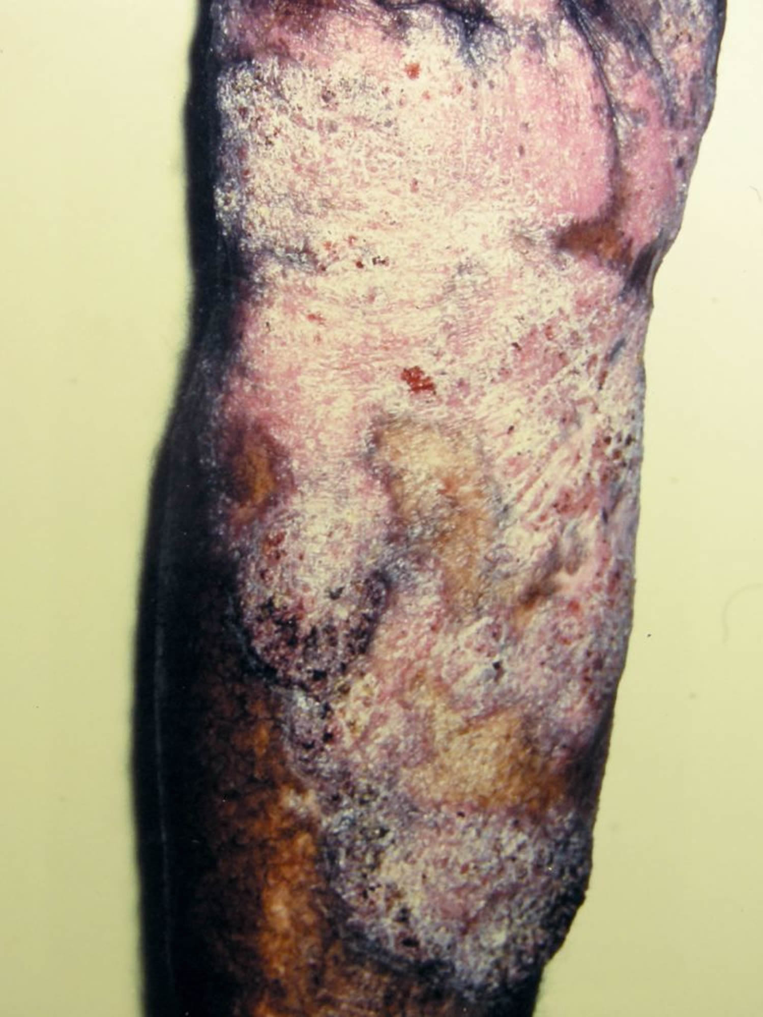
The fungi enter the skin following trauma, for example, a cut with a splinter when barefoot, evoking a granulomatous response. Usually, following trauma, a small warty papule appears. It enlarges very slowly to form a thick, hypertrophic, verrucous plaque. The lesion is, by and large, asymptomatic, though some patients may complain of occasional itching. Black dots (sclerotic bodies) will be seen on the surface of the plaque. These are fungal bodies eliminated transepidermally. They are usually solitary, but a few small lesions may also be seen in the periphery. These peripheral lesions are more common when there is itching. In some lesions, the plaque is flat and expands slowly with central scarring. The lesion is usually painless but can be painful in the presence of secondary infection. There can be lymphatic spread to adjacent areas 5.
Chromoblastomycosis histology
The epidermis shows pseudoepitheliomatous hyperplasia. In the dermis, a granuloma composed of epithelioid cells and Langhans giant cells are seen. The fungal elements can be seen as sclerotic bodies which are brown septate cells. The sclerotic bodies (medlar bodies/ muriform bodies/copper pennies) are extruded transepidermally, and they are seen as black dots on the surface of the lesion. This is characteristic of chromoblastomycosis 6.
Chromoblastomycosis histology is that of a foreign-body granuloma with isolated areas of microabscess formation. In the organized granuloma, mainly within giant cells, groups of fungal cells may be seen. They are chestnut or golden brown, and therefore can be easily distinguished in the infiltrate. The cells are characteristically divided into several planes of division by thick septa and are termed muriform or sclerotic cells. There is marked pseudoepitheliomatous hyperplasia of the epidermis, and in some areas, apparent transepidermal elimination of fungal cells, which can be found in the stratum corneum The tissue between the granulomatous nodules shows chronic fibrosis. When ulceration has occurred, there is usually a secondary bacterial infection 6.
Chromoblastomycosis symptoms
Chromoblastomycosis generally presents as a single lesion on an exposed site such as the foot or hand.
- It starts as a small firm red or grey bump.
- It grows very slowly: only about 2mm per year.
- Eventually, a warty dry nodule or plaque develops.
- There may be at least partial clearing with scarring in the centre of the lesion.
- The affected limb can enlarge generally (elephantiasis).
- New lesions may develop in time as satellites around the first one or the infection may be scratched into a new site.
- It may cause no discomfort but is frequently very itchy.
- Rarely, squamous cell carcinoma (SCC) develops within longstanding chromoblastomycosis.
The infection is sometimes confused with other skin conditions such as:
- Other fungal infections such as sporotrichosis
- Bacterial infections such as atypical mycobacterium infection, tuberculosis, leprosy and syphilis
- Protozoal infections such as leishmaniasis
- Squamous cell carcinoma
- Skin disorders such as psoriasis, discoid lupus erythematosus.
Chromoblastomycosis diagnosis
Microscopy and culture of scrapings or pus swabs suggest the diagnosis. Histopathology of chromoblastomycosis may show typical thick-walled dark-brown cells on skin biopsy confirming the presence of a dematiaceous fungus. It is dark coloured due to melanin in the walls of the organism.
Laboratory tests
Clusters of characteristic thick-walled brown ‘sclerotic’ (hard) cells are seen on microscopy.
Culture at 25-30 degrees celsius grows olive-green to black fungal colonies after one or two weeks. Naming the responsible fungus can be difficult. Phaeohyphomycosis is the name given to an infection caused by dematiaceous fungi.
Chromoblastomycosis treatment
Rarely, chromoblastomycosis resolves spontaneously leaving a scar.
Treatment is difficult and prolonged. It may include:
- Itraconazole, posaconazole or voriconazole, possibly in combination with terbinafine
- Flucytosine
- Thiabendazole
- Local heat
- Cryotherapy
- Surgery to remove the affected tissue completely.
Single small lesions can be excised followed by antifungal therapy. Surgical treatment, though highly effective, may not be feasible due to lesions being present over joints. If surgery is not feasible, oral antifungals alone can be given. The antifungal drugs of choice are, itraconazole 200-400 mg/day or terbinafine, 250-500 mg/day given for a period varying from 6 months to a year or more. Flucytosine alone or combined with amphotericin also may be effective. Oral supersaturated potassium iodide solution is another choice. Other approaches to treatment are cryotherapy or local application of heat 6.
Chromoblastomycosis prognosis
Chromoblastomycosis can be treated to the point of cure. Early intervention will yield better results with little morbidity. Long-standing cases which are over joints and with lymphatic involvement have relatively high morbidity. Disseminated disease with involvement of the central nervous system has the worst prognosis. Prolonged treatment with hepatotoxic drugs is another factor which has to be considered. Best results are seen in small lesions which are amenable to surgery and followed up with antifungal therapy.
References- Kurien G, Sugumar K, Chandran V. Chromoblastomycosis (Chromomycosis) [Updated 2019 Jun 11]. In: StatPearls [Internet]. Treasure Island (FL): StatPearls Publishing; 2019 Jan-. Available from: https://www.ncbi.nlm.nih.gov/books/NBK470253
- Verma S, Thakur BK, Raphael V, Thappa DM. Epidemiology of Subcutaneous Mycoses in Northeast India: A Retrospective Study. Indian J Dermatol. 2018 Nov-Dec;63(6):496-501
- Garzon LM, Rueda LJ, Celis AM, Cardenas M, Guevara-Suarez M. Exophiala psychrophila: A new agent of chromoblastomycosis. Med Mycol Case Rep. 2019 Mar;23:31-33.
- Queiroz-Telles F. CHROMOBLASTOMYCOSIS: A NEGLECTED TROPICAL DISEASE. Rev Inst Med Trop Sao Paulo. 2015;57 Suppl 19(Suppl 19):46–50. doi:10.1590/S0036-46652015000700009 https://www.ncbi.nlm.nih.gov/pmc/articles/PMC4711190
- Queiróz AJR, Pereira Domingos F, Antônio JR. Chromoblastomycosis: clinical experience and review of literature. Int. J. Dermatol. 2018 Nov;57(11):1351-1355.
- Brito AC, Bittencourt MJS. Chromoblastomycosis: an etiological, epidemiological, clinical, diagnostic, and treatment update. An Bras Dermatol. 2018 Jul-Aug;93(4):495-506.


