Livedoid vasculopathy
Livedoid vasculopathy is a rare, chronic blood vessel disorder characterized by persistent painful ulcers and scarring (atrophie blanche) on the feet and lower legs. Livedoid vasculopathy occurs chiefly but not exclusively on the lower leg or foot. These symptoms can persist for months to years and the ulcers often recur. Livedoid vasculopathy lesions appear as painful red or purple marks and spots that may progress to small, tender, irregular ulcers. Symptoms tend to worsen in the winter and summer months, and affect women more often then men. Livedoid vasculopathy may occur alone or in combination with another condition, such as lupus or thrombophilia 1.
Livedoid vasculopathy was also known as ‘livedo vasculitis’, ‘livedoid vasculitis’ and ‘livedo reticularis with summer ulceration’. It is now clear that it is not primarily a vasculitis (inflammation of the blood vessel wall), but to occlusion of small blood vessels, hence the change in name.
Livedoid vasculopathy occurs most commonly in middle-aged women but has been reported at all ages including childhood. There is often an increased incidence during the summer months and pregnancy.
Some patients with livedoid vasculopathy have an associated condition that predisposes them to occlusion of the small vessels of the lower leg. These include:
- Coagulation disorders such as antiphospholipid syndrome with lupus anticoagulant in 18% and anticardiolipin antibodies in 29% in one study
- Fibrinolytic disorders such as protein C deficiency (13%) and factor V mutation (Leiden) in 22% in one study
- Arteriosclerosis
- Homocysteinaemia (14%)
- High levels of lipoprotein-a
Livedoid vasculopathy causes
The exact cause of livedoid vasculopathy remains unclear, and various theories have been published referring to abnormalities within the blood vessel wall and in the circulating blood. It is likely that several different abnormalities may lead to clotting within small blood vessels of the lower legs. The thrombi result in necrosis of overlying skin, ulceration and very slow healing. There is no primary vasculitis.
Prothrombin G20210A gene mutation has been found in about 8% of patients.
Livedoid vasculopathy symptoms
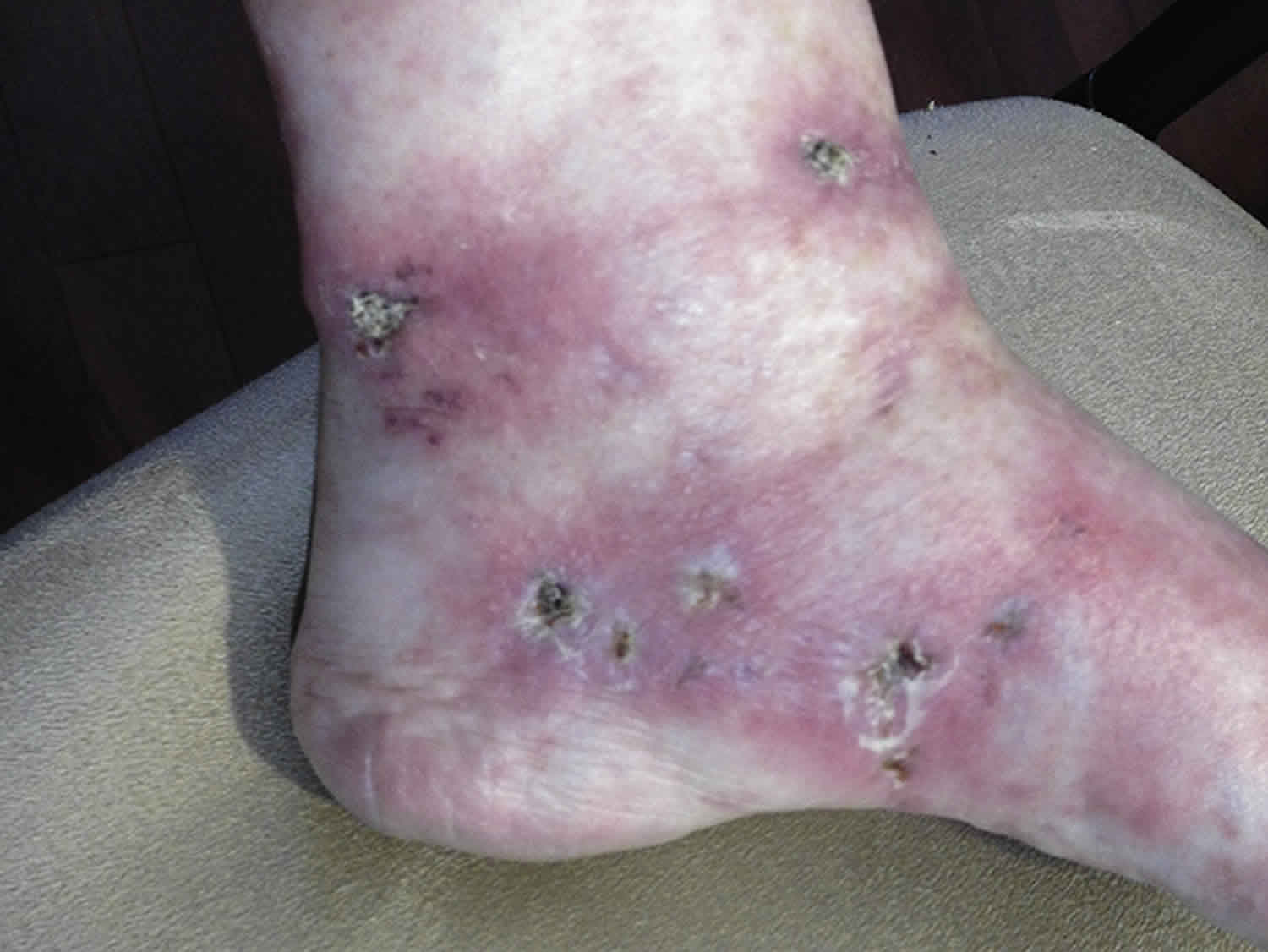
Livedoid vasculopathy affects lower legs, ankles, and upper surfaces of the feet. It is nearly always bilateral. Characteristics include:
- Mild to moderately painful red or purple marks and spots that progress to small, tender, irregular ulcers (30% of cases)
- Painless, irregular atrophie blanche (porcelain-white, stellate scars with red dots due to prominent capillaries)
- Livedo reticularis, Raynaud phenomenon and acrocyanosis may be present in some patients
Livedoid vasculopathy diagnosis
After taking a history, the patient should undergo a thorough general examination to identify any underlying associated condition.
Livedoid vasculopathy is a clinical diagnosis, supported by skin biopsy of a red papule or the edge of a new ulcer. Histopathology reveals hyalinisation, thickened blood vessel walls, fibrin deposition, vascular occlusion by thrombosis and minimal inflammation.
Direct immunofluorescence often shows deposition of immunoglobulin and complement components in the superficial, mid-dermal, and deep dermal vasculature (non-diagnostic).
Investigations are not diagnostic for livedoid vasculopathy. They should include:
- Complete blood count
- Coagulation studies
- Connective tissue antibodies
- Lupus coagulant and anticardiolipin antibodies
- Homocysteine levels
In many cases of livedoid vasculopathy, the results of these tests are normal.
Imaging studies for peripheral vascular disease may be carried out. Transcutaneous oximetry shows reduced oxygen flow in most patients.
Differential diagnosis includes ulceration and atrophie blanche arising in other diseases:
- Venous insufficiency with stasis dermatitis and leg ulceration
- Arterial insufficiency, arterial ulceration and diabetic foot ulcers
- Rheumatoid arthritis
- Systemic lupus erythematosus
- Systemic sclerosis
- Small vessel vasculitis
- Cutaneous polyarteritis nodosa
- Malignant atrophic papulosis
Livedoid vasculopathy treatment
Treatment of livedoid vasculopathy aims to reduce pain, ulceration and and the development of atrophie blanche. General treatment measures may involve protecting the skin from injury and irritants, removing dead tissue from the ulcers, treating infection with antibiotics, elevating legs, compression therapy, and avoiding smoking and hormonal contraceptives.[1][2] Treatments will also be given to address any co-occurring conditions such as lupus or thrombophilia. Drugs that aim to improve blood flow or prevent blood clotting may also be considered. Examples of these treatments, include:
- Protect the area from all knocks, abrasions, known allergens/irritants and ill-fitting shoes to avoid ulceration.
- Remove slough and dead tissue from the ulcers.
- Treat infection (cellulitis) with antibiotics.
- Leg elevation, compression therapy, bed rest, and occlusive wound dressings may aid healing.
- Stop smoking, if relevant, as smoking reduces peripheral blood flow.
Various drug therapies may be prescribed to enhance blood flow and prevent blood clotting:
- Pentoxifylline (alters blood viscosity and red cell flexibility)
- Antiplatelet agents (eg, aspirin, dipyridamole)
- Fibrinolytic agents (eg, danazol, tissue plasminogen activator). Low-dose danazol (200 mg/day orally)
- Anticoagulant agents (eg, subcutaneous heparin injections, oral warfarin)
- Vasodilating agents (eg, nifedipine, nicotinic acid)
- Anti-inflammatory agent (eg, prednisone, intravenous immunoglobulin)
- Phenformin and ethyloestrenol (dissolve blood clots)
- Folic acid, vitamin B-12, and vitamin B-6 (cofactors of homocysteine metabolism) for hyperhomocysteinemia
- Pentoxifylline
- Hyperbaric oxygen
- Pulsed intravenous immunoglobulin
- Iloprost
- Ketanserin
- Psoralen plus ultraviolet A (PUVA) therapy
- Niacin (nicotinic acid)
- Sulfapyridine
- Guanethidine
Currently there are no established guidelines for treatment. Decisions for treatment are made based on the clinicians clinical experience and specific patient characteristics. We strongly recommend that you discuss this information and your treatment options further with a trusted healthcare professional.
Livedoid vasculopathy prognosis
Livedoid vasculopathy is a chronic disorder, with spontaneous remissions and exacerbations. Reports of disease duration have ranged from 2.5 months to 21 years.
Once livedoid vasculopathy is in remission, over time the white patches of atrophie blanche become less defined and capillaries less prominent.
References


