Congenital nevus
Congenital nevus is a type of birthmark that is essentially colored skin markings that develop before or shortly after birth. Nevus is sometimes called hamartoma, which is disordered proliferations of cells within the tissue of origin, and are due to a developmental error.
Benign developmental skin lesions that develop later in life are called ‘acquired’ nevus.
Nevus may be derived from the outside layers of the skin (epithelial nevus) or from the deeper layers (dermal/subcutaneous nevus). Nevus are further classified based on the cell type involved. Melanocytic and vascular nevi are generally the most common types of birthmarks.
Some congenital nevi are given specific descriptive names. Some of these are listed here.
- Speckled lentiginous nevus
- Also called nevus spilus
- Dark spots on a flat tan background
- The number of spots may increase or decrease over time
- Satellite lesions
- Found on the periphery of central congenital melanocytic nevus or elsewhere on the body
- Smaller melanocytic naevi similar in appearance to
- Present in > 70% of patients with a large congenital melanocytic nevus
- Tardive nevus
- Melanocytic nevus that appears after birth
- Slower growth and less synthesis of melanin than congenital nevus 1
- Histopathology is similar to true congenital melanocytic naevi
- Garment nevus
- The name relates to the anatomical location of nevus
- Bathing trunk nevus involves central areas usually covered by a bathing costume, for example, buttocks
- Coat sleeve nevus involves an entire arm and proximal shoulder
- Halo nevus
- Affects some congenital and tardive melanocytic naevi
- Surrounding skin becomes lighter or white
- The central lesion may also become lighter and smaller and may disappear
- Due to immune destruction of melanocytes
Congenital melanocytic nevus
A congenital melanocytic nevus is a nests of benign melanocytes (cells that produce pigment) that are present at birth or develop shortly after birth 2. This form of a congenital nevus is also known as a brown birthmark. Congenital melanocytic nevi occur in approximately 1 in 100 live births.
Congenital melanocytic nevus are classified according to their predicted adult size 3:
- Small – reach less than 1.5cm
- Medium – reach between 1.5cm and 19.9cm
- Large (giant) – reach at least 20cm (40cm). A giant nevus is one which covers a large portion of an anatomical site, for example scalp, face, arm, leg or back. Another definition is if it covers more than 2% of a patients total body area or will measure over about twenty centimeters when the patient is fully grown. Many large or giant nevi are far more extensive than this.
- Satellite nevi: none, 1–20, > 20–50, and > 50 satellites.
Congenital melanocytic nevi should be described according to their body site, colors, surface features and whether or not there is hypertrichosis (hairs).
Similar melanocytic nevi or moles that were not present at birth, are often called ‘congenital melanocytic nevus-like’ nevi, ‘congenital type’ nevi or ‘tardive’ nevi.
Nevus may also form from other skin cells (eg, vascular nevi, which are formed from blood vessels). Some of these are also congenital (present at birth).
Congenital melanocytic nevus can exist on any part of the body, and usually grow in proportion to body growth with the child. As a rough guide, the likely adult size of a congenital nevus can be calculated as follows:
- Lower limbs: adult size is x 3.3 size at birth
- Upper limbs/torso: adult size is x 2.8 size at birth
- Head: adult size is x 1.7 size at birth.
Many patients also have multiple other nevi called satellite nevi on other parts of their body, and some will continue to develop these over their lifespan. Most nevi are brown or black in color, and have an increased tendency for hair growth. The skin texture can be smooth or leathery and because oil and sweat glands do not form properly within the nevus, overheating can be a problem. The skin is often dry and fragile. Many nevi have tumors just below the skin that give the nevus a lumpy or folded appearance.
Congenital melanocytic nevus treatment requires an individualised approach, based on the potential risk factors for complications, psychosocial and cosmetic considerations and the expectations of those affected with this condition and their families.
Infants with large lesions are usually managed by a multidisciplinary team and have regular follow-up by a dermatologist because of the increased risk of complications.
It is important that those with symptoms suggestive, or at high risk, of neurocutaneous melanocytosis have magnetic resonance imaging (MRI) to detect the disease. Those at high risk should have an MRI in the first 6 months of life.
Complete surgical excisions may reduce the risk of melanomas. However, this is associated with complications, and total removal may be impractical for very large lesions.
Non-surgical treatments such as dermabrasion may produce some cosmetic improvement. However, these treatments do not reduce the risk of melanoma as melanomas can occur deep in the skin. Options are best discussed with the dermatologist and plastic surgeon involved.
Figure 1. Congenital melanocytic nevus of the abdomen
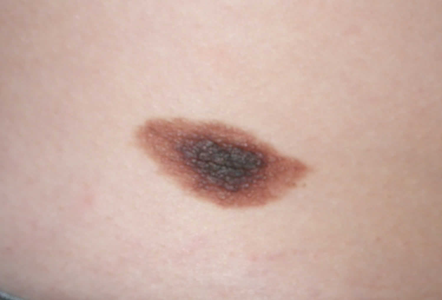
Figure 2. Congenital melanocytic nevus of the thigh
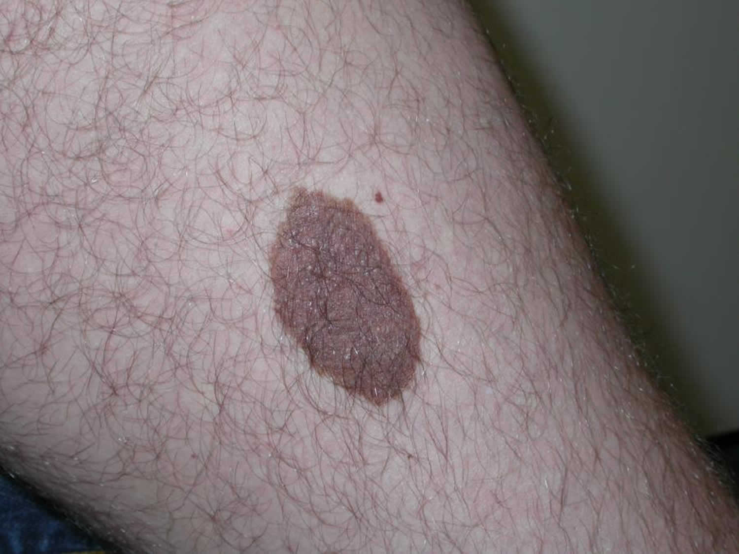
Figure 3. Congenital melanocytic nevus of the shoulder
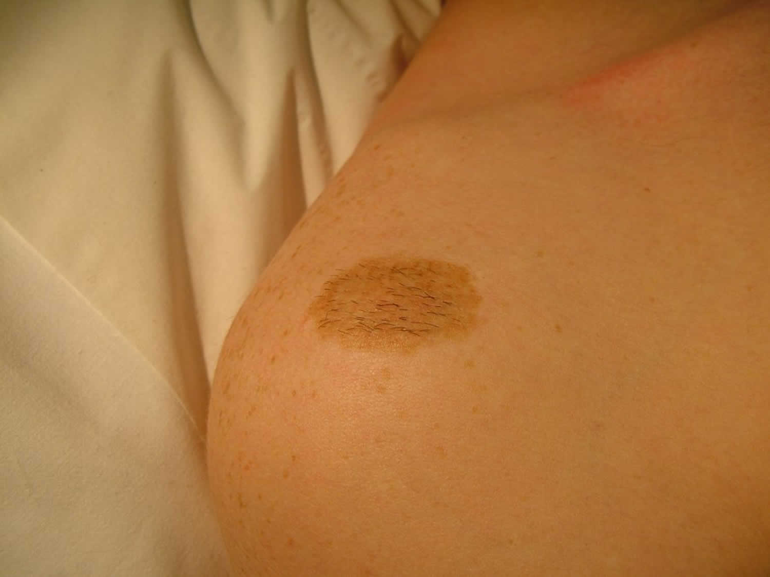
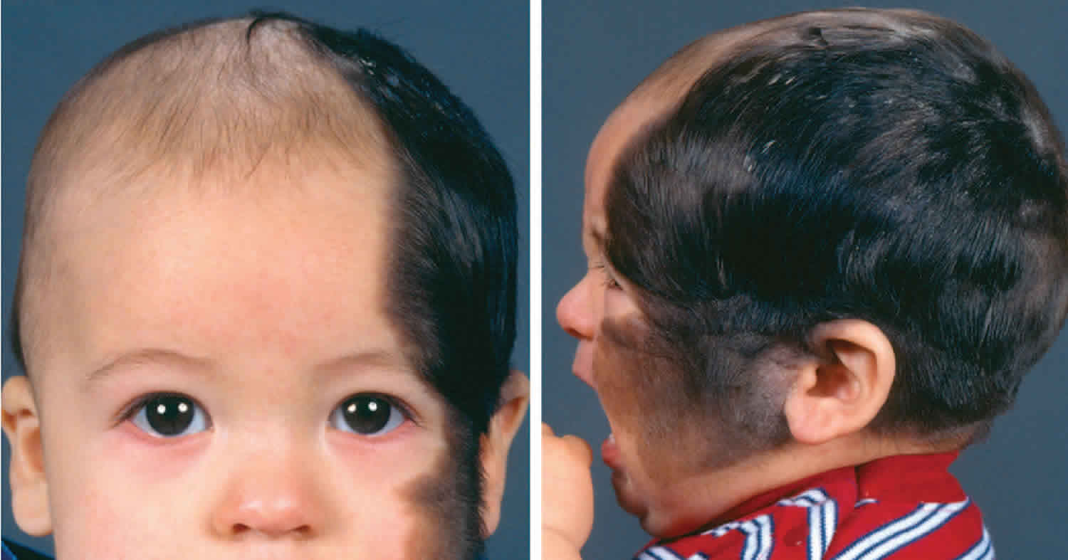
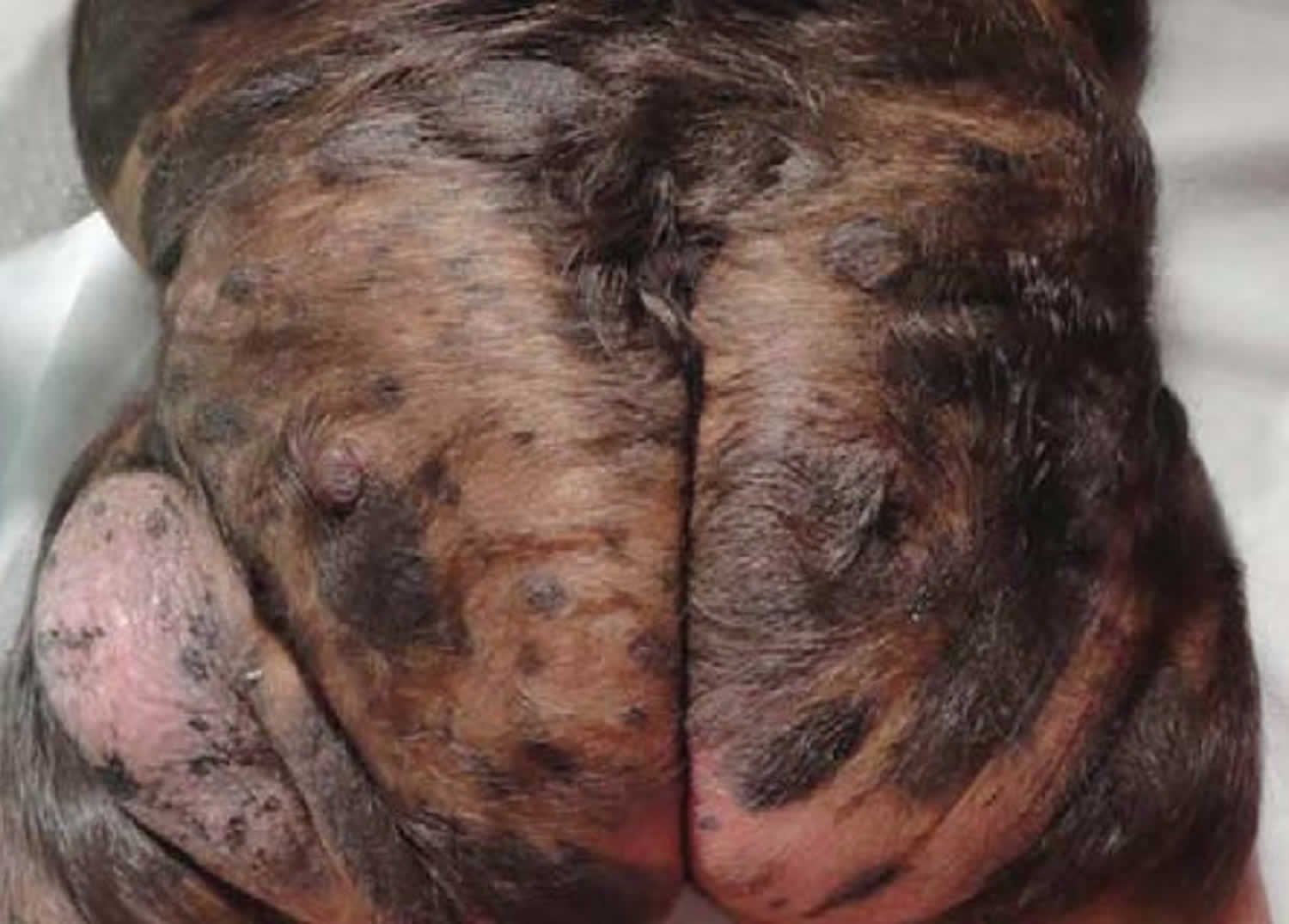
Figure 4. Giant congenital melanocytic nevus in “bathing trunk” with evident hypertrichosis, irregular surface and nodular areas
How common are congenital melanocytic nevi?
- Small congenital nevi occur in 1 in 100 births 1.
- Medium congenital nevi occur in 1 in 1000 births 1.
- Giant congenital melanocytic nevi are much rarer (1 in 20,000 live births) 1.
They occur in all races and ethnic groups, and males and females are at equal risk.
What is the likely outcome of a congenital melanocytic nevus?
Congenital melanocytic nevus usually grow proportionally as the child grows. Some may become lighter with time. However, they generally persist for life.
What other problems can occur with a congenital melanocytic nevus?
Due to their appearance, congenital melanocytic nevus can cause significant psychosocial consequences if they occur on prominent sites.
Giant congenital melanocytic nevus are associated with a risk of melanoma in the early years and over a lifetime. The risk increases with the size of the lesion and number of lesions. Large lesions (larger than 20 cm and especially larger than 40 cm predicted adult size) are associated with a 2-5% risk of melanoma over a lifetime. The risk of melanoma with small and medium congenital melanocytic nevus is not significant.
Large or giant lesions have been associated with other cancers.
Another complication of large or multiple lesions is neurocutaneous melanocytosis (melanocytes in the central nervous system). The risk is between 2.5% and 45% depending on factors such as size of the congenital melanocytic nevus, trunk location and number of satellite lesions. There may be no symptoms. However, a small percentage of children with large congenital melanocytic nevus and many satellites may experience neurological symptoms such as headaches and seizures.
Risk of developing melanoma within a congenital melanocytic nevus
The following characteristics of congenital melanocytic nevus are associated with the increased risk of development of melanoma (a skin cancer).
- Large or giant size
- Axial or paravertebral location (crossing the spine)
- Multiple congenital satellite nevi
- Neurocutaneous melanosis.
The risk of melanoma is mainly related to the size of the congenital melanocytic nevus. Small and medium-sized congenital melanocytic nevi have a very small risk, well under 1%. Melanoma is more likely to develop in giant congenital nevi (lifetime estimates are 5–10%), particularly in lesions that lie across the spine or where there are multiple satellite lesions. Melanoma can start deep inside the nevus or within any neuromelanosis found in the brain and spinal cord. Very rarely, other tissues that contain melanocytes may also be a source of melanoma such as the gastrointestinal tract mucosa. In 24% of cases, the origin of the melanoma cannot be identified 4..
Melanoma associated with a giant congenital melanocytic nevus or neuromelanosis can be very difficult to detect and treat.
The risk of development of melanoma is greater in early childhood; 70% of melanomas associated with giant congenital melanocytic nevi are diagnosed by the age of ten years 5.
Rarely, other types of tumour may develop within giant congenital melanocytic nevi including benign tumours (lipomas, schwannomas) and other malignant tumours (including sarcomas).
Melanoma can also develop within a small congenital melanocytic nevus. This is rare and likely to occur on the periphery of the nevus during adult life.
Prognosis of melanoma associated with congenital melanocytic nevus
Unfortunately, when a rare melanoma arises within a giant congenital melanocytic nevus, the prognosis is unfavourable. This is due to the deeper origin of the tumor rendering it more difficult to detect on clinical examination, resulting in a later stage at presentation. The deeper location also facilitates earlier spread through blood and lymph vessels. In 24% of cases, the melanoma has already spread to other sites (metastases) at the time of the first diagnosis.
Is regular follow-up recommended?
- It can be useful to have a close-up photograph of the congenital nevus with a ruler beside it to assess for changes in size.
- Digital surveillance using dermoscopic images (mole mapping) may also be helpful to detect changes in structure. However, such changes are normal in childhood and should not usually give rise to concern.
- It is advisable to continue neurodevelopmental observation in those at risk of neurocutaneous melanosis 5.
Congenital melanocytic nevus causes
Congenital melanocytic nevus usually occur sporadically. The condition is generally not inherited but arises from a mutation in the body’s cells that occurs after conception.
Congenital melanocytic nevi are caused by localized genetic abnormalities resulting in the proliferation of melanocytes; these are cells in the skin responsible for normal skin color. This abnormal proliferation is thought to occur between the 5th and 24th weeks of gestation. If proliferation starts early in development, giant and medium-sized congenital melanocytic nevi are formed 2. Smaller congenital melanocytic nevi are formed later in development after the melanoblasts (immature melanocytes) have migrated from the neural crest to the skin 2.
In some cases, there is also overgrowth of hair-forming cells and epidermis, forming an organoid nevus.
Very early onset of congenital nevus before the separation of the upper and lower eyelids results in kissing nevi, that is one part of the nevus is on the upper lid and the other part is on the lower eyelid.
NRAS gene mutations cause most cases of giant congenital melanocytic nevus. Rarely, mutations in the BRAF gene are responsible for this condition. The proteins produced from these genes instruct the cell to grow and divide (proliferate) or to mature and take on specialised functions (differentiate). The NRAS or BRAF gene mutations responsible for giant congenital melanocytic nevus are somatic, meaning that they are acquired after conception.
A somatic mutation in one copy of the NRAS or BRAF gene is sufficient to cause this disorder.
These mutations occur early in embryonic development during the growth and division (proliferation) of cells that develop into melanocytes. The overactive protein may contribute to the development of giant congenital melanocytic naevus by allowing cells that develop into melanocytes to grow and divide uncontrollably, starting before birth.
Molecular changes
Proto-oncogenes c-met and c-kit have important roles in the development of melanocytes. Hepatocyte growth factor, a cytokine (messenger protein) that regulates the proliferation and migration of melanocytes, may also be important in the development of congenital melanocytic nevi 2.
Neurocutaneous melanosis
Neurocutaneous melanocytosis is a rare syndrome defined by the proliferation of melanocytes in the central nervous system (brain and spinal cord) and the presence of a congenital melanocytic nevus 4. The majority of cases are associated with a giant congenital melanocytic nevus and satellite lesions.
It is estimated neurocutaneous melanosis affects 5–10% of people that have a giant congenital melanocytic nevus. However it is likely that the majority of cases remain asymptomatic, and the true incidence remains unknown 5. The melanocytes in the brain and spinal cord may often be detected by an MRI scan but the use of these scans is controversial because the condition is not easily treatable.
Neurocutaneous melanocytosis may present with symptoms of raised intracranial pressure, such as 4:
- A headache
- Vomiting
- Irritability
- Focal cranial nerve signs
- Seizures
- Hydrocephalus
- Delayed development.
Congenital melanocytic nevus signs and symptoms
Congenital melanocytic nevi are usually asymptomatic, however, some may be itchy, particularly larger lesions. It is thought there may be a reduced function of sebaceous (oil) and eccrine (sweat) glands, which may result in skin dryness and a heightened sensation of itch.
The overlying skin may become fragile and erode or ulcerate. Deep nests of melanocytes in the dermis may weaken the bonds between the epidermis and the dermis and account for skin fragility 1.
Congenital melanocytic nevi are often unsightly, especially when extensive, ie large or giant congenital melanocytic nevi. They may, therefore, result in anxiety and impaired self-image, especially when the lesions are in visible areas.
Giant melanocytic nevi, and to a lesser degree small lesions, are associated with increased risk of developing cutaneous melanoma, neurocutaneous melanoma and rarely other tumors.
What do congenital melanocytic nevi look like?
Congenital melanocytic nevi present as single or multi-shaded, round or oval-shaped pigmented patches 4. They may have increased hair growth (hypertrichosis). The surface may be slightly rough or bumpy.
Congenital nevi usually enlarge as the child grows but they may sometimes become smaller and less obvious with time. Rarely some may even disappear. However, they may also become darker, raised, more bumpy and hairy, particularly around the time of puberty.
Congenital melanocytic nevus diagnosis
The diagnosis of a congenital melanocytic nevus is usually based on the clinical appearance. If there is any doubt, examining the lesion with dermoscopy or taking a sample of the lesion for histology (biopsy) may show characteristic microscopic features.
Dermoscopy
Evaluation of the congenital melanocytic nevus by dermoscopy will reveal the pattern of pigmentation and its symmetry or lack of symmetry. The most common global pattern of congenital or tardive melanocytic nevus is globular, but reticular, structureless and mixed patterns may occur. The nevus may have differing structures across the lesion, sometimes leading to overall asymmetry of the structure.
Pathology
Congenital melanocytic nevi are usually larger than acquired nevi (which are melanocytic nevi that appear after 2 years of age), and the nevus cells often extend deeper into the dermis, fat layer, and deeper structures. The nevus cells characteristically cluster around blood vessels, hair follicles, sebaceous and eccrine glands, and other skin structures. Congenital nevus cells tend to involve collagen bundles in the deeper layers of the skin more than is the case in an acquired nevus 2.
Congenital melanocytic nevus treatment
Management of a congenital melanocytic nevus must take into account the age of the subject, the lesion size, the location and depth, and the risk of developing malignant change within the lesion.
Giant congenital melanocytic nevus
The only definite indication for surgery in a giant congenital melanocytic nevus is when melanoma develops within it 4..
Small congenital nevus
If a small congenital nevus is growing at the same rate as the child and is not changing in any other way, the usual practice is not to remove it until the child is old enough to co-operate with a local anesthetic injection, usually around the age of 10 to 12 years. Even then, removal is not essential.
Reasons to consider surgical removal may include:
- Unsightly appearance
- Difficulty in observing the mole (eg, scalp, back)
- A recent change in the lesion (darkening, lumpiness, increasing size)
- Melanoma-like appearance (irregular shape, variegated color).
Prophylactic surgical removal of a nevus
The following factors should be considered prior to prophylactic surgical removal of a nevus.
- Prophylactic excision of a small lesion may be delayed until an age when the patient is old enough to make an informed choice 1.
- Small or medium-sized congenital melanocytic nevi are at low risk for developing malignant change.
- Irregular, lumpy or thick lesions or lesions that are difficult to clinically assess may have a lower threshold for consideration of surgical excision, so as not to miss a melanoma.
- 50% of melanomas diagnosed in those with giant congenital melanocytic nevi occur at another body site such as within the central nervous system [2]. Therefore surgical excision of the lesion may not eliminate the risk of melanoma.
- Large or giant melanocytic lesions may be too large to excise completely.
- Large lesions may require a skin flap or graft to close the surgical defect.
Complications of surgery
Complications that may occur after surgery include:
- Graft or flap failure
- Infection
- Wound breakdown
- Bleeding or hematoma
- Hypertrophic or keloid scar
- Irritable or itchy scar.
Other treatment options for a congenital melanocytic nevus
Dermabrasion
Dermabrasion can allow partial removal of a large congenital nevus; deeper nevus cells may persist. Dermabrasion may lighten the colour of the nevus but may not reduce hair growth within it. It can cause scarring.
Tangential (shave) excision
Tangential or shave excision uses a blade to remove the top layers of the skin (epidermis and upper dermis). This may reduce the pigmentation but the lesion may not be completely removed. Shave excision may result in significant scarring.
Chemical peels
Chemical peels using trichloroacetic acid or phenol may lighten the pigmentation of a superficial (surface) congenital nevus that is located in the upper layers of the skin.
Laser ablation
Laser treatment is considered if surgical intervention is not possible. They may result in lightening of the lesion. Suitable devices include:
- Ruby Q-switched laser
- Carbon dioxide resurfacing laser
Techniques that result in partial removal of a congenital nevus can make the lesion more difficult to assess during long-term surveillance 4.
References- Kovalyshyn I, Braun R, Marghoob A. Congenital melanocytic nevi. Australas J Dermatol. 2009;50(4):231‐242. doi:10.1111/j.1440-0960.2009.00553_1.x
- Viana AC, Gontijo B, Bittencourt FV. Giant congenital melanocytic nevus [published correction appears in An Bras Dermatol. 2014 Jan-Feb;89(1):190]. An Bras Dermatol. 2013;88(6):863‐878. doi:10.1590/abd1806-4841.20132233 https://www.ncbi.nlm.nih.gov/pmc/articles/PMC3900335
- Krengel S, Scope A, Dusza SW, Vonthein R, Marghoob AA. New recommendations for the categorization of cutaneous features of congenital melanocytic nevi. J Am Acad Dermatol. 2013;68(3):441‐451. doi:10.1016/j.jaad.2012.05.043
- Eds. Bolognia J, Jorizzo J, Schaffer J. Dermatology. 3rd ed. Elsevier Saunders; 2012. P 1871-6
- Kovalyshyn I, Braun R, Marghoob A. Congenital melanocytic naevi. Australas J Dermatol. 2009;50(4):231‐242. doi:10.1111/j.1440-0960.2009.00553_1.x


