Cor triatriatum
Cor triatriatum also known as cor triatriatrum sinister, is when the left atrium is divided into two chambers by a fibromuscular membrane with an opening: one chamber with the pulmonary veins and the other with the mitral valve and atrial appendage (see Figure 1). This division of the left atrium results in three total atria (2 left, 1 right). The pulmonary veins drain abnormally into this second left atrial chamber, creating varying degrees of obstruction to pulmonary venous return. As a result, pulmonary artery and pulmonary venous hypertension (abnormally high pressures) occur, similar to patients with mitral stenosis. Cor triatriatum is one of the rarest forms of congenital (present at birth) heart defect with an estimated incidence of 0.1% of all the congenital heart diseases and is often associated with other congenital cardiac anomalies 1. In the pediatric population, Cor triatriatum may be associated with major congenital cardiac lesions such as tetralogy of Fallot, double outlet right ventricle, coarctation of the aorta, partial anomalous pulmonary venous connection, persistent left superior vena cava with unroofed coronary sinus, ventricular septal defect, atrioventricular septal (endocardial cushion) defect, and common atrioventricular canal 2. Rarely, asplenia or polysplenia has been reported in these patients. Although frequently an isolated finding, cor triatriatum in the adult 3 has been reported in association with ostium secundum atrial septal defect, dilated coronary sinus due to persistent left superior vena cava, and bicuspid aortic valve 3.
Another rarer form of cor triatriatum is cor triatriatum dextrum. Cor triatriatum dextrum is extremely rare and results from the complete persistence of the right sinus valve of the embryonic heart 4. The right valve of the sinus venosus of the embryonic heart divides the right atrium into a proximal (upper) and a distal (lower) chamber. The upper chamber receives the venous blood from both vena cavae and the lower chamber is in contact with the tricuspid valve and the right atrial appendage. This form presents similarly to Ebstein’s anomaly and is difficult to differentiate.
Most patients are identified shortly after birth with the evaluation of a distressed or cyanotic neonate. However, when presentation is delayed, primary symptoms
may mimic reactive airway disease 5.
In the normal heart, there are two atria, right and left (Figure 2). The right atrium pumps blood through the tricuspid valve to the right ventricle. The blood then leaves the right ventricle through the pulmonary artery to the lungs for oxygenation. Blood returns to the left atrium by way of the pulmonary veins. The oxygenated blood then travels to the left ventricle through the mitral valve and out to the body through the aorta.
Figure 1. Cor triatriatum
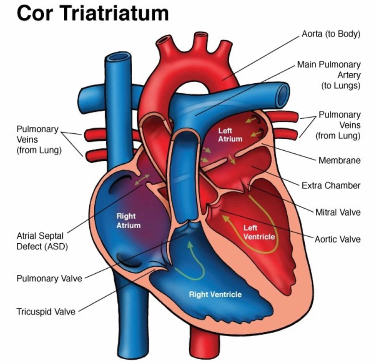 Figure 2. Normal heart
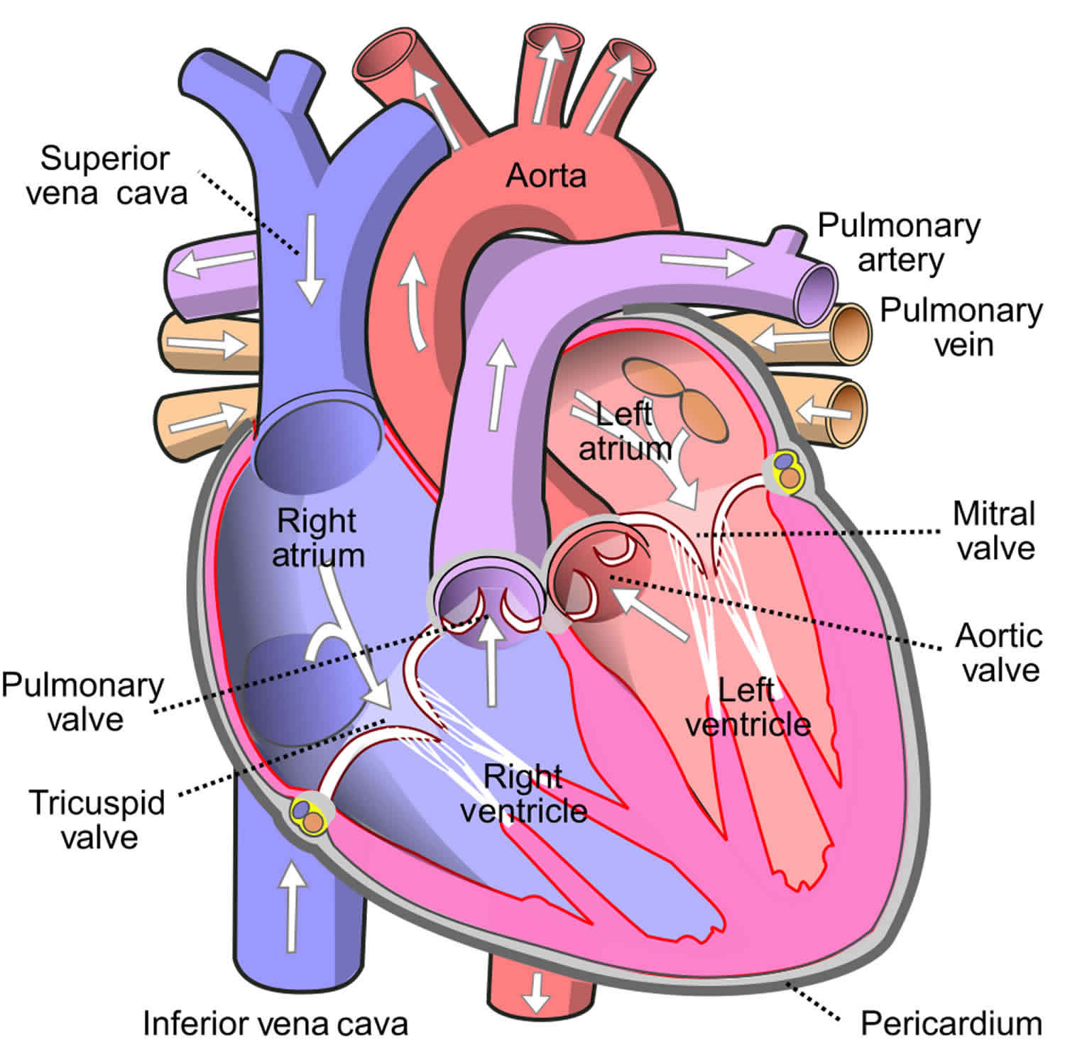
Cor triatriatum causes
Cor triatriatum results from the incomplete absorption of the common pulmonary vein, which is normally reabsorbed during the development of a fetus and becomes a part of the left atrium 6. The incomplete absorption results in the formation of an appendage which subdivides the left atrium into 2 chambers. No known genetic mutations or risk factors are found to be associated with the development of this condition.
A malincorporation theory 7 presented by Dr. Clifford G. Parsons explains how cor triatriatum sinister occurs. During normal fetal development, the pulmonary vein incorporates into the left atrium. If it fails to do so, the common pulmonary venous ostium remains narrow, resulting in a septum-like structure called the atrial appendage. The appendage then further divides the left atrium into 2 compartments. Although widely accepted, this theory fails to explain how fossa ovalis and atrial muscle fibers are also present in the proximal atrial chamber 8.
Two other theories, malseptation theory and entrapment theory, also explain the pathophysiology of cor triatriatum. The malseptation theory states that the fibro-muscular membrane is an abnormal growth of the septum primum; whereas, entrapment theory emphasizes the entrapment of the common pulmonary vein in the embryonic sinus venosus, thereby preventing its incorporation into the left atrium. The malincorporation theory remains the most widely accepted theory.
Cor triatriatum symptoms
Classically, cor triatriatum sinister presents in infancy with signs and symptom of pulmonary hypertension and pulmonary venous obstruction 6. Due to low cardiac output, children can show poor growth and weight gain, feeding difficulties, respiratory distress, and tet spells. In childhood and adulthood, the signs and symptoms of pulmonary venous hypertension and right heart failure dominate as the membrane calcifies, and the opening becomes smaller and smaller decreasing cardiac output even further. Mitral valve regurgitation and atrial fibrillation impose serious dangers. Most patients present with the following features 9:
- Dyspnea (difficulty breathing) and orthopnea (difficulty breathing when lying down)
- Easy fatigability
- Hemoptysis (coughing up blood)
- Exercise intolerance and shortness of breath
- Palpitations
Atrial fibrillation can cause systemic thromboembolism and present as pulmonary embolism or stroke. The left atrial enlarges due to backing up of blood and can present as life-threatening arrhythmias.
Cor triatriatum sinister that presents for the first time in adulthood is rare but possible. It presents similarly to mitral stenosis, but the absence of loud S1 and an opening snap helps to distinguish between the 2. Chest x-ray showing pulmonary congestion with an absence of left atrial enlargement is characteristic of cor triatriatum. A continuous gradient on Doppler echocardiography confirms the diagnosis 8.
The physical examination findings are due to the right heart failure and pulmonary congestion.
- Accentuated pulmonary component of second heart sound
- A soft mid-systolic murmur at the upper left sternal border with a wide and fixed splitting of an atrial septal defect.
- Right ventricular heave
- Rales at the lung base
- Diminished peripheral pulses
- Hepatomegaly and right upper quadrant tenderness
- Ascites
- Peripheral edema
- Distended peripheral veins
- Distended jugular venous and elevated jugular venous pressure
- Pallor
- Poor weight gain
Cor triatriatum diagnosis
The mainstay of evaluation and diagnosis includes imaging studies such as chest x-ray, ECG and echocardiography, angiography and left and right heart catheterization 10.
Chest X-Ray
A chest x-ray is the initial investigation of choice. The findings include:
- Pulmonary congestion with haziness (Kerley B-lines)
- Ground glass appearance of acute pulmonary edema
- Prominent pulmonary vessels
- Pleural effusion
- Left atrial enlargement
- Cardiomegaly
Electrocardiography (ECG)
ECG findings are non-specific in cor triatriatum. Can range from atrial fibrillation and no specific P-wave changes to right axis deviation due to pulmonary congestion and right ventricular hypertrophy.
Echocardiography
Echocardiography is the diagnostic modality of choice as it not only allows for definitive diagnosis, but the 3-dimensional reconstruction of echocardiographic images pinpoints the exact location of the defect and appendage, helps direct the surgical approach to the disease. The left atrial appendage and its fenestrations can be easily visualized along with the presence of an atrial septal defect, pulmonary stenosis, mitral valve stenosis, or regurgitation. Pulmonary arterial and venous drainage patterns can also be seen on echocardiography. Echocardiography differentiates between cor triatriatum and supravalvular mitral stenosis by clear visualization of the left atrial appendage in the left atrium.
Transesophageal echocardiography outlines the precise nature and anatomy of the defect; especially in older patients, but if unavailable, transthoracic echocardiography can be performed 11.
Angiography
Angiography helps determine the severity of obstruction and the time of surgical intervention needed.
Cor triatriatum treatment
Medical management
Asymptomatic patients
- Asymptomatic patients need no specific treatment. Observe the patients for the development of signs and symptoms. Your child will require life-long cardiology follow-up.
Symptomatic patients
Treatment option for symptomatic patients includes both medical/conservative management and surgical repair. Medical treatment includes:
- Hemodynamic stabilization of fluid overload and pulmonary edema
- Rate and rhythm control and anticoagulation for patients with atrial fibrillation
- Thromboembolic prophylaxis with anticoagulation against deep vein thrombosis, pulmonary embolism and stroke
- Obtain surgical consultation
- Diuretics are used in the short term to manage pulmonary congestion/edema and decrease work of breathing.
- If arrhythmias/atrial fibrillation occur, anticoagulation with heparin or coumadin may be needed.
Surgical management
Surgery is the definitive treatment. Complete surgical resection of atrial appendage/accessory membrane through a midline sternotomy under cardiopulmonary bypass and closure of atrial septum with a patch of the child’s own pericardium (sac that surrounds the heart) provides the optimum cure. The 10-year survival rate following surgery is 83%, while patients with coexisting congenital heart diseases have a greater risk of adverse outcomes and a lower survival rate 12.
Cor triatriatum prognosis
- Pulmonary hypertension usually improves rapidly if surgical correction occurs early.
- High survival rate post-repair.
- Atrial arrhythmias may occur postoperatively or later in life. Medications or a cardiac catheterization procedure called an ablation may be needed to treat/control arrhythmias.
- Growth and development post-repair are usually normal in the absence of other congenital heart disease or comorbidities.
A study by Saxena et al 13 indicated that surgery for cor triatriatum results in satisfactory rates of early and long-term survival. The study involved 25 patients in whom the cor triatriatum membrane was excised using cardiopulmonary bypass, with 20 of these patients also undergoing concomitant surgical procedures. The 10-year survival rate, using the Kaplan-Meier estimate, was 83%; at a mean follow-up of 12.8 years, all patients had a New York Heart Association classification of I or II. The investigators found that patients in whom cor triatriatum coexists with complex congenital anomalies may be at greater risk for adverse outcomes.
References- Hamdan R, Mirochnik N, Celermajer D, et al. Cor triatriatum sinister diagnosed in adult life with three-dimensional transesophageal echocardiography. BMC Cardiovasc Disord. 2010;10:54.
- Tuccillo B, Stumper O, Hess J, et al. Transoesophageal echocardiographic evaluation of atrial morphology in children with congenital heart disease. Eur Heart J. 1992 Feb. 13(2):223-31.
- Hamdan R, Mirochnik N, Celermajer D, Nassar P, Iserin L. Cor Triatriatum Sinister diagnosed in adult life with three dimensional transesophageal echocardiography. BMC Cardiovasc Disord. 2010 Oct 28. 10:54.
- Cor triatriatum. https://emedicine.medscape.com/article/154168-overview
- Pisanti A, Vitiello R. Wheezing as the sole clinical manifestation of cor triatriatum. Pediatr Pulmonol. 2000;30(4):346-9.
- Ather B, Siddiqui WJ. Cor Triatriatum. [Updated 2018 Nov 18]. In: StatPearls [Internet]. Treasure Island (FL): StatPearls Publishing; 2019 Jan-. Available from: https://www.ncbi.nlm.nih.gov/books/NBK534243
- ARSONS CG. Cor triatriatum; concerning the nature of an anomalous septum in the left auricle. Br Heart J. 1950 Oct;12(4):327-38.
- Narayanapillai J. Cor triatriatum sinister with severe obstruction: a rare presentation in an adult. BMJ Case Rep. 2016 Aug 05;2016
- Humpl T, Reineker K, Manlhiot C, Dipchand AI, Coles JG, McCrindle BW. Cor triatriatum sinistrum in childhood. A single institution’s experience. Can J Cardiol. 2010 Aug-Sep;26(7):371-6.
- Thakrar A, Shapiro MD, Jassal DS, Neilan TG, King ME, Abbara S. Cor triatriatum: the utility of cardiovascular imaging. Can J Cardiol. 2007 Feb;23(2):143-5.
- Hamdan R, Mirochnik N, Celermajer D, Nassar P, Iserin L. Cor Triatriatum Sinister diagnosed in adult life with three dimensional transesophageal echocardiography. BMC Cardiovasc Disord. 2010 Oct 28;10:54.
- Fuchs MM, Connolly HM, Said SM, Egbe AC. Outcomes in patients with cor triatriatum sinister. Congenit Heart Dis. 2018 Jul;13(4):628-632.
- Saxena P, Burkhart HM, Schaff HV, Daly R, Joyce LD, Dearani JA. Surgical repair of cor triatriatum sinister: the Mayo Clinic 50-year experience. Ann Thorac Surg. 2014 May. 97(5):1659-63.


