Desquamative gingivitis
Desquamative gingivitis is a descriptive term, first introduced by Prinz in 1932 1 that is synonymous with the presence of erythema, desquamation, erosion, and blistering of attached and marginal gingiva, signs that represent different mucocutaneous disorders 2. Desquamative gingivitis is characterized by the erythematous gingiva, desquamation and erosion of the gingival epithelium, and blister formation 3. The term “desquamation” is derived from the Latin word ‘Desquamare’, which means scraping fish flakes. As a word, desquamation means ‘loss of epithelial elements in small and large amounts, peeling of skin, and exfoliation’. Chronic desquamation of the gingiva is referred to as desquamative gingivitis 4. Chronic desquamative gingivitis was described for the first time by Tomes and Tomes in 1894 5. In 1932, Prinz 6 used the term “chronic diffuse desquamate gingivitis” for chronic diffuse inflammation cases, which were characterized by severe epithelial desquamation in the marginal gingiva. In actual use, the term “desquamative gingivitis” is used for a specific clinical symptom and it is not a diagnosis alone 3.
Desquamative gingivitis is a clinical finding, which progresses with vesicular formation, atrophy, erosion and desquamation, characterized with diffuse erythema of the marginal and keratinized gingiva 7. Lesions start with diffuse erythema and minimal desquamation. The affected gingiva epithelium is very fragile and tends to exfoliate easily, even with the slightest trauma 5. Large ulceration areas can be observed in some cases 8. The desquamative gingivitis is seen after puberty, especially in individuals over 30 years of age 5. Desquamative gingivitis appears more frequently in old women and menopause than in men. Desquamative gingivitis has been reported and can rarely be observed in children 7.
Desquamative gingivitis can be the clinical symptom of some dermatitis and mucocutaneous diseases. Several mucocutaneous diseases in which clinical desquamative gingivitis is observed have been reported in the literature (see differential diagnosis) 3. Desquamative gingivitis correspond more often to oral lichen planus, mucous membrane pemphigoid and pemphigus vulgaris, and in other cases with other immunological pathologies such as lupus erythematosus, erythema multiforme, graft versus host disease, epidermolysis bullosa acquisita, plasma cell gingivitis and collagen diseases 9. Contact allergic reactions to various oral hygiene products and chemical agents have also been reported to represent as desquamative gingivitis. The clinical condition generally exacerbates with plaque accumulation, trauma or improper brushing. The clinical picture worsens with the disruption of oral hygiene practices due to pain and bleeding 10. Desquamative gingivitis continues chronically with periods of remission and exacerbation. Recovery of the gingiva may take months 8. Although the intraoral presence of desquamative gingival lesions differ, various durations from 2 months to 25 years have been reported 11.
Desquamative gingivitis diagnosis involves taking detailed medical history, performing a careful intraoral examination and determining the presence or absence of similar lesions at other sites of the body are the most important steps in clinical practice. Definitive diagnosis of desquamative gingivitis should be made by incisional biopsy, histopathological examination and direct immune fluorescent (DIF). The management of desquamative gingivitis has been a major problem, largely because the cause of the disease has been elusive. Gingival lesions are controlled by improving oral hygiene and the use of topical corticosteroids. If there is an underlying systemic disease, the case should be consulted with the physician.
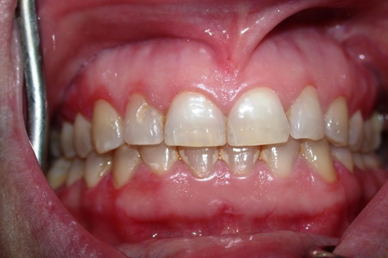
Figure 1. Desquamative gingivitis (mucosal and gingival desquamation developing as a result of an allergic reaction against toothpaste)
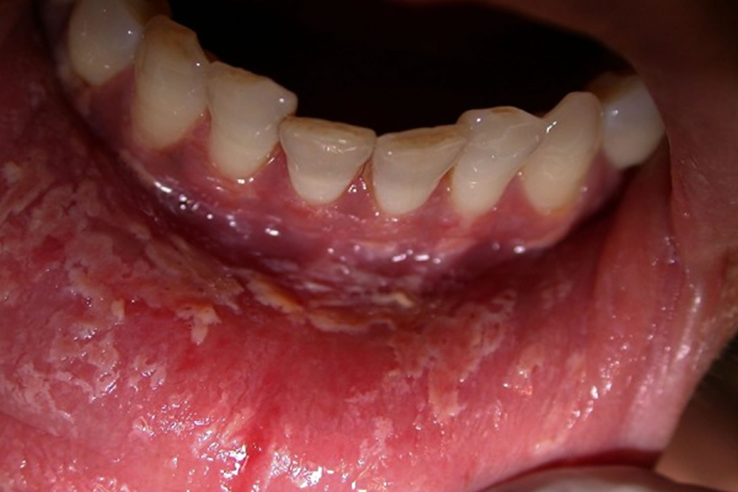
Figure 2. Lichen planus desquamative gingivitis
Footnote: Atrophic form of Lichen Planus creates a typical desquamative gingivitis appearance at the gingiva.
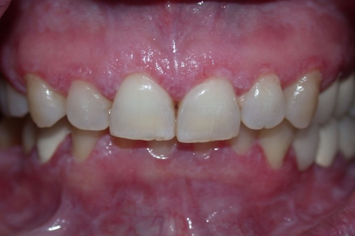
[Source 3 ]Figure 3. Mucous membrane pemphigoid
Footnote: The intraoral appearance of mucous membrane pemphigoid is generally similar to desquamative gingivitis.
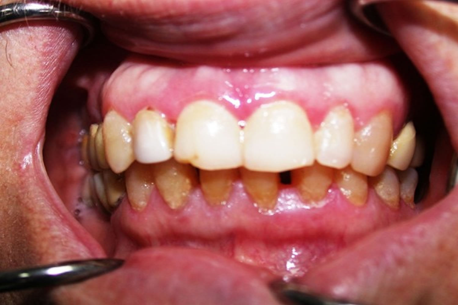
[Source 3 ]Figure 4. Chronic desquamative gingivitis

Footnote: Since the initial symptoms of pemphigus can begin as desquamative gingivitis, it is important to evaluate this clinical symptom to reach an early diagnosis.
[Source 3 ]Desquamative gingivitis causes
Glickman and Smulow 12 stated that desquamative gingivitis may be a clinical feature common to a wide number of disorders. A classification was proposed based on etiologic considerations, together with histologic and immunologic findings 13.
- Dermatological diseases
- Cicatricial pemphigoid
- Lichen planus
- Pemphigus
- Psoriasis
- Bullous pemphigoid
- Epidermolysis bullosa acquisita
- Contact stomatitis.
- Endocrine disturbances
- Estrogen deficiencies following oophorectomy and in postmenopausal stages
- Testosterone imbalance
- Hypothyroidism.
- Aging
- Abnormal response to bacterial plaque
- Idiopathic
- Chronic infections
- Tuberculosis
- Chronic candidiasis
- Histoplasmosis.
Overall, mucous membrane pemphigoid, oral lichen planus and pemphigus vulgaris have emerged as the most common causes of desquamative gingivitis, with the first two accounting for about 80% of cases 14.
Mucous membrane pemphigoid is a heterogeneous group of autoimmune, chronic inflammatory, subepithelial blistering disease of mucous membranes, oral, ocular, genital, nasopharyngeal, esophageal, and laryngeal mucosa are frequently affected, with rare skin involvement. Mucous membrane pemphigoid belongs to a group of mucocutaneous autoimmune blistering disorders often collectively referred to as subepithelial bullous dermatoses. Previously, mucous membrane pemphigoid was known as “benign mucous membrane pemphigoid,” “cicatricial pemphigoid” and “ocular or oral–gingival pemphigoid.” However, now the term “mucous membrane pemphigoid” is preferred 15.
Desquamative gingivitis, one of the clinical manifestation in mucous membrane pemphigoid is commonly seen among post-menopausal stages in females. Laskaris et al. 16 reported gingival involvement in 63.6% of 55 patients with mucous membrane pemphigoid. Silverman et al. 17 in a survey of 65 patients found gingival involvement in 100% of males and 92% of females. Gallagher and Shklar 18 compiled the data of 120 patients and observed gingival involvement in almost all cases. However, no direct relationship between mucous membrane pemphigoid and smoking or menopausal status has been cited in the literature 19. Autoantibodies against basement membrane proteins, together with complement (C3) and neutrophils cause a subepithelial split and resultant vesicle formation. mucous membrane pemphigoid antigens are usually present in lamina lucida of basement membrane, but lamina densa may also be the primary site of involvement in some cases. The majority of cases of mucous membrane pemphigoid demonstrate IgG directed against antigens on the epidermal side of the salt-split skin, which have been identified as BP180 (also called type XVII collagen). However, few cases of mucous membrane pemphigoid may have antigens on the dermal side of the split (epiligrin/laminin 5) 20.
The exact etiology of mucous membrane pemphigoid is not known. Known causative factors include-severe mucosal inflammatory injury 21, drugs (clonidine, indomethacin, D-penicillamine) 22, viruses, ultraviolet light and genetic predisposition such as HLA DQB1 * 0301 23.
Desquamative gingivitis differential diagnosis
The differential diagnosis of desquamative gingivitis includes a wide spectrum, such as chemical and electrical burns, allergic reactions, hormonal disorders and mucocutaneous diseases. Furthermore, a similar clinical pattern can be observed in reactions developing against mouthwashes (see Figure 1), chewing gums, cosmetic products, drugs, cinnamon and dental materials 10. It is suggested that desquamative gingivitis may be observed when there is lack of estrogen or progesterone 24. Additionally, there are idiopathic gingival desquamative lesions without any etiologic factors 5. There are conflicting arguments on whether desquamative gingivitis is a symptom of oral lichen planus (Figure 2), mucous membrane pemphigoid (Figure 3), or a clinical manifestation of these diseases 25. In many articles, it has been reported that desquamative gingivitis is related to lichen planus, mucous membrane pemphigoid and pemphigus vulgaris (88% – 98%) (Figure 4) 26, 27, 11, 10.
Disease in which desquamative gingivitis is clinically observed 3:
- Bullous pemphigoid
- Paraneoplastic pemphigus
- Dermatitis herpetiformis
- Chronic ulserative stomatit
- Lineer Ig A disease
- Psoriazis
- Pyostomatitis vegetans
- Erythema multiforme
- Discoid lupus erythematosus
- Dyskeratosis congenita
- Epidermolysis bullosa
- Graft-versus-Host disease
- Plasma cell gingivitis
- Foreign body gingivitis
- Kindler syndrome
- Ulserative colitis
- Hepatit C positive
- Acute myeloid leukemia (AML)
- Dermatomitosis, mixed connective tissue disorders
- Crohn disease
- Sarcoidosis
- Drugs or chemicals implicated include various oral oral health care products
- Sodium lauryl sulphate
- Magnesium monoperoxyphthalate.
Desquamative gingivitis signs and symptoms
Desquamative gingivitis is characterized by the erythematous gingiva, desquamation and erosion of the gingival epithelium, and blister formation 3. Clinically, desquamative gingivitis presents with moderate pain, partly due to the deposit of plaque in gingival margin 28, being in some cases the first manifestation of the disease 29. The study of periodontal status in patients with desquamative gingivitis suggests that in patients with mucous membrane pemphigoid the gingivo-periodontal status is worse than control health 30, in the same case, patients with oral lichen planus and pemphigus vulgaris present deeper pockets and higher loss of the clinical attachment level 31.
Desquamative gingivitis diagnosis
The definitive diagnosis of desquamative gingivitis is very difficult and complicated 3. Determination of the etiologic factors that cause the lesions or making the diagnosis of the underlying systemic disease can take a long time. Detailed history of the patient, systemic symptoms, presence of similar lesions at other sites of the body, medications used, contact with chemical materials and the family history should be questioned 5. If there is suspicion of allergy, a patch test against dental materials can be performed on the patient 10. The definitive diagnosis can be made by histopathological, direct immune fluorescent (DIF) and indirect immune fluorescent (IIF) examinations of the tissues obtained from the lesions, in addition to examination of autoantibodies in the circulation 32.
Desquamative gingivitis treatment
As the term “desquamative gingivitis” is a clinical term that represents an oral expression of different mucocutaneous diseases, its treatment is diversified and it involves treating the underlying cause 33. If there are previously determined causative factors (allergen materials, etc.) that cause desquamative gingivitis, those should be eliminated and oral hygiene practices should be improved. Subgingival and supragingival plaques should be removed and proper teeth brushing with a soft brush in addition to flossing should be recommended 10. Besides, patients should be warned about mechanical and chemical trauma. Intraoral restorations or prosthesis should be removed 7. Systemic and topical corticosteroids are used for the medical treatment of desquamative gingivitis. Topical corticosteroids are commonly used to treat desquamative gingivitis. However, their effects are limited due to the saliva volume and the tongue movements which decreases the effectiveness of the treatment. Direct application of chlobetasole-17- propionate to the affected site is recommended 8. Custom built silicone or acrylic carriers which provide long term contact of the drug with the gingival lesion can be prepared to increase the effectiveness of the topical treatment.
As for local lesions, beclomethasone dipropionate inhaler (50-100 microgram/spray), fluticasone propionate (50 microgram/spray) nasal spray can be directly applied onto the lesions four times a day. Furthermore, 0.1% triamcinolone orabase can also be used. For generalized lesions, prednisolone (5-10 mg), betamethasone (0.5-1 g) or fluticasone tablets dissolved in water can be used as a mouthwash for at least two minutes 2-3 times a day 10. Use of 0.15% benzydamine hydrochloride mouthwash is also recommended for its analgesic and anti-inflammatory effects. Topical use of sicatrizing drugs as supportive treatment accelerates regression of lesions 10. There are cases that have been reported concerning the successful use of topical tacrolimus (0.0%3, 0.1%, 0.3%). However, its use is not preferred, due to the necessity of controlling serum tacrolimus levels at certain intervals and because of the side effects in some patients 8. Use of drugs such as cyclosporine, azathioprine, and dapsone has also been mentioned in the literature 8. Estrogen support for the treatment of desquamative gingivitis has been recommended based on the presence of estrogen-sensitive receptors in the human gingiva and estrogen destruction 24. The idea of estrogen therapy has been rejected since the estrogen receptors expressions in the gingival tissues are not related to the presence or absence of estrogen as well as the side effects of estrogen 34.
References- Prinz H. Chronic diffuse desquamative gingivitis. Dent Cosm. 1932;74:332–3.
- Glickman I, Smulow JB. Histopathology and histochemistry of chronic desquamative gingivitis. Oral Surg Oral Med Oral Pathol. 1966;21:325–32.
- Karagoz G, Bektas-Kayhan K, Unur M. Desquamative gingivitis: A review. J Istanb Univ Fac Dent. 2016;50(2):54-60. Published 2016 Apr 1. doi:10.17096/jiufd.57228 https://www.ncbi.nlm.nih.gov/pmc/articles/PMC5573533
- Scully C, Porter SR. The clinical spectrum of desquamative gingivitis. Semin Cutan Med Surg. 1997;16(4):308–313.
- Guiglia R, Di Liberto C, Pizzo G, Picone L, Lo Muzio L, Gallo PD, Campisi G, D’Angelo M. A combined treatment regimen for desquamative gingivitis in patients with oral lichen planus. J Oral Pathol Med. 2007;36(2):110–116. doi: 10.1111/j.1600-0714.2007.00478.x
- Prinz H. Chronic diffuse desquamative gingivitis. Dental Cosmos. 1932;74:331–333.
- Endo H, Rees TD. Diagnosis and Management of Desquamative Gingivitis. In: Panagakos F, Davies RM, editors. Gingival Diseases – Their Aetiology, Prevention and Treatment. Rijeka, Crotia: InTech d.o.o; 2011. pp. 171–189.
- Corrocher G, Di Lorenzo G, Mansueto P, Martinelli N, Esposito-Pellitteri M, Gelio S, Lombardo G, Pacor ML. Comparison of topical tacrolimus 0.1 % in pectin ointment with clobetasol 0.5% ointment in adults with moderate to severe desquamative gingivitis: A 4-week, randomized, double-blind clinical trial. Clin Ther. 2006;28(9):1296–1302. doi: 10.1016/j.clinthera.2006.09.022
- Maderal AD, Lee Salisbury P 3rd, Jorizzo JL. Desquamative gingivitis: Clinical findings and diseases. J Am Acad Dermatol. 2018;78:839–48. https://doi.org/10.1016/j.jaad.2017.05.056
- Richards A. Desquamative gingivitis: investigation, diagnosis and therapeutic management in practise. Perio. 2005;2(3):183–190.
- Leao JC, Ingafou M, Khan A, Scully C, Porter S. Desquamative gingivitis: retrospective analysis of disease associations of a large cohort. Oral Dis. 2008 Feb 20;14(6):556–560. doi: 10.1111/j.1601-0825.2007.01420.x
- Glickman I, Smulow JB. Chronic desquamative gingivitis – Its nature and treatment. J Periodontol. 1964;35:397–405.
- Nisengard RJ, Neiders M. Desquamative lesions of the gingiva. J Periodontol. 1981;52:500–10.
- Scully C, Porter SR. The clinical spectrum of desquamative gingivitis. Semin Cutan Med Surg. 1997 Dec;16(4):308-13. doi: 10.1016/s1085-5629(97)80021-1
- Chan LS, Ahmed AR, Anhalt GJ, Bernauer W, Cooper KD, Elder MJ, et al. The first international consensus on mucous membrane pemphigoid: Definition, diagnostic criteria, pathogenic factors, medical treatment, and prognostic indicators. Arch Dermatol. 2002;138:370–9.
- Laskaris G, Sklavounou A, Stratigos J. Bullous pemphigoid, cicatricial pemphigoid, and pemphigus vulgaris. A comparative clinical survey of 278 cases. Oral Surg Oral Med Oral Pathol. 1982;54:656–62.
- Silverman S, Jr, Gorsky M, Lozada-Nur F, Liu A. Oral mucous membrane pemphigoid. A study of sixty-five patients. Oral Surg Oral Med Oral Pathol. 1986;61:233–7.
- Gallagher G, Shklar G. Oral involvement in mucous membrane pemphigoid. Clin Dermatol. 1987;5:18–27.
- Shklar G. Desquamative gingivitis and oral mucous membrane diseases. In: Carranza FA, Newman GM, editors. Clinical Periodontology. 8th ed. Philadelphia: W B Saunders; 1996. pp. 259–75.
- Greenberg MS, Glick M, Ship J. 11th ed. Hamilton BC Decker Inc; 2008. Burkitts Oral Medicine Diagnosis and Treatment.
- Chan LS, Soong HK, Foster CS, Hammerberg C, Cooper KD. Ocular cicatricial pemphigoid occurring as a sequela of Stevens-Johnson syndrome. JAMA. 1991;266:1543–6.
- Vassileva S. Drug-induced pemphigoid: Bullous and cicatricial. Clin Dermatol. 1998;16:379–87.
- Challacombe SJ, Setterfield J, Shirlaw P, Harman K, Scully C, Black MM. Immunodiagnosis of pemphigus and mucous membrane pemphigoid. Acta Odontol Scand. 2001;59:226–34.
- Yih WY, Richardson L, Kratochvil FJ, Avera SP, Zieper MB. Expression of estrogen receptors in desquamative gingivitis. J Periodontol. 2000;71(3):482–487. doi: 10.1902/jop.2000.71.3.482
- Robinson NA, Wray D. Desquamative gingivitis: a sign of mucocutaneous disorders a review. Aust Dent J. 2003;48(4):206–211. doi: 10.1111/j.1834-7819.2003.tb00033.x
- Lo Russo L, Fierro G, Guiglia R, Compilato D, Testa NF, Lo Muzio L, Campisi G. Epidemiology of desquamative gingivitis: evaluation of 125 patients and review of the literature. Int J Dermatol. 2009;48(10):1049–1052. doi: 10.1111/j.1365-4632.2009.04142.x
- Rogers 3rd RS, Sheridan PJ, Nightingale SH. Desquamative gingivitis: clinical, histopathologic, immunopathologic, and therapeutic observations. J Am Acad Dermatol. 1982;7(6):729–735.
- Lo Russo L, Fierro G, Guiglia R, Compilato D, Testa NF, Lo Muzio L. Epidemiology of desquamative gingivitis: Evaluation of 125 patients and review of the literature. Int J Dermatol. 2009;48:1049–52.
- Shklar G, McCarthy PL. Oral lesions of mucous membrane pemphigoid. A study of 85 cases. Arch Otolaryngol. 1971;93:354–64.
- Arduino PG, Farci V, D’Aiuto F, Carcieri P, Carbone M, Tanteri C. Periodontal status in oral mucous membrane pemphigoid: initial results of a case-control study. Oral Dis. 2011;17:90–4.
- Azizi A, Rezaee M. Comparison of periodontal status in gingival oral lichen planus patients and healthy subjects. Dermatol Res Pract. 2012;2012:561232. doi:10.1155/2012/561232 https://www.ncbi.nlm.nih.gov/pmc/articles/PMC3337596
- Challacombe SJ, Setterfield J, Shirlaw P, Harman K, Scully C, Black MM. Immunodiagnosis of pemphigus and mucous membrane pemphigoid. Acta Odontol Scand. 2001;59(4):226–234. doi: 10.1080/00016350152509256
- Maderal AD, Lee Salisbury P 3rd, Jorizzo JL. Desquamative gingivitis: Diagnosis and treatment. J Am Acad Dermatol. 2018;78:851–61.
- Chlebowski RT, Wactawski-Wende J, Ritenbaugh C, Hubbell FA, Ascensao J, Rodabough RJ, Rosenberg CA, Taylor VM, Harris R, Chen C, Adams-Campbell LL, White E. Estrogen plus progestin and colorectal cancer in postmenopausal women. N Engl J Med. 2004;350(10):991–1004. doi: 10.1056/NEJMoa032071


