
What is Eagle syndrome
Eagle syndrome also called stylohyoid ligament syndrome or styloid–carotid artery syndrome, is characterized by recurrent pain in the middle part of the throat (oropharynx) and face due to an elongated styloid process or disfigured styloid process or calcified stylohyoid ligament 1. The styloid process is a slender outgrowth at the base of the temporal bone immediately posterior to the mastoid apex that serves as an anchor point for several muscles associated with the tongue and larynx. The styloid process lies caudally, medially, and anteriorly toward the maxillo-vertebro-pharyngeal recess (which contains carotid arteries, internal jugular vein, facial nerve, glossopharyngeal nerve, vagal nerve, and hypoglossal nerve) 2.
“Classic Eagle syndrome” is typically seen in patients after throat trauma or tonsillectomy. Symptoms include dull and persistent throat pain that may radiate to the ear and worsen with rotation of the head. Other symptoms may include difficulty swallowing, feeling that there is something stuck in the throat, tinnitus, and neck or facial pain. A second form of Eagle syndrome unrelated to tonsillectomy causes compression of the vessel that carries blood to the brain, neck, and face (carotid artery). This form can cause headache 1.
Clinically, Eagle syndrome is most frequently seen in third or fourth decades. Although there is no significant sex predilection in the occurrence of mineralization of the styloid process, symptoms are more common in women. In Eagle’s syndrome, the symptoms range from mild discomfort to acute neurologic and referred pain. Symptoms are divided into two groups. The first group of symptoms also called the “classic Eagle syndrome” is usually encountered in patients after pharyngeal trauma or tonsillectomy. It characterized by pain located in the areas where the fifth, seventh, eighth, ninth and tenth cranial nerves are distributed.As mentioned earlier pain is presumably a consequence of stretching or compression of the cranial nerves V, VII, VIII, IX, or X or their nerve endings in the tonsillar fossa during healing (scar tissue). Although an elongated styloid process may cause episodic tic-like pain attacks that are typical of glossopharyngeal neuralgia, most patients present with a constant dull pharyngeal pain, focused in the ipsilateral tonsillar fossa, that can be referred to the ear and aggravated by rotation of the head. Other symptoms include the sensation of a foreign body in the pharynx (55%), difficulty swallowing(dysphagia), painful swallowing, otalgia, headache, pain along the distribution of the external and internal carotid arteries, pain on cervical rotation or mastication, facial pain and tinnitus. Further, a mass or bulge may be palpated in the ipsilateral tonsillar fossa, exacerbating the patient’s symptoms. Classic Eagle syndrome usually presents on only one side, however, it may rarely have a bilateral presentation. The other group of symptoms is attributed to vascular Eagle syndrome, or stylocarotid syndrome which is a consequence of compression of the internal or external carotid artery (along with their perivascular sympathetic fibers) by a laterally or medially deviated styloid process.In these cases, turning the head can cause compression of the artery which causes pain along the distribution of the artery. This can potentially lead to transient ischemic attacks (TIAs or mini strokes), vertigo, and syncope. Usually, there is no history of tonsillectomy. In the case of impingement of the internal carotid artery, pain is often referred to the supraorbital region. In the case of external carotid artery irritation, the pain radiates to the infraorbital region.
The mainstay treatment for Eagle syndrome is surgery to shorten the styloid process (styloidectomy) 3. Medical management may include the use of pain and anti-inflammatory medications, antidepressants, and/or steroids 3. The overall success rate for treatment (medical or surgical) is about 80% 4.
Figure 1. Styloid process
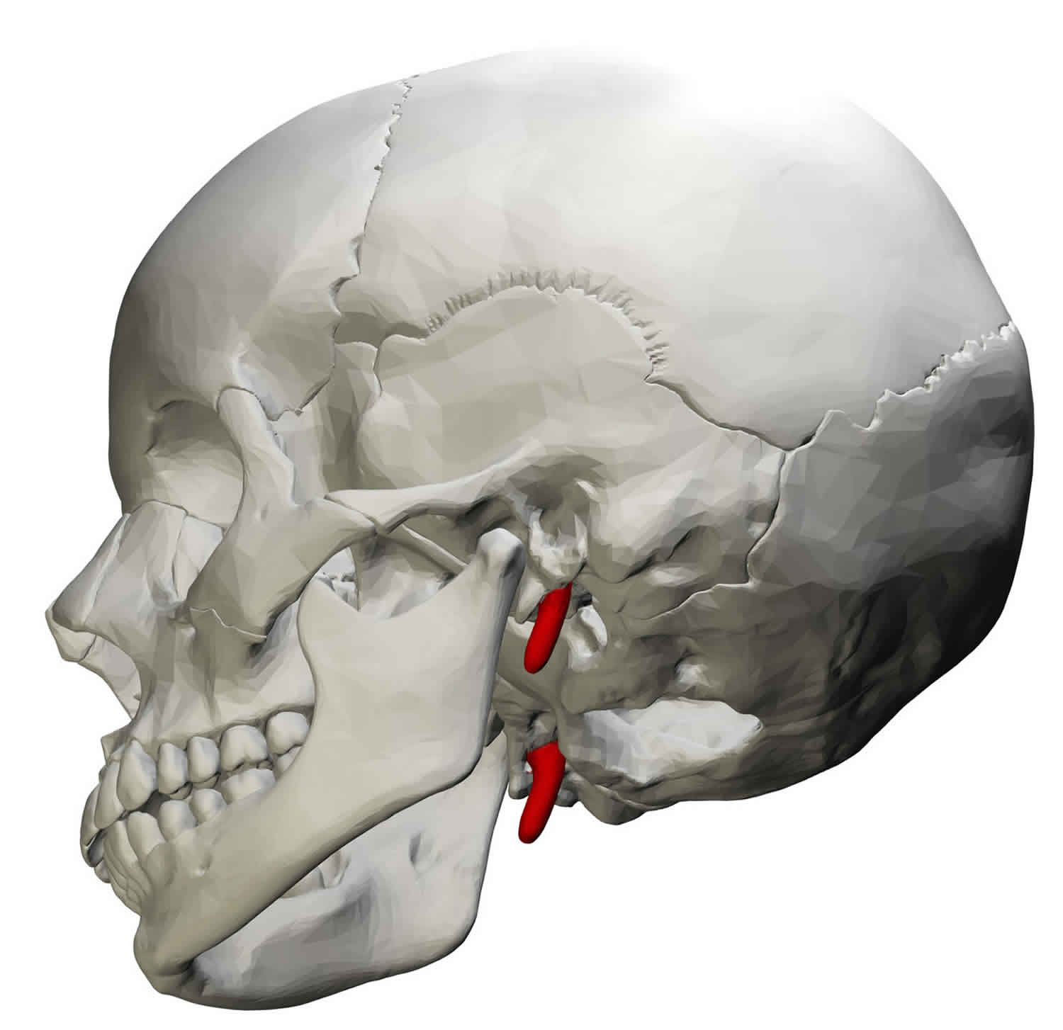
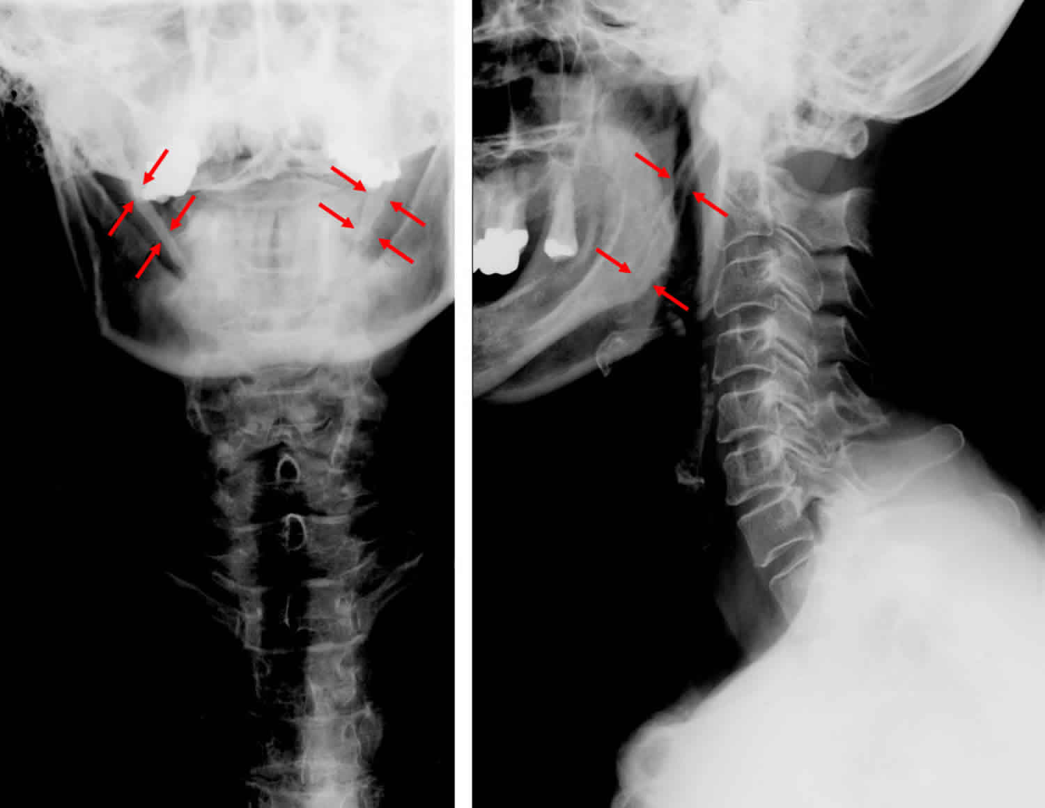
Figure 2. Eagle syndrome
Is Eagle syndrome life threatening?
No. In many Eagle syndrome cases, once the cause of pain has been attributed to the styloid process rather than a more sinister entity, no further treatment is required, or perhaps analgesics can be offered for pain alleviation.
Eagle syndrome causes
There is debate regarding the cause of Eagle syndrome 2. Dr. Watt Eagle 5 proposed that surgical trauma (tonsillectomy) or local chronic irritation causes osteitis, periostitis, or tendonitis of the styloid process and the stylohyoid ligaments which resulted in reactive, ossifying hyperplasia. Later Lentini 6 suggested the hypothesis that persistent mesenchymal elements, also known as Reichert cartilage residues, could undergo osseous metaplasia in the setting of an appropriate traumatic or stressful event. Epifanio in 1962 7 considered that the ossification of the styloid process was also corresponding to endocrine disorders in women at menopause, who also had ossification of other ligaments in the body. Gokce C et al. 8 reported that in patients with the end-stage renal disease having abnormal calcium, phosphorus, and vitamin D metabolism had heterotopic calcification which caused elongation of styloid process and thus the presentation of Eagle Syndrome. Finally, a retrospective study by Sekerci in 2015 9 indicated that relationship exists between the presence of an arcuate foramen and an elongated styloid process. Results were derived from data from 542 patients employing three-dimensional CT scans.
Early on Dr. Watt Eagle reported that the normal styloid process was ~2.5 cm in length, and any process longer than 2.5 cm might be considered abnormally elongated. Later in a postmortem study of 80 cadavers, the length of the styloid process was found to range from 1.52 to 4.77 cm. An elongated styloid process is incidental in about 4% of the general population, but of these, only about 4% present with symptoms that are attributable to elongation of styloid; therefore, the true incidence is about 0.16%, with a female-to-male predominance of 3:1. Patients are usually greater than 30 years of age, and this is usually a bilateral process (although unilateral cases are also seen).
Previously it was hypothesized that formation of scar tissue around the styloid apex after tonsillectomy caused compression and straining of the neurovascular structures present in the retro styloid compartment. However, Eagle syndrome also presents in patients who have never been operated for tonsillectomy. Several possible mechanisms for the pathogenesis of pain in Eagle syndrome have been proposed. The first considers that the elongated styloid process causes compression of cranial nerves, most commonly the glossopharyngeal nerve, with subsequent throat and neck pain. Alternatively, there is the possibility of compression of the internal carotid artery by the styloid process, which can cause transient ischemic attacks or compression of the sympathetic nerves running along the artery, leading to an array of symptoms. The pain in Eagle syndrome often resembles glossopharyngeal neuralgia but is typically more dull and constant, however, cases with sharp intermittent pain along the path of the glossopharyngeal nerve have also been reported. Furthermore, theories of reactive hyperplasia and reactive metaplasia exist which associate the elongation with either overgrowth of the styloid process itself or ossification of the stylohyoid ligament complex as a consequence of trauma. This phenomenon may explain the incidence of Eagle syndrome in patients after tonsillectomy, as it was originally described by Eagle. Other possible considered causes are the abnormal angulation associated with abnormally lengthy styloid process causing irritation of adjacent musculature or mucosa. Stretching and fibrosis involving the fifth, seventh, ninth, and tenth cranial nerves in the post-tonsillectomy period could also be a possible etiology. Finally, the symptoms may be a result of the normal process of aging. As normal aging is associated with a decrease in elasticity of soft tissues, degenerative and inflammatory changes in the tendinous portion of the stylohyoid insertion, a condition called insertion tendinosis, may cause pain in the distribution of glossopharyngeal nerve resembling Eagle syndrome. To avoid confusion, this manifestation better is called pseudo-stylohyoid syndrome.
Eagle syndrome symptoms
Eagle syndrome symptoms vary and establishing a causative relationship between the styloid process/stylohyoid ligament and symptoms can be challenging. Classically, the pain develops following tonsillectomy, presumably due to distortion of the local anatomy following surgery; however, it is frequently found in patients who have not had regional surgery 10.
Eagle syndrome symptoms can be divided into two main subtypes 10:
- due to compression of cranial nerves
- due to compression of the carotid artery
Cranial nerve impingement
Patients can have symptoms related to compression and irritation of cranial nerves in the region (cranial nerves V, VII, IX and X) such as 1,3:
- facial pain when turning the head
- difficulty swallowing (dysphagia)
- foreign body sensation
- pain on extending tongue
- change in voice
- sensation of hypersalivation
- tinnitus or ear pain (otalgia)
On palpation of the styloid process tip, symptoms should ideally be exacerbated.
Arterial impingement
Compression of the carotid artery may produce vascular/ischemic symptoms as well as pain along the artery to the supplied territory (thought to be mediated by the sympathetic plexus), including 10:
- mechanical compression
- visual symptoms
- fainting (syncope)
- carotid dissection has also been described 11
- sympathetic plexus irritation (carotidynia)
- eye pain
- parietal pain
Eagle syndrome diagnosis
Blood work is required to exclude possible systemic diseases. A complete blood count (CBC) is obtained if infection is suspected.
Lateral view radiographs of the skull can be substituted for panoramic radiographs of the mouth; the disadvantage of this view is the overlapping between styloid processes of both sides and/or with adjoining bony structures. An advantage of orthopantomogram (panoramic view) is that the entire length of the process can be seen very distinctly and its deviation can also be made out clearly.
Computed tomography (CT) scanning (and in particular three-dimensional [3-D] CT scanning) represents an extremely valuable imaging tool in patients with Eagle syndrome, offering an accurate evaluation of the styloid process in relation to its anatomical relationship with the other head and neck structures.
Additionally, reproduction of the patient’s pain on palpation of the tonsil or tonsillar fossa and relief of this discomfort by injection of local anesthetic are diagnostic.
Eagle syndrome treatment
The elongated styloid process Eagle syndrome can be managed either conservatively or surgically. Conservative treatments including analgesics, antidepressant medications, anticonvulsant, transpharyngeal injection of steroids and lidocaine, diazepam, nonsteroidal anti-inflammatory drugs, and the application of topical heat 2. Trans-pharyngeal manipulation with manual fracture the elongated styloid process does not usually relieve symptoms and risks damage to adjacent neurovascular structures. Patients who fail to respond to multiple medications may require surgical manipulations. The most effective treatment is the surgical shortening of the styloid process either via an intraoral or external approach as it produces better long-term results. The advantages of an intraoral approach are the simplicity of the technique, reduced operation time, achievability under local anesthesia, and absence of any visible external scar. However, the main disadvantages are a lack of access, particularly if there are a subsequent hemorrhage and deep neck infections, poor visualization of the surgical field especially in patients with significantly reduced jaw opening, the risk of iatrogenic injury to major neurovascular structures, alterations of speech and swallowing from postoperative edema. The most significant advantage of an external approach is enhanced exposure of the styloid process and the adjacent structures which overshadow all other benefits. It also aids the removal of a partially ossified stylohyoid ligament. Major disadvantages include more time-consumption, the hazard of injury to facial nerve and its branches, disfiguring neck scar and longer recovery period. In most cases, however, this choice is usually based on the surgical specialty of the conducting surgeon. Surgical failures in up to 20% of patients have been reported 2.
The overall success rate for treatment (medical or surgical) is about 80% 4.
Eagle syndrome surgery
Only severe cases, which do not respond to analgesics and anti-inflammatory medications, require surgery.
Intraoral approach
- Advantages
- Avoids external scarring
- Less time consuming
- Disadvantages
- Risk of deep space neck infection
- Poor visualization of the surgical field
- Major risk of iatrogenic injury to main neurovascular structures
- Poor hemorrhage control
- Alterations of speech and swallowing for postoperative edema
- Difficult in patients with markedly decreased jaw opening
Extraoral approach
- Advantages
- Better visualization of the surgical field
- Greater intraoperative sterility
- Disadvantages
- More time-consuming
- Risk of injury of facial nerve structures
- Neck scar
- Longer recovery
The mainstay treatment for Eagle syndrome is surgery to shorten the styloid process (styloidectomy) 10. Traditionally, this surgery has been done using either an intraoral (through the mouth) or extraoral (through the neck) approach 3.
The intraoral approach usually requires tonsillectomy, and access to the styloid process is limited 10. There is also risk of injury to major vessels. However, this method reportedly is safe, more simple, and an external scar is avoided 4.
The extraoral approach may provide better exposure of the process and its surrounding structures. However, this approach results in a scar, requires going through connective tissue and may carry an increased risk of trauma to surrounding structures 10.
In recent years, more minimally-invasive techniques have been used for head and neck surgery. Some patients with Eagle syndrome have undergone an endoscope-assisted approach. An endoscope is a long, thin tube with a camera attached at the end. According to the authors of a study published in 2017, this approach reportedly has the benefits of providing direct surgical access, satisfactory exposure, and minimal invasion 10.
The main surgical complications associated with styloidectomy are as follows:
- Deep space neck infection
- Injury to main neurovascular structures
- Hemorrhage
- Temporary alterations of speech and swallowing
- Injury of the facial nerve
- People with questions about personal treatment options and recommendations for Eagle syndrome should speak with their doctor.
Eagle syndrome prognosis
The overall success rate for Eagle syndrome treatment (medical or surgical) is about 80%.
The failure of treatment may be associated with the presence of other causes involved in the pathogenesis of the problem (multifactorial etiology).
References- Eagle Syndrome. https://emedicine.medscape.com/article/1447247-overview
- Bokhari MR, Mohseni M. Eagle Syndrome. [Updated 2018 Dec 2]. In: StatPearls [Internet]. Treasure Island (FL): StatPearls Publishing; 2019 Jan-. Available from: https://www.ncbi.nlm.nih.gov/books/NBK430789
- Chen R, Liang F, Han P, Cai Q, Yu S, Huang X. Endoscope-Assisted Resection of Elongated Styloid Process Through a Retroauricular Incision: A Novel Surgical Approach to Eagle Syndrome. J Oral Maxillofac Surg. January 26, 2017; https://www.ncbi.nlm.nih.gov/pubmed/28215854
- Eagle syndrome. https://emedicine.medscape.com/article/1447247-overview
- Eagle W. Elongated styloid process: Further observation and a new syndrome. Arch Otolaryngol. 1948. 47:630-640.
- Lentini A. Gli aspetti clinici e radiologici delle anomalie dell’apparato stilo-joideo. Radiol Med. 1975. 61:337-3640.
- Epifanio G. Processi stiloidei lunghi e ossificazione della catena stiloidea. Rad Prat. 1962. 12:127-132.
- Gokce C, Sisman Y, Sipahioglu M. Styloid Process Elongation or Eagle’s Syndrome: Is There Any Role for Ectopic Calcification?. Eur J Dent. 2008 Jul. 2 (3):224-8
- Sekerci AE, Soylu E, Arikan MP, Aglarci OS. Is there a relationship between the presence of ponticulus posticus and elongated styloid process?. Clin Imaging. 2015 Mar-Apr. 39 (2):220-4
- Chuang WC, Short JH, McKinney AM et-al. Reversible left hemispheric ischemia secondary to carotid compression in Eagle syndrome: surgical and CT angiographic correlation. AJNR Am J Neuroradiol. 2007;28 (1): 143-5
- Faivre A, Abdelfettah Z, Rodriguez S et-al. Neurological picture. Bilateral internal carotid artery dissection due to elongated styloid processes and shaking dancing. J. Neurol. Neurosurg. Psychiatr. 2009;80 (10): 1154-5. doi:10.1136/jnnp.2008.159954


