
Echogenic intracardiac focus
Echogenic intracardiac focus is a small bright spot seen inside the baby’s heart chambers on a routine fetal heart ultrasound exam 1. Echogenic intracardiac focus is commonly seen inside the left ventricle being the most frequent location 2. Echogenic intracardiac focus is thought to represent mineralization, or small deposits of calcium, in the muscle of the heart. Echogenic intracardiac focuss are a found in about 3-5% of normal pregnancies and cause no health problems. Echogenic intracardiac focus may be more common in the Asian population 3.
Echogenic intracardiac focus themselves have no impact on health or heart function. Often the echogenic intracardiac focus is gone by the third trimester. If there are no problems or chromosome abnormalities, echogenic intracardiac focuss are considered normal changes, or variants.
Although echogenic intracardiac focus probably represent a normal variant of papillary muscle development their presence should be interpreted as a possible risk for congenital heart defects. Researchers have noted an association between an echogenic intracardiac focus and a chromosome problem in the baby 4. Types of chromosome problems that are occasionally seen include Trisomy 18 or Trisomy 21 (Down syndrome). In the case of an isolated echogenic intracardiac focus, and no other ultrasound findings, some studies show that the risk for a chromosome abnormality is approximately two times a woman’s background risk. Other studies report up to a 1% risk for Down syndrome when an echogenic intracardiac focus is seen on a second trimester fetal ultrasound exam 5.
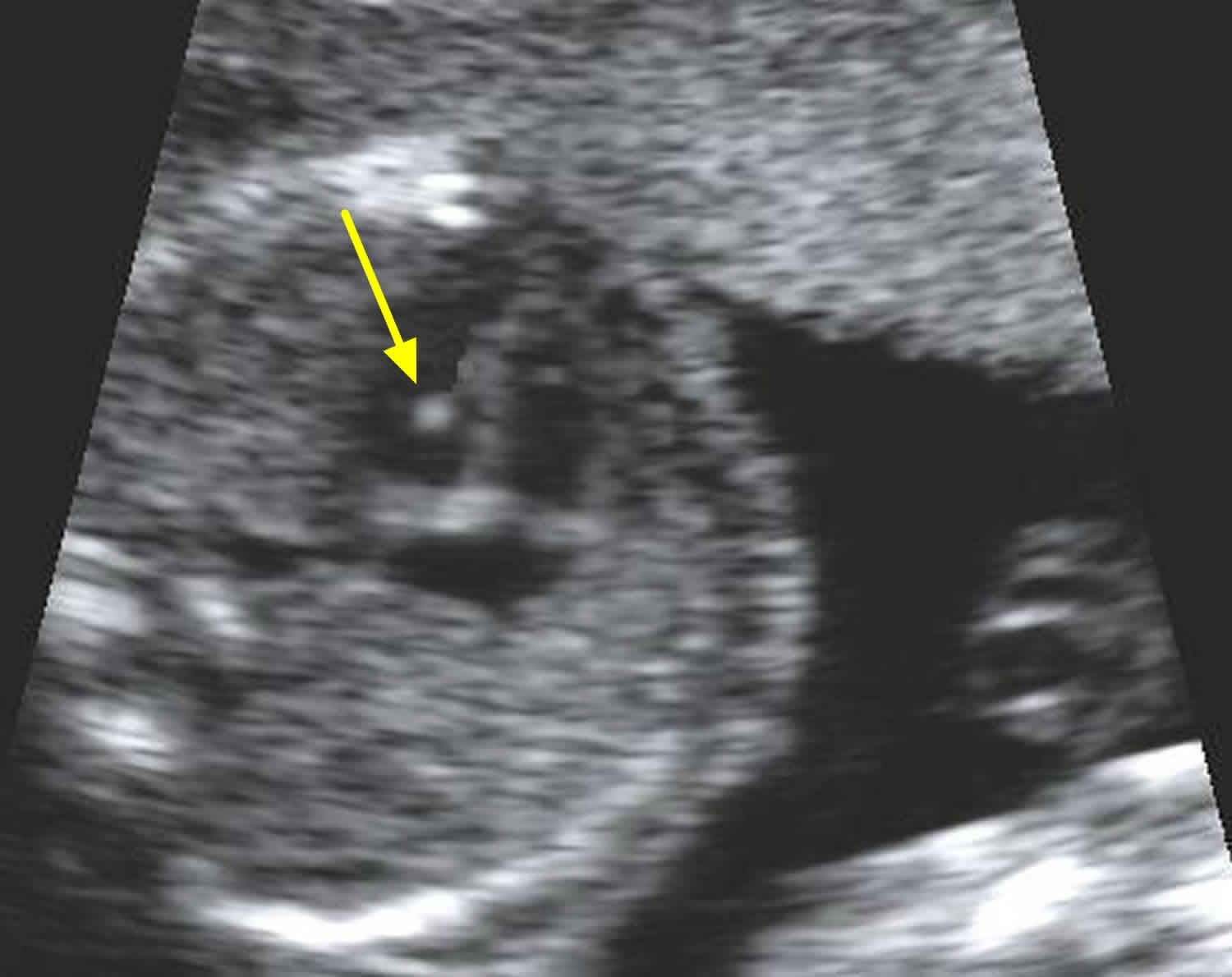
Figure 1. Echogenic intracardiac focus left ventricle
Echogenic intracardiac focus causes
No one knows for sure why echogenic intracardiac focus is seen in some babies and not others. It is thought that echogenic intracardiac focus is due to an area of the heart muscle where there is a little more calcium than average. Calcium is a natural mineral found in the body. Areas in the body with more calcium, like bones and muscles, look brighter on the ultrasound screen.
Echogenic intracardiac focus is often seen in babies whose mothers have Asian ancestry. However, echogenic intracardiac focus can be seen in any pregnancy, regardless of the ancestry of the parents.
Can an echogenic intracardiac focus cause problems for the baby?
An echogenic intracardiac focus is considered a normal variation in fetal development. It has not been found to have any long term health problems or heart problems for the baby. Most of the time, echogenic intracardiac focus is seen during the routine prenatal ultrasound done around 18 to 20 weeks in pregnancy. If there are no other ultrasound findings, the echogenic intracardiac focus is considered an “isolated” finding. While it is impossible to be completely certain that no birth defects are present in the baby, most pregnancies with isolated echogenic intracardiac focus result in a healthy baby.
Will the echogenic intracardiac focus go away?
Most echogenic intracardiac focus seen in the middle of the pregnancy will not go away before delivery. Since they do not cause problems for the baby, there is no special concern if they are still visible at a later time. For this reason, no ultrasound follow-up is needed to watch for changes in the echogenic intracardiac focus.
Could an echogenic intracardiac focus mean the baby has Down syndrome?
Some studies raised concerns about a small risk for Down syndrome with echogenic intracardiac focus. However, most studies do not find a higher risk for Down syndrome when an echogenic intracardiac focus is the only ultrasound finding. Blood tests or amniocentesis are a better way to look for Down syndrome during pregnancy.
Are additional tests needed?
There is no special testing recommended for pregnancies found to have an isolated echogenic intracardiac focus. However, routine prenatal testing is available to all pregnant women. Prenatal screening tests, like Integrated Screening and cell-free DNA screening (also called non-invasive prenatal testing or NIPT), include blood tests that help find out if there is a higher or lower chance of having a baby with certain problems, including Down syndrome. A test called amniocentesis can accurately diagnose Down syndrome and other chromosome conditions during pregnancy. This is an optional test for women of all ages. Amniocentesis is usually done until about 22 weeks of pregnancy. This procedure has a very small risk for miscarriage (1 in 500 or less). It is important to remember that prenatal tests will not test for all birth defects.
References- Echogenic intracardiac focus. https://radiopaedia.org/articles/echogenic-intracardiac-focus
- How important is a cardiac echogenic focus in a routine fetal examination? Rev Port Cardiol. 2004 Mar;23(3):459-61. https://www.ncbi.nlm.nih.gov/pubmed/15185567
- Shipp, T. D., Bromley, B. , Lieberman, E. and Benacerraf, B. R. (2000), The frequency of the detection of fetal echogenic intracardiac foci with respect to maternal race. Ultrasound Obstet Gynecol, 15: 460-462. doi:10.1046/j.1469-0705.2000.00138.x
- Rebarber A, Levey KA, Funai E, Monda S, Paidas M. An ethnic predilection for fetal echogenic intracardiac focus identified during targeted midtrimester ultrasound examination: A retrospective review. BMC Pregnancy Childbirth. 2004;4(1):12. Published 2004 Jun 25. doi:10.1186/1471-2393-4-12 https://www.ncbi.nlm.nih.gov/pmc/articles/PMC449713
- Echogenic intracardiac focus: a sonographic sign for fetal Down syndrome. Obstet Gynecol. 1995 Dec;86(6):998-1001. DOI:10.1016/0029-7844(95)00323-j https://www.ncbi.nlm.nih.gov/pubmed/7501356


