What is ectopia cordis
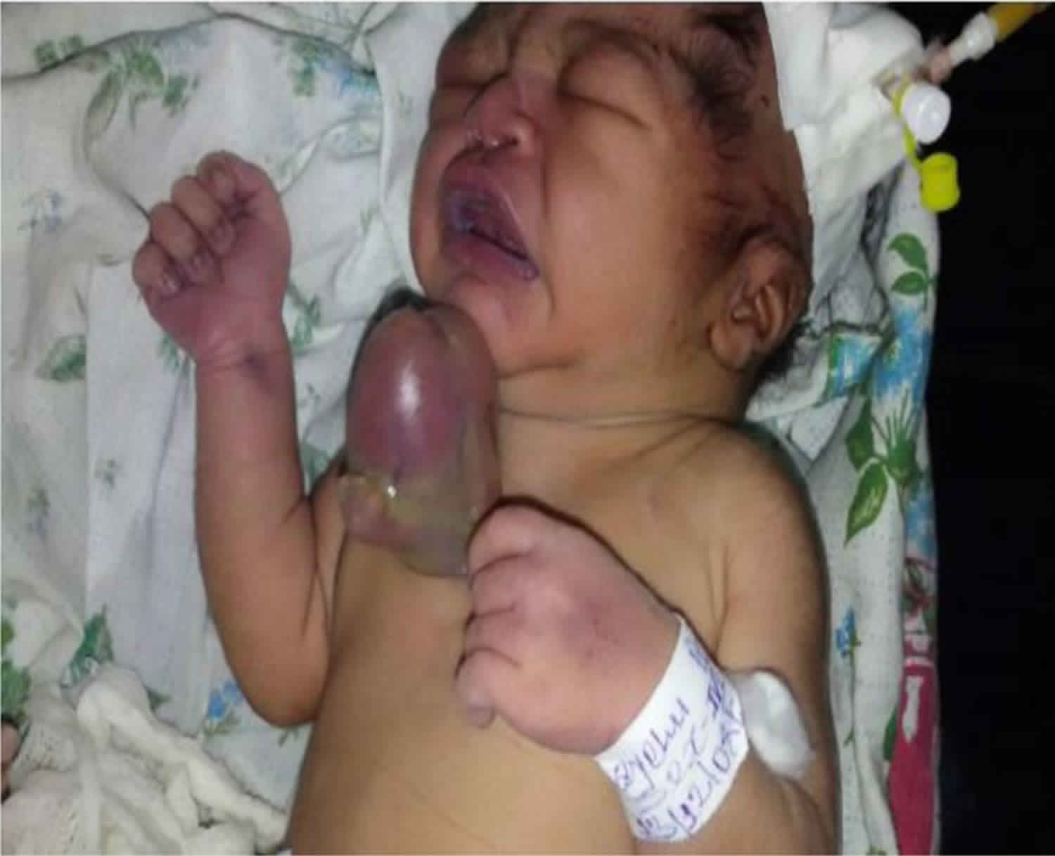
Ectopia cordis is defined as complete or partial displacement of the heart outside the thoracic cavity 1. Ectopia cordis is a rare congenital (present at birth) defect with failure of fusion of the sternum with extra thoracic location of the heart 1. The estimated prevalence of ectopia cordis is 5.5 to 7.9 per million live births 2.
Prenatal diagnosis is established by ultrasonography by visualizing the heart outside the thoracic cavity 3.
Depending upon the location of the heart, ectopia cordis can be classified into five types 4:
- Cervical (5%),
- Cervicothoracic and thoracic (65%),
- Thoracoabdominal (20%) and
- Aabdominal (10%).
The combination of thoracoabdominal ectopia cordis, lower sternal defect, anterior diaphragmatic hernia, midline supraumblical defect along with pericardial and intracardiac defects constitute the Pentology of Cantrell 5. The thoracic ectopia cordis has been reported to have worst prognosis with <5% surviving beyond the first month of life 6.
Ectopia cordis is classified into cervical, thoracic, thoracoabdominal and abdominal. Pentalogy of Cantrell is considered when a combination of thoracoabdominal ectopia cordis, anterior diaphragmatic hernia, lower sternal defect and midline supraumblical defect occurs. Although surgical techniques have evolved, the prognosis and survival are limited; thoracic type has the worst prognosis while the thoracoabdominal ectopia cordis has a better prognosis 7.
Ectopia cordis causes
Thoracic ectopia cordis was explained embryonically by the rupture of the chorion at 3 weeks of gestation with resultant compression of the thoracic cavity and failure of descent of the heart at this stage. The possibility of amniotic bands is also ascribed. Ectopia cordis may occur in isolation or in association with other ventral body wall defects 7.
The development of the ventral body wall begins by eighth day of embryonic life with differentiation and proliferation of mesoderm followed by its lateral migration. The heart originally develops in a cephalic location and reaches its definitive position by the lateral folding and ventral flexing of the embryo at about 16th–17th day. Midline fusion and formation of the thoracic and abdominal cavities is complete by the 9th embryonic week 8. Complete or incomplete failure of midline fusion at this stage result in disorders varying from isolated ectopia cordis to complete ventral evisceration. Genesis of ectopia cordis has not been fully explained, although several theories have been offered.16 Popular theories are early rupture of the chorion and/or yolk sac, and amniotic band syndrome 9.
The amnion rupture theory states that during early embryonic development, the amnion surrounding the embryo ruptures, and stringy, sticky, fibrous bands of amnion becomes ‘entangled’ with the forming embryo, and causes a disruption in the developing parts of the fetus which may lead to various deformities like ectopia cordis, midline sternal cleft, frontonasal dysgenesis, a midfacial cleft, limb deformities etc. The spectrum of defect corresponds to the timing of its rupture. The findings in the literature suggest that its rupture in the third week of gestation causes an arrest of cardiac descent which may be the cause of such defects. ectopia cordis with amniotic bands appears to have aetiology distinct from isolated ectopia cordis. This suggests several different aetiologies for ectopia cordis 9. Ectopia cordis has also been attributed to intrauterine drug exposure in animal models 10.
Cyllosomas, another name for limb-body wall complex is defined as anomaly consisting of two of the following three fetal anomalies: a) thoraco-abdominoschisis (opening extends from the chest through the abdomen) or abdominoschisis (opening starts at a lower point and only extends through the abdomen), b) limb defects, c) cranio-facial defects- cleft lip/palate, encephalocele, exencephaly, etc. and others 11.
Ectopia cordis diagnosis
Ectopia cordis can be diagnosed by routine prenatal ultrasonography as early as in 10-12 weeks of pregnancy 12. Of those not diagnosed antenataly, most result in stillbirth or die shortly after birth due to their frequent association with intrinsic cardiac and other congenital defects 12.
Ectopia cordis treatment
Newborns with ectopia cordis complex and life threatening deformity require intensive care right from birth. They require immediate resuscitation and coverage to the exposed heart and viscera with saline-soaked gauze pads wrapping to prevent desiccation and heat loss. For this, Harrison et al have reported use of adherent plastic drapes also 13. Thoracic ectopia cordis presents a formidable surgical challenge. In recent reviews of the literature the reported survival of this variety after birth averages 36 h; intracardiac defects were associated in 80.2% of the cases, and all unoperated patients died 14. Besides intrinsic cardiac defects, the increased morbidity in these patients may be attributed to the abnormal course, length and positioning of great vessels of the heart making them prone to kinking and further compromising circulation, as also must have been in our case. During surgical closure, in most of the cases the thoracic cavity is small with little mediastinal space for the heart. Attempts to close the chest wall often result in intolerable haemodynamic embarrassment secondary to kinking of the great vessels possibly due to their long length and abnormal course, or compression of the heart. Therefore, a staged repair is often necessary. The strategy for repair is divided in two stages: (1) urgent soft tissue coverage and haemodynamic palliation if necessary; and (2) intracardiac repair with concomitant chest wall reconstruction and reduction of the heart into the thoracic cavity.
Ectopia cordis surgery
The overall surgical objective of ectopia cordis (all variety) management includes: 1) closure of the chest wall defect (either by doing primary chest wall closures or by using bone/cartilage as tissue graft or artificial prosthesis like acrylic plaques, marlex mesh), 2) closure of the sternal defect, 3) repair of the associated omphalocele, 4) placement of the heart into the thorax, 5) repair of the intracardiac defect.
Key points
- Success of repair is dictated by the presence and severity of the intrinsic cardiac defects and associated congenital anomalies, rather than the type of surgical approach.
- Surgical technique is evolving, and surgical options should be considered in all cases, taking comorbidities into account.
- Antenatal ultrasonography plays an important role in detecting such anomalies which may be important for planning its further management.
The first attempted repair of ectopia cordis was performed in 1925 by Cutler and Wilens.25 Koop in 1975 achieved the first successful repair of thoracic ectopia cordis in two stages.26 Amato et al reported successful single stage repair of thoracic ectopia cordis in 1995.27 Cristofer Wall operated by Koop in August-1975 is the oldest live pt of ectopia cordis. Other surviving operated cases which could be traced on literature search are:
- Aytag and Sayam successfully operated partial ectopia cordis in 1976 15,
- Dobell et al 16, reported two stage correction
- Kim KA et al 17, – Los Angeles – 1996, did two – stage operation.
- Morales et al 18 – reported successful 2-stage repair of ectopia cordis on female baby.
- Khaled et al 19 from France reported successful single stage repair in 2003.
- Gonçalves et al at 20, from Brazil reported successful repair of uncomplicated ectopia cordis in June 2007.
Hence it may be concluded that ectopia cordis is a lethal anomaly requiring prompt medical and surgical interventions. Surgery on these patients with life-threatening complex intracardiac anomalies, owes the only chance of survival, which should still be attempted despite heretofore poor outcomes. Although the available literature is scant, but with the advance in all aspects of medicine, the number of patients who undergo successful surgical repair and survive should steadily increase, with the combined efforts to understand and formulate treating protocol of this extremely rare anomaly.
Ectopia cordis survival rate
In this case report 21, death occurred within twenty minutes of the initial surgical intervention. Earlier studies showed a lethal course of the thoracic ectopia cordis. Survival of thoracic ectopia cordis case is limited despite advances in care, and its management is challenging. Aggressive surgical procedures are recommended to increase the survival 22. The thoracic ectopia cordis has been reported to have worst prognosis with <5% surviving beyond the first month of life 6. Only few patients with thoracic type have survived and the thoracoabdominal ectopia cordis has a better prognosis.
References- Tadele H, Chanie A. Thoracic Ectopia Cordis in an Ethiopian Neonate. Ethiop J Health Sci. 2017;27(2):203–205. doi:10.4314/ejhs.v27i2.14
- Pamidi Narendra, Vollala Venkata R, Nayak Satheesha, Bhat Seetharama. Ectopia Cordis and amniotic band syndrome. Arch Med Sci. 2008;4(2):208–211.
- Rare occurrence of ectopia cordis in a Congolese neonate. Ucima N, Tuka DD, Kimbongila MM, Lumbala PK, Biselele T, Tady BM, Nkidiaka ED, Aloni MN. Pediatr Neonatol. 2015 Apr; 56(2):132-3.
- Anderson RH, Shinebourne EA, Macartney FJ, et al. Abnormal positions and relationships of the heart. Anderson RH, Shinebourne EA, editors. Paediatric Cardiology. London: Churchill Livingstone; 1987:1057–72.
- Fonkalsrud EW. Chest wall abnormalities. Bove AE, Geha AS, Hammond GL, Laks H, Naunheim KS, editors. , eds. Glenn’s Thoracic and Cardiovascular Surgery. East Norwalk, Connecticut: Appleton and Lange 1991:507–16.
- Shamberger RC, Welch KJ. Sternal defects. Pediatr Surg 1990;5:156–64.
- Engum SA. Embryology, sternal clefts, ectopia cordis, and Cantrell’s pentalogy. Semin Pediatr Surg. 2008 Aug;17(3):154–160. doi: 10.1053/j.sempedsurg.2008.03.004
- Ectopia cordis in man. KANAGASUNTHERAM R, VERZIN JA. Thorax. 1962 Jun; 17():159-67.
- Ectopia cordis and cleft sternum: evidence for mechanical teratogenesis following rupture of the chorion or yolk sac. Kaplan LC, Matsuoka R, Gilbert EF, Opitz JM, Kurnit DM. Am J Med Genet. 1985 May; 21(1):187-202.
- Limb body wall complex: a critical review and a nosological proposal. Russo R, D’Armiento M, Angrisani P, Vecchione R. Am J Med Genet. 1993 Nov 1; 47(6):893-900.
- Extrathoracic ectopia cordis. Case report. Morello M, Quaini E, Nenov G, Pomé G. J Cardiovasc Surg (Torino). 1994 Dec; 35(6):511-5.
- Fetal echocardiography in ectopia cordis. Repondek-Liberska M, Janiak K, Wloch A. Pediatr Cardiol. 2000 May-Jun; 21(3):249-52.
- Prenatal diagnosis and management of omphalocele and ectopia cordis. Harrison MR, Filly RA, Stanger P, de Lorimier AA. J Pediatr Surg. 1982 Feb; 17(1):64-6.
- Ectopia cordis (ectocardia) and gastroschisis induced in rats by maternal administration of the lathyrogen, beta-aminopropionitrile (BAPN). Barrow MV, Willis LS. Am Heart J. 1972 Apr; 83(4):518-26.
- Aytag A, Sayam A. Successful surgical repair of congenital total cleft sternum with partial ectopia cordis. Thorax 1976;31:466–9
- Staged repair of ectopia cordis. Dobell AR, Williams HB, Long RW. J Pediatr Surg. 1982 Aug; 17(4):353-8.
- Kim KA, Vincent WR, Muenchow SK, et al. Successful repair of ectopia cordis using alloplastic materials. Ann Plast Surg 1997;38:518–22.
- Morales JM, Patela SG, Duff JA, et al. Ectopia cordis and other midline defects. Ann Thorac Surg 2000;70:111–4.
- Ectopia cordis, a successful single stage thoracoabdominal repair. Samir K, Ghez O, Metras D, Kreitmann B. Interact Cardiovasc Thorac Surg. 2003 Dec; 2(4):611-3.
- Gonçalves FD, Novaes FR, Maia MA, et al. Thoracic ectopia cordis with anatomically normal heart. Rev Bras Cir Cardiovasc 2007;22:no.2 São José does Rio Preto April/June. http://dx.doi.org/10.1590/S0102-76382007000200015
- Tadele H, Chanie A. Thoracic Ectopia Cordis in an Ethiopian Neonate. Ethiop J Health Sci. 2017;27(2):203–205. doi:10.4314/ejhs.v27i2.14 https://www.ncbi.nlm.nih.gov/pmc/articles/PMC5440836
- Engum SA. Embryology, sternal clefts, ectopia cordis, and Cantrell’s pentalogy. Semin Pediatr Surg. 2008 Aug;17(3):154–160. doi: 10.1053/j.sempedsurg.2008.03.004.


