What is epiretinal membrane
An epiretinal membrane also called surface-wrinkling retinopathy, macular pucker, cellophane maculopathy or preretinal macular fibrosis, is formation of a thin fibrotic membrane (scar tissue) found on the inner surface of the retina that contracts, wrinkling the underlying retina and interfering with vision 1. Epiretinal membrane is a semi-translucent, avascular, fibrocellular membrane and proliferates on the inner retinal surface along the internal limiting membrane of the retina. Contracture of epiretinal membranes produces distortion and wrinkling of the inner surface of the retina. It is also called “cellophane maculopathy” or “pre-retinal macular fibrosis” when mild; “surface wrinkling retinopathy” or “retinal striae” when moderate; or “macular pucker” when severe. Epiretinal membrane typically occurs after age 50 and is most common among people > 75. As you get older, the vitreous begins to shrink and pull away from the retina. Usually the vitreous pulls away with no problems. But sometimes the vitreous can stick to the retina. Scar tissue forms, causing the retina and macula to wrinkle or bulge.
Risk factors for epiretinal membrane are the following:
- Diabetic retinopathy
- Uveitis
- Retinal tear or detachment
- Eye injury
Most epiretinal membrane cases are idiopathic.
Symptoms may include blurred vision or distorted vision (eg, straight lines may appear wavy). Many patients say that it seems like they are looking through plastic wrap or cellophane. Diagnosis is by funduscopy. Fluorescein angiography and optical coherence tomography may also be helpful.
Most people need no treatment. If problems with vision are significant, the membrane can be removed surgically with vitrectomy and membrane peel.
What causes epiretinal membrane
Idiopathic epiretinal membranes is the most common presentation. Secondary epiretinal membranes occur in association with retinal vascular diseases including diabetic retinopathy, retinal vein occlusion, ocular inflammatory disease, trauma, intraocular surgery, intraocular tumors, and retinal tear or detachment.
Other risk factors include age, posterior vitreous detachment, and history of epiretinal membrane in the fellow eye.
Age is the most common cause of epiretinal membrane. As you get older, the vitreous begins to shrink and pull away from the retina. Usually the vitreous pulls away with no problems. But sometimes the vitreous can stick to the retina. Scar tissue forms, causing the retina and macula to wrinkle or bulge.
The mean age of epiretinal membrane diagnosis is 65 years old 2. The incidence of developing an epiretinal membrane in the primary eye is 1.1% per year. The incidence of developing an epiretinal membrane in the fellow eye is 2.7% per year.
Retinal glial and retinal pigment epithelial cells are the major components. Fibrous astrocytes, fibrocytes, myofibrocytes, and macrophages can also be identified in pathological analysis.
It has been hypothesized that residual cortical vitreous secondary to a posterior vitreous detachment or partial separation of the posterior hyaloid allows proliferation of glial cells.
Inflammatory mediators also promote fibrocellular growth especially in the setting of secondary epiretinal membrane formation.
Who is at risk for epiretinal membrane?
Aging is the most common risk factor for epiretinal membrane. People who have other eye problems may also get a epiretinal membrane. These problems include:
- Posterior vitreous detachment, where the eye’s vitreous pulls away from the retina
- Torn or detached retina
- Swelling inside the eye
- Serious damage to the eye (from surgery or injury)
- Problems with blood vessels in the retina
- Diabetic retinopathy
Epiretinal membrane symptoms
With epiretinal membrane, things can look wavy, or you may have trouble seeing details. You might notice a gray or cloudy area in your central vision. You may even have a blank spot in your central vision. Epiretinal membrane will not affect your peripheral (side) vision.
Epiretinal membrane symptoms
Metamorphopsia, blurred vision, monocular diplopia, and micropsia can be noted with any macular pathology. The vast majority of patients with epiretinal membranes are asymptomatic.
Epiretinal membrane signs
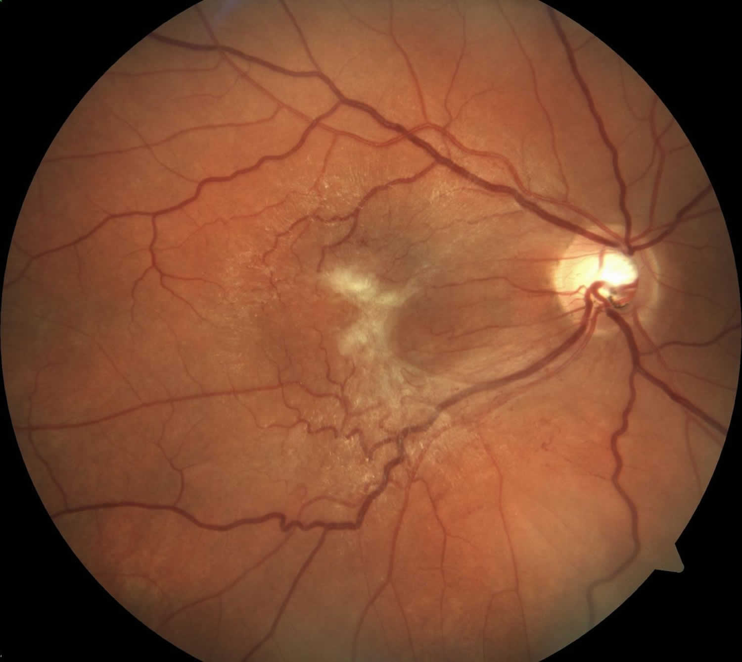
A sheen or abnormal reflectivity of the macular surface is suggestive of an epiretinal membrane. More advanced epiretinal membranes can become opaque.
Epiretinal membrane diagnosis
Epiretinal membrane is a clinical diagnosis based on history and clinical exam, including slit lamp and dilated fundus examination. In some cases, Optical Coherence Tomography (OCT) is useful in the diagnosis, quantification of retinal thickness, and management of epiretinal membrane. With OCT, a machine scans the back of your eye. This provides very detailed pictures of the retina and macula. Your ophthalmologist studies these pictures to check for problems.
Patients with epiretinal membranes typically present over the age of 50 and both sexes are equally affected. A careful history should be obtained to investigate for any of the risk factors mentioned above.
Physical examination
Slit lamp examination with dilated fundus examination and scleral depression (to rule out peripheral breaks/lesions) are important to determine the presence and evaluate the severity of an epiretinal membrane. Careful examination of the macular area is important to evaluate the epiretinal membrane. However, paying attention the vitreous, retinal vasculature, and peripheral retina can provide insight as to the cause of the epiretinal membrane in secondary cases.
Careful examination of the fellow eye is also recommended given that epiretinal membranes are bilateral in approximately 10-20% of patients. The clinical appearance of an epiretinal membrane is fairly distinctive. However, macular hole, parafoveal telangiectasia, and macular edema must also be considered.
Idiopathic epiretinal membranes affect the architecture of the macula. There can be blunting of the foveal contour or wrinkling on the retinal surface from membrane contracture. Most commonly it involves the foveal and parafoveal area.
Macular edema and/or pseudohole can be seen in association with an epiretinal membrane. As the name implies, a pseudohole is not a full-thickness macular hole, but rather a hole or gap in the epiretinal membrane that appears to be a retinal hole. The inner retina around the pseudohole is thickened. The pseudohole may not be exactly round and may have oval or irregular shape. Other associated signs include vascular tortuousity in the region of the epiretinal membrane and/or intraretinal hemorrhages.
A posterior vitreous detachment is often noted which supports the pathophysiology of this entity.
Diagnostic procedures
Fluorescein angiography can be helpful in secondary cases of epiretinal membrane including retinal vascular occlusions or intraocular tumors. Macular edema can be confirmed with angiography, as well.
Optical Coherence Tomography (OCT) has become increasingly helpful in the diagnosis and management of this disorder. This high-resolution image can allow evaluation of the macula in cross section and three-dimensionally. OCT can be helpful detecting subtle epiretinal membranes as well as when associated with macular edema or other macular pathology.
Optical Coherence Tomography (OCT) can also help guide management. Some cases of epiretinal membrane with vitreomacular traction are subtle clinically and better detected with OCT. One of the great advantages of the OCT is the assessment of the vitreoretinal interface. This can provide additional information regarding therapeutic options and prognosis. In surgical cases, evaluation of each scan can elucidate the best approach for removal. Spectral domain OCT can also allow evaluation of the outer retinal layers/structure which may have barring on the physiologic outcome after an epiretinal membrane removal.
Epiretinal membrane treatment
How you are treated depends on your symptoms. The most important issue in the management of idiopathic epiretinal membranes is the presence of visual complaints. Visual symptoms can be variable and sometimes independent of clinical severity.
If your symptoms are mild, you might not need any treatment. Instead, your ophthalmologist may change your glasses or contact lens prescription to improve your vision. You might also choose to wear bifocals when you are looking at something close. Eye drops, medicine, and laser surgery do not help vision if you have macular pucker.
If your symptoms are more serious, your ophthalmologist may recommend a surgery called vitrectomy. Your ophthalmologist will remove some of the vitreous and scar tissue on your macula. This flattens the macula, returning it to its proper position. It is likely your vision will slowly improve. However, your sight may not be as good as it was before macular pucker.
Epiretinal membrane surgery
Epiretinal membrane surgery is the most common vitreoretinal surgery performed as reported by the Centers of Medicare and Medicaid Services 3. Surgery involves a pars plana vitrectomy procedure with membrane peel. A number of different instruments can be used to facilitate removal including intraocular forceps, pick, diamond dusted instruments, as well as other instruments.
Epiretinal membrane surgery complications
Like all surgery, vitrectomy has some risks. They include:
- Eye infection
- Bleeding in your eye
- A detached retina (where the retina lifts away from the back of the eye)
- Having the macular pucker happen again
- Cataract, when the lens in your eye becomes cloudy
The complications are similar to all eyes undergoing pars plana vitrectomy (including cataract in phakic, retinal break, retinal detachment, dissociated optic nerve fiber layer). In addition, macular surgery complications include intraoperative macular trauma and light toxicity.
Your ophthalmologist will talk about these risks and how vitrectomy surgery may help you.
Epiretinal membrane surgery recovery
Surgery is indicated if patient has visual complaints (visual decline/metamorphopsia).
The follow up is similar for most eyes following pars plana vitrectomy surgery. Visual acuity improvement does not occur immediately in some patients. This is highly dependant on preoperative characteristics, duration of the epiretinal membrane, as well as other factors. Most patients improve by 3-6 months postoperatively. However, some may experience improvement 1-2 years postoperatively.
Epiretinal membrane surgery success rate
The mean preoperative and postoperative visual acuity has been reported to be 20/110 and 20/55 4. This data was a metanalysis of three studies reporting surgical results following small incision pars plana vitrectomy.
The recurrence of epiretinal membrane has been estimated to be 1% 4.
References- Epiretinal membrane. https://eyewiki.org/Epiretinal_Membrane
- Fraser-Bell S, Guzowski M, Rochtchina E, et al. Five-year cumulative incidence and progression of epiretinal membranes: the Blue Mountains Eye Study. Ophthalmology 2003;110(1):34-40.
- Gillis K. Medicare Physician Payment Schedule Services for 2001 – A Summary of Claims Data. In: Physician Marketplace Report. Chicago: American Medical Association, 2003.
- Gupta OP, Brown GC, Brown MM. A value-based medicine cost-utility analysis of idiopathic epiretinal membrane surgery. Am J Ophthalmol 2008;145(5):923-8.


