
What are Epstein pearls
Epstein pearls are small, firm, white, keratin-filled cysts located on the mid-palatal raphe at the junction of the hard and soft palates 1. Some authors use the terms Epstein pearls, Bohn nodules, and dental lamina cysts interchangeably. However, Epstein pearls have been labeled as epithelial debris of the tooth follicle, gingival glands of Serres, and as abortive enamel organs on the Palatine area. On the other hand, Bohn’s nodules are those found along the buccal and lingual aspects of the dental ridges.
Epstein pearls are observed in nearly 60% to 85% of newborn infants. Among the different racial groups, Japanese newborns are most commonly affected (up to 92%), followed by Caucasians and African-Americans 1. No gender predilection has been observed through the years 1.
In one study, Epstein pearls were more common in infants born to multigravida mothers, also with those with higher birth weight. One study done in Turkey found that Epstein’s pearls were less frequent in post-term babies in comparison with pre-term and term ones. A greater rate seen in term babies was reported. 1.
Epstein pearls are a clinical diagnosis. No laboratory or imagining is needed.
Epstein pearls are small, opaque whitish-yellow lesions adjacent to the mid palatine raphe with no mucous glands in it. Epstein pearls are firm in consistency, size range from less than a millimeter to several millimeters in diameter. Size does not progress over time. Epstein pearls can be palpated during sucking by the examiner.
- Epstein pearls can be seen in cluster groups of 2 to 6 cysts, although they can present as an isolated lesion. Their distribution varied from mouth to mouth, barely being identical even in twins.
- Occasionally these cysts show communication with the mucosal surface.
- Cleft children appear to have cysts in the identical distribution pattern as non-cleft children except for those with a cleft palate which are located at the margins of the palatal shelves instead of the midline.
- Parents of newborn infants sometimes worriedly visit a dental surgeon or pediatrician, complaining of the presence of these abnormal structures in the mouth of their infants.
- The clinician should explain that Epstein pearls are benign and asymptomatic, with no interference with feedings or tooth eruption.
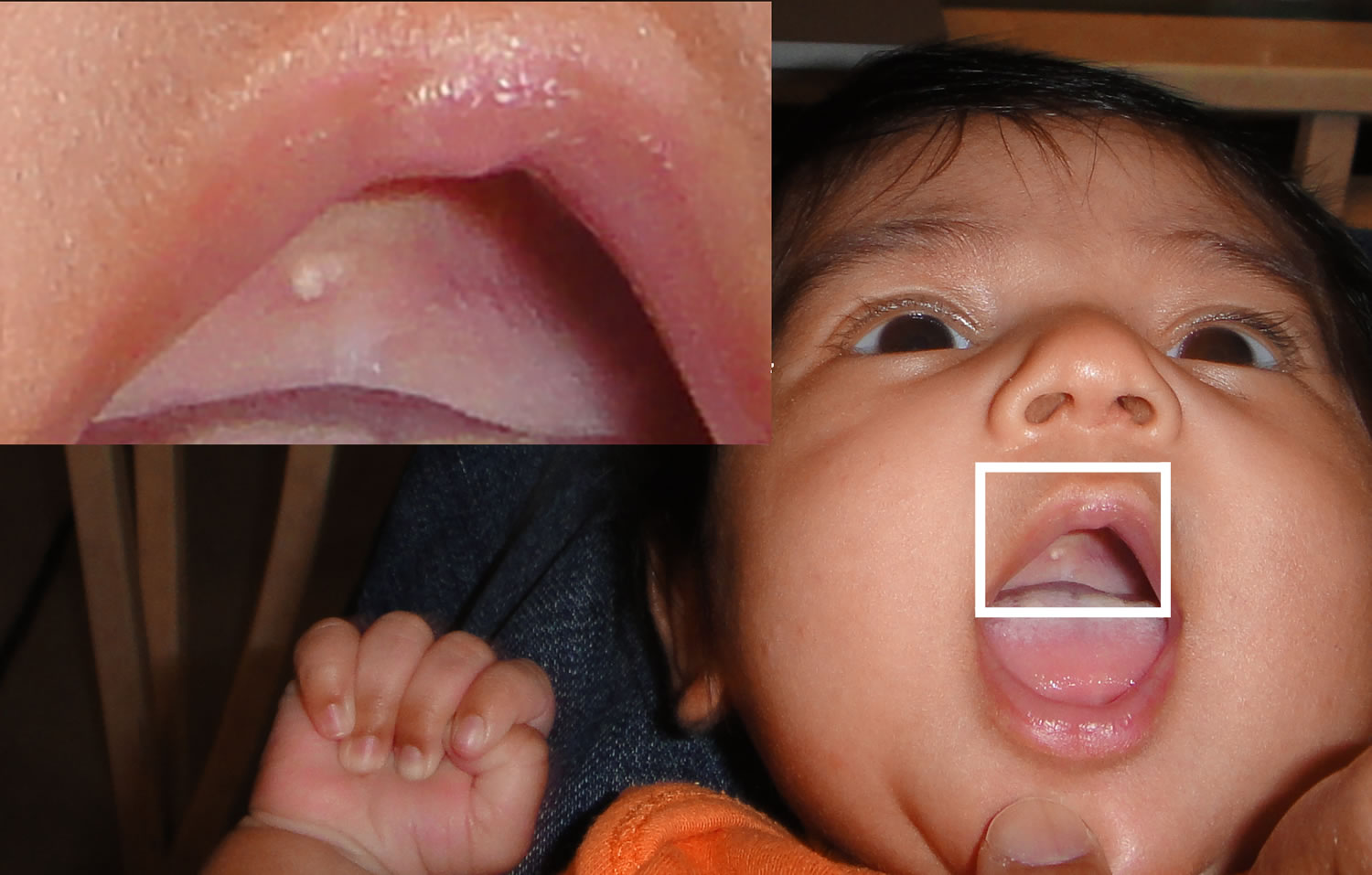
- Epstein pearls resemble the equivalent of milia (white papules produced by retention of sebum and keratin in the hair follicles), which are frequently seen on the faces of neonates.
Figure 1. Epstein pearls on hard palate
What causes Epstein pearls?
Near the end of the 8 weeks in utero, the palate begins its development. Each maxillary process generates a lateral palatine process within the mouth. These processes are horizontal and shelf like, growing from the lateral aspect of the mouth toward the midline and downward. Between the 10 to the 11 weeks in utero, the lateral palatine processes meet and fuse with each side and with the much smaller premaxillary process and the nasal septum; palatal fusions normally are completed by the end of the 4 months of gestation.
In this stage, there is a theory that states that epithelium entrapped between the palatal shelves and the nasal process formed cysts called Epstein pearls. Another theory expressed that these cysts may come from epithelial remnants that have arisen from the formation of the minor salivary glands of the palate.
Epstein pearls treatment
No treatment or removal is required 1. Most of these cysts involute or spontaneously rupture eliminating their keratin contents into the oral cavity within the first few weeks to months of postnatal life. Epstein pearls are hardly ever seen after three months of age. However, it has been suggested that part of the cystic epithelium may remain inactive even in the adult gingiva.
Epstein pearls differential diagnosis
After a lesion is found in the oral cavity, it is important to formulate a differential diagnosis since this will help lead any additional evaluation of the condition and managing the patient.
Some of the differential diagnosis for Epstein pearls include:
- Bohn nodules which are mucous gland cysts, frequently located on the buccal or lingual aspects of the alveolar ridges and rarely on the palate. Histologically consist of mucous glands and ducts. They are numerous, grayish white and firm in consistency.
- Dental lamina cyst (gingival cyst of newborn) are remnants of dental lamina epithelium found on the crest of the alveolar mucosa. Dental lamina cyst is a benign oral mucosal lesion of transient nature. Dental lamina cysts (gingival cysts) are yellow-white cystic lesion over the alveolar crest that arises from epithelial remnants of the degenerating dental lamina. Dental lamina cysts (gingival cysts) may be greater in size, more transparent, solitary lesion and fluctuant. Dental lamina cysts (gingival cysts) are usually multiple but do not increase in size. Since the lesions are self-limiting and spontaneously shed a few weeks or months after birth no treatment is required. Clinical diagnoses of these conditions are important in order to avoid unnecessary therapeutic procedure and provide suitable information to parents about the nature of the lesion. In addition, it may be incorrectly diagnosed as natal teeth if present in mandibular anterior region.
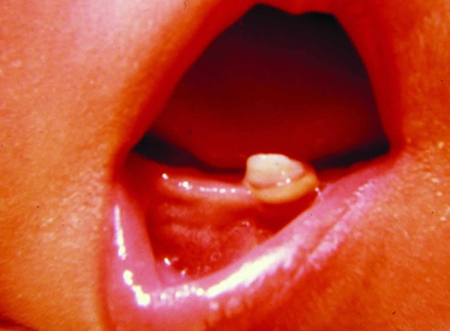
- Natal and Neonatal teeth are rare and usually located in the lower incisors region. Humans are usually born edentulous (without teeth), but tooth eruption may occur before birth (natal teeth) or within the first month of life (neonatal teeth) at a rate of 1:2,000 to 1:3,000 live births (Figure 4) 2. Central mandibular incisors are most likely to prematurely erupt; these are usually primary dentition and not extra teeth 3, so they should not be extracted without cause. Management is usually observation, although extraction may be considered if teeth are mobile and present an aspiration risk, interfere with breastfeeding or cause Riga-Fede ulceration 3. Natal teeth are most commonly an isolated finding but can be associated with genetic syndromes of chondroectodermal dysplasia (Ellis-van Creveld syndrome), oculomandibulofacial syndrome (Hallermann-Streiff syndrome) 4 and recognized syndromes including Rubinstein-Taybi, Hallerman-Streiff, Ellis-van Creveld, Pierre-Robin, pachyonychia congenita, short rib-polydactyly type 2, steatocystoma multiplex, cyclopia, Pallister-Hall, and many others 1. Therefore if the patient has any other dysmorphic features, genetic evaluation is indicated.
- Congenital epulis is pedunculated, soft nodule from 1 mm to several cms in diameter located on gingival margin.
A congenital ranula is a translucent, firm papule or nodule found on the anterior floor of mouth, lateral to lingual frenulum. - Alveolar lymphangioma is bluish fluid-filled lesions on the alveolar ridges. No seen in the palate. Predominant in black neonates. These lesions can be isolated or multiples.
Bohn nodules
Bohn’s nodules are keratin cysts derived from remnants of odontogenic epithelium over the dental lamina or may be remnants of minor salivary glands. They occur on the alveolar ridge, more commonly on the maxillary than mandibular.
Bohn’s nodules usually rupture spontaneously and disappear within a few weeks to a few months. Counseling of the family members regarding its benign and self-limiting nature is all that is required in the management.
Figure 2. Bohn’s nodules
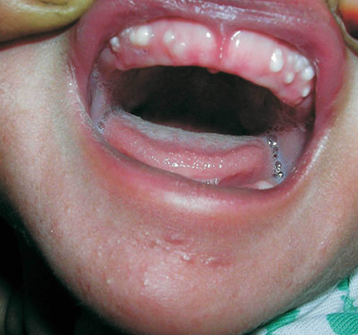
Footnote: A full term newborn boy, weighing 3 kg, born out of an uncomplicated pregnancy, was brought to us for evaluation of a few small, white and round bumps on the gingival surface. Examination of the oral cavity showed multiple, firm, pearly-white papules measuring 2 to 4 mm in diameter, grouped over the vestibular aspect of the alveolar ridge of the maxillary arch (Figure 2). Two similar lesions were seen on the mandibular area. These lesions were asymptomatic, non-tender, and fixed to the mucosa. Oral mucosa was otherwise normal. A few milia were noted on his chin. Detailed systemic examination was normal. No specific therapy was prescribed. Within a couple of months, most of the lesions subsided spontaneously. Based on the clinical features and the natural course of the disease, a diagnosis of Bohn’s nodule was made.
[Source 5 ]Figure 3. Dental lamina cyst (gingival cyst)
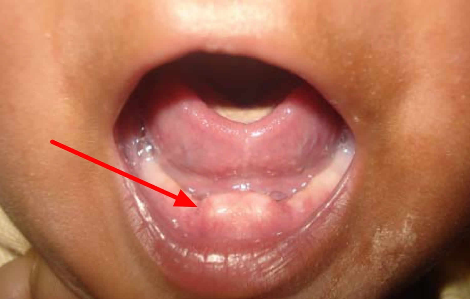
Footnote: A 14-day-old male newborn infant presented with nodular papules in the deciduous lower central incisor region. No treatment of any kind was
done, except for parental counseling and reassurance. It is important to note that management of all oral inclusion cysts (dental lamina cysts, Epstein pearls and Bohn’s nodules) remains the same, as all these have a self-limiting nature and require no treatment.
Figure 4. Natal tooth
- Diaz de Ortiz LE, Mendez MD. Epstein Pearls. [Updated 2018 Oct 27]. In: StatPearls [Internet]. Treasure Island (FL): StatPearls Publishing; 2018 Jan-. Available from: https://www.ncbi.nlm.nih.gov/books/NBK493177
- Cunha RF, Boer FA, Torriani DD, Frossard WT. Natal and neonatal teeth: review of the literature. Pediatr Dent. 2001. Mar-Apr;23(2):158-162.
- Bodenhoff J, Gorlin RJ. Natal and neonatal teeth: folklore and fact. Pediatrics. 1963. December;32(6):1087-1093.
- Hattab FN, Yassin OM, Sasa IS. Oral manifestations of Ellis-van Creveld syndrome: report of two siblings with unusual dental anomalies. J Clin Pediatr Dent. 1998. Winter;22(2):159-165.
- Bohn’s Nodules. Indian Pediatr 2014;51: 849-850 https://indianpediatrics.net/oct2014/oct-849-850.htm
- Singh RK, Kumar R, Pandey RK, Singh K. Dental lamina cysts in a newborn infant. BMJ Case Rep. 2012;2012:bcr2012007061. Published 2012 Oct 9. doi:10.1136/bcr-2012-007061 https://www.ncbi.nlm.nih.gov/pmc/articles/PMC4544414/
- Clark MB, Clark DA. Oral Development and Pathology. Ochsner J. 2018;18(4):339-344. https://www.ncbi.nlm.nih.gov/pmc/articles/PMC6292461/


