Evans syndrome
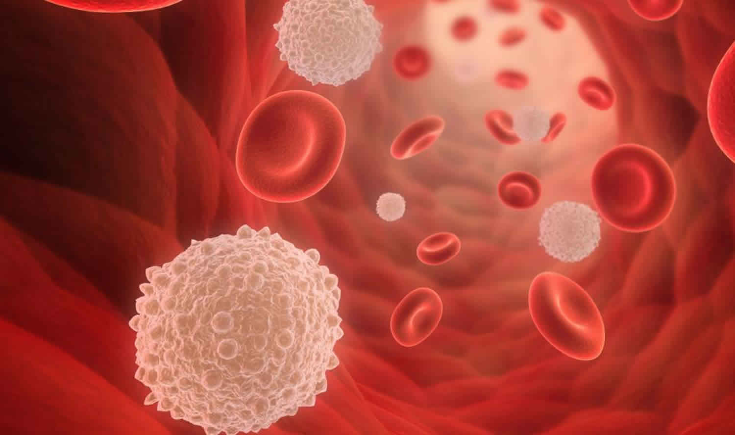
Evans syndrome is a rare autoimmune disorder in which the body’s immune system produces antibodies that mistakenly destroy red blood cells, platelets and sometimes certain white blood cell known as neutrophils 1. This leads to abnormally low levels of these blood cells in the body (cytopenia). The premature destruction of red blood cells (hemolysis) is known as autoimmune hemolytic anemia. Thrombocytopenia refers to low levels of platelets (idiopathic thrombocytopenia purpura). Neutropenia refers to low levels of certain white blood cells known as neutrophils. Evans syndrome is defined as the association of autoimmune hemolytic anemia along with immune thrombocytopenia; neutropenia occurs less often. In some cases, autoimmune destruction of these blood cells occurs at the same time (simultaneously); in most cases, one condition develops first before another condition develops later on (sequentially). The type of autoimmune hemolytic anemia that presents in Evans syndrome is warm autoimmune hemolytic anemia, in which IgG antibodies react with red blood cell surface antigens at body temperature, as opposed to cold autoimmune hemolytic anemia. In immune-mediated thrombocytopenia, the immune system is directed against GPIIb/IIIa on the platelets. The symptoms and severity of Evans syndrome can vary greatly from one person to another. Evans syndrome can potentially cause severe, life-threatening complications. Evans syndrome may occur by itself as a primary (idiopathic) disorder or in association with other autoimmune disorders or lymphoproliferative disorders as a secondary disorder. (Lymphoproliferative disorders are characterized by the overproduction of white blood cells.) The distinction between primary and secondary Evans syndrome is important as it can influence treatment. Presentations of Evans syndrome can vary, being self-controlled in some cases and relapsing-remitting in others. Many cases respond to treatment transiently, but relapses are very common.
Recently, a proposition has been laid out to classify the condition as primary (idiopathic) or secondary (associated with an underlying disorder) 2. Secondary Evans syndrome has been associated with diseases such as systemic lupus erythematosus (SLE), common variable immunodeficiency and autoimmune lymphoproliferative syndrome in Non-Hodgkin’s lymphoma in patients older than 50 years, chronic lymphocytic leukemia, viral infections (such as HIV, hepatitis C) and following allogeneic hematopoietic cell transplantation 3. Identifying Evans syndrome as secondary when associated with a disease is important because cytopenias have been observed to be more severe when with Evans syndrome in contrast to when presenting alone as autoimmune hemolytic anemia or immune thrombocytopenia. Also, the treatment options differ according to the classification.
Evans syndrome is rare, diagnosed in less than 5% of all patients with either autoimmune hemolytic anemia or immune thrombocytopenia at onset. Mean age at the time of diagnosis is 52 years and like most of the other autoimmune conditions, it is more prevalent in females, sex ratio is 3:2 in females to males 2. Although usually considered a disease of children, adults are also be affected by Evans syndrome. It is generally a sporadic condition, and there are no genes identified to associate with the condition. It can rarely be inherited when it is called “familial Evans syndrome.” Some of these cases have been reported to co-occur with other disorders, such as heart defects 4 or with inherited disorders such as hereditary spastic paraplegia 5.
Evans syndrome causes
The exact cause of Evans syndrome is unknown, which is why it is usually considered an idiopathic condition. Evans syndrome is an autoimmune disease, in which B cells produce auto-antibodies that attack own cells, in this case, red blood cells, platelets, and white blood cells. More recently, there has been speculation that Evans syndrome is likely a result of excessive immune dysregulation.
The immune system normally responds to foreign substances by producing specialized proteins called antibodies. Antibodies work by destroying foreign substances directly or coating them with a substance that marks them for destruction by white blood cells. When antibodies target healthy tissue they may be referred to as autoantibodies. Researchers believe that a triggering event (such as an infection or an underlying disorder) may induce the immune system to produce autoantibodies in Evans syndrome.
Evans syndrome may occur in combination with another disorder as a secondary condition. Secondary Evans syndrome can be associated with other disorders including autoimmune lymphoproliferative syndrome (ALPS), lupus, antiphospholipid syndrome, Sjogren’s syndrome, common variable immunodeficiency, IgA deficiency, certain lymphomas, and chronic lymphocytic leukemia.
Evans syndrome symptoms
Signs and symptoms of Evans syndrome are variable and depend on the type of blood cell lines that are affected. In the presence of autoimmune hemolytic anemia, they can present with fatigue, pallor, dizziness, shortness of breath, and limitation of physical activity. Physical examination usually shows pallor and jaundice. The spleen can be enlarged. Easy bruising or bleeding on minor injuries, petechiae, and purpura occur in those with immune thrombocytopenia and recurrent infections in those with neutropenia.
Other individuals may first present with low levels of platelets, known as thrombocytopenia. Thrombocytopenia may cause tiny reddish or purple spots on the skin (petechiae), larger purplish discoloration on the skin caused by bleeding from ruptured blood vessels into subcutaneous tissue (ecchymosis), and purpura, a rash consisting of purple spots cause by internal bleeding from small blood vessels. Affected individuals may be more susceptible to bruising following minimal injury and spontaneous bleeding from the mucous membranes.
Immune thrombocytopenia in Evans syndrome, in some cases, has been reported to be severe enough to lead to a life-threatening hemorrhage 2. There have been cases of increased risk of ischemic complications such as events related to the acute coronary syndrome or cerebrovascular events secondary to autoimmune hemolytic anemia, frequently in those older than 60 years 2.
Low levels of white blood cells, known as neutropenia, occurs less frequently in individuals with Evans syndrome than anemia or thrombocytopenia. Individuals with neutropenia may be susceptible to recurrent infections. General symptoms may include fever, a general feeling of poor health (malaise) and sores (ulcers) on the mucous membranes of the mouth.
Additional symptoms that may occur in individuals with Evans syndrome include enlargement of the lymph nodes, spleen and liver. These findings may come and go or, in some cases, may only occur during acute episodes.
All too often, patients with Evans syndrome may not respond to treatment (refractory Evans syndrome) and can eventually progress to cause life-threatening complications including sepsis, severe bleeding (hemorrhaging) episodes, and significant cardiovascular problems including heart failure.
Evans syndrome complications
Potential complications of Evans syndrome include the following:
- Hemorrhage with severe thrombocytopenia – The national survey reported hemorrhage in 29% of patients, with 2 deaths resulting from severe gastrointestinal bleeding and a third death from acute intracranial bleeding 6
- Serious infection in patients with neutropenia – The national survey showed invasive infections in 29% of patients, including pneumonia, sepsis, meningitis with Streptococcus pneumoniae, localized abscess, and osteomyelitis 6; one patient died of presumed sepsis and liver failure 9 years after splenectomy
Evans syndrome diagnosis
Once anemia is diagnosed on complete blood count and differential, if Evans syndrome is suspected, further workup such as levels of lactate dehydrogenase, haptoglobin, bilirubin, and reticulocyte count is usually required to evaluate for hemolysis. Positive direct antiglobulin test and spherocytes on peripheral smear further confirm warm autoimmune hemolytic anemia. Evans syndrome is a diagnosis of exclusion. Therefore, ruling out common etiologies such as cold agglutinin disease, thrombotic thrombocytopenic purpura through careful evaluation of the peripheral blood smear, infectious causes (such as HIV, Hepatitis C), other autoimmune conditions and malignancies is required before the diagnosis of Evans syndrome can be made.
There are no established guidelines regarding which tests should be performed in patients suspected to have secondary Evans syndrome to look for an underlying disease. However, with common disorders such as SLE (systemic lupus erythematosus), autoimmune lymphoproliferative syndrome in young patients, a minimal workup to evaluate for malignancy, including a chest and abdominal computed tomography scan should be performed.
Evans syndrome treatment
Management of Evans syndrome is challenging as many patients are refractory to common treatments that work very well with isolated autoimmune hemolytic anemia. Treatment depends on various factors, including the severity of the condition, presenting signs and symptoms, and patient co-morbidities. Symptomatic management such as transfusions is required in those with low blood counts presenting with symptoms secondary to anemia or bleeding in those with thrombocytopenia.
For definitive management, first-line treatment is usually corticosteroids or intravenous immunoglobulin (IVIG). Steroids are given at 1 to 2 mg/kg per day tapered over weeks in case of isolated immune thrombocytopenia or over months when warm autoimmune hemolytic anemia is present. In the presence of immune thrombocytopenia, IVIG (intravenous immunoglobulin) is used relatively more frequently compared with patients with isolated autoimmune hemolytic anemia.
Although most of the patients have been observed to respond to corticosteroids initially, the duration of response can vary, and more than half relapse, making the use of additional or alternative treatment options imperative 2.
Rituximab or splenectomy may be considered in those refractory to the standard treatment or if steroid-dependant (that is, at least prednisone greater than or equal to 15 mg required daily to prevent relapse). Again, the responses can be variable. Rituximab is usually preferred due to increased response and particularly when Evans syndrome is likely secondary to an underlying condition such as a malignancy or SLE, and also in those at increased risk of infections due to co-morbidities making it necessary to avoid splenectomy 7. Its combined use with steroids has been reported to have remission rates as high as 76% according to one study 8. Splenectomy is becoming less frequent now, usually reserved for those refractory to medical treatment due to low response rates, higher relapse and increased risk of increased sepsis. Danazol has frequently been used as a second-line treatment option especially with its corticosteroid-sparing effects 2.
Immunosuppressive drugs can be used in those unresponsive to corticosteroids or rituximab. Various immunosuppressants have been tried, but cyclosporin A 8 and mycophenolate mofetil 9 are the preferred ones due to increased efficacy in autoimmune conditions. Others that have also been used include cyclophosphamide, 10 azathioprine 11, and sirolimus 12. The choice of immunosuppressant is dependant on patient factors, co-morbidities, and disease severity. Hematopoietic stem cell transplant has been used very rarely as a last resort in those unresponsive to all medical treatments. Both autologous and allogeneic stem cell transplantation has been tried in a small number of patients, with mixed results 11.
Evans syndrome prognosis
Those with Evans syndrome mostly require some kind of treatment. Even with treatment, responses can be variable, and relapses are common. They are more prone to developing other autoimmune disorders such as SLE 2. According to a study done on patients with Evans syndrome, only 32% were in remission off treatment at a median follow-up of 4.8 years 2.
The characteristic clinical course of Evans syndrome includes periods of remission and exacerbation. Patients rarely do well without treatment, and responses to therapy are variable and often disappointing. On occasion, Evans syndrome can be fatal.
Recurrences of thrombocytopenia and anemia are common, as are episodes of hemorrhage and serious infections. In a national survey by Mathew et al, recurrences of thrombocytopenia were documented in 60% of Evans syndrome patients; the number of reported recurrent episodes was 1-20 6. Autoimmune hemolytic anemia recurred in 31% of patients; the number of episodes ranged from 1 to 8. Neutropenia recurred in 15% of patients.
Treatment occasionally provides complete resolution. In a median follow-up study of 42 patients (age, 4 months to 18.9 years) that spanned 3 years, 3 patients (7%) had died, 20 (48%) had active disease and remained on some treatment, and 5 (12%) had persistent disease but were not receiving any treatment 6. The remaining 14 (33%) had no evidence of disease for 1.5 months to 5 years (median, 1 year).
In the national survey, each patient received a median of 5 (range, 1-12) treatment modalities, either in combination or sequentially 6. Only 1 patient received no treatment; this patient’s hemoglobin levels were 9-13.2 g/dL and platelet counts were 9-208,000/µL during follow-up examinations over 11 years.
Long-term survival data are limited. In patients followed for a median range of 3-8 years, mortality ranged from 7-36% 6. The main causes of death were hemorrhage and sepsis. None of these patients developed any malignancy.
References- Shaikh H, Zulfiqar H, Mewawalla P. Evans Syndrome. [Updated 2019 Jun 9]. In: StatPearls [Internet]. Treasure Island (FL): StatPearls Publishing; 2019 Jan-. Available from: https://www.ncbi.nlm.nih.gov/books/NBK519015
- Michel M, Chanet V, Dechartres A, Morin AS, Piette JC, Cirasino L, Emilia G, Zaja F, Ruggeri M, Andrès E, Bierling P, Godeau B, Rodeghiero F. The spectrum of Evans syndrome in adults: new insight into the disease based on the analysis of 68 cases. Blood. 2009 Oct 08;114(15):3167-72.
- Seif AE, Manno CS, Sheen C, Grupp SA, Teachey DT. Identifying autoimmune lymphoproliferative syndrome in children with Evans syndrome: a multi-institutional study. Blood. 2010 Mar 18;115(11):2142-5.
- Ahmed FE, Albakrah MS. Neonatal familial Evans syndrome associated with joint hypermobility and mitral valve regurgitation in three siblings in a Saudi Arab family. Ann Saudi Med. 2009 May-Jun;29(3):227-30.
- Ahmed FE, Qureshi IM, Wooldridge MA, Pejaver RK. Hereditary spastic paraplegia and Evans’s syndrome. Acta Paediatr. 1996 Jul;85(7):879-81.
- Mathew P, Chen G, Wang W. Evans syndrome: results of a national survey. J Pediatr Hematol Oncol. 1997 Sep-Oct. 19(5):433-7.
- Crowther M, Chan YL, Garbett IK, Lim W, Vickers MA, Crowther MA. Evidence-based focused review of the treatment of idiopathic warm immune hemolytic anemia in adults. Blood. 2011 Oct 13;118(15):4036-40.
- Bader-Meunier B, Aladjidi N, Bellmann F, Monpoux F, Nelken B, Robert A, Armari-Alla C, Picard C, Ledeist F, Munzer M, Yacouben K, Bertrand Y, Pariente A, Chaussé A, Perel Y, Leverger G. Rituximab therapy for childhood Evans syndrome. Haematologica. 2007 Dec;92(12):1691-4.
- Emilia G, Messora C, Longo G, Bertesi M. Long-term salvage treatment by cyclosporin in refractory autoimmune haematological disorders. Br. J. Haematol. 1996 May;93(2):341-4.
- Kotb R, Pinganaud C, Trichet C, Lambotte O, Dreyfus M, Delfraissy JF, Tchernia G, Goujard C. Efficacy of mycophenolate mofetil in adult refractory auto-immune cytopenias: a single center preliminary study. Eur. J. Haematol. 2005 Jul;75(1):60-4.
- Mathew P, Chen G, Wang W. Evans syndrome: results of a national survey. J. Pediatr. Hematol. Oncol. 1997 Sep-Oct;19(5):433-7.
- Chang DK, Yoo DH, Kim TH, Jun JB, Lee IH, Bae SC, Kim IS, Kim SY. Induction of remission with intravenous immunoglobulin and cyclophosphamide in steroid-resistant Evans’ syndrome associated with dermatomyositis. Clin. Rheumatol. 2001;20(1):63-6.


