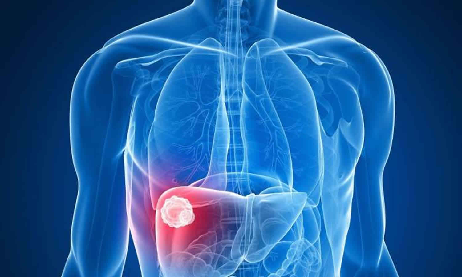
Fibrolamellar hepatocellular carcinoma
Fibrolamellar carcinoma also known as fibrolamellar hepatocellular carcinoma, is a rare form of liver cancer (hepatocellular carcinoma) characterized on microscopy by laminated fibrous layers between the tumor cells, which is generally diagnosed in adolescents and young adults (before age 40). Fibrolamellar carcinoma tends to develop in people in their 20’s or 30’s, and it’s not usually linked with cirrhosis or infection with hepatitis B or C. Many people with early fibrolamellar carcinoma have no signs or symptoms of the condition. When present, symptoms are often nonspecific (i.e. abdominal pain, weight loss, malaise) and blamed on other, more common conditions. Currently, there are no known causes of fibrolamellar hepatocellular carcinoma, and it is very difficult to diagnose because its symptoms can be attributed to many things, such as abdominal pain and fatigue. Unlike hepatocellular carcinoma, there is no blood test available to raise a doctor’s suspicion that the symptoms may be caused by something other than the flu. For most patients with fibrolamellar hepatocellular carcinoma, the diagnosis is made after a CT-scan or MRI of the abdomen is performed, long after all other tests are completed and other causes ruled out. At that point, the images often reveal a large liver mass. Fibrolamellar carcinoma is typically treated with surgical resection 1. This surgery can involve either a resection, where part of the liver is taken out, or a liver transplant, where the whole liver is taken out and replaced with a donor liver. If the surgeon does a resection, you will be able to live and function well even with part of your liver missing. When surgery is not possible or when the cancer has spread, chemotherapy will be used to treat the fibrolamellar carcinoma. Depending on where the fibrolamellar hepatocellular carcinoma is, embolization therapy can be used. This type of therapy cuts off the blood supply to the part of the liver where the fibrolamellar hepatocellular carcinoma is located. This causes the fibrolamellar hepatocellular carcinoma to die because it can’t get oxygen and nutrients.
How common is fibrolamellar carcinoma?
Fibrolamellar hepatocellular carcinoma is so rare that there is little data on how many people have it. It is thought to make up 1% to 5% of all liver cancers. Fibrolamellar hepatocellular carcinoma affects both men and women and is thought to occur in about one in five million people in the US.
Fibrolamellar hepatocellular carcinoma causes
The exact underlying cause of fibrolamellar carcinoma is poorly understood. Unlike other forms of liver cancer (hepatocellular carcinoma), fibrolamellar carcinoma typically occurs in the absence of underlying liver inflammation, scarring (cirrhosis), hepatotoxins, α1-antitrypsin deficiency or hemochromatosis; thus, specific risk factors for fibrolamellar carcinoma remain unidentified.
Scientists know that in fibrolamellar hepatocellular carcinoma, a chromosome (the part of your cells that contains your genes) breaks apart and gets put back together in the wrong way. This can cause cells to not function like they should. In fibrolamellar hepatocellular carcinoma, a gene called DNAJB1 joins with a gene called PRKACA. This happens in almost all cases, so it may be very important for how fibrolamellar hepatocellular carcinoma forms. Scientists are trying to figure out how this works, so they can invent new therapies. Your doctor may test your tumor for DNAJB1 and PRKACA to confirm it is fibrolamellar hepatocellular carcinoma.
Fibrolamellar hepatocellular carcinoma symptoms
Patients with fibrolamellar carcinoma typically present with nonspecific symptoms or no symptoms at all. When symptoms develop, they are most commonly the following 2:
- Abdominal pain
- Weight loss
- Malaise
Uncommonly, presenting signs and symptoms may include the following:
- Migratory thrombophlebitis (Trousseau syndrome) or venous thrombosis as a result of direct invasion of the hepatic veins and inferior vena cava (IVC) or mass effect on the IVC 3
- Pain and fever simulating a hepatic abscess 4
- Obstructive jaundice due to biliary compression 5
- Gynecomastia, due to aromatase production by fibrolamellar carcinoma cells and resultant conversion of circulating androgens to estrogens 6
The most common physical finding is an abdominal mass or fullness due to hepatomegaly 7.
Unlike patients with typical hepatocellular carcinoma, the stigmata of chronic liver disease and portal hypertension are usually absent in patients with fibrolamellar carcinoma.
Fibrolamellar hepatocellular carcinoma diagnosis
Laboratory studies
Hepatic enzymes
Mild elevations of serum aspartate aminotransferase (AST) and alanine aminotransferase (ALT) 8 and alkaline phosphatase may be identified but are commonly absent 9.
Elevation of serum bilirubin is rarely identified, but subclinical biliary obstruction may be relatively common, as 40% of cases demonstrate intrahepatic biliary ductal dilatation on imaging studies 10.
Other circulating tumor markers
Alpha fetoprotein (AFP) levels are typically normal but may be elevated. Less than 10% of patients with fibrolamellar carcinoma (fibrolamellar hepatocellular carcinoma) have alpha fetoprotein levels greater than 200 ng/mL, [44] and even this finding may in part represent misclassification of hepatocellular carcinoma (HCC) as fibrolamellar hepatocellular carcinoma 11.
Serum carcinoembryonic antigen (CEA) can be elevated on occasion.
An association between fibrolamellar hepatocellular carcinoma and increased serum binding capacity of vitamin B-12 has been reported 12. Likewise, elevated serum neurotensin levels have been reported in association with fibrolamellar hepatocellular carcinoma 13. Neither test can accurately differentiate fibrolamellar hepatocellular carcinoma from other liver tumors, and neither test is useful as a screening modality. However, these levels may have utility for following a tumor’s response to chemotherapy or for monitoring disease recurrence in patients following tumor resection.
Imaging studies
Fibrolamellar carcinoma (fibrolamellar hepatocellular carcinoma) is typically found as a solitary mass in an otherwise normal-appearing liver. fibrolamellar hepatocellular carcinomas can be quite large, and a visible central scar is noted in 20-60% of cases 14. On abdominal radiographs, calcifications in a nodular or stellate pattern may be seen in up to 40% of cases.
fibrolamellar hepatocellular carcinoma must be carefully differentiated from focal nodular hyperplasia because their management differs. Epidemiologic characteristics are similar in that both lesions occur in young, healthy patients who usually do not have a history of cirrhosis or liver disease. However, fibrolamellar hepatocellular carcinoma requires aggressive treatment by liver resection, while focal nodular hyperplasia, a benign disease, does not require treatment unless the patient is symptomatic.
Computed tomography (CT) scan
The imaging technique of choice for staging is computed tomography (CT).
Fibrolamellar hepatocellular carcinoma usually appears as a hypoattenuated, well-defined, solitary mass on a nonenhanced CT scan. When a dynamically enhanced CT scan is used with arterial- and portal venous-phase contrast, the cellular portion enhances prominently and is heterogeneous, consistent with its vascular characteristics.
The central scar, if present, can be viewed on nonenhanced and arterial-phase scans; however, it is best viewed on delayed images because the unscarred portion of the mass becomes more homogeneous. Note that the fibrous scar in fibrolamellar hepatocellular carcinoma typically does not enhance 15. This is in contrast to focal nodular hyperplasia, in which the central scar, which is in reality a vascular entity, enhances on arterial-phase CT scan images 16. This difference generally distinguishes fibrolamellar hepatocellular carcinoma from focal nodular hyperplasia but is not absolute, as up to 25% of fibrolamellar hepatocellular carcinomas may in fact demonstrate delayed enhancement of the central scar.
Calcifications are present in 33-55% of fibrolamellar hepatocellular carcinoma and are usually located within the central scar but may less commonly be located at the tumor periphery 17.
Pseudoencapsulation of the tumor, caused by compression of adjacent liver parenchyma, is present in up to 15% of cases. However, true encapsulation is more characteristics of typical hepatocellular carcinoma.
Retraction of the adjacent Glisson capsule can occur in up to 10% of fibrolamellar hepatocellular carcinoma cases, but it is also found with other primary liver malignancies. However, Glisson capsule retraction argues against a benign lesion such as focal nodular hyperplasia 18.
As noted above, the ability to differentiate fibrolamellar hepatocellular carcinoma from other lesions that also demonstrate central scars is useful. In a blinded retrospective review of CT scans of 64 patients with liver tumors (20 fibrolamellar hepatocellular carcinoma, 29 focal nodular hyperplasia, and 15 hemangiomas), findings that were useful in differentiating fibrolamellar hepatocellular carcinoma from focal nodular hyperplasia and large hemangiomas were as follows 19:
- Tumor size greater than 10 cm (focal nodular hyperplasia is usually < 5 cm)
- Nodular centripetal enhancement (typical of hemangioma)
- Invasion of hepatic vessels or bile ducts (rules out focal nodular hyperplasia and hemangioma)
- Scar width greater than 2 cm (suggests fibrolamellar carcinoma or hemangioma over focal nodular hyperplasia)
- Heterogeneity (typical of fibrolamellar hepatocellular carcinoma, also found in hemangioma but in a characteristic pattern of nodular centripetal enhancement, uncommon in focal nodular hyperplasia)
- Isoattenuation with blood vessels (suggests focal nodular hyperplasia over fibrolamellar hepatocellular carcinoma or hemangioma)
- Extrahepatic metastases (rules out focal nodular hyperplasia and hemangioma)
- Calcification (suggests fibrolamellar hepatocellular carcinoma over focal nodular hyperplasia or hemangioma)
- Surface lobulation (suggests fibrolamellar hepatocellular carcinoma or hemangioma over focal nodular hyperplasia)
- Almost isoattenuating on portal venous phase images (favors fibrolamellar hepatocellular carcinoma or focal nodular hyperplasia over hemangioma)
Ultrasonography
Ultrasonography, which is often the initial study performed for right upper quadrant abdominal pain, commonly shows fibrolamellar hepatocellular carcinoma as a solitary, well-defined mass with variable echotexture. Tumors with mixed echotexture are most common (60%) and are predominantly hyperechoic or isoechoic. The sensitivity of ultrasonography for detecting a central scar is only 33-60% (as compared with CT and pathological analysis) 14 and when present is visualized as a central area of hyperechogenicity. Ultrasonography is less accurate than CT scan or MRI for evaluation of lymph node involvement and staging 20.
Angiography
Angiography may demonstrate a hypervascular lesion with a dense tumor blush and an avascular region corresponding to the central scar. However, it is of limited value in diagnosing fibrolamellar hepatocellular carcinoma. On the other hand, it does have utility for defining portal venous and hepatic arterial anatomy and may aid in preoperative planning by allowing assessment of vascular invasion.
Fine-needle aspiration biopsy
In some patients, fine needle aspiration biopsy (FNAB) can allow a histopathologic diagnosis to be made prior to operative intervention 21.
Fine needle aspiration biopsy (FNAB) should not be performed if the tumor is deemed resectable based on imaging studies. But, if the tumor is unresectable, FNAB may facilitate a tissue diagnosis in order for the oncologist to select the appropriate chemotherapy regimen.
Fine needle aspiration biopsy should be used in cases when the diagnosis is unclear, such as when focal nodular hyperplasia is considered in an asymptomatic patient. This is important because distinguishing fibrolamellar carcinoma from focal nodular hyperplasia has implications for treatment and prognosis.
The cytologic findings of fibrolamellar carcinoma are very characteristic 22:
- Large polygonal tumor cells: A single cell aspirated from a fibrolamellar carcinoma is 3 times the size of a normal hepatocyte and 1.6 times the size of a single cell aspirated from a well-differentiated typical hepatocellular carcinoma.
- Large nuclei and prominent nucleoli
- Abundant eosinophilic, granular cytoplasm, such that the N/C ratio is not as high as that for typical hepatocellular carcinoma (despite the large nucleus)
- Parallel bands of fibrous tissue may be seen in tumor fragments.
Core needle biopsy
The indications for core needle biopsy and the cytologic appearance of the tumor are as for FNAB. Core needle biopsy is more likely than FNAB to preserve the fibrous lamellae that are important to distinguish fibrolamellar carcinoma from typical hepatocellular carcinoma. As such, core needle biopsy is preferred over fine needle aspiration biopsy when a percutaneous biopsy is needed 23.
Fibrolamellar hepatocellular carcinoma staging
CT scan is the imaging study of choice for the staging of fibrolamellar carcinoma. The American Joint Commission on Cancer (AJCC) tumor-node-metastasis (TNM) staging system for fibrolamellar carcinoma is the same as for typical hepatocellular carcinoma. Metastatic spread to regional lymph nodes may be more common in fibrolamellar carcinoma than in typical hepatocellular carcinoma.
Fibrolamellar hepatocellular carcinoma treatment
Surgical resection or transplantation is the standard of care for fibrolamellar carcinoma for eligible patients. Trans-arterial chemo-embolization (TACE) may be a useful option in patients who have unresectable disease 24.
The role of neoadjuvant or adjuvant chemotherapy is controversial. Fibrolamellar hepatocellular carcinoma is not typically responsive to chemotherapy. In some cases, chemotherapy had no effect on survival or recurrence in patients who received chemotherapy after surgery. Contrary to this, in other studies, patients who had chemotherapy in neoadjuvant and adjuvant settings fared better than those with surgery only 23.
While not well studied, radiation therapy has been used to treat recurrent fibrolamellar hepatocellular carcinoma in isolated case reports. Peacock et al 25 demonstrated an 85.9% decrease in tumor volume of fibrolamellar hepatocellular carcinoma metastases using 40 Gy in 10 fractions over a 13-day period.
In addition to systemic chemotherapy, recent research has focused on taking advantage of the new understanding of the pathogenesis and molecular genetics of fibrolamellar hepatocellular carcinoma. One current multi-institutional, randomized controlled trial is evaluating mammalian target of rapamycin (mTOR) inhibition in combination with estrogen suppression in the treatment of fibrolamellar hepatocellular carcinoma 26.
Hepatic resection
Cirrhosis often limits the feasibility of liver resection in patients with typical hepatocellular carcinoma (hepatocellular carcinoma) because cirrhotic patients have increased perioperative risk and require a larger future liver remnant to be left behind to allow adequate liver function. In contrast, fibrolamellar carcinoma (fibrolamellar hepatocellular carcinoma) is rarely associated with cirrhosis, and patients are often young and otherwise healthy. For this reason, aggressive liver resections can be more readily undertaken in patients with fibrolamellar hepatocellular carcinoma than in those with typical hepatocellular carcinoma.
Several factors are associated with a better prognosis following surgery, including younger age at diagnosis, earlier tumor stage at diagnosis, and absence of large vessel invasion or thrombosis 27. The ability to perform complete resection has been reported to be one of the most important and well-described prognostic factors for fibrolamellar hepatocellular carcinoma 28. Factors associated with a particularly poor prognosis include lymph node metastasis, multiple tumors, metastatic disease at presentation, and vascular invasion 27.
Patients with resected fibrolamellar hepatocellular carcinoma generally have a prognosis similar to that for noncirrhotic patients with resected typical hepatocellular carcinoma 29. Aggressive surgical approaches, even for recurrent disease, can be undertaken with good results 9.
Orthotopic liver transplantation
Total hepatectomy followed by orthotopic liver transplantation should be considered in patients with unresectable fibrolamellar hepatocellular carcinoma that is confined to the liver. An analysis of 63 patients who received liver transplanation for fibrolamellar hepatocellular carcinoma between October 1988 and January 2013 had an overall survival at 1, 3, and 5 years of 96%, 80%, and 48%, respectively, while comparable rates in hepatocellular carcinoma patients were 89%, 77%, and 68%. Six patients had tumor recurrence (10%). The Cox Model demonstrated that Model for End-stage Liver Disease (MELD) score and cold ischemic time were the strongest predictors of overall survival in fibrolamellar hepatocellular carcinoma patients 30.
Even among patients who develop tumor recurrence, the time to recurrence is significantly longer in patients with fibrolamellar hepatocellular carcinoma than in those with typical hepatocellular carcinoma 31. This may be a result of more indolent behavior by fibrolamellar hepatocellular carcinoma versus typical hepatocellular carcinoma. Alternatively, it may result from the fact that accompanying cirrhosis in typical hepatocellular carcinoma increases the risk of recurrence (including development of a second primary tumor) via a field effect.
Orthotopic liver transplantation can result in long-term survival but is a resource-intensive treatment approach. In typical hepatocellular carcinoma, orthotopic liver transplantation confers the advantage of removing the at-risk cirrhotic liver in addition to the tumor itself. In fibrolamellar hepatocellular carcinoma, this theoretical advantage is likely not present because no such field effect is present (due to the lack of cirrhosis in most cases).
As such, it is not clear that the use of donor organs in patients with fibrolamellar hepatocellular carcinoma is an appropriate use of this scarce resource 32. Newer innovative approaches (eg, adult living-related donations, split-liver techniques, and potential use of marginal donor organs) may allow for a future increase in the use of orthotopic liver transplantation for fibrolamellar hepatocellular carcinoma.
Chemotherapy
Chemotherapy for metastatic fibrolamellar carcinoma is as for metastatic typical hepatocellular carcinoma. Single-agent chemotherapy or combinations of chemotherapeutic drugs give responses of no more than 25%, with questionable benefit for overall survival.
Sorafenib is an orally delivered small molecule that inhibits several different protein kinases, including Raf-1 and B-Raf, platelet-derived growth factor β (PDGFR-β), and vascular endothelial growth factor receptors (VEGFRs) 1, 2, and 3. Sorafenib targets both tumor growth (Raf-MEK-ERK pathway) and neoangiogenesis (VEGFRs, PDGFR-β), both of which are thought to be important in the pathogenesis of typical hepatocellular carcinoma.
A randomized controlled trial of sorafenib in 602 patients with advanced typical hepatocellular carcinoma and well-compensated cirrhosis demonstrated a survival benefit with sorafenib as compared with placebo, corresponding to a 3-month increase in median survival (10.7 versus 7.9 months) 32. A similar study will likely never be performed in fibrolamellar hepatocellular carcinoma due to the rarity of the disease. It is unclear whether the data on sorafenib for typical hepatocellular carcinoma are generalizable to fibrolamellar hepatocellular carcinoma. There are not yet any reports in the literature describing sorafenib use in fibrolamellar hepatocellular carcinoma.
A phase II clinical trial reported in 2003 evaluated 21-day courses of systemic continuous 5-fluorouracil repeated monthly and thrice-weekly subcutaneous recombinant interferon α-2b in a mixed population of 43 patients with fibrolamellar hepatocellular carcinoma or typical hepatocellular carcinoma 33. All of these patients were ineligible for surgical therapy due to extent of hepatic disease as determined by CT scan, but none had extrahepatic disease. Of nine patients with fibrolamellar hepatocellular carcinoma, eight were radiologically assessed for response, which showed one complete response (CR), four partial responses (PR), and one minor response (MR). The median survival of the fibrolamellar hepatocellular carcinoma patients was 23 months, compared with 15.5 months for typical hepatocellular carcinoma (statistical significance not reported).
Of note, a major reason the authors chose this treatment regimen was that they anticipated it would be well tolerated by patients with cirrhosis. Although the regimen was in fact well tolerated in this trial, none of the fibrolamellar hepatocellular carcinoma patients had cirrhosis. Because fibrolamellar hepatocellular carcinoma patients generally do not have cirrhosis, chemotherapeutic regimens that might not be appropriate for typical hepatocellular carcinoma might be of use in fibrolamellar hepatocellular carcinoma.
Fibrolamellar hepatocellular carcinoma prognosis
The prognosis (long-term outlook) for people with fibrolamellar hepatocellular carcinoma is generally better than other forms of liver cancer, particularly when treated with surgical resection 34. Prognosis after surgical therapy is superior to that with any medical management alone. For people with fibrolamellar hepatocellular carcinoma who are treated with surgery, 44% to 68% survive for five years. When people with fibrolamellar hepatocellular carcinoma get other treatment without surgery, 2% to 17% survive for five years. Unfortunately, the prognosis is less favorable in people with unresectable metastatic fibrolamellar carcinoma. The median survival of these patients is approximately 14 months 35.
Using data from the Surveillance, Epidemiology, and End Results (SEER) program, Mayo et al 36 studied surgical treatment and prognosis in patients with fibrolamellar carcinoma or hepatocellular carcinoma; 7,135 (99%) had hepatocellular carcinoma and 90 (1%) had fibrolamellar carcinoma. Patients with fibrolamellar carcinoma were younger (25 years vs 59 years) and more were women (44% vs 27%). Regional disease was nearly twice as common in patients with fibrolamellar carcinoma (42.2%) than in those with hepatocellular carcinoma (22.1%).
Patients with fibrolamellar carcinoma were more often treated with a hemihepatectomy compared with patients with hepatocellular carcinoma, who were more often managed with a liver transplant. However, despite a higher likelihood of advanced disease at the time of diagnosis, surgically treated fibrolamellar carcinoma patients had better long-term outcomes than patients with conventional hepatocellular carcinoma, surviving a median of 75 months vs 43 months, respectively 36.
A multi-institutional study that included 35 patients (13 female; median age, 32 years) with fibrolamellar carcinoma who underwent hepatectomy found that for curative-intent surgery (n=30), overall and recurrence-free survivals at 5 years were 62% and 45%, respectively. None of the patients who achieved a 4-year disease-free interval developed recurrence, suggesting possible freedom from disease thereafter. These authors concluded that in patients with recurrent resectable disease, repeat surgery should be strongly considered 37.
Fibrolamellar hepatocellular carcinoma survival rate
Reported 5-year survival rates for fibrolamellar hepatocellular carcinoma range from 51 to 70% following liver resection 38.
Fibrolamellar hepatocellular carcinoma life expectancy
The population-based relative survival of patients with fibrolamellar carcinoma in the United States is 73% at 1 year and 32% at 5 years. [19] In contrast, hepatocellular carcinoma relative survival is 26% at 1 year and 7% at 5 years 27. However, these figures include death from all causes. For example, because hepatocellular carcinoma patients tend to be older than those with fibrolamellar carcinoma, they may be at higher risk of death related to medical comorbidities (eg, heart disease or stroke). Nevertheless, even when only patients younger than 40 years are analyzed, fibrolamellar carcinoma still appears to portend a better prognosis than typical hepatocellular carcinoma 27.
More importantly, fibrolamellar carcinoma usually occurs in the setting of a normal liver parenchyma, while 80% of typical hepatocellular carcinoma occurs in the setting of a cirrhotic liver. Because cirrhosis is an independent risk factor for mortality, the observed difference in survival between fibrolamellar carcinoma and typical hepatocellular carcinoma may not in fact reflect differences in tumor biology or cancer-specific mortality 39.
Two studies have found that fibrolamellar carcinoma and hepatocellular carcinoma in children have similar prognoses and responses to therapy 40. Two studies in adults have found that fibrolamellar carcinoma and typical hepatocellular carcinoma in noncirrhotics have similar prognoses after resection 29, supporting the viewpoint that the survival differences seen in older groups may be confounded by competing causes of death.
References- Lim II, Farber BA, LaQuaglia MP. Advances in fibrolamellar hepatocellular carcinoma: a review. Eur J Pediatr Surg. December 2014; 24(6):461-466.
- El-Gazzaz G, Wong W, El-Hadary MK, et al. Outcome of liver resection and transplantation for fibrolamellar hepatocellular carcinoma. Transpl Int. 2000. 13 Suppl 1:S406-9.
- Kanai T, Takabayashi T, Kawano Y, et al. A case of postoperative recurrence of fibrolamellar hepatocellular carcinoma with increased vitamin B12 binding capacity in a young Japanese female. Jpn J Clin Oncol. 2004 Jun. 34(6):346-51.
- Debray D, Pariente D, Fabre M, et al. Fibrolamellar hepatocellular carcinoma: report of a case mimicking a liver abscess. J Pediatr Gastroenterol Nutr. 1994 Nov. 19(4):468-72.
- Soyer P, Roche A, Levesque M. Fibrolamellar hepatocellular carcinoma presenting with obstructive jaundice. A report of two cases. Eur J Radiol. 1991 Nov-Dec. 13(3):196-8.
- Agarwal VR, Takayama K, Van Wyk JJ, et al. Molecular basis of severe gynecomastia associated with aromatase expression in a fibrolamellar hepatocellular carcinoma. J Clin Endocrinol Metab. 1998 May. 83(5):1797-800.
- Saab S, Yao F. Fibrolamellar hepatocellular carcinoma. Case reports and a review of the literature. Dig Dis Sci. 1996 Oct. 41(10):1981-5.
- Craig JR, Peters RL, Edmondson HA, et al. Fibrolamellar carcinoma of the liver: a tumor of adolescents and young adults with distinctive clinico-pathologic features. Cancer. 1980 Jul 15. 46(2):372-9.
- Hemming AW, Langer B, Sheiner P, et al. Aggressive surgical management of fibrolamellar hepatocellular carcinoma. J Gastrointest Surg. 1997 Jul-Aug. 1(4):342-6.
- Ichikawa T, Federle MP, Grazioli L, et al. Fibrolamellar hepatocellular carcinoma: pre- and posttherapy evaluation with CT and MR imaging. Radiology. 2000 Oct. 217(1):145-51.
- Darcy DG, Chiaroni-Clarke R, Murphy JM, Honeyman JN, Bhanot U, LaQuaglia MP, et al. The genomic landscape of fibrolamellar hepatocellular carcinoma: whole genome sequencing of ten patients. Oncotarget. 2015 Jan 20. 6 (2):755-70.
- Kanai T, Takabayashi T, Kawano Y, et al. A case of postoperative recurrence of fibrolamellar hepatocellular carcinoma with increased vitamin B12 binding capacity in a young Japanese female. Jpn J Clin Oncol. 2004 Jun. 34(6):346-51.
- Collier NA, Weinbren K, Bloom SR, et al. Neurotensin secretion by fibrolamellar carcinoma of the liver. Lancet. 1984 Mar 10. 1(8376):538-40.
- Brandt DJ, Johnson CD, Stephens DH, et al. Imaging of fibrolamellar hepatocellular carcinoma. AJR Am J Roentgenol. 1988 Aug. 151(2):295-9.
- Corrigan K, Semelka RC. Dynamic contrast-enhanced MR imaging of fibrolamellar hepatocellular carcinoma. Abdom Imaging. 1995 Mar-Apr. 20(2):122-5.
- Gabata T, Matsui O, Kadoya M, et al. Delayed MR imaging of the liver: correlation of delayed enhancement of hepatic tumors and pathologic appearance. Abdom Imaging. 1998 May-Jun. 23(3):309-13.
- Soyer P, Roche A, Levesque M, et al. CT of fibrolamellar hepatocellular carcinoma. J Comput Assist Tomogr. 1991 Jul-Aug. 15(4):533-8.
- Soyer P, Bluemke DA, Vissuzaine C, et al. CT of hepatic tumors: prevalence and specificity of retraction of the adjacent liver capsule. AJR Am J Roentgenol. 1994 May. 162(5):1119-22.
- Blachar A, Federle MP, Ferris JV, et al. Radiologists’ performance in the diagnosis of liver tumors with central scars by using specific CT criteria. Radiology. 2002 May. 223(2):532-9.
- Stevens WR, Johnson CD, Stephens DH, et al. Fibrolamellar hepatocellular carcinoma: stage at presentation and results of aggressive surgical management. AJR Am J Roentgenol. 1995 May. 164(5):1153-8.
- Crowe A, Knight CS, Jhala D, Bynon SJ, Jhala NC. Diagnosis of metastatic fibrolamellar hepatocellular carcinoma by endoscopic ultrasound-guided fine needle aspiration. Cytojournal. 2011 Jan 31. 8:2.
- Kojiro M. Histopathology of liver cancers. Best Pract Res Clin Gastroenterol. 2005 Feb. 19(1):39-62
- Kassahun WT. Contemporary management of fibrolamellar hepatocellular carcinoma: diagnosis, treatment, outcome, prognostic factors, and recent developments. World J Surg Oncol. 2016 May 23. 14 (1):151.
- Sitzmann JV. Conversion of unresectable to resectable liver cancer: an approach and follow-up study. World J Surg. 1995 Nov-Dec. 19(6):790-4.
- Peacock JG, A Call J, R Olivier K. Radiotherapy for metastatic fibrolamellar hepatocellular carcinoma. Rare Tumors. 2013. 5 (3):e28.
- Riehle KJ, Yeh MM, Yu JJ, Kenerson HL, Harris WP, Park JO, et al. mTORC1 and FGFR1 signaling in fibrolamellar hepatocellular carcinoma. Mod Pathol. 2015 Jan. 28 (1):103-10.
- El-Serag HB, Davila JA. Is fibrolamellar carcinoma different from hepatocellular carcinoma? A US population-based study. Hepatology. 2004 Mar. 39(3):798-803.
- Stipa F, Yoon SS, Liau KH, et al. Outcome of patients with fibrolamellar hepatocellular carcinoma. Cancer. 2006 Mar 15. 106(6):1331-8.
- Kakar S, Burgart LJ, Batts KP, et al. Clinicopathologic features and survival in fibrolamellar carcinoma: comparison with conventional hepatocellular carcinoma with and without cirrhosis. Mod Pathol. 2005 Nov. 18(11):1417-23.
- Atienza LG, Berger J, Mei X, Shah MB, Daily MF, Grigorian A, et al. Liver transplantation for fibrolamellar hepatocellular carcinoma: A national perspective. J Surg Oncol. 2017 Mar. 115 (3):319-323.
- Schlitt HJ, Neipp M, Weimann A, et al. Recurrence patterns of hepatocellular and fibrolamellar carcinoma after liver transplantation. J Clin Oncol. 1999 Jan. 17(1):324-31.
- Llovet JM, Ricci S, Mazzaferro V, et al. Sorafenib in advanced hepatocellular carcinoma. N Engl J Med. 2008 Jul 24. 359(4):378-90.
- Patt YZ, Hassan MM, Lozano RD, et al. Phase II trial of systemic continuous fluorouracil and subcutaneous recombinant interferon Alfa-2b for treatment of hepatocellular carcinoma. J Clin Oncol. 2003 Feb 1. 21(3):421-7.
- Fibrolamellar Carcinoma. https://emedicine.medscape.com/article/278354-overview
- Bartlett D, Di Bisceglie A, Dawson L. Cancer of the Liver. De Vita VT, Lawrence TS, Rosenberg S. Cancer: Principles & Practice of Oncology. 8th ed. Philadelphia, Pa: Lippincott, Williams and Wilkins; 2008. 1: 1129-56.
- Mayo SC, Mavros MN, Nathan H, Cosgrove D, Herman JM, Kamel I, et al. Treatment and prognosis of patients with fibrolamellar hepatocellular carcinoma: a national perspective. J Am Coll Surg. 2014 Feb. 218(2):196-205.
- Groeschl RT, Miura JT, Wong RK, Bloomston M, Lidsky ML, Clary BM, et al. Multi-institutional analysis of recurrence and survival after hepatectomy for fibrolamellar carcinoma. J Surg Oncol. 2014 Sep. 110(4):412-5.
- Darcy DG, Malek MM, Kobos R, Klimstra DS, DeMatteo R, La Quaglia MP. Prognostic factors in fibrolamellar hepatocellular carcinoma in young people. J Pediatr Surg. Jan 2015; 50(1):153-156.
- Torbenson M. Review of the clinicopathologic features of fibrolamellar carcinoma. Adv Anat Pathol. 2007 May. 14(3):217-23.
- Katzenstein HM, Krailo MD, Malogolowkin MH, et al. Fibrolamellar hepatocellular carcinoma in children and adolescents. Cancer. 2003 Apr 15. 97(8):2006-12.


