What is fluorosis
Dental fluorosis is a condition that causes changes in the appearance of tooth enamel due to hypomineralization of tooth enamel, caused by excessive exposure to fluoride during tooth development affecting its organization, composition and structure 1. Fluorosis is a dental lesion caused by the destructive effect of fluoride on the tooth making cells in the forming and mineralizing stages. The affected enamel is also called mottled enamel 2. Severity of fluorosis depends on the duration of exposure to fluoride. Specific body response, weight, activity, diet, and bone growth factors result in variable severity of dental fluorosis in response to similar dose of fluoride 3.
Dental fluorosis may result when children regularly consume fluoride during the teeth-forming years, age 8 and younger. In high dosages fluorides produce a lower dental mineral content and increased porosity in the enamel and dentin 4. In the world, at least 25 countries suffer from endemic fluorosis, only in China 26 million people present fluorosis 5. Most dental fluorosis in the U.S. is very mild to mild, appearing as white spots on the tooth surface that may be barely noticeable and do not affect dental function. Moderate and severe forms of dental fluorosis, which are far less common, cause more extensive enamel changes. In the rare, severe form, pits may form in the teeth. The severe form hardly ever occurs in communities where the level of fluoride in water is less than 2 milligrams per liter.
Endemic skeletal fluorosis is a chronic metabolic bone and joint disease caused by ingesting large amounts of fluoride either through water or rarely from foods of endemic areas 6. Fluoride is a cumulative toxin which can alter accretion and resorption of bone tissue. Fluoride also affects the homeostasis of bone mineral metabolism. The total quantity of ingested fluoride is the single most important factor which determines the clinical course of the skeletal fluorosis disease which is characterized by immobilization of joints of the axial skeleton and of the major joints of the extremities. A combination of osteosclerosis, osteomalacia and osteoporosis of varying degrees as well as exostosis formation characterizes the bone lesions 6. In a proportion of cases secondary hyperparathyroidism is observed with associated characteristic bone changes. Contrary to earlier thinking, severe crippling forms of skeletal fluorosis are seen in paediatric age group too. Increased metabolic turnover of the bone, impaired bone collagen synthesis and increased avidity for calcium are features in fluoride toxicity. Osteosclerotic picture is evident when small doses of fluoride are ingested over a long period of time during which calcium intakes are apparently normal while osteoporotic forms are common in paediatric age group and with higher body load of the element. Alterations in hormones concerned with bone mineral metabolism are seen in fluorosis. Kidney is the primary organ of excretion for fluorides. Age, sex, calcium intake in the diet, dose and duration of fluoride intake and renal efficiency in fluoride handling are the factors which influence the outcome. Serum parameters rarely help in the diagnosis. Elevated urinary fluoride and increased bone fluoride content are indicators of fluoride toxicity. Fluorosis is a preventable crippling disease. No effective therapeutic agent is available which can cure fluorosis. Industrial fluorosis is on the increase on a global basis. Bone density measurement is a tool for early diagnosis.
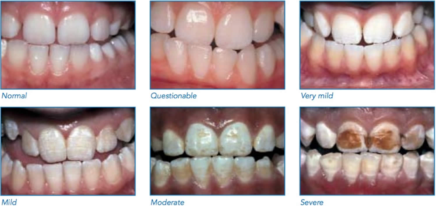
Figure 1. Dental fluorosis

Figure 2. Normal teeth
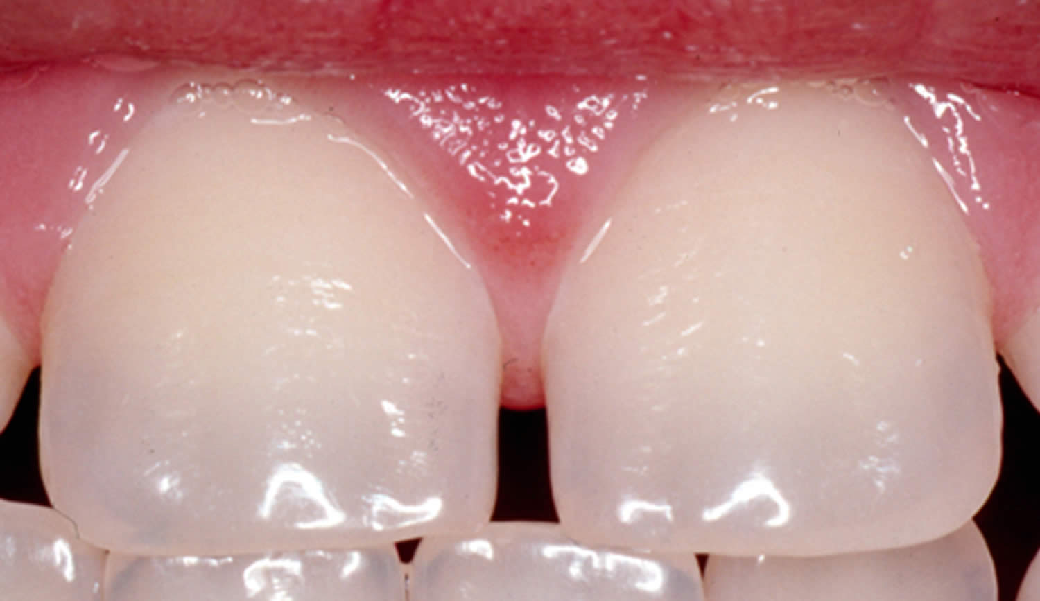
Figure 3. Questionable dental fluorosis
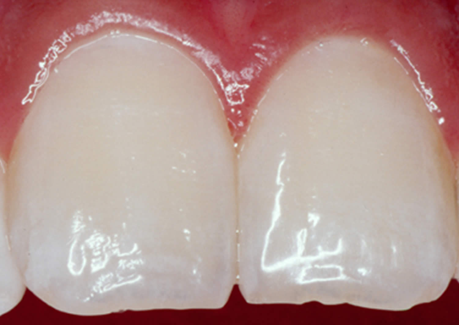
Figure 4. Very mild dental fluorosis
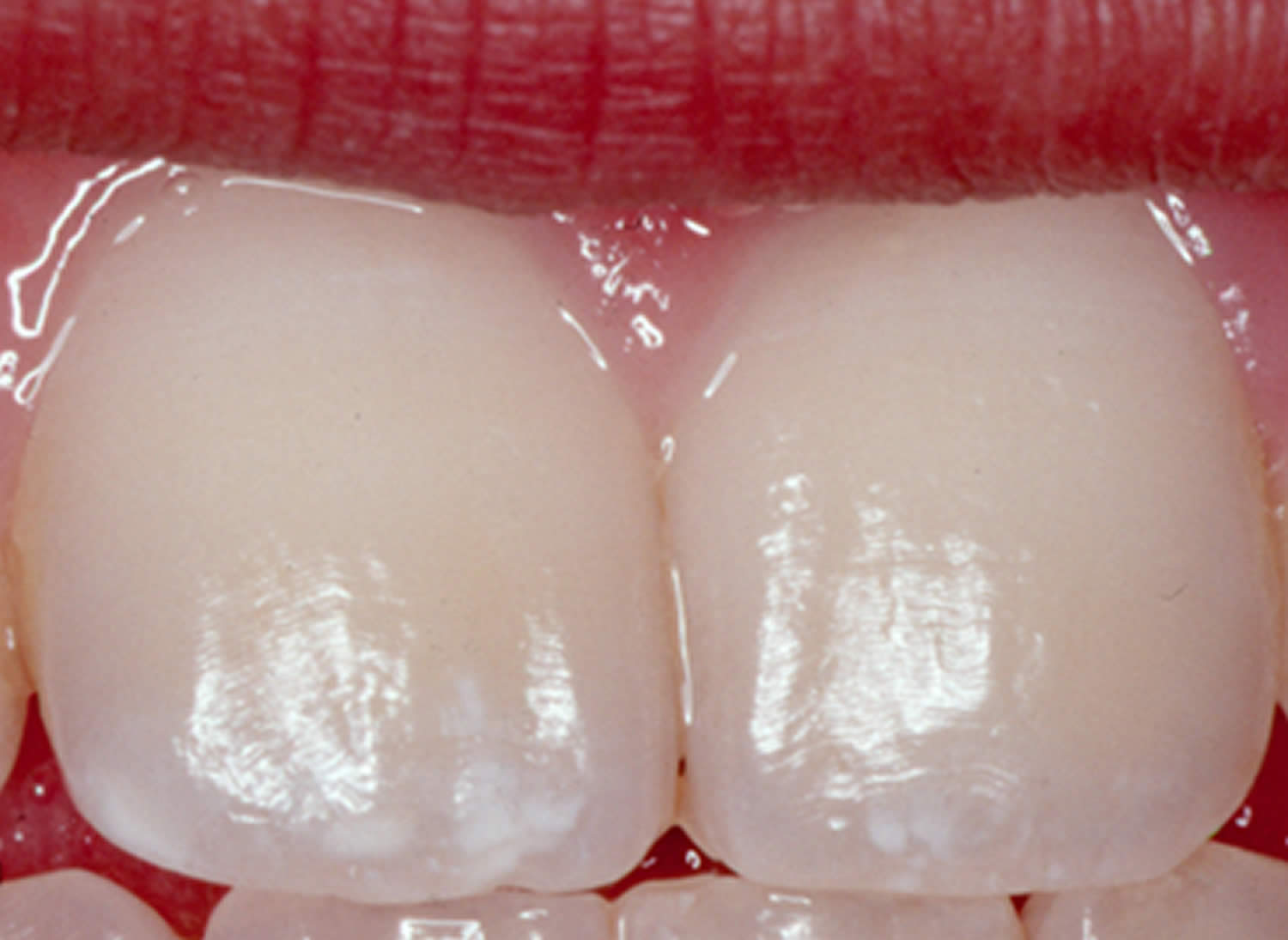
Figure 5. Mild dental fluorosis
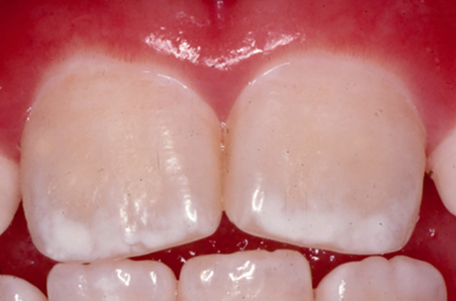
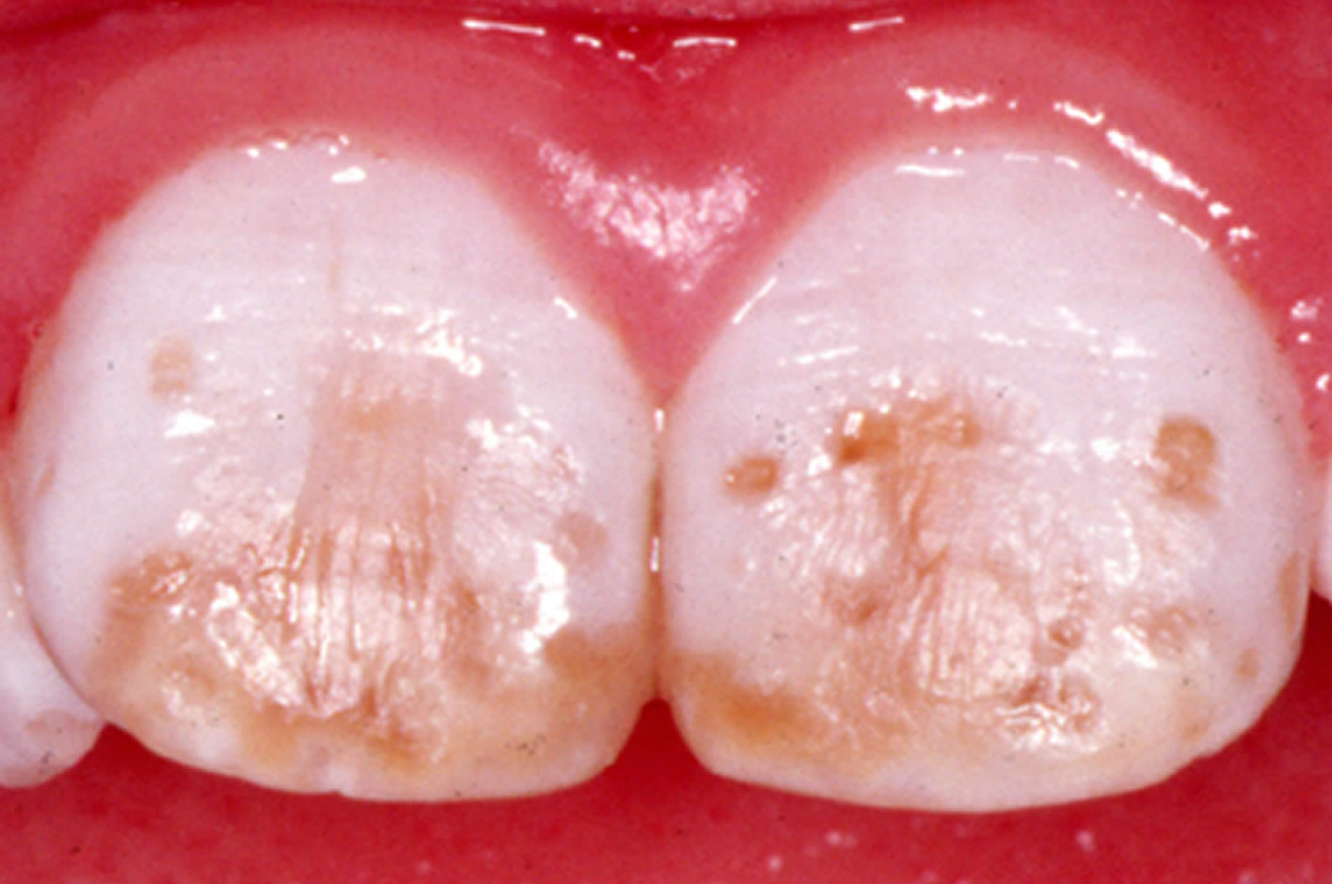
Figure 6. Moderate dental fluorosis
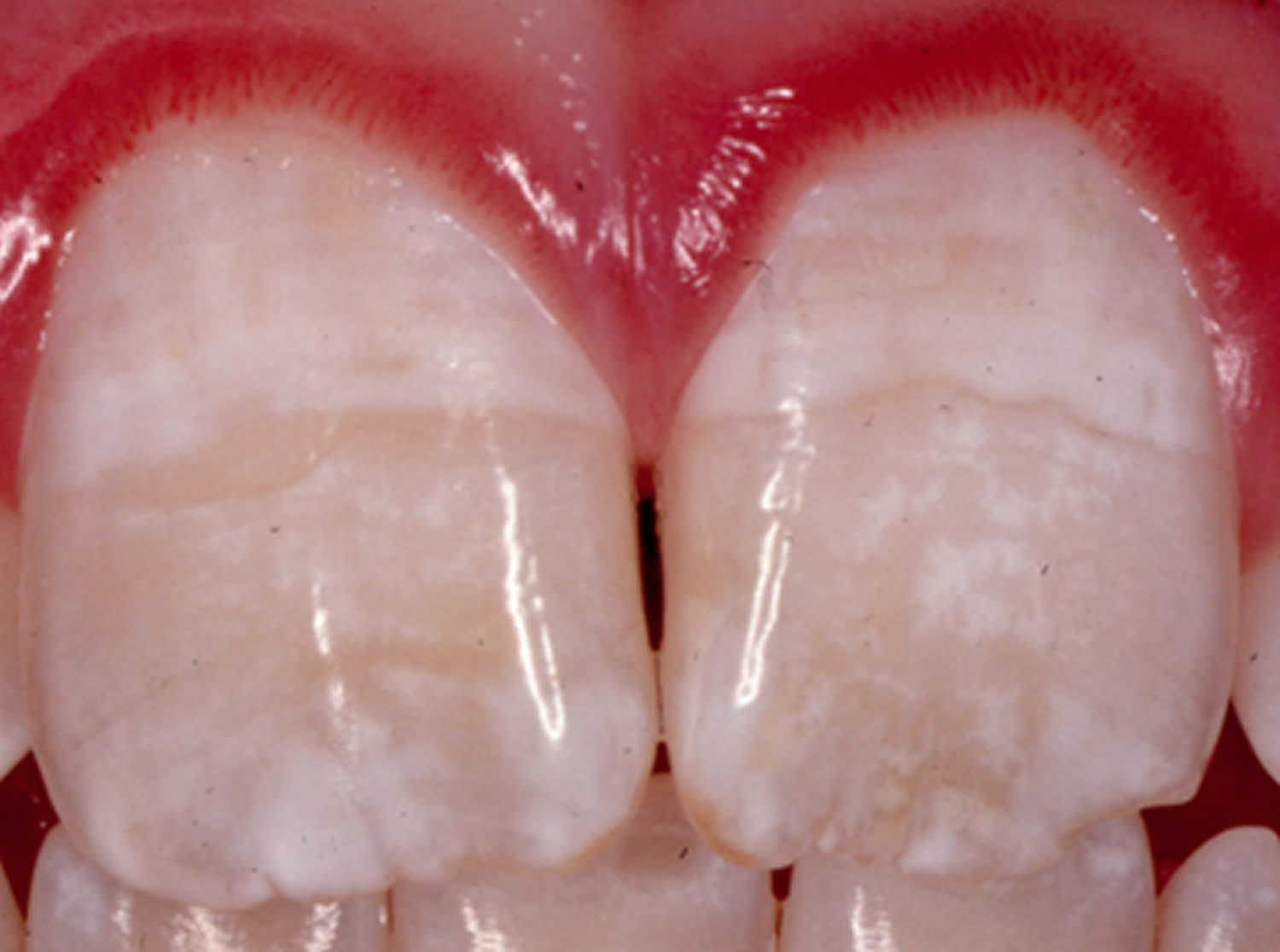
Figure 7. Severe dental fluorosis
What causes dental fluorosis?
Dental fluorosis is caused by taking in too much fluoride over a long period when the teeth are forming under the gums. Only children aged 8 years and younger are at risk because this is when permanent teeth are developing; children older than 8 years, adolescents, and adults cannot develop dental fluorosis. The severity of the condition depends on the dose (how much), duration (how long), and timing (when consumed) of fluoride intake.
Increases in the occurrence of mostly mild dental fluorosis were recognized as more sources of fluoride became available to prevent tooth decay. These sources include drinking water with fluoride, fluoride toothpaste—especially if swallowed by young children—and dietary prescription supplements in tablets or drops (particularly if prescribed to children already drinking fluoridated water).
Is my child at increased risk of fluorosis if they are being fed infant formula?
Breastfeeding is ideal for infants. If breastfeeding is not possible, formula can be used. You should speak with your pediatrician about what type of infant formula is best for your child.
Three types of infant formula are available in the United States: powdered formula, which comes in bulk or single-serve packets, concentrated liquid, and ready-to-feed formula. Ready-to-feed formula contains little fluoride and does not cause dental fluorosis. The kinds of formula that must be mixed with water—powdered or liquid concentrates—may increase the chance of dental fluorosis if they are the child’s main source food and if the water is fluoridated.
Can I use bottled water to mix infant formula?
Yes, you can use bottled water to reconstitute (mix) powdered or liquid concentrate infant formulas, but be aware that the fluoride content in bottled water varies. If your child is exclusively consuming infant formula reconstituted with water that contains fluoride, there may be an increased chance for mild dental fluorosis (a change in the appearance of tooth enamel creating barely visible lacy white markings). To lessen this chance, parents may choose to use low-fluoride bottled water some of the time to mix infant formula. These bottled waters are labeled as de-ionized, purified, demineralized, or distilled and are without any fluoride added after purification treatment (FDA requires the label to indicate when fluoride is added). Some water companies make available bottled waters marketed for infants and for the purpose of mixing with formula. When water is labeled as intended for infants, the water must meet tap water standards established by the EPA and indicate that the water is not sterile.
Dental fluorosis prevention
Know the fluoride concentration of your drinking water
You should know the fluoride concentration in your primary source of drinking water, especially if you have young children. This information should help with decisions about using other fluoride products, particularly fluoride tablets or drops that your physician or dentist may prescribe for your young child. Fluoride tablets or drops should not be used at all if your drinking water has the recommended fluoride concentration of 0.7 mg/L or higher.
If you live in a state that participates in Centers for Disease Control and Prevention (CDC) My Water’s Fluoride (https://nccd.cdc.gov/DOH_MWF/Default/Default.aspx), you can find out your water system’s fluoridation status online. If you are on a public water system, you can call the water utility company and request a copy of the utility’s most recent Consumer Confidence Report.
Use an alternative source of water for children aged 8 years and younger if your primary drinking water contains greater than 2 mg/L of fluoride.
In some regions of the United States, public water systems and private wells contain a natural fluoride concentration of more than 2 mg/L; at this concentration, children 8 years and younger have a greater chance for developing dental fluorosis, including the moderate and severe forms. These children should have an alternative source of drinking water that contains fluoride at the recommended level.
Fluoride supplements can be prescribed for children at high risk for tooth decay and whose primary source of drinking water has a low fluoride level. If the children are younger than 6 years, however, then the dentist or physician should weigh the risks for developing decay without supplements with the possibility of developing dental fluorosis. Access to other sources of fluoride, especially drinking water, should be considered when determining this balance. Parents and caregivers should be informed of both the benefits and risks of fluoride supplements.
Fluoride supplements can be prescribed for persons as appropriate or used in school-based programs. When practical, supplements should be prescribed as chewable tablets or lozenges to maximize the topical effects of fluoride.
For very young children, less than 2 years old
Do not use fluoride toothpaste unless advised to do so by your doctor or dentist. You should clean your child’s teeth as soon as the first tooth appears by brushing without toothpaste with a small, soft-bristled toothbrush and plain water.
Check with the doctor or dentist about your child’s specific fluoride needs. After age 2, most children get the right amount of fluoride to help prevent cavities if they drink water that contains fluoride and brush their teeth with a pea-sized amount of fluoride toothpaste twice a day.
For children aged 2 to 6 years
Fluoride is important for fighting cavities. But if children younger than 6 years old swallow too much fluoride, their permanent teeth may have white spots. To keep this from happening, use only a small amount of toothpaste, apply no more than a pea-sized amount of fluoride toothpaste to the brush and supervise their tooth brushing, encouraging the child to spit out the toothpaste rather than swallow it. Until about age 6, children have poor control of their swallowing reflex and frequently swallow most of the toothpaste placed on their brush.. Teach your child to spit out the toothpaste rather than swallow it and to rinse well after brushing.
Brush your child’s teeth twice a day until your child has the skill to handle the toothbrush alone. Then continue to closely watch brushing to make sure the child is doing a thorough job and using only a small amount of toothpaste.
Do not let a child younger than 6 years old use a fluoride mouth rinse unless the child’s doctor or dentist recommends it. Because fluoride mouth rinses have resulted in only limited reductions in tooth decay among children, especially as their exposure to other sources of fluoride has increased, their use should be targeted to individuals and groups at high risk for decay.
Fluorosis symptoms
Very mild and mild forms of dental fluorosis—teeth have scattered white flecks, occasional white spots, frosty edges, or fine, lacy chalk-like lines. These changes are barely noticeable and difficult to see except by a dental health care professional.
Moderate and severe forms of dental fluorosis—teeth have larger white spots and, in the rare, severe form, rough, pitted surfaces.
Dental fluorosis treatment
Dental fluorosis may be managed by bleaching, micro‐abrasion, veneering or crowning. The choice between these treatments depends on the severity of the fluorosis.
McInnes solution has been used for treating mild grade fluorosis for a long time and successfully 7. McInnes solution consists of one part anesthetic ether, five parts hydrochloric acid (36%), five parts hydrogen peroxide (30%). The solution was freshly mixed and applied onto tooth using a cotton applicator. Each bleaching session consisted of application of bleaching solution for five minutes with one minute interval under rubber dam application followed by polishing of teeth with prophylaxis paste 8.
Micro and macro abrasion technique has been employed successfully for mild to moderate grade fluorosis 9. The teeth were abraded using water cooled fine diamond finishing flame shaped points, with diamond abrasive particle size of 20 – 30μm with a high speed hand piece to remove surface enamel layer of 0.5mm thickness. Removal of surface enamel was done with intermittent pressure under water coolant. Final polishing of teeth was done with polishing discs 10.
Composite resin has been used for treatment of dental fluorosis 11. The treatment involved veneer preparation with window design 12, composite resin used was nano-composite Ceram-X Duo enamel shade E1 and dentin shade D2 bonding agent employed was Prime and Bond NT. Polishing of composite restoration was accomplished with Super Snap.
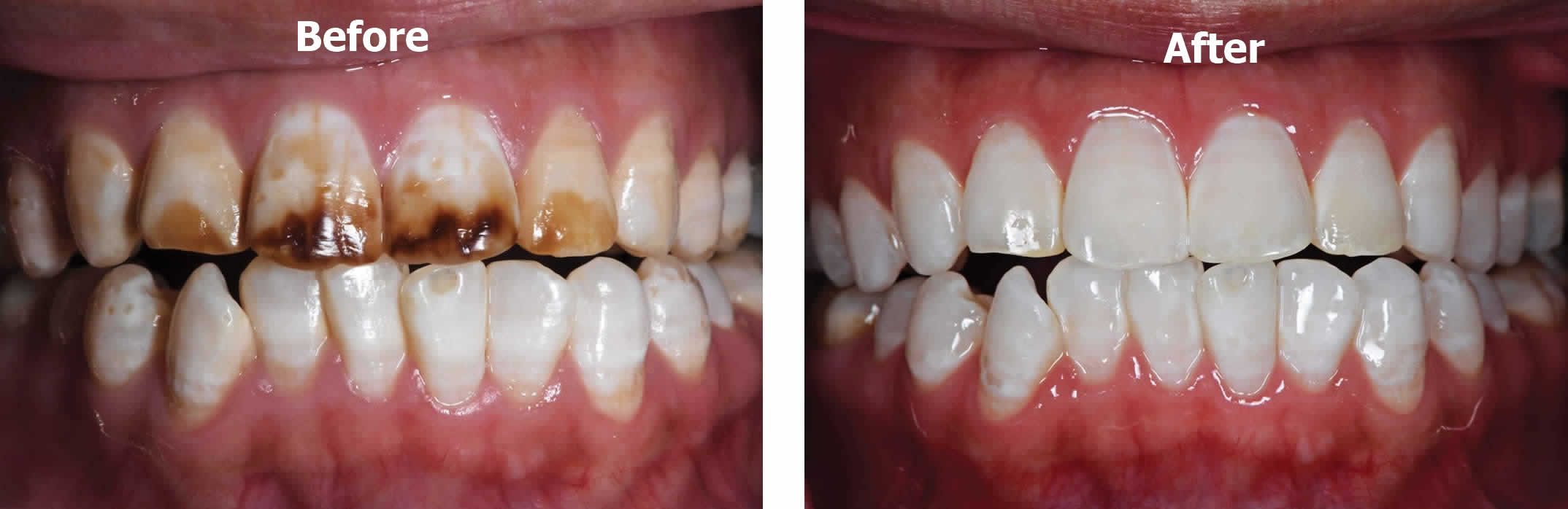
Severe dental fluorosis can be treated with enamel abrasion followed by tooth whitening and infiltration application 13. First, mega-abrasion was performed using a high-speed handpiece with a 105-μm fine diamond bur (ML524, Diatech, Altstatten, Switzerland) to remove the superficial 200–400 μm enamel. After that, medium to fine abrasive discs (Sof-Lex, 3M ESPE, St Paul, MN, USA) were used to reshape the enamel surface and remove the sharp angles. Subsequently, a photopolymerizable resin dam (Beyond Technology Inc, Santa Clara, CA, USA) was applied, and a small amount of abrasive paste (Opalustre, Ultradent Products, South Jordan, UT, USA) containing silicon carbamide microparticle paste and 6.6% hydrochloric acid was painted on the affected teeth. The tooth surfaces were then microabraded using a specific rubber cup (Oralcups, Ultradent Products) with slight pressure for about 120 seconds. After microabrasion, an in-office bleaching agent (Opalescence Boost, 38% H2O2 Ultradent Products) was performed to alleviate the dark brown tooth color. Then a desensitizing agent (Fluorinated protector, Beyond Technology Inc) was painted and left undisturbed on the surfaces of bleached teeth for five minutes. Subsequently, agent removal with suction, thorough water rinsing, and removal of the photopolymerizable rubber dam were conducted. To better harmonize color, at-home bleaching was suggested to the patient. After applying eight syringes of 10% carbamide peroxide (Ultradent Products), the patient was satisfied with the bleaching effect. Considering the hypomineralization structure of dental fluorosis, a standard resin infiltration approach (Icon, DMG Products, Hamburg, Germany) was performed to prevent potential enamel caries two weeks after at-home bleaching therapy. Finally, improvement of the esthetic appearance was achieved and remained stable until the 12-month follow-up.
Figure 8. Dental fluorosis treatment before and after minimally invasive treatment
Severe fluorosis can also be treated with porcelain fused to metal crowns. This treatment option of restoring vertical dimensions of occlusion for severe fluorosis patients requires careful investigations and preparation. This treatment option is limited to cases with severe fluorosis and loss of inter-occlusal space. Advantage of this procedure is that it is an extensive procedure by which the desired aesthetic results and functional efficiency is achieved. The main disadvantage is also its extensiveness in treatment procedure which requires extensive lab procedure and operator skill, knowledge.
References- Aguilar-Díaz FC, Irigoyen-Camacho ME, Borges-Yáñez SA. Evaluation of a fluorosis prevention educational program: A randomized field trial. J Clin Exp Dent. 2018;10(5):e469-e476. Published 2018 May 1. doi:10.4317/jced.54225 https://www.ncbi.nlm.nih.gov/pmc/articles/PMC5971072/
- Basir L, Khaneh Masjedi M, Haghighi M, Neamatiasl S. Comparative investigation on the prevalence of fluorosis and DMFT and their relations with the amount of fluoride in all the three drinking water resources (Maroon, Karoon and Karkheh rivers) among 10-12 school students in Khuzestan in 2002. J Dent Shahid Beheshti Univ Med Sci. 2006;24:14–23
- Guidelines for Fluoride Intake: First Discussant. Martinez-Mier EA. Adv Dent Res. 2018 Mar; 29(2):177-178.
- Den Besten PK, Thariani H. Biological Mechanisms of Fluorosis and Level and Timing of Systemic Exposure to Fluoride with Respect to Fluorosis. J Dent Res. 2002;71:1238–43
- Petersen PE, Lennon MA. Effective use of fluorides for the prevention of dental caries in the 21st century: the WHO approach. Community Dent Oral Epidemiol 2004;32:319–21
- Skeletal fluorosis in humans: a review of recent progress in the understanding of the disease. Prog Food Nutr Sci. 1986;10(3-4):279-314. https://www.ncbi.nlm.nih.gov/pubmed/3295994
- McInnes JW. Removing brown stain from teeth. Arizona Dent J. 1966;12:13–4. https://www.ncbi.nlm.nih.gov/pubmed/5220601
- Grossman, Oliet, DelRio . Bleaching of discolored teeth. 11th ed. India: Varghese Publication; 1991. Endodontic practice; p. 276.
- Hardy Limeback, Viera, Lawrence Improving esthetically objectionable human enamel fluorosis with simple microabrasion technique. Eur J Oral Sci. 2006;114:123–6. https://www.ncbi.nlm.nih.gov/pubmed/16674673
- Fluorosis varied treatment options. J Conserv Dent. 2010;13(1):47-53. https://www.ncbi.nlm.nih.gov/pmc/articles/PMC2883808/
- Akapata ES. Occurrence and management of dental fluorosis. Int Dent J. 2001;51:325–33. https://www.ncbi.nlm.nih.gov/pubmed/11697585
- Roberson, Heymann, Swift . Additional conservative esthetic procedures. 4th ed. Missouri: Mosby; 2002. Sturdevant’s Art and Science of Operative dentistry; pp. 610–20.
- Minimally invasive treatment for esthetic management of severe dental fluorosis: a case report. Oper Dent. 2013 Jul-Aug;38(4):358-62. doi: 10.2341/12-238-S. Epub 2012 Dec 4. http://www.jopdentonline.org/doi/pdf/10.2341/12-238-S


