Hs and Ts
Hs and Ts is a mnemonic used to aid in remembering the possible reversible causes of cardiac arrest 1. Many different traumatic and medical conditions can lead to cardiac arrest in both adults and children. This includes electrical abnormalities, inherited disorders and structural changes in the heart. Determining and treating the cause of cardiac arrest is critical to improving patient outcomes. Fortunately, many causes of cardiac arrest are reversible, including the conditions listed below. These conditions are often referred to by the mnemonic “H’s and T’s” 2. Being able to think through the reversible causes of sudden cardiac arrest will give your patient the best chance of survival as the appropriate diagnosis is made and interventions initiated.
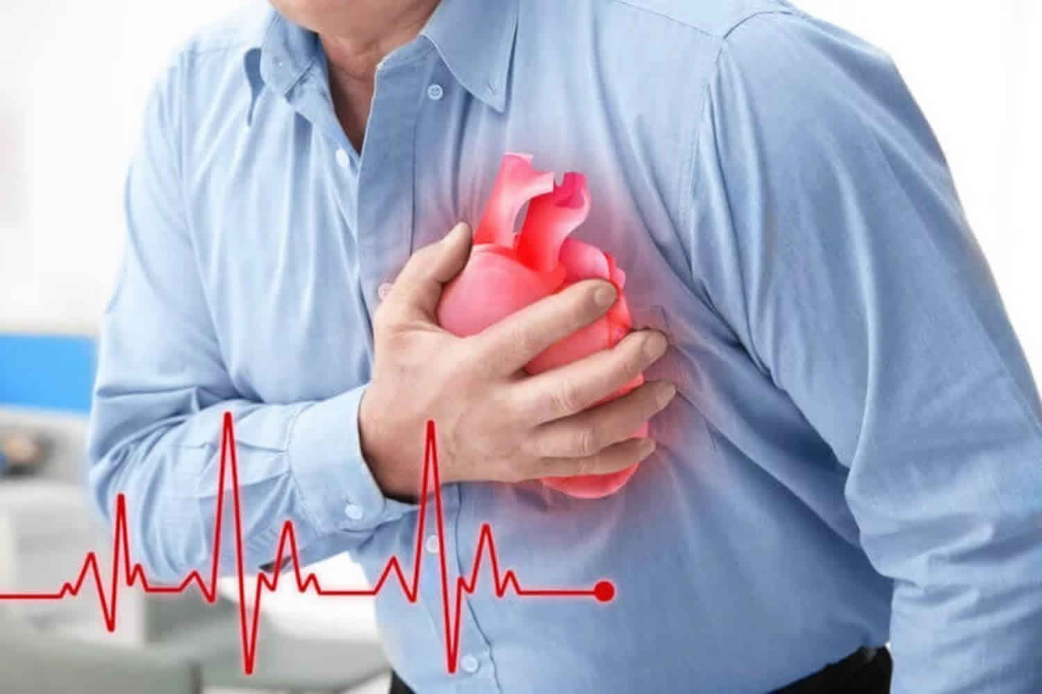
Hs and Ts of ACLS
Hs
Hypovolemia
One common cause of cardiac arrest is hypovolemia, which can develop due to a reduced intravascular volume (i.e. hemorrhage). This is usually (though not exclusively) caused by some form of bleeding, anaphylaxis, extreme sweating, severe diarrhea and/or vomiting, severe vasodilation or pregnancy with gravid uterus. Severe burns can also lead to hypovolemia. Hypovolemia from blood loss is a leading cause of death in traumatic cardiac arrest. External blood loss is usually obvious (e.g., trauma, hematemesis, hemoptysis), but may be more challenging to diagnose when occult (e.g., gastrointestinal bleeding or rupture of an aortic aneurysm) 3. Treatment of hypovolemia includes rapid infusion of preferably warmed crystalloids and/or blood products while treating the original cause of the hypovolemia. Controlling the source of any bleeding – by direct pressure for external bleeding, or emergency surgical techniques such as esophageal banding, gastroesophageal balloon tamponade (for treatment of massive gastrointestinal bleeding such as in esophageal varices), thoracotomy in cases of penetrating trauma or significant shear forces applied to the chest, or exploratory laparotomy in cases of penetrating trauma, spontaneous rupture of major blood vessels, or rupture of a hollow viscus in the abdomen.
Hypoxia
Hypoxemia is low levels of circulating oxygen in the blood, which can lead to hypoxia at the tissues (heart, brain and other vital organs). Hypoxemia is normally a consequence of asphyxia, which accounts for most of the non-cardiac causes of cardiac arrest 3. Rapid assessment of airway patency and respiratory effort must be performed. If the patient is mechanically ventilated, the presence of breath sounds and the proper placement of the endotracheal tube should be verified. Treatment may include providing oxygen, proper ventilation, and good CPR technique. In cases of carbon monoxide poisoning or cyanide poisoning, hyperbaric oxygen may be employed after the patient is stabilized.
Cardiac arrest caused by pure hypoxemia is uncommon. The following is a list of some of the causes of hypoxemia according to Truhlar 3:
- Airway obstruction: soft tissues (coma), laryngospasm, aspiration
- Anemia
- Asthma
- Avalanche burial
- Central hypoventilation – brain or spinal cord injury
- Chronic obstructive pulmonary disease
- Drowning
- Hanging
- High altitude
- Impaired alveolar ventilation from neuromuscular disease
- Pneumonia
- Tension pneumothorax
- Trauma
- Traumatic asphyxia or compression asphyxia (e.g. crowd crush)
Treating the cause of hypoxia/hypoxemia must be done quickly, because this is one of the potentially reversible causes of cardiac arrest. Proper oxygenation and ventilation are key to restoring adequate amounts of oxygen into the system and negating the lethal cardiac rhythm.
Hydrogen ions (acidosis)
Acidosis can be either metabolic or respiratory acidosis. Either cause can lead to cardiac arrest. Acidosis of any kind is most likely detrimental to the circulation as it causes peripheral vasodilatation, negative inotropy and impaired oxygen uptake in the lungs 4. Although severe acidemia frequently occurs in patients during and after cardiac arrest, the prognostic value of severe acidemia for neurologic outcomes is unknown 5. An arterial blood gas is a quick and accurate method to determine if a patient is acidotic. If a patient has respiratory acidosis, they can be treated by providing adequate ventilation. Metabolic acidosis is one of the most common abnormalities in patients suffering from serious diseases, and there have been numerous etiologies and treatments of the underlying disease as the basis of therapy 6. A common intervention to treat metabolic acidosis may be by the administration of sodium bicarbonate and in select cases may require emergent hemodialysis.
Hyperkalemia or hypokalemia
Electrolyte abnormalities can cause cardiac arrhythmias or cardiac arrest, and life-threatening arrhythmias are associated most commonly with potassium disorders, particularly hyperkalemia 3. Potassium is an electrolyte which plays a role in maintaining normal contraction of the myocardium. If levels become too high or too low, cardiac arrest may ensue. The precise values that trigger treatment decisions will depend on the patient’s clinical condition and rate of change of electrolyte values 3. Evaluation of serum potassium must take into consideration the effects of changes in serum pH. When serum pH decreases (acidemia), serum potassium increases because potassium shifts from the cellular to the vascular space; the process that is reversed when serum pH increases (alkalemia). Causes of hypokalemia include excessive vomiting/diarrhea or use of diuretics. Chronic kidney disease can also lead to potassium loss. Treatment may include a controlled but rapid infusion of potassium. Hyperkalemia may be caused by kidney disease, diabetes and as a side effect of certain drugs. Hyperkalemia can be treated by administering sodium bicarbonate or calcium chloride or by performing dialysis.
Hyperkalemia
Hyperkalemia is the most common electrolyte disorder associated with cardiac arrest. It is usually caused by impaired excretion by the kidneys, drugs or increased potassium release from cells and metabolic acidosis with hyperkalemia occurring in up to 10% of hospitalized patients 7. There is no steadfast numeric limit universally used to define hyperkalemia, but 5.5 mmol is commonly recognized. As the potassium concentration increases above this value the risk of adverse events increases and the need for urgent treatment increases 3. The main causes of hyperkalemia are 8:
- Renal failure (i.e., acute kidney injury or chronic kidney disease)
- Drugs (e.g., angiotensin converting enzyme inhibitors (ACE-I), angiotensin II receptor antagonists (ARB), potassium-sparing diuretics, non-steroidal anti-inflammatory drugs, beta-blockers, trimethoprim)
- Tissue breakdown (e.g., rhabdomyolysis, tumor lysis, hemolysis)
- Metabolic acidosis (e.g., renal failure, diabetic ketoacidosis)
- Endocrine disorders (e.g., Addison’s disease)
- Diet (may be sole cause in patients with advanced chronic kidney disease)
The treatment for hyperkalemia involves five key strategies 9:
- Cardiac protection
- Shifting potassium into cells
- Removing potassium from the body
- Monitoring serum potassium and blood glucose
- Prevention of recurrence
A common presentation of hyperkalemia is in the patient with end-stage renal disease who has missed a dialysis appointment and presents with weakness, nausea, and broad QRS complexes on the electrocardiogram. (Note however that patients with chronic kidney disease are often more tolerant of high potassium levels as their body often adapts to it.) Several medications, for example the antibiotic trimethoprim/sulfamethoxazole or an ACE inhibitor, can also lead to the development of significant hyperkalemia. The electrocardiogram will show tall, peaked T waves (often larger than the R wave) or can degenerate into a sine wave as the QRS complex widens. Immediate initial therapy is the administration of calcium, either as calcium gluconate or calcium chloride. This stabilizes the electrochemical potential of cardiac myocytes, thereby preventing the development of fatal arrhythmias. This is, however, only a temporizing measure. Other temporizing measures may include nebulized salbutamol, intravenous insulin (usually given in combination with glucose), and sodium bicarbonate which all temporarily drive potassium into the interior of cells. Definitive treatment of hyperkalemia requires actual excretion of potassium, either through urine (which can be facilitated by administration of loop diuretics such as furosemide) or in the stool (which is accomplished by giving sodium polystyrene sulfonate enterally, where it will bind potassium in the GI tract.) Severe cases will require emergent hemodialysis.
Hypokalemia
Hypokalemia is the most common electrolyte disturbance in clinical practice 10. It is seen in up to 20% of hospitalized patients 11. Hypokalemia increases the incidence of arrhythmias and sudden cardiac death 12. Hypokalemia is defined as a serum potassium level <3.5 mmol and severe hypokalemia is a serum potassium <2.5 mmol 8. The main causes of hypokalemia include 3:
- Gastrointestinal loss (e.g., diarrhea)
- Drugs (e.g., diuretics, laxatives, steroids)
- Renal losses (e.g., renal tubular disorders, diabetes insipidus, dialysis)
- Endocrine disorders (e.g., Cushing’s syndrome, hyperaldosteronism)
- Metabolic alkalosis
- Magnesium depletion
- Poor dietary intake
Treatment of hypokalemia depends on the severity and the presence of symptoms and ECG abnormalities. The electrocardiogram may show flattening of T waves and prominent U waves. Hypokalemia is an important cause of acquired long QT syndrome, and may predispose the patient to torsades de pointes. Digitalis use may increase the risk that hypokalemia will produce life-threatening arrhythmias. Hypokalemia is especially dangerous in patients with ischemic heart disease. The best course of action is the gradual replacement of potassium to normal serum levels. In an emergency, intravenous potassium is warranted, with the knowledge that many patients who are hypokalemic are also hypomagnesimic. Repletion of magnesium stores will facilitate more rapid correction of hypokalemia and is recommended in severe cases of hypokalemia 13.
Hypothermia
A low core body temperature, defined clinically as a temperature of less than 35 degrees Celsius (95 degrees Fahrenheit). Every year approximately 1,500 people die of primary accidental hypothermia in the United States 14. Accidental hypothermia is defined as an involuntary drop of the core body temperature <35 degrees Celcius (<95 degrees Fahrenheit) 3. Hypothermia can be estimated and further subdivided by using the Swiss staging system 15:
- Hypothermia 1 – mild hypothermia (conscious, shivering, core temperature 32–35 °C [<89.6– 95 °F])
- Hypothermia 2 – moderate hypothermia (impaired consciousness without shivering, core temperature 28–32 °C [<82.4– 89.6 °F])
- Hypothermia 3 – severe hypothermia (unconscious, vital signs present, core temperature 24–28 °C [<75.2–82.4 °F])
- Hypothermia 4 – cardiac arrest or low flow state (no or minimal vital signs, core temperature <24 °C [<75.2 °F])
- Hypothermia 5 – death due to irreversible hypothermia (core temperature <13.7 °C [<56.6 °F])
The risk of hypothermia is increased by alcohol or drug ingestion, exhaustion, illness, injury or neglect especially when there is a decrease in the level of consciousness 3. As core temperature decreases, sinus bradycardia tends to give way to atrial fibrillation followed by ventricular fibrillation (VF) and finally asystole 16. Arrhythmias other than VF tend to revert spontaneously as core temperature increases, and usually do not require immediate treatment 3. Unless the patient goes into VF, rewarm using active external methods (e.g., forced warm air) and minimally invasively methods (e.g., warm IV infusions) 3.
Hypoglycemia
There is an unclear association between hypoglycemia and sudden cardiac death. In the NICE-SUGAR trial 17, moderate and severe hypoglycemia were both associated with increased mortality. However, administration of dextrose is also associated with worse outcomes 17.
Hypoglycemia was removed from the Hs and Ts by the American Heart Association in their 2010 ACLS update 18.
Ts
Tablets or toxins
Airway obstruction and respiratory arrest secondary to a decreased conscious level is a common cause of death after self-poisoning (benzodiazepines, alcohol, opiates, tricyclics, barbiturates) 19. Early tracheal intubation of unconscious patients by trained personnel may decrease the risk of aspiration 3. Drug-induced hypotension usually responds to IV fluids, but occasionally vasopressor support (e.g., noradrenaline infusion) is required. Some of the most common drugs involved in an overdose are benzodiazepines, opioids, tricyclic antidepressants, local anesthetics, beta-blockers, and calcium channel blockers.
Benzodiazepines
Overdose of benzodiazepines can cause loss of consciousness, respiratory depression, and hypotension 3. The drug of choice for the treatment of benzodiazepine overdose is Flumazenil. Flumazenil is a competitive antagonist of benzodiazepines and can be used when the patient does not have a history of risk of seizures.
Opioids
Excess opioid consumption via any route can lead to respiratory depression, respiratory insufficiency, and/or respiratory arrest. The opiate antagonist naloxone can reverse the respiratory effects of an opioid overdose. The preferred route for giving naloxone depends on the skills of the rescuer: intravenous (IV), intramuscular (IM), subcutaneous (SC), intraosseous (IO) and intranasal (IN) routes are all suitable 20. The initial doses of naloxone are 0.4–2 mg IV, IO, IM or SC, and may be repeated every 2–3 minutes. Additional doses may be needed every 20–60 minutes. Intranasal dosing is 2 mg IN (1 mg in each nostril), which may be repeated every 5 minutes. Titrate the dose until the victim is breathing adequately and has protective airway reflexes 3.
Tricyclic antidepressants
Self-poisoning with tricyclic antidepressants is common and can cause hypotension, seizures, coma and life-threatening arrhythmias. Cardiac toxicity mediated by anticholinergic and Na+channel-blocking effects can produce a wide complex tachycardia (VT) 3. Give sodium bicarbonate (1–2 mmol/kg) for the treatment of tricyclic-induced ventricular arrhythmias 21.
Local anesthetics
Local anesthetic systemic toxicity (LAST) is a serious but rare consequence of regional anesthesia and most commonly results from an inadvertent vascular injection or absorption of large amounts of drug from certain nerve blocks requiring large volume injections 22. Severe agitation, loss of consciousness, seizures, bradycardia, asystole or ventricular tachyarrhythmias can all occur 3. When local anesthetic systemic toxicity (LAST) is suspected, benzodiazepines are the drug of choice because they are an anticonvulsant without causing significant cardiac depression 22. Although there are many case reports and case series of patients who were resuscitated after administration of IV lipid emulsion, evidence for its benefit in treating local anesthetic-induced cardiac arrest is limited. Despite the paucity of data, patients with both cardiovascular collapse and cardiac arrest attributable to local anesthetic toxicity may benefit from treatment with intravenous 20% lipid emulsion in addition to standard ACLS 23.
Beta-blockers
Beta-blocker toxicity causes bradyarrhythmias and negative inotropic effects that are difficult to treat and can lead to cardiac arrest 3. Improvement has been reported with glucagon (50–150 mcg/kg), high-dose insulin and glucose, lipid emulsions, phosphodiesterase inhibitors, extracorporeal and intra-aortic balloon pump support, and calcium salts 24.
Calcium channel blockers
Calcium channel blocker overdose is emerging as a common cause of prescription drug poisoning deaths 25. Overdose of short-acting drugs can rapidly progress to cardiac arrest and overdose by sustained-release formulations can result in delayed onset of arrhythmias, shock, and sudden cardiac collapse 3. Treatment can include the administration of calcium chloride 10% in boluses of 20 ml (or equivalent dose of calcium gluconate) every 2-5 minutes in severe bradycardia or hypotension followed by an infusion as needed 26.
Cardiac tamponade
Cardiac tamponade occurs when the pericardial sac is filled with fluid under pressure, which leads to compromise of cardiac function and ultimately cardiac arrest 3. Cardiac tamponade may be caused by trauma to the chest such as a gunshot wound or by inflammation of the pericardium. Thoracotomy or pericardiocentesis is used to treat cardiac arrest associated with suspected traumatic or non-traumatic cardiac tamponade. The use of ultrasound guidance during pericardiocentesis is preferred, if available.
Tension pneumothorax
A tension pneumothorax develops when there is a buildup of air in the pleural space. The buildup causes a shift in the mediastinum and venous return to the heart is obstructed, which can lead to cardiac arrest. When this happens, the great vessels (particularly the superior vena cava) become kinked, which limits blood return to the heart. Tension pneumothorax can be recognized by severe air hunger, hypoxia, jugular venous distension, hyperresonance to percussion on the affected side, and a tracheal shift away from the affected side. The tracheal shift often requires a chest x-ray to appreciate (although treatment should be initiated prior to obtaining a chest x-ray if this condition is suspected). Tension pneumothorax is a treatable cause of cardiac arrest and should be excluded during CPR 27.Tension pneumothorax can occur in a variety of clinical situations including trauma, asthma and other respiratory disease, but can also be iatrogenic following invasive procedures (e.g., attempts at central venous catheter insertion) 3. Diagnosis of tension pneumothorax in a patient with cardiac arrest or hemodynamic instability must be based on clinical examination. The symptoms include hemodynamic compromise (hypotension or cardiac arrest) in conjunction with signs suggestive of a pneumothorax (preceding respiratory distress, hypoxia, absent unilateral breath sounds on auscultation, subcutaneous emphysema) and mediastinal shift (tracheal deviation and jugular venous distention)19. Treatment of a tension pneumothorax is either needle compression and/or thoracostomy with chest tube placement.
Thrombosis (myocardial infarction)
Coronary heart disease is the most frequent cause of out-of-hospital cardiac arrest. Although proper diagnosis of the cause may be difficult in a patient already in cardiac arrest, if the initial rhythm is VF it is most likely that the cause is coronary artery disease with an occluded large coronary vessel 3. Treatment options include immediate coronary angiography, primary percutaneous coronary intervention (PPCI) or other interventions such as (more rarely) pulmonary embolectomy. Ongoing CPR and immediate access to the catheterization laboratory may be considered if a prehospital and in-hospital infrastructure is available with teams experienced in mechanical or hemodynamic support and rescue PPCI with ongoing CPR 3.
Thromboembolism (pulmonary embolism)
Cardiac arrest from acute pulmonary embolism is the most serious clinical presentation of venous thromboembolism, in most cases originating from a deep venous thrombosis (DVT) 28. The 2014 European Society of Cardiology Guidelines on the diagnosis and management of acute pulmonary embolism define “confirmed pulmonary embolism” as a probability of pulmonary embolism high enough to indicate the need for specific treatment 28. Common symptoms preceding cardiac arrest are sudden onset of dyspnea, pleuritic or substernal chest pain, cough, hemoptysis, syncope and signs of DVT (e.g., unilateral, low extremity swelling). However, pulmonary embolism may not be symptomatic until it presents as sudden cardiac arrest 29. Specific treatments for cardiac arrest resulting from pulmonary embolism include administration of fibrinolytics, surgical embolectomy and percutaneous mechanical thrombectomy 3.
Trauma
Cardiac arrest can also occur after a hard blow to the chest at a precise moment in the cardiac cycle, which is known as commotio cordis. Other traumatic events such as high speed car crashes can cause sufficient structural damage to induce arrest.
References- Sudden Cardiac Arrest and the H’s and T’s. https://acls.com/free-resources/knowledge-base/pea-asystole/reversible-causes-of-cardiac-arrest-hs-and-ts
- ACLS: Principles and Practice. p. 71-87. Dallas: American Heart Association, 2003. ISBN 0-87493-341-2.
- Truhlar, A., Deakin, C.D., Soar, J., Khalifa, G.E., Alfonzo, A., Bierens, J.J., Brattebo, G., Brugger, H., Dunning, J., Hunyadi-Anticevic, S., Koster, R.W., Lockey, D.J., Lott, C., Paal, P., Perkins, G.D., Sandroni, C., Thies, K.C., Zideman, D.A., & Nolan, J.P. (2015). European Resuscitation Council guidelines for resuscitation 2015. Resuscitation, 95, 148-201.
- Spindelboeck, W., Gemes, G., Strasser, C., Toescher, K., Kores, B., Metnitz, P., Haas, J., & Prause, G. (2016). Arterial blood gases during and their dynamic changes after cardiopulmonary resuscitation: A prospective clinical study. Resuscitation, 106, 24-29.
- Tetsuhara, K., Kato, H., Kanemura, T., Okada, I., & Kiriu, N. (2015). Severe acidemia on arrival not predictive of neurologic outcomes in post–cardiac arrest patients. American Journal of Emergency Medicine, 34, 425-428.
- Rubens de Nadai, T., Nunes de Nadai, M., Albuquerque, A., Menezes de Carvalho, M., Celotto, A.C., & Evora, P.R. (2013). Metabolic Acidosis Treatment as Part of a Strategy to Curb Inflammation. International Journal of Inflammation, 1-4.
- Einhorn, L.M., Zhan, M., & Hsu, V.D., et al. (2009). The frequency of hyperkalemia and its significance in chronic kidney disease. Arch Intern Med, 169, 1156–1162.
- Asirvatham, J.R., Moses, V., & Bjornson, L. (2013). Errors in potassium measurement: a lab-oratory perspective for the clinician. North American Journal of Medical Sciences, 5, 255–259.
- UK Renal Association. (2014). Treatment of acute hyperkalemia in adults. Clinical practice guidelines. London: UK Renal Association.
- El-Sherif, N., & Turitto, G. (2011). Electrolyte disorders and arrhythmogenesis. Cardiology Journal, 18, 233-245.
- Paice, B.J., Paterson, K.R., Onyanga-Omara, F., Donnelly, T., Gray, J.M., & Lawson, D.H. (1986) Record linkage study of hypokalemia in hospitalized patients. Postgraduate Medical Journal, 162, 187–191.
- Kjeldsen, K. (2010). Hypokalemia and sudden cardiac death. Experimental and Clinical Cardiology, 15, e96–99.
- Cohn, J.N., Kowey,. P.R., Whelton, P.K., & Prisant, L.M. (2000). New guidelines for potassium replacement in clinical practice: a contemporary review by the National Council on Potassium in Clinical Practice. Archives of Internal Medicine, 160, 2429–2436.
- Brown, D.J., Brugger, H., Boyd, J., & Paal, P. (2012). Accidental hypothermia. New England Journal of Medicine, 367, 1930-1938.
- Pasquier, M., Zurron, N., & Weith, B., (2014). Deep accidental hypothermia with core temperature below 24 degrees C presenting with vital signs. High Altitude Medical Biology 15,58–63.
- Paal, P., Strapazzon, G., & Braun, P., et al. (2013). Factors affecting survival from avalanche burial – a randomised prospective porcine pilot study. Resuscitation, 84, 239–243.
- Finfer S, Liu B, Chittock DR, et al. Hypoglycemia and risk of death in critically ill patients. N Engl J Med. 2012;367(12):1108-18.
- Part 7: Adult Advanced Cardiovascular Life Support. https://eccguidelines.heart.org/circulation/cpr-ecc-guidelines/part-7-adult-advanced-cardiovascular-life-support/?strue=1&id=5-2-2-1
- Thompson, T.M., Theobald, J., Lu, J., & Erickson, T.B. (2014). The general approach to the poisoned patient. Disease a Month, 60, 509–524.
- Robertson, T.M., Hendey, G.W., Stroh, G., & Shalit, M. (2009). Intranasal naloxone is a viable alternative to intravenous naloxone for prehospital narcotic overdose. Prehospital Emergency Care, 13, 512–515.
- Bradberry, S.M., Thanacoody, H.K., Watt, B.E., Thomas, S.H., & Vale, J.A. (2005). Management of the cardiovascular complications of tricyclic antidepressant poisoning: role of sodium bicarbonate. Toxicology Review, 24,195–204.
- Nagelhout, J.J. (2018). Local Anesthetics. 6th Edition. Nurse Anesthesia (pp. 119-125). St. Louis: Elsevier.
- Association of Anaesthetists of Great Britain and Ireland. (2010). Management of Severe Local Anaesthetic Toxicity.
- Engebretsen, K.M., Kaczmarek, K.M., Morgan, J., & Holger, J.S. (2011) High-dose insulin therapy in beta-blocker and calcium channel-blocker poisoning. Clinical Toxicology, 49, 277–283.
- Bronstein, A.C., Spyker, D.A., Cantilena, Jr. L.R., Green, J.L., Rumack, B.H., & Giffin, S.L. (2009). 2008 annual report of the American Association of Poison Control Centers’ National Poison Data System (NPDS): 26th annual report. Clinical Toxicology, 47, 911–1084.
- Gunja, N., & Graudins, A. (2011). Management of cardiac arrest following poisoning. Emergency Medicine of Australia, 23, 16–22.
- Barton, E.D. (1999). Tension pneumothorax. Current Opinion in Pulmonary Medicine, 5, 269–274.
- Konstantinides, S.V., Torbicki, A., Agnelli, G., et al. (2014) ESC guidelines on the diagnosis and management of acute pulmonary embolism. European Heart Journal, 35, 3033–69.
- Heit, J.A., Silverstein, M.D., Mohr, D.N., Petterson, T.M., O’Fallon, W.M., & Melton, III L.J. (2000). Risk factors for deep vein thrombosis and pulmonary embolism: a population-based case–control study. Archives of Internal Medicine, 160, 809–815.


