
Hyperpigmentation
Hyperpigmentation also called hypermelanosis, is a common, usually harmless condition in which patches of skin become darker in color than the normal surrounding skin. This darkening occurs when an excess of melanin, the brown pigment that produces normal skin color, forms deposits in the skin. Hyperpigmentation or increase in melanin in your skin can be due to an increased number of pigment cells (melanocytes) or from increased production of melanin. Hyperpigmentation can be localized (focal) or generalized (diffused). Hyperpigmentation can affect the skin color of people of any race.
Localized (focal) hyperpigmentation is most often postinflammatory in nature, occurring after injury (e.g., cuts and burns) or other causes of inflammation (eg, acne, lupus). Focal linear hyperpigmentation is commonly due to phytophotodermatitis, which is a phototoxic reaction that results from ultraviolet light combined with psoralens (specifically furocoumarins) in plants (eg, limes, parsley, celery—see Chemical photosensitivity). Focal hyperpigmentation can also result from neoplastic processes (eg, lentigines, melanoma), melasma, freckles, or café-au-lait macules. Acanthosis nigricans causes focal hyperpigmentation and a velvety plaque most often on the axillae and posterior neck.
Generalized (diffused or widespread) hyperpigmentation can result from drugs and also has systemic and neoplastic causes (especially lung carcinomas and melanoma with systemic involvement). After eliminating drugs as a cause of diffuse hyperpigmentation, patients should be tested for the most common systemic causes. These causes are Addison disease, hemochromatosis, and primary biliary cholangitis. Skin findings are nondiagnostic; therefore, a skin biopsy is not necessary or helpful. The search for underlying cancer should be based on a review of systems.
Age or “liver” spots are a common form of hyperpigmentation. They occur due to sun damage, and are referred to by doctors as solar lentigines. These small, darkened patches are usually found on the hands and face or other areas frequently exposed to the sun.
Melasma or chloasma spots are similar in appearance to age spots but are larger areas of darkened skin that appear most often as a result of hormonal changes. Pregnancy, for example, can trigger overproduction of melanin that causes the “mask of pregnancy” on the face and darkened skin on the abdomen and other areas. Women who take birth control pills may also develop hyperpigmentation because their bodies undergo similar kind of hormonal changes that occur during pregnancy. If one is really bothered by the pigment, the birth control pills should be stopped.
Changes in skin color can result from outside causes. For example, skin diseases such as acne may leave dark spots after the condition clears. Other causes of dark spots are injuries to the skin, including some surgeries. Freckles are small brown spots that can appear anywhere on the body, but are most common on the face and arms. Freckles are an inherited characteristic.
Freckles, age spots, and other darkened skin patches can become darker or more pronounced when skin is exposed to the sun. This happens because melanin absorbs the energy of the sun’s harmful ultraviolet rays in order to protect he skin from overexposure. The usual result of this process is skin tanning, which tends to darken areas that are already hyperpigmented. Wearing a sunscreen is a must. The sunscreen must be “broad spectrum” (i.e. it blocks both UVA and UVB). A single day of excess sun can undo months of treatment.
Most prescription creams used to lighten the skin contain hydroquinone. Bleaches lighten and fade darkened skin patches by slowing the production of melanin so those dark spots gradually fade to match normal skin coloration. Prescription bleaches contain twice the amount of hydroquinone, the active ingredient, as over-the-counter skin bleaches. In more severe cases prescription creams with tretinoin and a cortisone cream may be used. These may be somewhat irritating to sensitive skin and will take 3-6 months to produce improvement.
There are now several highly effective laser treatments. The Q-switched ruby and other pigmented lesion lasers often remove pigment without scarring. A test spot in an inconspicuous place will need to be done as they sometimes make things worse instead of better.
Hyperpigmentation key points
- Common causes of focal hyperpigmentation include injury, inflammation, phytophotodermatitis, lentigines, melasma, freckles, café-au-lait macules, and acanthosis nigricans.
- Common causes of widespread hyperpigmentation include melasma, drugs, cancers, and other systemic disorders.
- Test patients who have widespread hyperpigmentation not caused by drugs for primary biliary cholangitis, hemochromatosis, and Addison disease.
- Treat melasma initially with a combination of hydroquinone 2 to 4%, tretinoin 0.05 to 1%, and a class V to VII topical corticosteroid.
- If lentigos or lentigines are a cosmetic concern, treat with cryotherapy or laser.
Hyperpigmentation causes
Generalized hyperpigmentation
- Acromegaly
- Addison disease
- Drug-induced pigmentation (phenothiazines, silver, chemotherapy)
- Haemochromatosis
- Ichthyosis
- Sézary syndrome
- Porphyria cutanea tarda
- Systemic sclerosis
- Wilson disease
Localized hyperpigmentation
- Acanthosis nigricans
- Alkaptonuria
- Becker nevus
- Berloque dermatitis
- Chloasma (melasma)
- Confluent and reticulated papillomatosis
- Congenital nevi
- Dark circles under the eyes
- Diffuse melanosis cutis
- Dowling-Degos disease
- Fixed drug eruption
- Erythema ab igne
- Erythrasma
- Flagellate erythema
- Freckles, lentigines
- Gastrointestinal stromal tumour (GIST) — cutaneous hyperpigmentation disease associated with mastocytosis and c-kit mutation
- Idiopathic eruptive macular pigmentation
- Laugier-Hunziker syndrome
- Lichen planus pigmentosa
- Lipodermatosclerosis
- Melanoma
- Moles
- Mongolian spot
- Morphoea (morphea)
- Naevi of Ota and Ito
- Neurofibromatosis
- Ochronosis
- Pigmented contact cheilitis
- Pigmented purpura
- Pityriasis versicolor (tinea versicolor)
- Poikiloderma of Civatte
- Postinflammatory (trauma, burns, skin disease)
- Prurigo pigmentosa
- Purpura
- Systemic sclerosis
- Terra firma-forme dermatosis
- Tinea nigra
- Urticaria pigmentosa, mastocytosis
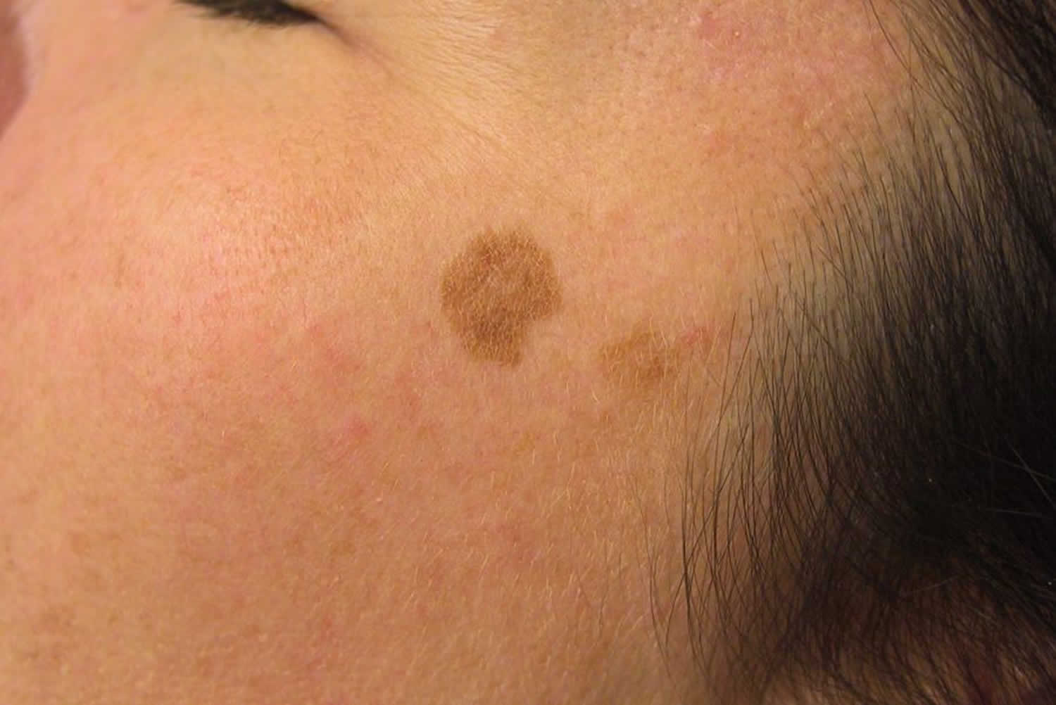
Lentigo
Lentigos also called lentigines are flat, tan to brown, oval macules. Lentigos are commonly due to chronic sun exposure (solar lentigines; sometimes called liver spots) and occur most frequently on the face and back of the hands. They typically first appear during middle age and increase in number with age. Although progression from lentigines to melanoma has not been established, lentigines are an independent risk factor for melanoma.
If lentigines are a cosmetic concern, they are treated with cryotherapy or laser; hydroquinone is not effective.
Nonsolar lentigines are sometimes associated with systemic disorders, such as Peutz-Jeghers syndrome (in which profuse lentigines of the lips occur), multiple lentigines syndrome (or LEOPARD syndrome, which stands for multiple Lentigines, Electrocardiogram (ECG) conduction abnormalities, Ocular hypertelorism, Pulmonic stenosis, Abnormal genitals, Retardation of growth, and sensorineural Deafness), or xeroderma pigmentosum.
Figure 1. Lentigo
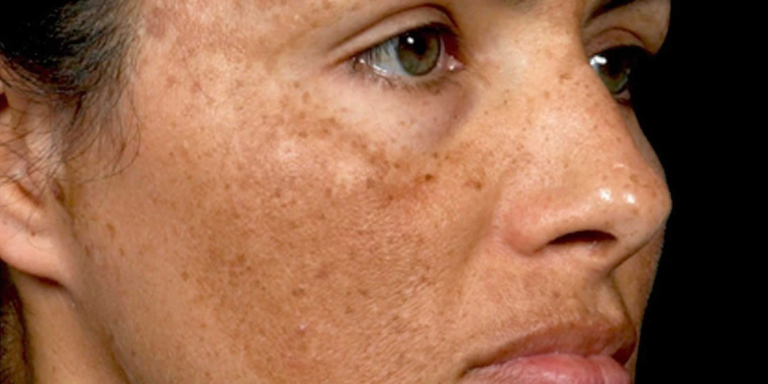
Melasma (chloasma)
Melasma consists of dark brown, sharply marginated, roughly symmetric patches of hyperpigmentation on the face (usually on the forehead, temples, cheeks, upper lip, or nose). It occurs primarily in pregnant women (melasma gravidarum, or the mask of pregnancy) and in women taking oral contraceptives. Ten percent of cases occur in nonpregnant women and dark-skinned men. Melasma is more prevalent among and lasts longer in people with dark skin.
Because melasma risk increases with increasing sun exposure, the mechanism probably involves overproduction of melanin by hyperfunctional melanocytes. Other than sun exposure, aggravating factors include:
- Autoimmune thyroid disorders
- Photosensitizing drugs
In women, melasma fades slowly and incompletely after childbirth or cessation of hormone use. In men, melasma rarely fades.
The mainstay of melasma management is strict sun protection. Patients should use sunscreen with a sun protection factor (SPF) of 30 or higher, wear protective clothing and hats, and avoid direct sun exposure. During and after therapy, strict sun protection must be maintained.
Other treatment depends on whether the pigmentation is epidermal or dermal; epidermal pigmentation becomes accentuated with a Wood light (365 nm) or can be diagnosed with biopsy. Only epidermal pigmentation responds to treatment. Most topical melasma treatments are used in combination rather than individually.
Triple topical therapy is first-line treatment that is often effective and consists of a combination of:
- Hydroquinone 2 to 4%
- Tretinoin 0.05 to 1%
- A class V to VII topical corticosteroid
Hydroquinone depigments the skin by blocking the enzymatic oxidation of tyrosine 3,4-dihydroxyphenylalanine (DOPA) and inhibiting melanocyte metabolic processes. Hydroquinone should be tested behind one ear or on a small patch on the forearm for 1 week before use on the face because it may cause irritation or an allergic reaction. Tretinoin promotes keratinocyte turnover and can exfoliate skin that contains epidermal pigment. Corticosteroids help block synthesis and secretion of melanin. Two promising technologies being tried in conjunction with triple topical therapy are the Q-switched Nd:YAG (1064 nm) laser and nonablative fractional resurfacing.
If triple topical therapy is not available, then hydroquinone 3 to 4% applied twice daily for up to 8 weeks at a time (chronic continuous use can theoretically increase the risk for exogenous ochronosis, which is a permanent form of hyperpigmentation) may be considered; 2% hydroquinone is useful as maintenance.
Azelaic acid 15 to 20% cream can be used in place of or with hydroquinone and/or tretinoin. Azelaic acid is a tyrosinase inhibitor that reduces melanin production (and is considered safe for use during pregnancy). In addition, topical kojic acid has been increasingly used; it is a chelating agent that blocks tyrosine conversion to melanin.
Second-line treatment options for patients with severe melasma unresponsive to topical bleaching agents include chemical peeling with glycolic acid or 30 to 50% trichloroacetic acid. Laser treatments also have been used but are not standard therapy.
Oral therapies have been studied. A recent randomized study has shown improvement with oral tranexamic acid in patients with moderate-to-severe melasma 1.
Figure 2. Melasma
Drug-induced hyperpigmentation
Drug-induced skin pigmentation accounts for 10–20% of all cases of acquired hyperpigmentation or secondary hyperpigmentation (see Tables 1 and 2 below). Hyperpigmentation may be induced by a wide variety of drugs; the main ones implicated include non-steroidal anti-inflammatory drugs (NSAIDs), phenytoin, antimalarials, amiodarone, antipsychotic drugs, cytotoxic drugs, tetracyclines, and heavy metals.
Some drugs may cause fixed drug eruption, which is followed by localized hyperpigmentation and gradually fades. For example, focal hyperpigmentation frequently occurs after drug-induced lichen planus (also known as lichenoid drug eruption).
Changes are usually diffuse but sometimes have drug-specific distribution patterns or hues (see Tables 1 and 2 below). Mechanisms include:
- Increased melanin in the epidermis (tends to be more brown)
- Increased melanin in the epidermis and high dermis (mostly brown with hints of gray or blue)
- Increased melanin in the dermis (tends to be more grayish or blue)
- Dermal deposition of the drug, metabolite, or drug–melanin complexes (usually slate or bluish gray)
In fixed drug eruptions, red plaques or blisters form at the same site each time a drug is taken; residual postinflammatory hyperpigmentation usually persists. Typical lesions occur on the face (especially the lips), hands, feet, and genitals. Typical inciting drugs include antibiotics (sulfonamides, tetracyclines, trimethoprim, and fluoroquinolones), nonsteroidal anti-inflammatory drugs, and barbiturates.
Figure 3. Drug-induced hyperpigmentation

Table 1. Hyperpigmentation effects of some drugs and chemicals
| Drugs | Effect | |
| Amiodarone | Slate-gray to violaceous discoloration of sun-exposed areas; yellowish brown deposits in the dermis | |
| Antimalarials | Yellow-brown to gray to bluish black discoloration of pretibial areas, face, oral cavity, and nails; drug–melanin complexes in the dermis; hemosiderin around capillaries | |
| Bleomycin | Flagellate hyperpigmented streaks on the back, often in areas of scratching or minor trauma | |
| Cancer chemotherapy drugs, including busulfan, cyclophosphamide, dactinomycin, daunorubicin, and 5-fluorouracil (5-FU) | Diffuse hyperpigmentation | |
| DesipramineImipramine | Grayish blue discoloration on sun-exposed areas; golden-brown granules in upper dermis | |
| Hydroquinone | Bluish black discoloration of ear cartilage and face after years of use (“rebound hyperpigmentation”) | |
| Phenothiazines, including chlorpromazine | Grayish blue discoloration on sun-exposed areas; golden-brown granules in upper dermis | |
| Tetracyclines, particularly minocycline | Grayish discoloration of teeth, nails, sclerae, oral mucosa, acne scars, face, forearms, and lower legs | |
| Heavy metals | Effect | |
| Bismuth | Blue-gray discoloration of face, neck, and hands | |
| Gold | Blue-gray deposits around the eyes (chrysiasis) | |
| Mercury | Slate-gray discoloration of skinfolds | |
| Silver | Diffuse slate-gray discoloration (argyria), especially in sun-exposed areas | |
Drug-induced hyperpigmentation causes
Several mechanisms may be involved in the drug-induced hyperpigmentation of the skin.
- Certain heavy metals, such as iron, silver, and gold, may be deposited in the dermis following damage to dermal vessels. If deposited in sufficient quantities a distinctive change in skin colour may be seen without any significant increase in melanin.
- Some drugs react with melanin to form a drug-pigment complex. Exposure to sunlight often stimulates sun-induced melanin synthesis forming these complexes.
- Some drugs will induce hypermelanosis (accumulation of melanin) as a non-specific post-inflammatory change in predisposed individuals. This is often worsened by sun exposure.
- Some drugs induce pigmentation directly by accumulating and/or reacting with other substances in the skin.
Drug-induced hyperpigmentation signs and symptoms
The clinical features of drug-induced skin pigmentation are very variable according to the drug involved. A large range of patterns and shades may be formed.
Table 2. Drug-induced hyperpigmentation clinical features
| Drug/drug group | Clinical features |
|---|---|
| Heavy metals |
|
| Tetracyclines (minocycline) |
|
| Antipsychotics (chlorpromazine and related phenothiazines) |
|
| Phenytoin/anticonvulsants |
|
| Antimalarials |
|
| Cytotoxic drugs |
|
| Amiodarone |
|
| NSAIDs |
|
Drug-induced hyperpigmentation treatment
Drug-induced hyperpigmentation can become cosmetically disfiguring. In many cases, once the offending drug has been stopped, fading of the hyperpigmentation occurs. However, the pigmentation may last a long time or become permanent. Because many drugs that induce skin pigmentation also cause photosensitivity reactions, sun protection is usually recommended.
Laser treatment has been successful in treating amiodarone-induced hyperpigmentation.
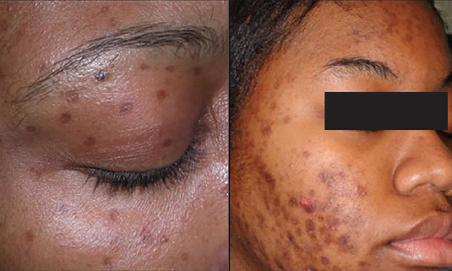
Post inflammatory hyperpigmentation
Postinflammatory pigmentation also called acquired melanosis, is temporary pigmentation that follows injury (e.g. thermal burn) or inflammatory disorder of the skin (e.g. dermatitis, infection). More severe injury results in postinflammatory hypopigmentation, which is usually permanent. Postinflammatory pigmentation can occur in anyone, but is more common in darker skinned individuals, in whom the color tends to be more intense and persist for a longer period than in lighter skin colors 2.
Postinflammatory hyperpigmentation tends to more pronounced in sun-induced skin conditions such as phytophotodermatitis and lichenoid dermatoses (skin conditions with lichen planus-like inflammation, such as erythema dyschromicum perstans).
Some medications may also darken postinflammatory pigmentation. These include antimalarial drugs, clofazimine, tetracycline, anticancer drugs such as bleomycin (flagellate erythema), doxorubicin, 5-fluorouracil and busulfan.
Figure 4. Post-inflammatory hyperpigmentation
Post inflammatory hyperpigmentation causes
Postinflammatory hyperpigmentation follows damage to the epidermis and/or dermis with deposition of melanin within the keratinocytes (skin cells) and/or dermis.
Inflammation in the epidermis stimulates melanocytes to increase melanin synthesis and to transfer the pigment to surrounding keratinocytes (epidermal melanosis). If the basal layer is injured (e.g. lichen planus), melanin pigment is released and subsequently trapped by macrophages in the papillary dermis (dermal melanosis or pigment incontinence).
Post inflammatory hyperpigmentation signs and symptoms
Postinflammatory hyperpigmented patches are located at the site of the original disease after it has healed. The lesions range from light brown to black in colour. The patches may become darker if exposed to sunlight (UV rays).
Post inflammatory hyperpigmentation diagnosis
Postinflammatory hyperpigmentation is diagnosed by taking a careful history and examining the skin. Dermal melanosis gives a characteristic hue to the skin colour (grey-purple-brown).
Sometimes the diagnosis is only made after skin biopsy. Histopathology reveals patchy epidermal melanosis and/or dermal melanosis.
Post inflammatory hyperpigmentation treatment
If pigmentation affects an exposed site, daily application of SPF 50+ broad-spectrum sunscreen is important to minimize darkening caused by UV radiation. Cosmetic camouflage can be used.
Topical treatments for postinflammatory hyperpigmentation
A variety of topical treatments are available to lighten/bleach hyperpigmented lesions in epidermal hypermelanosis. Varying degrees of success are achieved but combinations of the treatments below are usually required for significant improvement.
- Hydroquinone
- Azelaic acid
- Cysteamine cream
- Vitamin C cream
- Tretinoin cream
- Corticosteroid creams
- Glycolic acid peels
- Others: kojic acid, arbutin, licorice extracts, mequinol, niacinamide, N-acetyl glucosamine, soy
Physical treatments for postinflammatory hyperpigmentation
Chemical peels, laser treatments and intense pulsed light therapies (IPL) may be helpful for epidermal pigmentation, but physical treatments may also aggravate it by injuring the epidermis.
These treatments are not effective in dermal hypermelanosis.
Best treatment for hyperpigmentation
The best treatment for hyperpigmentation involves treating the underlying cause of your hyperpigmentation.
Treatment of drug-induced hyperpigmentation involves stopping the causative drug; the hyperpigmentation fades very slowly in some if not all cases. Because many drugs that cause skin pigmentation also cause photosensitivity reactions, patients should avoid sun exposure.
Localized hyperpigmentation treatment
Bleaching cream
Bleaching or skin lightening creams or ointments are widely used worldwide either to attempt to remove localized dark patches (eg, melasma or postinflammatory hyperpigmentation) or as a fashion trend aiming to reduce normal melanin in the skin.
A bleaching cream may contain a variety of ingredients. Some of these are more effective than others. In many areas, unregulated products are sold, often without listing their contents or they are labelled incorrectly. They may be safe but completely ineffective, or the chemicals may result in side effects and toxicity. The risks depend on which ingredient is being applied to the skin, in what concentration, over what area, and for how long it is used.
Skin lightening agents may include:
- Hydroquinone
- Cysteamine
- Topical retinoids
- Botanicals
- Other agents
- Topical corticosteroids
- Mercury. Mercury was used as mercurous chloride, oxide and ammoniated mercury in many cosmetics and toiletries in the early part of the 20th century before it was realized it caused toxicity. It is still found in some skin lightening creams because mercury inactivates the enzyme that leads to the production of melanin. These products should NOT be used and are illegal in many countries including the USA. Longterm application of mercurial products to the skin makes the skin and nails darker because the mercury is deposited in the epidermis, hair follicles and dermis. Mercury poisoning results in acute and chronic toxicity including acrodynia, as well as neurological and kidney damage.
Hydroquinone
Hydroquinone is an effective skin lightening agent. It is no longer available in some parts of the world because of the damaging effects of long-term use. The recommended concentration over the counter is 2%, but up to 4% is available from a dermatologist in some countries. It should be used daily for no more than 6 months.
Its initial effect of inhibiting pigmentation is lost with prolonged application and sun stimulation.
Exogenous ochronosis is the main risk of continued use of hydroquinone. This results in an irregular blue-black staining affecting sun-exposed skin and nails. It is due to deep deposition of the same pigment that occurs in alkaptonuria (endogenous ochronosis). Exogenous ochronosis may also occur from phenol, quinine or resorcinol.
Ochronosis may also result in loss of elasticity of the skin and impaired wound healing.
In some subjects, excessive use of hydroquinone in combination with certain foods in the diet (fish, eggs, offal, beans) can result in an unpleasant fish odor in the body secretions such as sweat and urine (trimethylaminuria).
Hydroquinone is sometimes given another name, such as:
- 1, 4-Benzenediol
- Quinol
- Benzene-1
- 4-Diol
- p-Diphenol
- p-Dihydroxyl benzene
- Hydrochinone
- p-Hydroxylphenol
- Hydrochinonium
- Hydroquinol
- Tequinol.
Monobenzyl ether of hydroquinone is a strong derivative of hydroquinone that almost always causes nearly irreversible complete depigmentation of the skin (white patches).
Topical retinoids
Tretinoin is the main topical retinoid that has been used in skin lightening products. It thins the skin, increasing the penetration of other agents. as well as having a direct effect in reducing melanisation. It is a prescription medication because of potential risk in pregnancy. It can be quite irritating and may cause contact irritant dermatitis.
Cysteamine cream
Cysteamine cream is applied for 15 minutes each day for up to 12 weeks, then twice weekly for maintenance. It can cause temporary burning and redness, and may also cause dryness and irritation, relieved by applying an emollient.
Botanicals
New active skin lightening compounds isolated from plants are being added to modern cosmetics. They appear to inhibit the production of melanin without being toxic to the melanocyte (tyrosinase inhibitors). It is not yet known which preparations are the most effective. Active ingredients include:
- Arbutin 1% (a glycosylated hydroquinone)
- Paper mulberry 1%
- Glabridin 0.5% (licorice extract)
- Arctostaphylos patula and Arctostaphylos viscida
- Aloesin
- Gentisic acid
- Flavonoids
- Hesperidin
- Ascorbic acid or its derivative, magnesium ascorbyl phosphate 10%
- Niacinamide
- Yeast derivatives
- Polyphenols
- Soy proteins.
Other agents
Other agents in use for their skin lightening effect include:
- Azelaic acid 20%, produced by the yeast, Malassezia
- Kojic acid 1-4% (5-hydroxy-4-pyran-4-one-2-methyl), produced by a fungus (may cause contact irritant or contact allergic dermatitis)
- Mequinol 5-20% (4-hydroxyanisole)
- Isopropylcatechol
- N-acetyl-4-cysteaminylphenol
- N-acetyl glucosamine
- Piceatannol.
Unregulated skin lightening creams may include many other ingredients. These may be relatively safe (for example, lemon juice), toxic (camphor), irritating (detergents) or likely to provoke allergy (hair dye). Complications may include:
- Irritant contact dermatitis
- Allergic contact dermatitis.
Topical corticosteroids
Steroid creams used for bleaching skin color
Topical corticosteroids lighten the skin by the following mechanisms:
- Initial blanching due to vasoconstriction
- Slowing down skin cell turnover so reducing the number and activity of melanocytes (pigment cells)
- Reducing production of precursor steroid hormones thus reducing production of melanocyte stimulating hormone (MSH).
In the United States and many other countries, stronger topical corticosteroids are regulated and can only be obtained with a doctor’s prescription. Products containing betamethasone valerate, fluocinonide and clobetasol propionate can be purchased over the counter from a pharmacy or from drug vendors in a marketplace in other countries.
Potent topical steroids have a wide range of local side effects including skin thinning and atypical fungal infections (tinea incognita). When used over large areas for prolonged periods, they may risk serious internal disease from hypopituitarism. Steroid addiction syndrome results in folliculitis and steroid rosacea.
References- Del Rosario E, Florez-Pollack S, Zapata L Jr, et al: Randomized, placebo-controlled, double-blind study of oral tranexamic acid in the treatment of moderate-to-severe melasma. J Am Acad Dermatol 78(2):363–369, 2018. doi: 10.1016/j.jaad.2017.09.053.
- Postinflammatory pigmentation. https://dermnetnz.org/topics/postinflammatory-hyperpigmentation/


