Kasabach Merritt syndrome
Kasabach-Merritt syndrome also known as hemangioma thrombocytopenia syndrome, is defined as diffuse cavernous hemangioma along with the combination of thrombocytopenia, microangiopathic hemolytic anemia and consumptive coagulopathy 1. Kasabach-Merritt syndrome is only associated with two rare vascular tumors: kaposiform hemangioendotheliomas and tufted angiomas 2. Kasabach-Merritt syndrome can be life threatening secondary to the risk of bleeding and progression to DIC (disseminated intravascular coagulopathy). The blood clotting disorder results from platelets and other clotting factors of the blood being used up within the rapidly growing vascular tumor.
The association of hemangioma, thrombocytopenia, and hypofibrinogenemia was first described in 1940 by Haig Haigouni Kasabach and Katharine Krom Merritt 3, who took care of an infant with a giant capillary hemangioma and thrombocytopenic purpura. Kasabach-Merritt syndrome is a rare disorder that can affect infants from the time of birth, or may appear later in infancy as the vascular malformation grows. Diagnosis of Kasabach Merritt syndrome is made based on the constellation of a vascular lesion, thrombocytopenia, consumptive coagulopathy, and microangiopathic hemolytic anemia. Unlike true capillary hemangiomas that regress in childhood and are a cosmetic nuisance, the lesions in Kasabach Merritt syndrome are distinctive vascular tumors that include tufted angiomas and kaposiform hemangioendotheliomas 4.
The hemangioma is often within the skin but can be present anywhere, including retroperitoneal organs, the mediastinum, the pelvis, visceral organs, or the mesentery. For skin lesions, the mortality rate, with treatment, is under 10%, but retroperitoneal tumors have a mortality rate of approximately 60% 5. The overall mortality rate is between 12 and 50% with death occurring from severe haemorrhage related to disseminated intravascular coagulation, local invasion of vital structures, high output cardiac failure, multi-organ failure, or sepsis 5.
Kaposiform hemangioendotheliomas are typically solitary tumors which appear in the soft tissues of the limbs, head and neck or retroperitoneum 6. They are usually seen in infants less than 2 years of age, although cases have been reported in adults. They do not spread (metastasize) but can cause serious problems because of local growth, cardiac failure or the associated Kasabach-Merritt phenomenon 6.
There are few reports of kaposiform hemangioendotheliomas without Kasabach-Merritt syndrome. Kaposiform haemangioendotheliomas usually regress with time but do not completely disappear. Tufted angiomas usually present before 5 years of age, although they can occur throughout life. They present as brown, red or purple areas of skin and are firm to touch. They are often painful. Spontaneous regression is unusual. Most tufted angiomas do not cause Kasabach-Merritt syndrome and metastasis is rare.
The pathophysiology is believed to be consumption of platelets and fibrinogen by intralesional thrombosis 7. The lesions are typically superficial and solitary, but may involve internal structures such as the liver. Cardiac failure may result from high-volume arteriovenous shunting. Shock, intracranial bleeding, or other internal hemorrhages may result in mortality rates as high as 30% 7.
Kasabach Merritt syndrome is a rare disorder that affects males and females equally The diagnosis is most often made during infancy but older children have been reported with this phenomenon. Kaposiform hemangioendothelioma and tufted angioma tumors can occur without Kasabach Merritt syndrome. The reason for this is still unknown and may be secondary to a smaller size of the tumor, an older age at presentation or other clinical features.
Treatment aims to involute the tumor to prevent significant morbidity or mortality, or in response to a life-threatening event. Surgical excision is curative but most lesions are not amenable to this option. Historically, the first-line of treatment has been high-dose systemic corticosteroids. However, up to two-thirds of lesions will not respond to corticosteroids, or will quickly relapse once treatment is discontinued 8. Also, this treatment is not without its own troubling adverse effects. A number of alternative therapies have been tried with variable results, including interferon α−2a and 2b 9, radiation therapy and chemotherapeutic agents such as vincristine and actinomycin. The most promising recent option available for treatment of infantile hemangiomas is propranolol 10.
Kasabach Merritt syndrome causes
The cause of Kasabach Merritt syndrome is unknown. It is believed to be secondary to sequestration or trapping of platelets into the tumor. These tumors are made up of abnormal endothelial cells (spindle cells) and also lymphatic malformation. It is unclear why the Kasabach Merritt syndrome occurs and if it is caused by the spindle cells or the lymphatic component.
Kasabach Merritt syndrome symptoms
Signs and symptoms in Kasabach Merritt syndrome include the following:
- Visible cutaneous giant hemangioma or multiple smaller hemangiomas, usually on the extremities
- Enlarged abdomen
- Hepatomegaly or jaundice
- Petechiae, bruising, and frank bleeding
- Painful lesions
- Anemia
Physical findings may include the following:
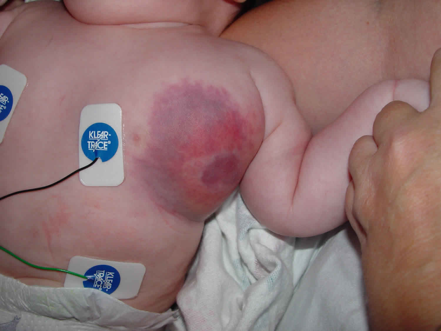
- Cutaneous hemangioma, often appearing as a large irregular bruise anywhere on the body and often circumscribed by widespread, overlying, shiny and dusky, purple skin
- Kaposiform hemangioendothelioma or tufted angioma – Blue or reddish-brown discoloration and skin induration
- Petechiae and bruising
- Painful, tender lesions
- Bleeding from thrombocytopenia and coagulopathy (locally and, at times, distantly [disseminated intravascular coagulation (DIC)])
- Tachycardia, feeding difficulty, and shock (signs of high-output cardiac failure)
- Pallor (suggestive of anemia)
Initially a vascular lesion is noted on the skin which can be firm, indurated and purpuric. Areas of petechiae (tiny red dots) can appear around the lesion or on other parts of the body. If the vascular lesion is internal, these petechiae can be seen on the skin. Bruising and spontaneous bleeding can also occur. These tumors are not hemangiomas. They usually present in young infants, less than three months of age, but have rarely been reported in older children. These tumors occur in the extremities, chest, neck, abdomen and pelvis. They infiltrate across tissue plans and can be aggravated by interventions, infection and trauma. When these tumors with Kasabach Merritt syndrome are internal such as in the pleural or retroperitoneum, they can cause significant morbidity and mortality. The morbidity and mortality is caused by bleeding.
Kasabach Merritt syndrome diagnosis
The diagnosis of Kasabach Merritt syndrome is based on the diagnosis of Kaposiform hemangioendothelioma/tufted angioma and this coagulopathy as noted above. If this diagnosis is suspected blood work including a complete blood count (CBC) with differential and platelets, fibrinogen, D-dimer, prothrombin time (PT), and activated partial thromboplastin time (aPTT) should be ordered. The best imaging modality to assess the extent of the lesion is a MRI with contrast. A biopsy will confirm the diagnosis.
Kasabach Merritt syndrome treatment
Patients diagnosed with Kasabach Merritt syndrome need to be treated at multidisciplinary vascular anomaly centers. There is no known standard of therapy for Kasabach Merritt syndrome. Medical management has included corticosteroids, interferon, chemotherapeutic agents such as vincrisitne, aspirin, and antiplatelet drugs such as Ticlopidine. Sometimes a combination of medications has been used. Other adjuvant therapies have included interventional embolization. If the lesion can be surgically removed that is the treatment of choice.
Agents that have been tried (most of them not specifically FDA-approved for this application), with varying success, include the following:
- Corticosteroids (most commonly used)
- Interferon alfa
- Aminocaproic acid (to treat bleeding)
- Aspirin
- Dipyridamole
- Ticlopidine
- Pentoxifylline
- Cryoprecipitate
- Heparin
- Vincristine (80% response reported)
- Cyclophosphamide
- Actinomycin D
- Propranolol (unlike in infantile hemangioma, response is poor)
Nonpharmacologic treatment modalities include the following:
- Surgical resection (when lesions are not too large or surgically inaccessible) – Wide local excision is recommended but may be difficult; amputation may be necessary for intractable lesions involving a limb
- Interventional radiologic procedures (when surgical treatment is not feasible)
- Intermittent pneumatic compression (most useful for a vascular lesion located on an extremity)
- Radiation therapy (now largely abandoned because of long-term adverse effects)
Kasabach Merritt syndrome prognosis
When Kasabach Merritt syndrome is promptly recognized and properly treated, the prognosis is usually excellent because the DIC (disseminated intravascular coagulopathy) resolves as the vascular lesion recedes and because Kasabach Merritt syndrome does not recur. Therefore, most children do well if they reach age 2 years. When Kasabach Merritt syndrome goes untreated, mortality is 10-37%, primarily due to bleeding secondary to the consumption coagulopathy 11. Even after failed treatment, however, the hematologic abnormalities may spontaneously resolve over a few months.
Mortality and morbidity are influenced by the anatomic location, depth, and extent of the vascular lesion. Besides bleeding, other conditions associated with morbidity and mortality are as follows:
- Visceral involvement (particularly in the retroperitoneal area and mediastinum)
- Profound thrombocytopenia
- DIC (disseminated intravascular coagulopathy)
- Severe infection
- Iatrogenic complications (eg, from procedures such as arterial ligation, arterial embolization, or surgical excision)
Hemolytic anemia resulting from physical damage to the red blood cells (RBCs) may be mild, moderate, or severe. The results of the direct antibody test or the Coombs test is negative in patients with anemia, and anemia is secondary to microangiopathic destruction of the red blood cells 12, manifested by the presence of schistocytes on peripheral blood smear examination (a helpful tool in suggesting the presence of viseral hemangiomas).
Heart failure often occurs in affected infants as a result of the large volume of blood flowing through the giant hemangioma 13.
An association of Kasabach Merritt syndrome with trisomy 21 mosaicism is uncommon; however, 43% of children with Down syndrome have cutaneous vascular lesions. One child with Down syndrome and Kasabach Merritt syndrome has been reported 14.
References- Haque PD, Mahajan A, Chaudhary NK, Jain D. Kasabach-Merritt Syndrome Associated With a Large Cavernous Splenic Hemangioma Treated With Splenectomy: A Surgeon’s Introspection of an Uncommon, Little Read, and Yet Complex Problem-Review Article. Indian J Surg. 2015;77(Suppl 1):166–169. doi:10.1007/s12262-015-1232-9 https://www.ncbi.nlm.nih.gov/pmc/articles/PMC4425793
- Kasabach-Merritt phenomenon. https://rarediseases.org/rare-diseases/kasabach-merritt-phenomenon/
- Kasabach HH, Merritt KK. Capillary hemangioma with extensive purpura: report of a case. American Journal of Diseases of Children. 1940; 59: 1063–70.
- Beutler E, Lichtman MA, Coller BS, Williams WJ. Williams Hematology 6th edn Newyork: McGraw-Hill; 2001.
- Maguiness S, Guenther L. Kasabach-Merritt syndrome. Journal of Cutaneous Medicine & Surgery. 2002; 6: 335–9.
- Osman NM. Kasabach – Merritt syndrome: A case report. Sudan J Paediatr. 2013;13(1):49–52. https://www.ncbi.nlm.nih.gov/pmc/articles/PMC4949964
- Larsen EC, Zinkham WH, Eggleston JC & Zitelli BJ. Kasabach-Merritt syndrome: therapeutic considerations. Pediatrics 1987; 79: 971–980.
- Moore J, Lee M, Garzon M, et al. Effective therapy of a vascular tumor of infancy with vincristine. Journal of Pediatric Surgery. 2001; 36: 1273–6.
- Wananukul S, Nuchprayoon I, Seksarn P. Treatment of Kasabach-Merritt syndrome: A stepwise regimen of prednisolone, dipyridamole, and interferon. International Journal of Dermatology. 2003; 42: 741–8.
- Arunachalam P, Kumar VRR, Swathi D. Kasabach–Merritt syndrome with large cutaneous vascular tumors. J Indian Assoc Pediatr Surg 2012; 17(1): 33–36.
- Szlachetka DM. Kasabach-Merritt syndrome: a case review. Neonatal Netw. 1998 Feb. 17(1):7-15.
- Payne LG, Hayward CPM, Kelton JG. Destruction of red cells by the vasculature and reticuloendothelial system. Hematology of Infancy and Childhood. 1998. 523-43.
- Berman B, Lim H. Concurrent cutaneous and hepatic hemangiomata in infancy: report of a case and a review of the literature. J Dermatol Surg Oncol. 1978 Nov. 4(11):869-73.
- Ram SP. Kasabach-Merritt syndrome and Down’s syndrome. J R Soc Med. 1997 Mar. 90(3):159-60.


