Lepromatous leprosy
Lepromatous leprosy also known as multibacillary leprosy, is a type of leprosy with a much more widespread disease, that involves the skin, thickening of many peripheral nerves, and at times involvement of other organs, such as eyes, nose, testicles, and bone 1. Lepromatous leprosy is characterized by multiple skin lesions that are smaller than those observed in tuberculoid leprosy (paucibacillary leprosy). Although the sites of skin lesions are similar to those of tuberculoid leprosy, the multiple lesions of lepromatous leprosy are often symmetrically distributed. Lepromatous macules may have poorly defined borders and no loss of sensation; local nerve enlargement is not characteristic. In addition to macules, lepromatous skin lesions may be nodules or plaques, or they may diffusely infiltrate the skin, especially on the face which may cause loss of eyebrows and “Leonine Facies” (see Figure 1).
Leprosy also called Hansen disease, is a chronic bacterial infection caused by slow-growing bacteria called Mycobacterium leprae, primarily affecting the skin, peripheral nerves, skin, eyes, and lining of the nose (nasal mucosa). In 2008, Mycobacterium lepromatosis was found to be the cause of diffuse lepromatous leprosy endemic in Mexico and the Caribbean 1. These bacteria grow very slowly and it may take up to 20 years to develop signs of the infection.
The clinical presentation of leprosy can vary based on the patient’s immune status. Traditionally patients were classified according to the Ridley-Jopling scale, which included indeterminate leprosy, tuberculoid leprosy, borderline tuberculoid leprosy, borderline leprosy, borderline lepromatous leprosy and lepromatous leprosy 2. The Ridley-Jopling scale has been replaced by the 2017 World health Organization (WHO) system, which classifies leprosy on the basis of clinical manifestations and skin smear results 3. In the WHO revised definitions, patients with 1 to 5 skin lesions with negative slit-skin smears for bacilli are said to have paucibacillary leprosy (tuberculoid leprosy); while those with more than five skin lesions or with nerve involvement (pure neuritis, or any number of skin lesions and neuritis) or with the demonstrated presence of bacilli in a slit-skin smear, irrespective of the number of skin lesions are said to have multibacillary leprosy (lepromatous leprosy) 3.
Tuberculoid leprosy represents a sufficient cell-mediated immune response. Patients have one or two, hypopigmented, erythematous, and anesthetic macules with a raised margin 4. Lepromatous leprosy may occur in the setting of immune dysfunction or if the patient is anergic to Mycobacterium leprae. These patients may have widespread disease that involves the skin, upper respiratory tract, anterior chamber of the eye, testes, lymph nodes, periosteum, and superficial sensory and motor nerves 5. Cutaneous findings include diffuse, erythematous macules, papules, and nodules4. A difference in cytokine response also has been demonstrated in subtypes of leprosy, with a primarily TH1 profile expressed in tuberculoid leprosy and borderline tuberculoid leprosy tissue specimens and a TH2 profile in lepromatous leprosy tissue specimens 5.
If leprosy is left untreated, the nerve damage can result in paralysis of hands and feet. In very advanced cases, the person may have multiple injuries due to lack of sensation, and eventually the body may reabsorb the affected digits over time, resulting in the apparent loss of toes and fingers. Corneal ulcers and blindness can also occur if facial nerves are affected. Other signs of advanced Hansen’s disease may include loss of eyebrows and saddle-nose deformity resulting from damage to the nasal septum.
Early diagnosis and treatment usually prevent disability that can result from the disease, and people with leprosy can continue to work and lead an active life. Once treatment is started, the person is no longer contagious. However, it is very important to finish the entire course of treatment as directed by the doctor.
Leprosy is diagnosed by finding at least one of the following cardinal signs 6:
- Definite loss of sensation in a pale (hypopigmented) or reddish skin patch;
- Thickened or enlarged peripheral nerve, with loss of sensation and/or weakness of the muscles supplied by that nerve;
- Presence of acid-fast bacilli in a slit-skin smear.
Leprosy is curable with a combination of drugs known as multidrug therapy, as the treatment of leprosy with only one antileprosy drug (monotherapy) will result in development of drug resistance to that drug. The combination of drugs used in the multidrug therapy depends on the classification of the disease (paucibacillary leprosy or multibacillary leprosy).
- Paucibacillary leprosy – 2 antibiotics are used at the same time, daily dapsone and rifampicin once per month
- Multibacillary leprosy – daily clofazimine is added to rifampicin and dapsone.
Treatment usually lasts between one to two years. The illness can be cured if treatment is completed as prescribed.
Leprosy key facts
- In the U.S., leprosy is rare. Around the world, as many as 2 million people are permanently disabled as a result of Hansen’s disease (leprosy). Overall, the risk of getting leprosy for any adult around the world is very low. That’s because more than 95% of all people have natural immunity to the disease.
- Early diagnosis and treatment prevents nerve involvement, the hallmark of Hansen’s disease (leprosy), and the disability it causes.
- Without nerve involvement, Hansen’s disease (leprosy) would be a minor skin disease.
- You may be at risk for leprosy if you live in a country where the disease is widespread. Countries that reported more than 1,000 new cases of leprosy to WHO between 2011 and 2015 are:
- Africa: Democratic Republic of Congo, Ethiopia, Madagascar, Mozambique, Nigeria, United Republic of Tanzania
- Asia: Bangladesh, India, Indonesia, Myanmar, Nepal, Philippines, Sri Lanka
- Americas: Brazil
- 185 new cases were reported in the U.S. in 2018 (the most recent year for which data are available).
- Most (133 or 72%) of these new cases were reported in 7:
- Florida
- California
- Hawaii
- Louisiana
- New York
- Arkansas
- Texas
- You may also be at risk if you are in prolonged close contact with people who have untreated leprosy. If they have not been treated, you could get the bacteria that cause leprosy. However, as soon as patients start treatment, they are no longer able to spread the disease.
Figure 1. Lepromatous leprosy (Leonine Facies)
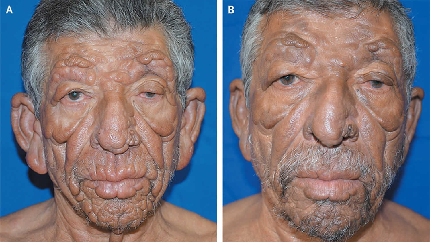
Footnote: A 57-year-old man presented with a 7-year history of diffuse skin infiltration associated with sensory loss in his left hand. His face had multiple nodular lesions that coalesced into plaques, especially on the forehead, ears, nose, and lips (Panel A). He had weakness in the muscles on the left side of his face and loss of eyebrow hair and eyelashes. The ulnar and tibial posterior nerves were enlarged. A skin smear stained according to the Ziehl–Neelsen method revealed acid-fast bacilli in clumps. Screening for IgM antibodies to phenolic glycolipid 1, which is specific for Mycobacterium leprae, was performed with the use of enzyme-linked immunosorbent assay. The highly positive results were consistent with a diagnosis of lepromatous leprosy. This anergic form of leprosy is characterized by an inefficient immune response. Nerve damage is slow to occur and is related to high levels of infection of the nerves. The patient underwent multidrug therapy, with the addition of prednisone, for 5 months. After 9 months of multidrug treatment, the skin infiltration and weakness in the left eyelid had diminished (Panel B). The treatment period for standard multibacillary leprosy is 12 months, but anergic polar forms, as occurred in this patient, usually require 24 months of treatment before cure.
[Source 8 ]Lepromatous leprosy causes
Leprosy is nearly always caused by an acid fast rod-shaped bacillus Mycobacterium leprae. In 2008, Mycobacterium lepromatosis was found to be the cause of diffuse lepromatous leprosy endemic in Mexico and the Caribbean 1. Mycobacterium lepromatosis was identified by genome sequencing of isolates in Mexico and has subsequently been identified in many other countries. It is believed to have reached the Americas with populations migrating from Asia across the Bering Strait, whereas Mycobacterium leprae traveled with colonists from the Old World. The organism multiplies very slowly (dividing approximately once every 13 days) and is an obligate intracellular pathogen that lacks several genes needed for independent survival, thus it has never been grown in bacteriologic media. However, it has been grown in mouse foot pads by injecting ground tissue from lepromatous nodules or nasal scrapings from leprosy patients into the foot pad of the animal. Typically, the granuloma appears at the inoculation site within 6 months. Armadillos can also be experimentally infected and will develop systemic disease, and are now the most common animal used to study Hansen’s disease and its treatment.
A genetic study at the National Hansen’s Disease Program reports that armadillos may be a source of infection in the southern United States.
The National Hansen’s Disease Program advises:
- The risk of transmission from animals to humans is low, but armadillos are wild animals and should be treated as such, with all proper precautions.
- Individuals should decide for themselves whether to interact with these animals and, if so, what precautions to take.
How do people get leprosy?
It is not known exactly how leprosy spreads between people. Scientists currently think it may happen when a person with leprosy coughs or sneezes, and a healthy person breathes in the droplets containing the Mycobacterium leprae bacteria. Prolonged, close contact with someone with untreated leprosy over many months is needed to catch the disease.
You cannot get leprosy from a casual contact with a person who has leprosy like:
- Shaking hands or hugging
- Sitting next to each other on the bus
- Sitting together at a meal
leprosy is also not passed on from a mother to her unborn baby during pregnancy and it is also not spread through sexual contact.
Due to the slow-growing nature of the bacteria and the long time it takes to develop signs of the disease, it is often very difficult to find the source of infection.
In the southern United States, some armadillos are naturally infected with the bacteria that cause leprosy in people and it may be possible that they can spread it to people. However, the risk is very low and most people who come into contact with armadillos are unlikely to get leprosy.
For general health reasons, avoid contact with armadillos whenever possible. If you had a contact with an armadillo and are worried about getting leprosy, talk to your healthcare provider. Your doctor will follow up with you over time and perform periodic skin examinations to see if you develop the disease. In the unlikely event that you have leprosy, your doctor can help you get treatment.
Lepromatous leprosy signs and symptoms
Leprosy is a highly variable disease, affecting different people in different ways, according to their immune response. Those at one end of the spectrum, with a high level of immunity, harbor a low number of bacilli and are referred to as patients with paucibacillary leprosy (tuberculoid leprosy). Those showing limited or low immunity with many bacilli in their body are referred to as patients with multibacillary leprosy (lepromatous leprosy).
- Paucibacillary leprosy (tuberculoid leprosy): a case of leprosy with 1 to 5 skin lesions, without demonstrated presence of bacilli in a skin smear.
- Multibacillary leprosy (lepromatous leprosy): a case of leprosy with more than five skin lesions; or with nerve involvement (pure neuritis, or any number of skin lesions and neuritis); or with the demonstrated presence of bacilli in a slit-skin smear, irrespective of the number of skin lesions.
In over 90% of patients, the first symptom noticed is numbness. Temperature is the first sensation lost, followed by light touch, pain, and then deep pressure. This may precede the development of cutaneous lesions by years. The initial skin lesions are usually of the indeterminate type, presenting as a solitary or small number of hypopigmented patches before evolving into borderline tuberculoid or lepromatous types.
Paucibacillary leprosy or tuberculoid leprosy
Paucibacillary leprosy or tuberculoid leprosy, is characterized by one or a few hypopigmented or hyperpigmented skin macules that exhibit loss of sensation (anesthesia) due to infection of the peripheral nerves supplying the region. The body’s immune response may also result in swelling of the peripheral nerves; these enlarged nerves may be palpated under the skin, and may or may not be tender to the touch.
Tuberculoid leprosy or paucibacillary leprosy is defined by:
- A few sharply defined red patches with raised borders or a single larger hypopigmented patch less than 10 cm in diameter
- Loss of sweating with dry hairless skin in the patches
- Loss of sensation in lesions
- Affected nerves are thickened and tender on palpation. The nerves most often found to have swelling are:
- Great auricular nerve
- Ulnar nerve above the elbow and dorsal cutaneous branches at the wrist
- Median nerve at the wrist (in the carpal tunnel)
- Radial nerve (superficial at wrist)
- Common peroneal nerve (also femoral cutaneous and lateral popliteal nerves where they wind around the neck of the fibula)
- Posterior tibial nerve, posterior to the medial malleolus
- Sural nerve.
Lepromatous leprosy or multibacillary leprosy
Multibacillary leprosy or lepromatous leprosy is characterized by generalized or diffuse involvement of the skin, a thickening of the peripheral nerves under microscopic examination, and has the potential to involve other organs, the eyes, nose, testes, and bone. The nodular form of this condition is the most advanced form of the disease. Ulcerated nodules contain large numbers of Mycobacterium leprae acid-fast bacilli packed in macrophages that appear as large foamy cells. Lepromatous leprosy (multibacillary leprosy) is associated with:
- multiple, symmetrically-distributed skin lesions that might not exhibit loss of sensation
- nodules
- plaques
- thickened dermis
- frequent involvement of the nasal mucosa resulting in nasal congestion and epistaxis
Lepromatous leprosy or multibacillary leprosy defined by:
- Early symptoms of nasal stuffiness, discharge, and bleeding
- Swelling and thickening of limbs, especially ankles and legs with subsequent ulceration
- Widespread hypopigmented and erythematous macules with a shiny surface and normal sensation
- Progression to widespread infiltration of skin forming nodules and plaques
- Characteristic leonine facies with thickening of the forehead, loss of eyebrows and eyelashes (madarosis), distortion of the nose, and thickening of the earlobes
- Involvement of other systems:
- Eyes — corneal anaesthesia, keratitis, corneal ulceration, uveitis, glaucoma, irreversible blindness
- Testes — orchitis, testicular atrophy, sterility
- Liver — hepatitis, hepatic amyloidosis
- Kidneys — glomerulonephritis, renal amyloidosis
- Bones — osteoporosis, resorption of digits.
Borderline leprosy
Borderline leprosy or dimorphous Hansen’s disease is the most common form. When compared to tuberculoid leprosy or lepromatous leprosy, it is of intermediate severity. The skin lesions seem to be of the tuberculoid type, but are more numerous, and may be found anywhere on the body. Peripheral nerves are affected as well, with ensuing weakness and anesthesia.
Borderline leprosy presents with:
- Similar lesions to tuberculoid leprosy but smaller in size, more numerous, and less well-defined.
- Anesthesia over the lesions is less pronounced compared to tuberculoid leprosy.
Borderline borderline leprosy is a rare unstable form of leprosy defined by:
- Multiple lesions of varying size, shape, and distribution
- Characteristic inverted saucer-shaped lesions with sloping edges and punched out center
Borderline lepromatous leprosy
Borderline lepromatous leprosy is characterized by:
- Widespread bilaterally symmetrical lesions
- Macules, papules, and nodules of variable size and shape
- Sensation and hair growth remain normal within a lesion
- Characteristic glove and stocking numbness
- Widespread peripheral nerve involvement.
Lepromatous leprosy diagnosis
Leprosy can be recognized by appearance of patches of skin that may look lighter or darker than the normal skin. Sometimes the affected skin areas may be reddish. Loss of feeling in these skin patches is common. You may not feel a light touch or a prick with a needle.
To confirm the diagnosis, your doctor will take a sample of your skin or nerve (through a skin or nerve biopsy) to look for the bacteria under the microscope and may also do tests to rule out other skin diseases.
In an endemic country or area, an individual should be regarded as having leprosy if he or she shows ONE of the following cardinal signs:
- Skin lesion consistent with leprosy and with definite sensory loss, with or without thickened nerves
- Positive skin smears
Leprosy diagnosis should be confirmed by one of the following investigations.
- Skin slit smear — a small slit is made using a sharp blade over the skin of the earlobe, forehead, or lesional skin, then a smear is made by scraping the exposed dermis onto a glass slide and examining for acid fast bacilli (AFB) under microscopy; useful for multibacillary leprosy only.
- Lepromin test — is an intradermal test for delayed type hypersensitivity to Mycobacterium leprae antigens; although not specific, it is helpful for classifying the type of leprosy.
- Skin biopsy — may show typical features, depending on the type of leprosy (see leprosy pathology); special stains may be required to demonstrate the bacilli.
- Mycobacterium leprae DNA PCR is very specific for detecting leprosy organisms.
Lepromatous leprosy treatment
The treatment of leprosy aims to stop active infection and minimize complications and deformity. Residual disabilities may require corrective reconstructive surgery to allow day-to-day activity.
Leprosy is treated with a combination of antibiotics. Typically, 2 or 3 antibiotics are used at the same time. Most endemic countries follow the WHO recommended multi-drug therapy (MDT) of antibiotics; the combination of drugs selected and duration of treatment depends on the type of leprosy. The 2018 WHO guidelines recommend three drugs: dapsone, rifampicin and clofazimine is added for 6 months in paucibacillary leprosy and 12 months for multibacillary disease. Other drug options include ofloxacin, moxifloxacin, minocycline, clarithromycin, rifapentine, and diarylquinolone. Vaccines and other forms of immunotherapy are being trialled.
- The standard adult treatment regimen for paucibacillary leprosy is:
- Rifampicin: 600 mg once a month
- Dapsone: 100 mg daily
- Duration: 6 months (6 blister packs of 28 days each)
- The standard adult treatment regimen for multibacillary leprosy is:
- Rifampicin: 600 mg once a month
- Clofazimine: 300 mg once a month, and 50 mg daily
- Dapsone: 100 mg daily
- Duration: 12 months (12 blister packs of 28 days each)
- The standard child (ages 10–14 years) treatment regimen for paucibacillary leprosy is:
- Rifampicin: 450 mg once a month
- Dapsone: 50 mg daily
- Duration: 6 months (6 blister packs of 28 days each)
- Standard child (ages 10–14 years) treatment regimen for multibacillary leprosy is:
- Rifampicin: 450 mg once a month
- Clofazimine: 150 mg once a month, and 50 mg every other day
- Dapsone: 50 mg daily
- Duration: 12 months (12 blister packs of 28 days each)
Treatment usually lasts between one to two years. The illness can be cured if treatment is completed as prescribed.
Antibiotics used during the treatment will kill the bacteria that cause leprosy. But while the treatment can cure the disease and prevent it from getting worse, it does not reverse nerve damage or physical disfiguration that may have occurred before the diagnosis. Thus, it is very important that the disease be diagnosed as early as possible, before any permanent nerve damage occurs.
If you are treated for leprosy, it’s important to:
- Tell your doctor if you experience numbness or a loss of feeling in certain parts of the body or in patches on the skin. This may be caused by nerve damage from the infection. If you have numbness and loss of feeling, take extra care to prevent injuries that may occur, like burns and cuts.
- Take the antibiotics until your doctor says your treatment is complete. If you stop earlier, the bacteria may start growing again and you may get sick again.
- Tell your doctor if the affected skin patches become red and painful, nerves become painful or swollen, or you develop a fever as these may be complications of Hansen’s disease that may require more intensive treatment with medicines that can reduce inflammation.
In the U.S., people with leprosy may be treated at special clinics run by the National Hansen’s Disease Program (https://www.hrsa.gov/hansens-disease/index.html). There are several federally supported outpatient clinics throughout the U.S. and Puerto Rico (https://www.hrsa.gov/hansens-disease/ambulatory-clinics.html).
Reactive episodes
At least one in four patients with leprosy experience reactive episodes or “reactions” – during their treatment, although reactions may occur before treatment begins or after it is completed. Reactions are not due to medications used to treat the disease, although people treated with clofazimine tend to have slightly fewer episodes. There are two main groups of reactions:
- Type 1 reactions also called reversal reactions, are typical in paucibacillary leprosy or tuberculoid leprosy, and also in its borderline presentations, and show up as edema and erythema of pre-existing lesions. In some cases, neuritis and rarely new lesions or fever may also occur.
- Type 2 reactions or erythema nodosum leprosum are most frequently seen in patients with multibacillary leprosy or lepromatous leprosy. Patients usually present with painful erythematous nodules, often distributed between existing lesions, and moderate to high fever. Inflammation of other tissues may be present, including peripheral neuritis, orchitis, lymphadenitis, iridocyclitis, nephritis, periostitis and arthralgias.
- Lucio’s phenomenon, a rare reaction characterized by multiple hard-to-heal ulcers of varying size, is usually seen in patients with diffuse lepromatous leprosy who are of Mexican ancestry.
Lepromatous leprosy prognosis
Once appropriate treatment has been commenced, the skin lesions slowly subside. However, nerve damage cannot be reversed.
References- Xiang Y. Han, MD, PhD, Yiel-Hea Seo, MD, PhD, Kurt C. Sizer, MD, Taylor Schoberle, MS, Gregory S. May, PhD, John S. Spencer, PhD, Wei Li, PhD, R. Geetha Nair, MD, A New Mycobacterium Species Causing Diffuse Lepromatous Leprosy, American Journal of Clinical Pathology, Volume 130, Issue 6, December 2008, Pages 856–864, https://doi.org/10.1309/AJCPP72FJZZRRVMM
- Booth, A. V, & Kovich, O. I. (2007). Lepromatous leprosy. Dermatology Online Journal, 13(1). Retrieved from https://escholarship.org/uc/item/75z6499z
- Leprosy (Hansen’s disease). https://www.who.int/health-topics/leprosy
- Hartzell J, et al. Leprosy: a case series and review. South Med J 2004; 97:1252
- Ooi W, Moschella S. Update on leprosy in immigrants in the United States: status in the year 2000. Clin Infect Dis 2001; 32:930
- Guidelines for the diagnosis, treatment and prevention of leprosy. World Health Organization October 2018. https://apps.who.int/iris/bitstream/handle/10665/274127/9789290226383-eng.pdf
- The National Hansen’s Disease (Leprosy) Program. https://www.hrsa.gov/hansens-disease/index.html
- Leonine Facies: Lepromatous Leprosy. N Engl J Med 2012; 366:1433. DOI: 10.1056/NEJMicm1106238


