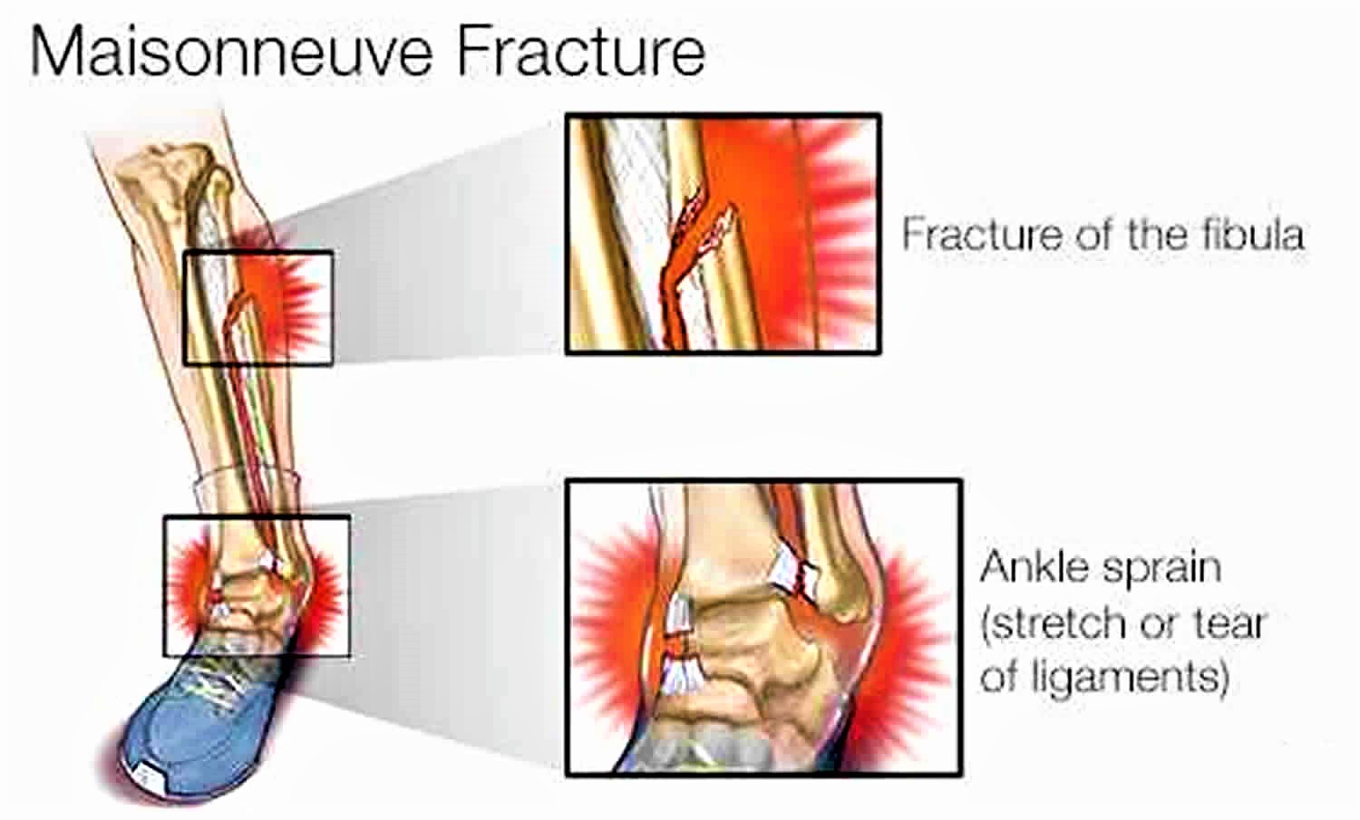
Maisonneuve fracture
Maisonneuve fracture refers to a combination of a fracture of the proximal or mid fibula together with an unstable ankle injury (widening of the ankle mortise on x-ray), often comprising ligamentous injury the medial ankle (distal tibiofibular syndesmosis, deltoid ligament) and/or fracture of the medial malleolus 1. Maisonneuve fracture is caused by pronation external-rotation mechanism. Maisonneuve fracture injury disrupts the tibiofibular syndesmosis and creates an unstable ankle joint. Maisonneuve fracture accounts for 7% of all ankle fractures 2. Maisonneuve fracture should be considered in all patients with medial ankle tenderness following trauma.
Two joints are involved in ankle fractures:
- Ankle joint – where the tibia, fibula, and talus meet
- Syndesmosis joint – the joint between the tibia and fibula, which is held together by ligaments
Multiple ligaments help make the ankle joint stable.
A Maisonneuve fracture is the result of two injuries that happen at the same time. The first is typically a very high break or fracture in the fibula — the smaller of the two bones between your ankle and your knee. The second is an ankle sprain — an injury that stretches or tears the tough bands of tissue, called ligaments, that help hold the bones of the ankle joint in place.
Maisonneuve fractures are most common in athletes who participate in sports such as skiing, gymnastics or dancing, where there is a risk of falling with the foot and leg hitting the ground at an awkward angle while they are rotating.
For this kind of complex ankle and leg injury, surgery often is necessary to stabilize the ankle joint and allow for proper healing. If a Maisonneuve fracture isn’t treated, the result can be long-term ankle instability and early-onset arthritis.
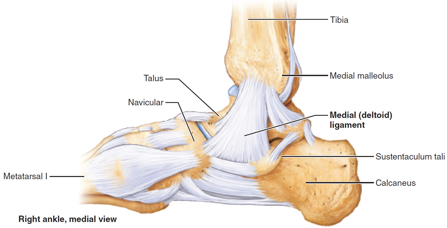
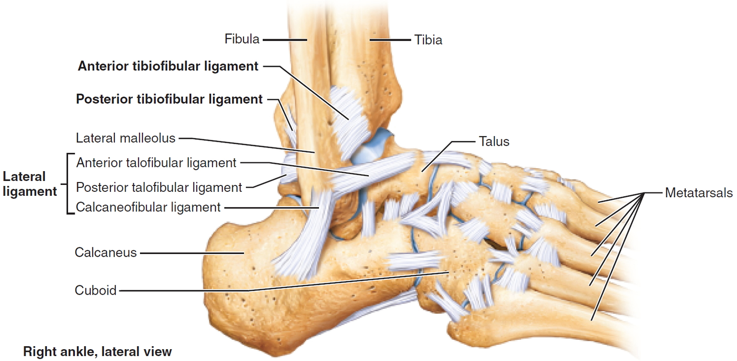
Ankle anatomy
- The tibia is the major weight-bearing bone of the lower leg
- Deltoid and medial collateral ligament: attach the distal medial tibia to multiple tarsal bones
- The deltoid ligament stabilizes the medial ankle joint and consists of the tibiocalcaneal, posterior tibiotalar, anterior tibiotalar, tibionavicular segments
- Tibiofibular syndesmosis: fibrous interosseous membrane attaching the tibia and fibula along the entire length of both bones
Figure 1. Ankle bones and ligaments
Maisonneuve fracture causes
The trauma mechanism involves an external rotation associated with ankle pronation, resulting in an injury of the distal tibiofibular ligament, syndesmotic complex, and proximal fibular fracture.:
- Sports-related injuries (most common)
- Motor vehicle crashes
The injury force typically starts at the medial ankle, then tears through the tibiofibular interosseous membrane, and then causes a fracture in the proximal tibia.
The injury force, if sufficient, may also cause a posterior tibial tubercle fracture.
Maisonneuve fracture symptoms
Symptoms of a Maisonneuve fracture include those you’d have with an ankle sprain, such as ankle pain, swelling, bruising and restricted range of motion. Symptoms also include ankle instability and pain higher in the leg at the site of the fibula fracture.
Maisonneuve fracture diagnosis
Diagnosing a Maisonneuve fracture can be challenging because it requires identifying several injuries. When health care providers suspect a Maisonneuve fracture, full-length X-rays of the fibula and the tibia (your other lower leg bone) are taken. Ankle joint, or mortise, X-rays are done, as well, to assess the joint damage and see if the bones in the joint are out of alignment.
X-rays taken while you aren’t bearing weight on your ankle may not reveal the full extent of a Maisonneuve fracture. Therefore, your health care provider may recommend that additional X-rays be taken while you put weight on your ankle. These images, also called stress films, can further assess possible damage to the ligaments in the ankle and evaluate the status of the ankle joint. In some cases, an MRI is used to assess the ankle joint, too.
Maisonneuve fracture treatment
Although management is variable depending on complexity of injuries, Maisonneuve fracture pattern is generally managed by operative treatment.
If the images show a Maisonneuve fracture, but the bones of the ankle joint are still in their proper place, or just slightly out of alignment, and the stress films show only minor ankle instability, then surgery may not be necessary. In such cases, using crutches, along with a brace, cast or splint as the ankle and leg heal, followed by physical therapy to regain your range of motion, may be sufficient treatment.
When a Maisonneuve fracture involves significant ligament injury, surgery is almost always required to stabilize the ankle joint and allow for proper healing. Depending on the specifics of the injury, your surgeon may place screws or other devices to hold the ankle joint in place while the ligaments heal. Screws may be removed when healing is complete, but that’s determined on a case-by-case basis.
Maisonneuve fracture surgery
Internal fixation of the distal tibiofibular syndesmosis 3 commonly achieved by trans-syndesmotic screws. Alternative stabilization mechanisms exist (e.g. bioabsorbable constructs, syndesmotic staples, hooks, or cerclage wires). In some cases, internal fixation of a posterior malleolar fracture fragment may result in sufficient stabilization. Fixation screws may or may not be removed after several weeks of healing 3.
Reduction and stabilization of medial malleolus fracture and/or ligamentous injuries 3. Ligamentous injuries may be managed non-operatively.
Reduction and stabilization of fibular fracture:
- fracture involving distal 2/3 of fibula may compromise ankle mortise, and so may benefit from surgery 3
- fracture involving proximal 1/3 fibula often managed non-operatively.
Maisonneuve fracture recovery
Standard postoperative recovery typically includes keeping weight off the affected ankle for 6 to 12 weeks. Following this, physical therapy may be used to regain flexibility, strength and balance. The goal is for the patient to be able to return to full activity and to regain the function he or she had prior to the injury.
If the ankle joint is not positioned properly during the healing process, the joint may be at risk for developing arthritis. Eventually, that can lead to chronic ankle pain, tenderness, stiffness and loss of flexibility.
To ensure the long-term health of the ankle joint following this injury, it is important to have a Maisonneuve fracture thoroughly assessed, properly diagnosed and promptly treated.
References- Maisonneuve Fracture – Emergency Management. https://www.dynamed.com/management/maisonneuve-fracture-emergency-management-16
- Alencar Neto JB, Cavalcante MLC, Pinto Neto LH, Lucena IF, Garrido RJ, Rocha PHMD. Maisonneuve Variant Lesion with Proximal Tibiofibular Dislocation. Rev Bras Ortop (Sao Paulo). 2019;54(3):339–342. doi:10.1055/s-0039-1692625 https://www.ncbi.nlm.nih.gov/pmc/articles/PMC6597427
- Stufkens SA, van den Bekerom MP, Doornberg JN, van Dijk CN, Kloen P. Evidence-based treatment of maisonneuve fractures. (2011) The Journal of foot and ankle surgery : official publication of the American College of Foot and Ankle Surgeons. 50 (1): 62-7. doi:10.1053/j.jfas.2010.08.017 https://doi.org/10.1053/j.jfas.2010.08.017


