
What is a mucous cyst
An oral mucous cyst is also called mucocele, is a painless, benign, mucous-containing cystic lesion on the inner surface of the mouth, especially the lower lip. A mucous cyst contains clear fluid. Mucous cyst forms when mucus or saliva escapes into surrounding tissues and a lining of granulation or connective tissue is formed to create a smooth, soft round fluid-filled lump. Mucous cysts are not true cysts (i.e, lined by epithelium). Mucous cysts are histologically characterized by pooled mucin lined with compressed reactive granulation tissue.
Mucous cysts most commonly occur on the inner surface of the lower lip (>80% of cases) but may also appear on the floor of the mouth (in this region they are known as ranula) or on the gingiva (gums), buccal mucosa (inner lining of the cheeks) and tongue.
Classic mucous cyst (Figure 1) account for 94% of lesions, are larger (most range from 5-10 mm), fluctuant, and nodular, with a deep blue hue or flesh toned color. Oral mucous cysts are typically painless. Other sites include the mouth floor, ventral tongue, or buccal wall. Rarely (1-3%), the upper lip is involved. Sublingual lesions (ventral tongue or floor of mouth) are also called ranula (Latin: little frog) from its resemblance to a frog’s air sack.
Superficial mucous cyst, a less common variant, are small (<5 mm), vesicular, frequently translucent, solitary or multiple oral papules. Women greater than 30 years of age tend to be most affected with this subtype. Frequent sites of involvement include the soft palate, retromolar pad and posterior buccal mucosa.
No significant gender predilection exists. More than 75% mucous cyst occurs in people younger than 40 years of age. Superficial mucous cysts tend to occur in females more than 30 years of age.
A mucous cyst often can be left alone. It usually will rupture on its own. If the cyst returns, it may need to be removed.
To remove a mucous cyst, your doctor may perform any of the following:
- Freezing the cyst (cryotherapy)
- Laser treatment
- Surgery to cut out the cyst
A ranula is usually removed using laser or surgery. The best outcome is removing both the cyst and the gland that caused the cyst.
To prevent infection and damage to the tissue, DO NOT try to open the sac yourself. Treatment should only be done by your doctor. Oral surgeons and some dentists can remove the sac.
Figure 1. Mucous cyst lip
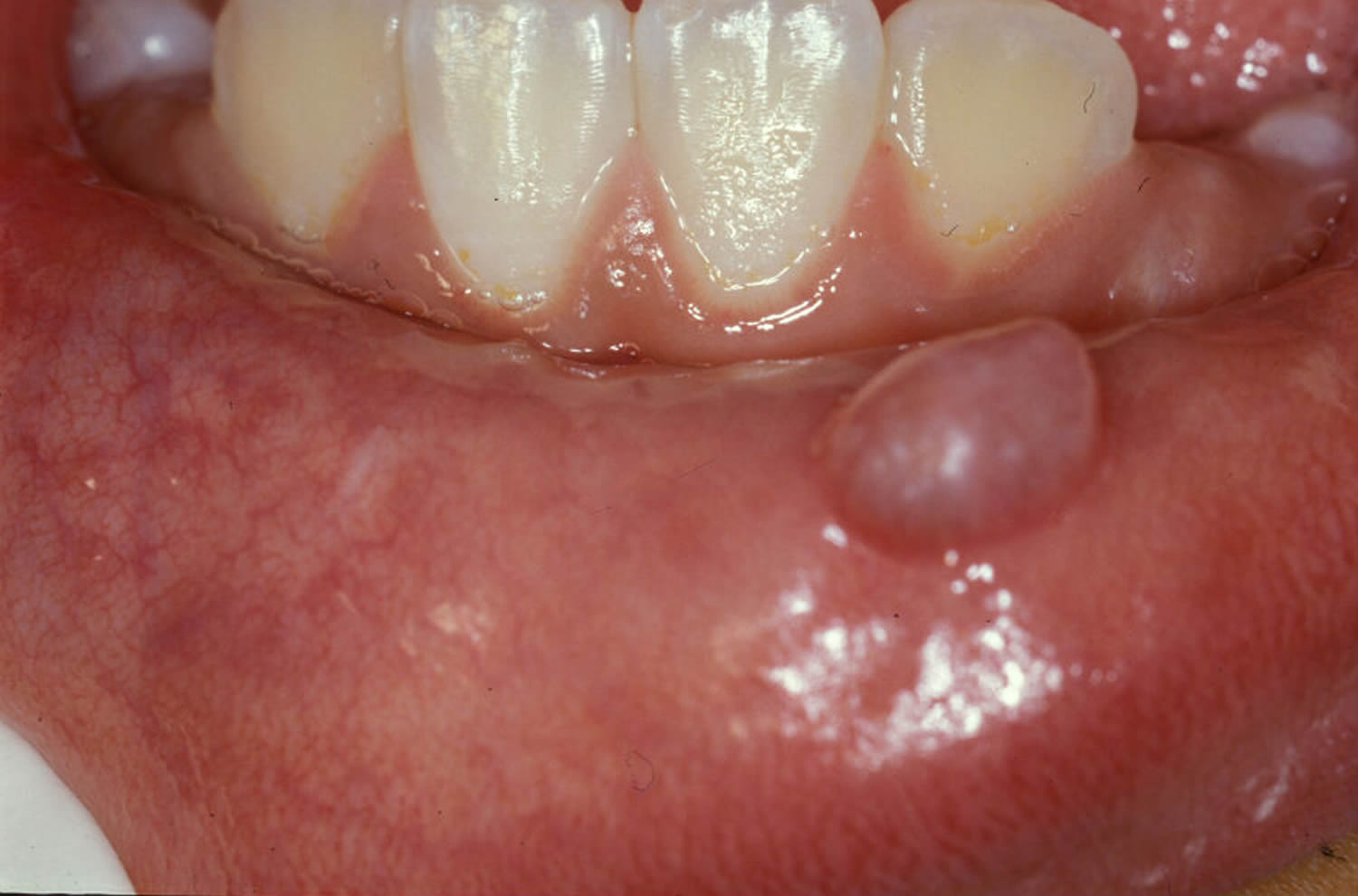
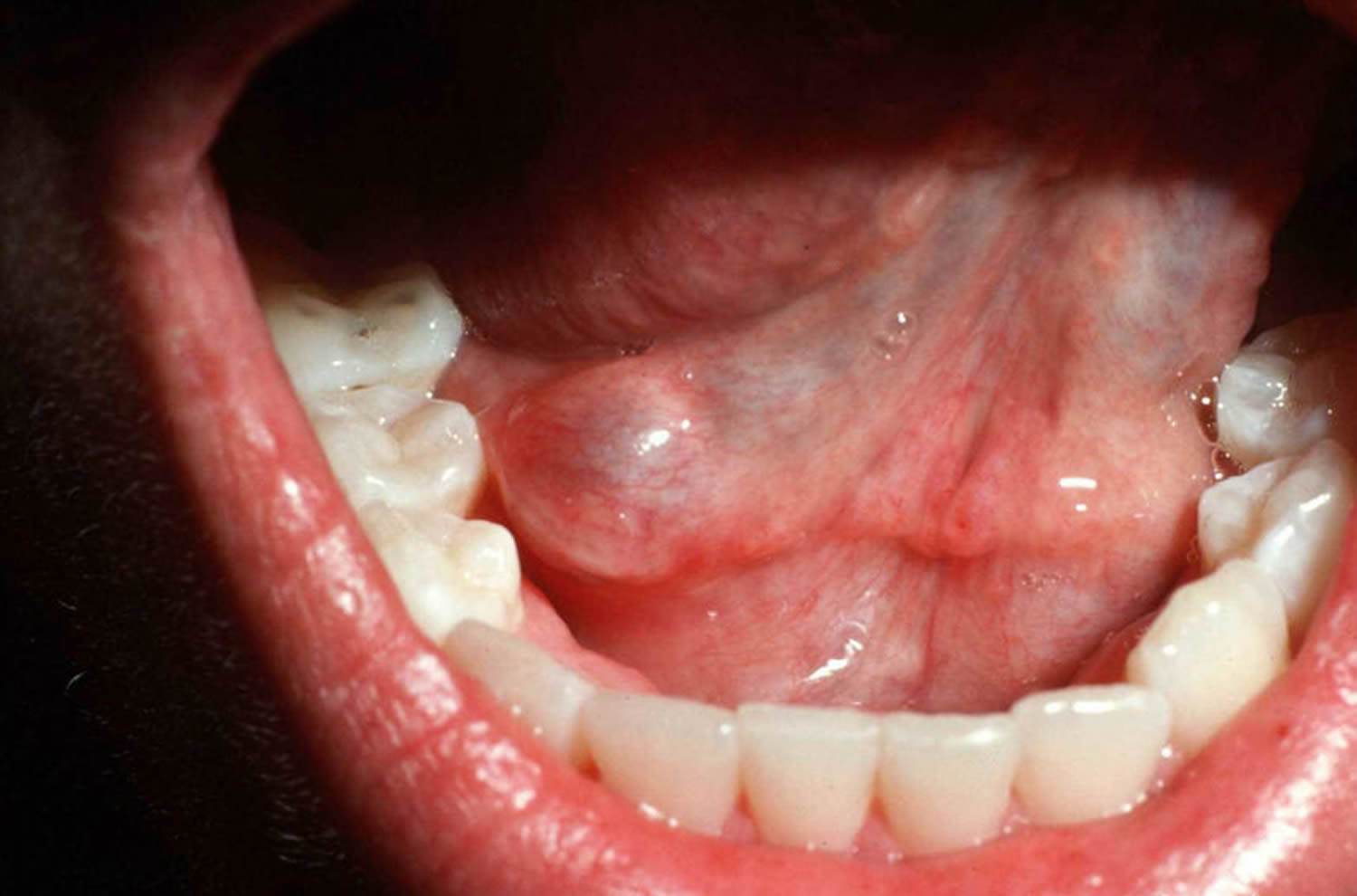
Figure 2. Mucous cyst on the floor of the mouth (ranula)
Contact your doctor if you:
- Notice a cyst or mass in your mouth
- Have difficulty swallowing or talking
- Pain, unusual growth, or any suspicious character of a clinically suspected mucous cyst may need histologic confirmation to exclude a malignant neoplastic process.
These may be a sign of more serious problem, such as mouth cancer.
Mucous cyst causes
Mucous cysts most often appear near salivary gland openings (ducts). Common sites and causes of mucous cysts include:
- Inner surface of the upper or lower lip, inside the cheeks, bottom surface of the tongue. These are called mucoceles. They are often caused by lip biting, lip sucking, or other trauma.
- Floor of the mouth. These are called ranula. They are caused by blockage of the salivary glands under the tongue.
Rupture of the minor salivary gland duct with subsequent escape of salivary mucous into the surrounding submucosa. Complete duct blockage was initially implicated as a cause but studies are not supportive. Reasons behind lower lip predilection are unknown. Prevailing theories include large tissue size of the lower lip leading to ease of trauma, greater kinematic demands with speech leading to increased manipulation, and increased density of glands leading to the simple likelihood of occurence.
Mucoceles are not true cysts (i.e, lined by epithelium). They are histologically characterized by pooled mucin lined with compressed reactive granulation tissue. Mucin and granulation can also be haphazardly or diffusely distributed throughout the lesion.
Mucous cyst prevention
Avoiding intentionally sucking the cheeks or biting the lips may help prevent some mucous cysts.
Mucous cyst symptoms
Symptoms of mucous cyst include:
- Usually painless, but can be bothersome because you’re aware of the bumps in your mouth.
- Often appears clear, bluish or pink, soft, smooth, round and dome-shaped.
- Vary in size up to 1 cm in diameter.
- May break open on their own, but may recur.
Symptoms of ranula include:
- Usually painless swelling on the floor of the mouth below the tongue.
- Often appears bluish and dome-shaped.
- If the cyst is large, chewing, swallowing, talking may be affected.
- If the cyst grows into the neck muscle, breathing can stop. This is a medical emergency.
Mucous cyst possible complications
Mucous cyst complications may include:
- Return of the cyst
- Injury of nearby tissues during removal of a cyst
Mucous cyst diagnosis
Diagnosis can be made on clinical grounds, especially with superficial vesicular variants. Deeper mucoceles typically need histopathologic examination to confirm or exclude other salivary gland tumors. Imaging studies such as ultrasound, computed tomography, and/or magnetic resonance imaging may rarely be needed. These can be used to identify form, size, and location relative to other organs. CT scan, usually for ranula that has grown into the neck.
Fine needle aspiration may be of use if excisional or incisional biopsy is contraindicated, particularly when a deep hemangioma is a clinical possibility. Large bore needle aspiration may also be confirmatory with the simple observation of mucous.
Distribution
Although mucous cyst can occur anywhere in the oral cavity where minor salivary glands are present, approximately 75-80% of cases occur on the lower lip, followed by the floor of the mouth (ranula), ventral tongue, and buccal mucosa.
Morphology
- About 75% of lesions are smaller than 1 cm in diameter
- Superficial mucous cysts take on a bluish to translucent hue, whereas deep lesions have normal mucosal coloration. Bleeding into the swelling may impart a bright red and vascular appearance
- Clinical features include a non-tender, mobile, dome-shaped enlargement with intact overlying epithelium
- Palpation reveals a fluctuant mass that does not blanch on compression
Histology overview of a mucous cyst
Mucophages (macrophages with engulfed foamy mucin) are commonly seen. These should be distinguished from salivary duct cysts, which are true cysts (eg, dilitations of the salivary ducts lined by ductal epithelium) and have different clinicopathological features. Salivary duct cysts tend to enlarge in size after eating, particularly with salivary stimulating foods such as lemon drops, grapefruit and pickles.
Mucous cyst treatment
Observation is a feasible approach to treatment. Five months is adequate time for spontaneous resolution to occur, particularly in children, and is recommended by some authorities prior to surgery in this age group.
Conservative management
- In general, no treatment is needed
- Mucous cysts may spontaneously resolve, especially in infants and young children. In a recent retrospective study, approximately 44% of mucous cysts in children spontaneously resolved after an average of three months
Surgery
- For larger, more troublesome lesions, surgical intervention such as marsupialization, complete excision, or dissection of the mucous cyst along with the adjacent associated minor salivary glands is recommended
- The risk for recurrence is minimal when appropriate surgical excision has been performed.
Surgical intervention is the most common therapy. Marsupialization, complete excision (mucocele with its associated minor salivary gland), or dissection can be performed. Trauma to the labial branch of the mental nerve is the greatest risk with excision and dissection techniques.
Marsupialization of the lesion is the first interventional approach to avoid mental nerve trauma. In this procedure, the lesion is unroofed, then packed with gauze for 7-10 days. Sutures may be placed laterally to keep the unroofed mucocele patent. Careful suture placement should taken with nearby glands to prevent iatrogenic mucocele formation. Sublingual or superficial lesions respond best with this technique. Deeper lesions tend to recur.
Micromarsupialization is a new novel technique used in children with lesions less than 1cm. Silk suture is placed through the internal part of the mucocele’s widest diameter, stitched, then left in place for 7 days with eventual resolution.
Complete excision simply requires removal of the lesion, some normal surrounding tissue and the underlying feeding gland en total. This technique can be performed with a scalpel, electrosurgery unit, or laser.
Dissection technique involves a superficial incision (does not penetrate the mucocele) at the lateral base of the lesion. Blunt dissection scissors are used to separate the overlying oral mucosal tissue from the underlying submucosal tissue to expose and localize the central mucocele. At the base, the feeding minor salivary gland is typically attached and should be removed.
Other treatment options
Other options with variable, or lower, success rates include aspiration, incision and drainage, cryosurgery, laser ablation, or sclerosing with OK-432 (Picibanil).
For superficial mucous cyst, intermittent use of high potency topical steroids can be used. Gamma-linolenic acid (oil of primrose) can also be employed.
With respect to variant techniques, aspiration or incision and drainage has a high recurrence rate but is a reasonable conservative interventional approach in children where lesions tend to involute with ease. Cryosurgery with a 30-second freeze and 1-minute thaw can be used. Damage to the lingual nerve and submandibular duct is still possible. Laser ablation (CO2 or Erbium:Yag) has been utilized with minimal complications and satisfactory aesthetic results.
OK-432 (Picibanil), a streptococcal-derived sclerosing agent, is a new alternative therapy that has been used with some success. Few studies highlight its use and it is experimental. Reported side effects include temporary local injection site discomfort, persistent fever and, rarely, shock (0.05%). It should be performed in a controlled setting by a practitioner with advanced life support experience, due to the low but possible risk of shock.
Superficial mucoceles are less problematic and can be treated conservatively with observation and intermittent use of high-dose topical steroids, such as Clobetasol ointment twice daily for 2 weeks then weekends only. Cessation can be adjusted with response and resumpton with disease flare. Gamma-linolenic acid (oil of primrose, a proglandin E precursor which has antiinflammatory and antiproliferative effects) can also be used.


