
What is the pericardium
The pericardium is a fibrous sac called serous membrane, that encloses the heart and great vessels within the pericardial cavity (pericardial sac). The pericardium keeps the heart in a stable location in the mediastinum, facilitates its movements and separates it from other mediastinal structures and the lungs 1. You can visualize the relationship of the serous membranes by pushing your fist into a limp balloon.
- The part of the balloon that clings to your fist represents the visceral pericardium (serous pericardium) on the heart’s (your fist’s) outer surface.
- The outer wall of the balloon represents the parietal pericardium (fibrous pericardium).
- The balloon’s thin airspace represents the pericardial cavity (pericardial sac) itself.
The visceral pericardium covers the surface of the heart; the parietal pericardium lines the chest wall. Between them is the pericardial cavity, filled with a small amount of lubricating serous fluid. This fluid is produced by both serous membranes. The slippery serous fluid allows the heart and great vessels to slide with little friction across the pericardial cavity walls as they carry out their routine functions. This freedom of movement is extremely important for the heart that move or change shape, such as the pumping heart.
The pericardium also supports physiological cardiac function 2.
The fibrous pericardium and the parietal part of the serosal pericardium are supplied by the phrenic nerve. The visceral pericardium is insensitive; therefore, the pain from the pericardium originates in the parietal layer only and is transmitted by the phrenic nerve.
Figure 1. Pericardium
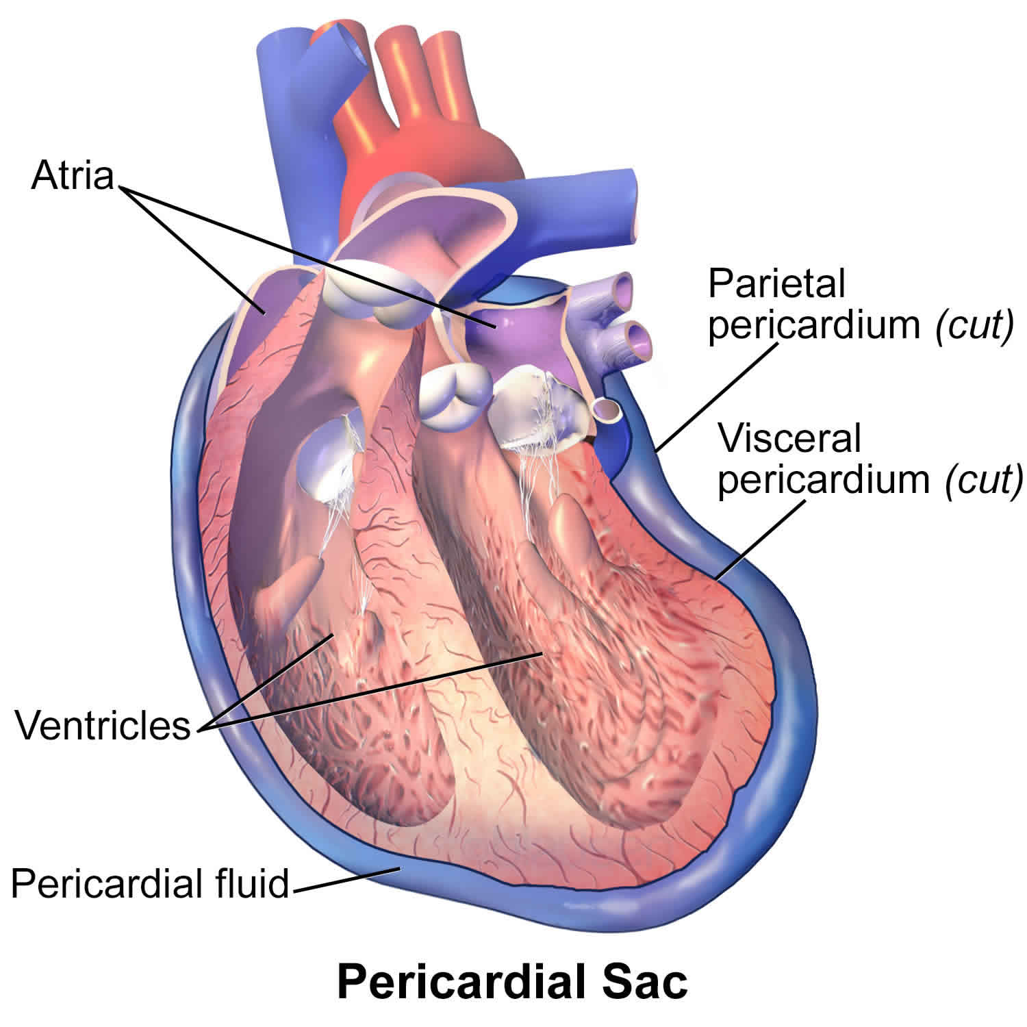
Figure 2. Heart pericardium
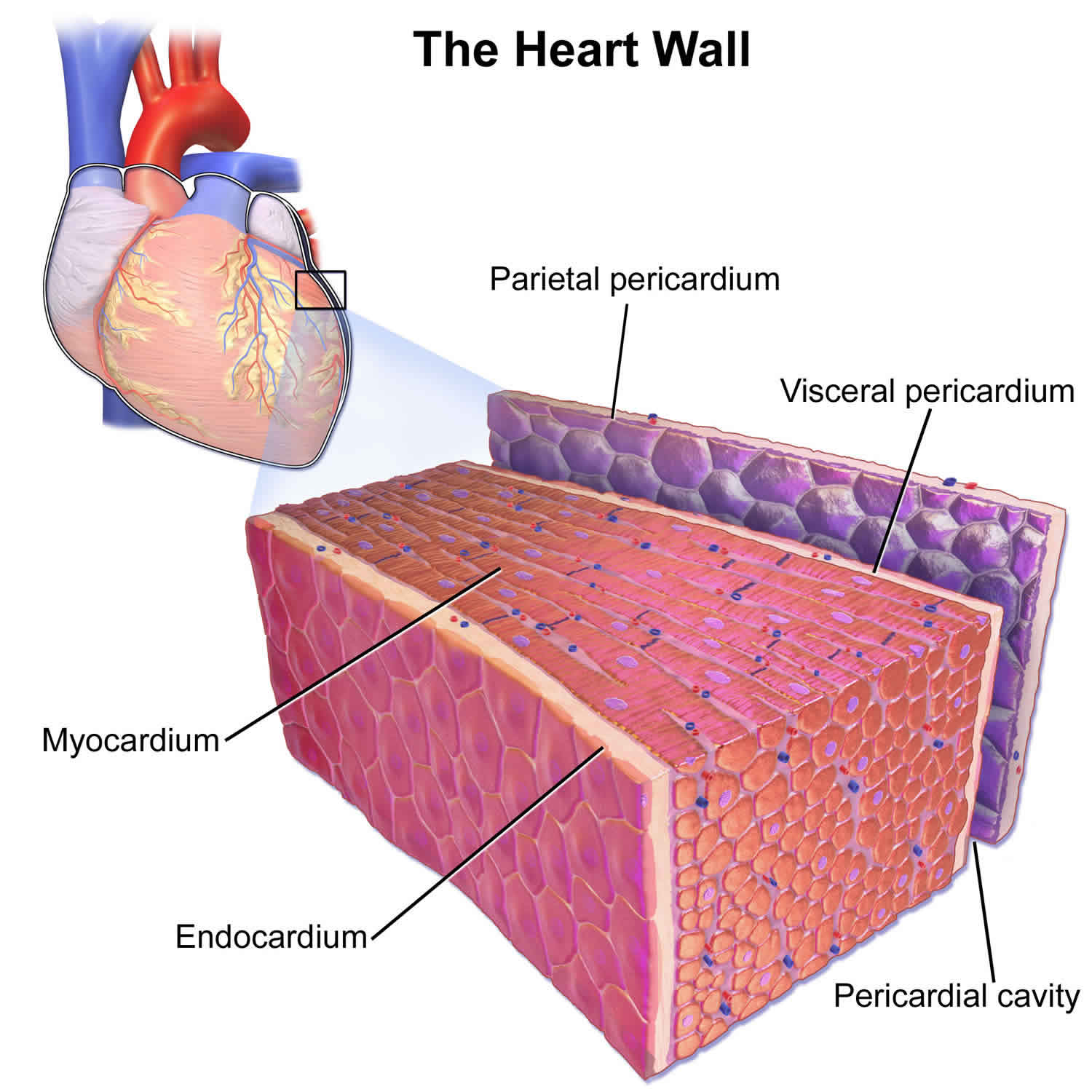
Figure 3. Pericardium location
Where is the pericardium located?
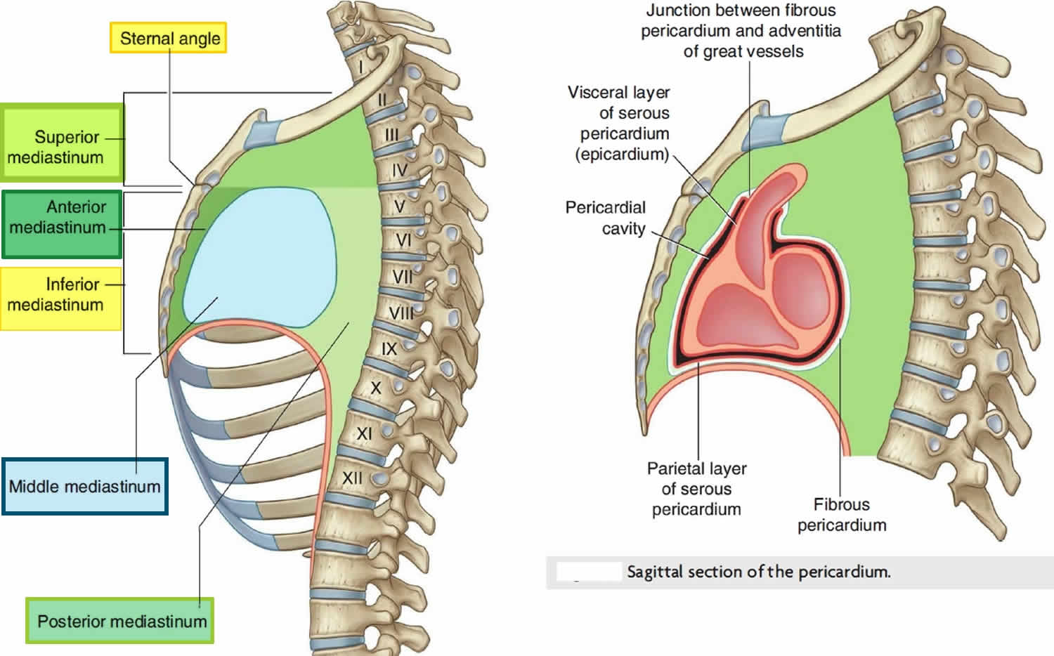
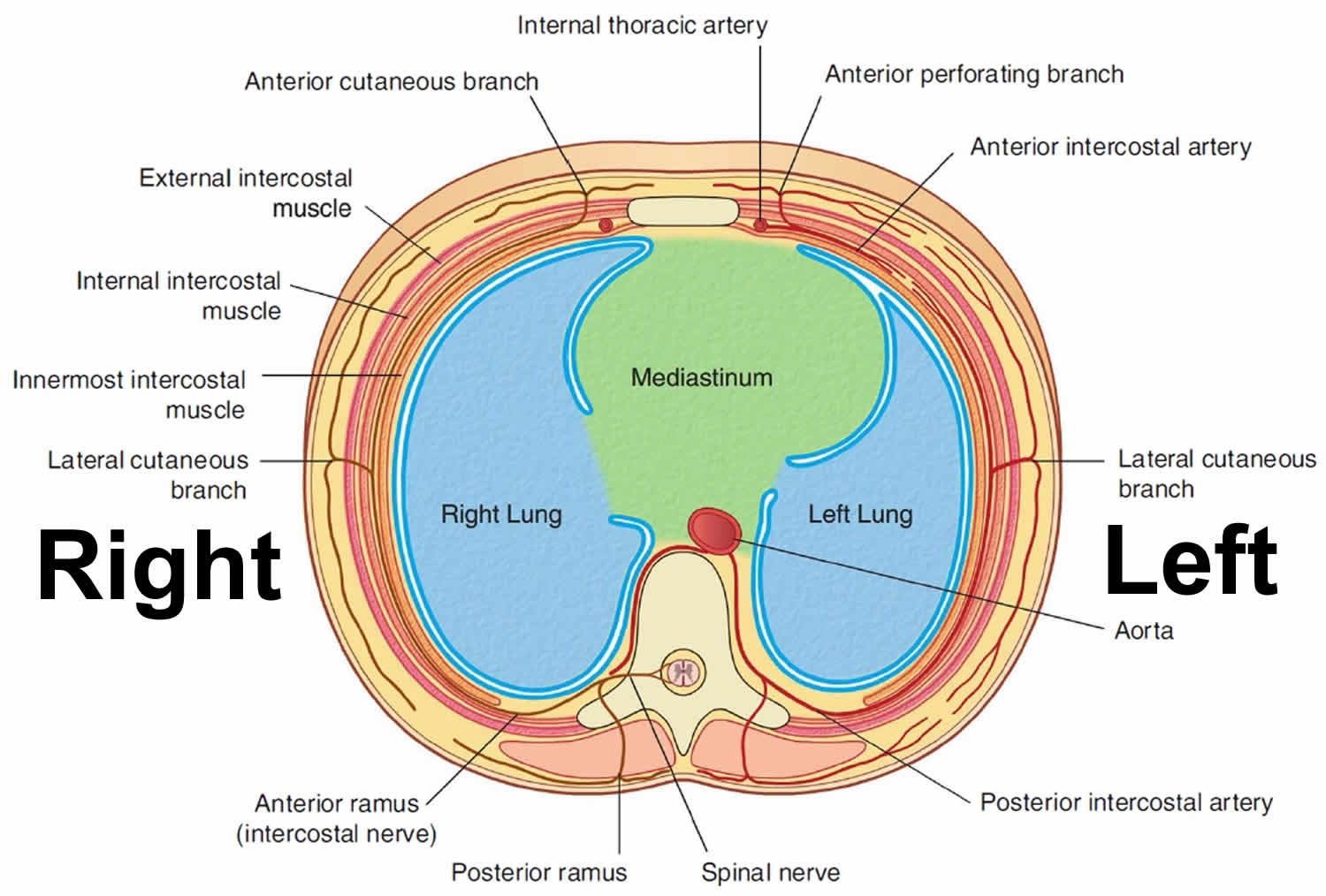
The pericardium is located within the middle mediastinum, which is part of the thoracic cavity surrounded by a pleural cavity on both sides (see Figure 3).
Pericardium layers
Pericardium consists of two layers: the fibrous and the serous pericardium.
The Fibrous Pericardium (parietal pericardium) is a conical-shaped sac. Its apex is fused with the roots of the great vessels at the base of the heart. Its broad base overlies the central fibrous area of the diaphragm with which it is fused. Weak sterno-pericardial ligaments connect the anterior aspect of the fibrous pericardium to the sternum.
The Serous Pericardium (visceral pericardium) is a layer of serosa that lines the fibrous pericardium (parietal pericardium), which is reflected around the roots of the great vessels to cover the entire surface of the heart (visceral pericardium). Between the parietal and visceral pericardium is a potential space that may be filled with a small amount of fluid. The part of the visceral pericardium that covers the heart, but not the great vessels is called the Epicardium.
As the serous pericardium (visceral pericardium) reflects off various cardiac structures, it forms two sinuses: the Transverse Sinus and the Oblique Sinus. The oblique sinus is a cul-de-sac extending superiorly from the inferior vena cava between the two left pulmonary veins on one side and the two right pulmonary veins on the other. Its anterior wall is formed by the posterior wall of the left atrium, between the four pulmonary veins. The transverse sinus is open at both ends and formed by the reflection of visceral serosal pericardium from the posterior aspects of the aortic and pulmonary trunks over to the anterior aspect of the atrium. Thus, a finger in the transverse sinus will pass behind the aortic and pulmonary trunks but in front of the superior vena cava on the right and the left atrial appendage on the left.
The parietal pleura is densely adherent to the outside of the fibrous pericardium, except for a small, bare area in the front from the midline to the apex and posteriorly where the pericardium touches the esophagus.
What is the function of the pericardium?
The pericardial sac positions the heart in the mediastinum and limits its motion while providing a lubricated slippery surface for the heart to beat inside and the lungs to move outside. The oblique sinus provides expansion space for the left atrium.
The pericardium prevents the excessive dilatation of the heart, and in pathological states, can limit the overfilling of the heart, which would result in a low cardiac out.
The pericardium also influences the pressure-volume relationships of cardiac chambers by providing a limited space for the heart as a whole. It equalizes hydrostatic, inertial, and gravitational forces, maintains the geometry of the left ventricle, and acts as a mechanical barrier to infection.
During cardiac surgery, the pericardium needs to be incised to access the heart. Such handling of the pericardium can result in inflammation and thickening of the pericardium. This may cause the pericardium to become attached to the back of the sternum around the incision site, making repeat surgery difficult.
A needle passed just below and to the left of the xiphoid process and pointed towards the left shoulder will pass above the diaphragm to enter the pericardial space next to the right ventricle. This approach often is used to drain pericardial effusions but also can be used as access for performing catheter-based procedures over the epicardial surface of the heart, such as catheter ablation for arrhythmias 3.
Pericardium disease
Pericarditis
Inflammation of the pericardium is called pericarditis. Its origin can be infectious, immunologic, metabolic, neoplastic, traumatic, or idiopathic. Myocardial infarction also may cause localized pericarditis of the area overlying the infarct. Pericarditis often produces “pleuritic” pain in the center of the chest that radiates to the back and varies with deep breathing. It increases upon lying flat and is relieved by sitting upright. The condition is usually short-lived and is treated with anti-inflammatory pain medications and colchicine.
Pericardial Effusion
Normally the pericardium contains only a few milliliters of fluid that act as a lubricant, but pericarditis and other conditions may lead to the pericardium filling with hundreds of milliliters of exudative or transudative fluid. This is called pericardial effusion and can be easily recognized on an echocardiogram. A chest x-ray may also show an enlarged and bag-like cardiac shadow when a significant pericardial effusion is present.
Cardiac Tamponade
Pericardial effusion or bleeding into the pericardium due to injury can limit the space available for the expansion of the heart. If this is severe enough to limit venous return, then cardiac output falls drastically as a result of the heart being not able to fill with blood. This condition is called cardiac tamponade. Cardiac tamponade is a life-threatening condition and often requires urgent attention. Treatment consists of draining the pericardial fluid using a needle, placement of a catheter in the pericardium for continuous drainage or surgical creation of a ‘window’ between the pericardial and pleural spaces.
Constrictive Pericarditis
Constrictive pericarditis occurs when long-term inflammation of the pericardium causes thickening, scarring, and often calcified pericardium, which limits the diastolic filling of the ventricles. This can be a complication of acute pericarditis, especially when the cause of pericarditis is neoplastic, bacterial, or radiotherapy.
References- Rehman I, Rehman A. Anatomy, Thorax, Pericardium. [Updated 2018 Dec 9]. In: StatPearls [Internet]. Treasure Island (FL): StatPearls Publishing; 2019 Jan-. Available from: https://www.ncbi.nlm.nih.gov/books/NBK482256
- Goizueta AA, Bordoni B. StatPearls [Internet]. StatPearls Publishing; Treasure Island (FL): Jan 13, 2019. Anatomy, Thorax, Lung Pleura And Mediastinum
- Abdelnaby M, Almaghraby A, Saleh Y, Abayazeed R. Pericardial sarcoma. BMJ Case Rep. 2018 Oct 07;2018


