What is scleredema
Scleredema is a form of cutaneous mucinosis of unknown cause, a diverse group of rare skin conditions that are characterized by an accumulation of mucin (a jelly-like complex carbohydrate substance) in the skin. Signs and symptoms of this condition include hardening and thickening of the skin which may restrict movement. Skin in affected areas may be red or brown and often has an ‘orange-skin’ appearance. There are three forms of the condition which vary by disease course and long term outlook (prognosis). Although the underlying cause is currently unknown, each form is associated with a different condition: infection (type 1), blood abnormalities (type 2), and diabetes (type 3). In some cases, scleredema resolves spontaneously on its own, while in other affected people, the condition persists for long periods of time. Due to the rarity of the condition, there is no standard treatment 1
Scleredema should not be confused with “scleroderma”, in which the skin is fibrotic (morphoea and systemic sclerosis). Histologically, scleredema differs from scleroderma due to the presence of adnexa and of large interfibrous spaces containing acid mucopolysaccharides 2. Clinically, scleredema differs from scleroderma due to its sudden onset, sometimes after an infection. Also its healing can be very rapid. Moreover, skin involvement in scleredema is more diffuse and prevails on the trunk and root of the limbs, although the head, forearms and hands can be affected. In scleredema the borders of the lesions are not well defined as compared with scleroderma. Above all in scleredema the skin is normally colored without erythema and porcelain-like sclerosis and, in case of healing, the skin does not show dyschromic residua.
Scleredema affects adults. In many cases, people with scleredema have an underlying systemic disease. These include:
- Diabetes mellitus
- Hyperparathyroidism
- Sjögren syndrome
- Rheumatoid arthritis
- Multiple myeloma
- Malignant insulinoma
- HIV infection.
There are three types of scleredema. All three types of scleroedema can restrict movement, but otherwise seldom have serious consequences. Occasionally swallowing and speech can be affected and other organs involved – eyes, tongue, parotid gland, muscles, joints and heart.
Type 1 scleredema
- Type one is the acute type of scleredema, and typically starts with an infection, most often Streptococcus pyogenes, the cause of tonsillitis. It mainly affects middle aged women and children. Hardening of the skin of the face and neck quickly develops and spreads to the upper trunk and arms. It usually improves spontaneously over six months to two years.
Type 2 scleredema
- Type 2 scleredema is not associated with infection. It starts more slowly and persists. Abnormal levels of an abnormal paraprotein (immunoglobulin) in the blood may occur, sometimes due to multiple myeloma.
Type 3 scleredema
- Type 3, scleredema adultorum of Buschke, also called scleredema diabeticorum, occurs in diabetics, particularly adult men. It is very persistent. The skin of the neck and upper back slowly thickens over months or years.
Scleredema vs Scleroderma
Scleroderma means hard skin, is the name for a group of rare diseases that cause abnormal growth of connective tissue resulting in patches of tight, hard skin and connective tissues. Some forms of scleroderma can also damage your blood vessels and internal organs. Connective tissue is the material inside your body that gives your tissues their shape and helps keep them strong. In scleroderma, the tissue gets hard or thick. It can cause swelling or pain in your muscles and joints.
Scleroderma results from an overproduction and accumulation of collagen in body tissues. Collagen is a fibrous type of protein that makes up your body’s connective tissues, including your skin.
Doctors don’t know exactly what causes the abnormal collagen production to begin, but the body’s immune system appears to play a role. Most likely, scleroderma is caused by a combination of factors, including immune system problems, genetics and environmental triggers.
Scleroderma affects women more often than men and most commonly occurs between the ages of 30 and 50. While there is no cure for scleroderma, a variety of treatments can ease symptoms and improve quality of life.
There are many different types of scleroderma. In some people, scleroderma affects only the skin. But in many people, scleroderma also harms structures beyond the skin, such as blood vessels, internal organs and the digestive tract (systemic scleroderma). Signs and symptoms vary, depending on which type of scleroderma you have.
Symptoms of scleroderma include:
- Calcium deposits in connective tissues (calcinosis)
- Raynaud’s phenomenon, a narrowing of blood vessels in the hands or feet in response to cold temperatures or emotional distress. When this happens, your fingers or toes may turn blue or feel painful or numb. Raynaud’s disease also occurs in people who don’t have scleroderma.
- Swelling of the esophagus, the tube between your throat and stomach
- Skin. Nearly everyone who has scleroderma experiences a hardening and tightening of patches of skin. These patches may be shaped like ovals or straight lines, or cover wide areas of the trunk and limbs. The number, location and size of the patches vary by type of scleroderma. Skin can appear shiny because it’s so tight, and movement of the affected area may be restricted.
- Red spots on your hands and face
- Digestive system. Scleroderma can cause a variety of digestive symptoms, depending on which part of the digestive tract is affected. If the esophagus is affected, you might have heartburn or difficulty swallowing. If the intestines are affected, you might have cramps, bloating, diarrhea or constipation. Some people who have scleroderma may also have problems absorbing nutrients if their intestinal muscles aren’t properly moving food through the intestines.
- Heart, lungs or kidneys. Scleroderma can affect the function of the heart, lungs or kidneys to varying degrees. These problems, if left untreated, can become life-threatening.
Scleroderma complications range from mild to severe and can affect your:
- Fingertips. The variety of Raynaud’s disease that occurs with systemic scleroderma can be so severe that the restricted blood flow permanently damages the tissue at the fingertips, causing pits or skin sores. In some cases, the tissue on the fingertips may die and require amputation.
- Lungs. Scarring of lung tissue can result in reduced lung function, which can impact your ability to breathe and tolerance for exercise. You may also develop high blood pressure in the arteries to your lungs.
- Kidneys. When scleroderma affects your kidneys, you can develop elevated blood pressure and an increased level of protein in your urine. More-serious effects of kidney complications may include renal crisis, which involves a sudden increase in blood pressure and rapid kidney failure.
- Heart. Scarring of heart tissue increases your risk of abnormal heartbeats and congestive heart failure, and can cause inflammation of the membranous sac surrounding your heart. Scleroderma can also raise the pressure on the right side of your heart and cause it to wear out.
- Teeth. Severe tightening of facial skin can cause your mouth to become smaller and narrower, which may make it hard to brush your teeth or to even have them professionally cleaned. People who have scleroderma often don’t produce normal amounts of saliva, so the risk of dental decay increases even more.
- Digestive system. Digestive problems associated with scleroderma can lead to heartburn and difficulty swallowing. It can also cause bouts of cramps, bloating, constipation or diarrhea.
- Sexual function. Men who have scleroderma may experience erectile dysfunction. Scleroderma may also affect the sexual function of women by decreasing sexual lubrication and constricting the vaginal opening.
No one knows what causes scleroderma. It is more common in women. It can be mild or severe. Doctors diagnose scleroderma using your medical history, a physical exam, lab tests, and a skin biopsy. There is no cure, but various treatments can control symptoms and complications.
Anyone can get scleroderma, but it does occur much more often in women than in men. Several combined factors appear to influence the risk of developing scleroderma:
- Genetics. People who have certain gene variations appear to be more likely to develop scleroderma. This may explain why a small number of scleroderma cases appear to run in families and why certain types of scleroderma are more common for certain ethnic groups. For example, Choctaw Native Americans are more likely to develop the type of scleroderma that affects internal organs.
- Environmental triggers. Research suggests that, in some people, scleroderma symptoms may be triggered by exposure to certain viruses, medications or drugs. Repeated exposure — such as at work — to certain harmful substances or chemicals also may increase the risk of scleroderma.
- Immune system problems. Scleroderma is believed to be an autoimmune disease. This means that it occurs in part because the body’s immune system begins to attack the connective tissues. In 15 to 20 percent of cases, a person who has scleroderma also has symptoms of another autoimmune disease, such as rheumatoid arthritis, lupus or Sjogren’s syndrome.
Scleroderma symptoms
There are 2 forms of localized scleroderma:
- Symptoms start with red patches of skin that thicken into hard, oval-shaped areas. The patches later become white in the middle with purple borders. Patches often appear on your chest, stomach, and back, but also can occur on your face, arms, and legs. You may have 1 or more patches as small as half an inch or as large as 12 inches in diameter.
- The main symptom is a line or band of skin that thickens and changes color. This line can appear on your arm, leg, or forehead. Linear scleroderma is more common in children.
There are 2 forms of systemic scleroderma:
- Limited scleroderma progresses gradually. It affects the skin on your fingers, hands, lower arms, legs, and face. It causes patches of skin to become thick and firm, and change color. People who have this also may have Raynaud’s disease or problems with frequent heartburn. Limited scleroderma also can affect your lungs, esophagus, and blood vessels.
- Diffuse scleroderma progresses quickly. Symptoms include fatigue, loss of appetite, and joint swelling and pain. It can affect the skin all over your body, causing it to swell and become tight, shiny, and itchy. Over time, your skin may go back to normal. Diffuse scleroderma also can damage internal organs, such as your intestines, lungs, kidneys, and heart.
Scleroderma treatment
Your doctor will choose the right treatment for you depending on your type and symptoms. Treatment focuses on relieving symptoms and preventing more damage. Possible treatments include medicine, such as creams for your skin, or dietary and lifestyle changes. Your doctor also may recommend physical or occupational therapy to help manage your pain. Cosmetic surgery may help to lessen the effects of scleroderma on your skin.
Localized scleroderma sometimes goes away on its own. If scleroderma has caused internal damage, your doctor may work with specialists to treat your condition. For example, if scleroderma affects your heart, they may want to work closely with a cardiologist.
Medications
There is no medication that can cure or stop the overproduction of collagen that is characteristic of scleroderma. But a variety of medications can help control scleroderma symptoms and prevent complications. For example, your doctor may recommend medications to:
- Treat or slow skin changes. Steroid creams or pills may help reduce swelling and joint pain, loosen stiff skin, and slow the development of new skin changes.
- Dilate blood vessels. Blood pressure medications that dilate blood vessels may help prevent lung and kidney problems and may help treat Raynaud’s disease.
- Suppress the immune system. Drugs that suppress the immune system, such as those taken after organ transplants, may help reduce scleroderma symptoms.
- Reduce digestive symptoms. Pills to reduce stomach acid can help relieve heartburn. Antibiotics and medications that help move food through the intestines may help reduce bloating, diarrhea and constipation.
- Prevent infections. Antibiotic ointment, cleaning and protection from the cold may help prevent infection of fingertip ulcers caused by Raynaud’s disease.
- Regular influenza and pneumonia vaccinations can help protect lungs that have been damaged by scleroderma.
- Relieve pain. If over-the-counter pain relievers don’t help enough, you can ask your doctor to prescribe stronger medications.
Therapies
Physical or occupational therapists can help you:
- Manage pain
- Improve your strength and mobility
- Maintain independence with daily tasks
Surgery
Used as a last resort, surgical options for scleroderma complications may include:
- Amputation. If finger sores caused by severe Raynaud’s disease have progressed to the point that the fingertip tissue begins to die, amputation may be necessary.
- Lung transplants. People who have developed severe lung problems may be candidates for lung transplants.
Scleredema causes
The cause of scleredema is currently unknown; however, it is associated with a number of conditions. Many patients have no history of an acute antecedent illness. Note the following:
- Febrile illness with streptococcal infections: An upper respiratory tract infection (typically pharyngitis) is the most common cause of scleredema in patients with type 1 scleredema 3. Scleredema following scabies infestation, as a result of superinfection with Streptococcus, has been reported 4.
- Other infections: The onset of scleredema has also been associated with cytomegalovirus infection, influenza, measles, pertussis, mumps, diphtheria, typhus fever, encephalitis, and dental abscesses.
- Myelomatous disorders: A subset of patients with type 2 scleredema has shown a well-established relationship between scleredema with paraproteins and multiple myeloma 5. One analysis of 52 patients with type 2 scleredema revealed 25% had plasma cell dyscrasia, including 3 patients with multiple myeloma and 10 with monoclonal gammopathy of unknown significance 6.
- Diabetes mellitus: Patients usually have a longstanding history of diabetes, often poorly controlled 7.
- Trauma: Rare reports exist of scleredema occurring after trauma to the affected area. One report 8 has described geometric scleredema due to mechanical stress.
- Drug reaction: In 2005, scleredema was reported in association with the use of infliximab; although rare, treating physicians should consider the possibility of an adverse drug reaction as the underlying etiology 9.
Scleredema symptoms
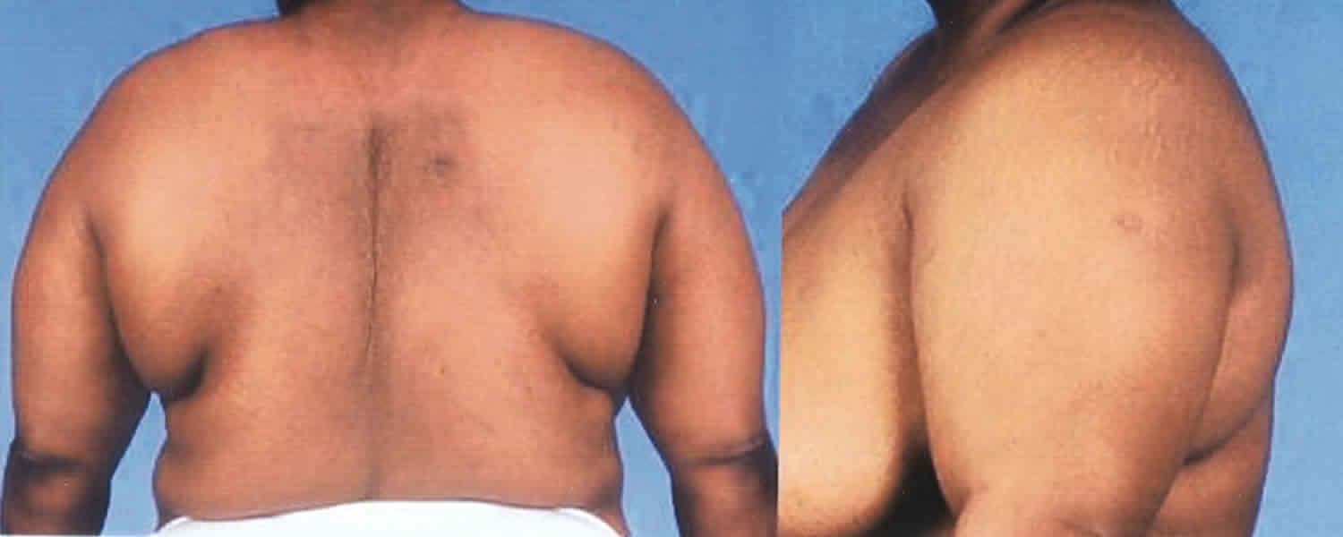
Patients report stiff or hard skin. Scleredema presents as ill-defined, woody, nonpitting, indurated plaques of the skin. The affected areas are firm and woody plaques, sometimes slightly red or brown and often with a ‘peau d’orange’ (orange-skin) appearance. The face may appear expressionless and there can be difficulty opening the mouth. The rapidity of onset and locations of involvement differ based on the clinical subgroup.
Scleredema is usually most evident in the upper part of the body, specifically the face, the neck, the trunk, and the extremities in type 1 and 2 subgroups. However, a case of scleredema confined to the thighs 10 and a case of scleredema confined to the periocular region 11 have been reported. Hands and feet are typically spared in scleredema, in contrast to systemic sclerosis, which has sclerodactyly. Scleredema patients with extensive facial involvement may appear expressionless and may have difficulty in opening their mouths. They may have difficulty with tongue protrusion.
A thorough history regarding preceding illnesses, history of diabetes, and a review of systems should be performed to help identify less commonly associated extracutaneous manifestations (lungs, heart, trouble eating or talking, or muscle weakness) or, rarely, reported associations with malignancies.
Physical examination should include evaluation of range of motion of the neck, upper extremities, and tongue. Auscultation of the heart and lungs should also be performed.
Scleredema can be categorized into three clinical subgroups. Each has a different history, course, and prognosis. Note the following:
Type 1 postinfectious scleredema
This subgroup was historically referred to as scleredema adultorum. However, this is considered by some to be a misnomer because most pediatric patients fall into this group. Patients report a hardening of the skin a few weeks after a febrile illness, most commonly an upper or lower respiratory tract streptococcal infection 3. The skin hardening progresses rapidly, first involving the face and neck, then spreading distally to involve the trunk and proximal upper limbs in a symmetric manner. Hands and feet are typically spared. Complications may include difficulty in smiling, opening the mouth, limited range of motion, and, in severe cases, involvement of the pharynx or tongue can lead to dysphagia or dysphonia. The condition usually clears spontaneously in 6 months to 2 years. The duration is not affected by the use of antibiotics.
Type 2 scleredema associated plasma dyscrasia
This subgroup includes patients whose disease tends to occur insidiously, progressing slowly over many years, with no history of preceding illness. Rimon et al identified 52 cases of type 2 scleredema in the world literature from 1963 to 1986, of which 25% had plasma cell dyscrasias, including 3 with multiple myeloma and 10 with monoclonal gammopathy of unknown significance. They also noted that the gammopathies were diagnosed on average 10 years after the onset of scleredema skin changes 6. IgG monoclonal gammopathy is most common, followed by an IgA type. Spontaneous remission is much less likely to occur than in the type 1 subgroup.
Type 3 scleredema diabeticorum
This subtype of scleredema tends to occur more often in middle-aged males (at a reported 10:1 ratio), often obese, with longstanding, often uncontrolled, diabetes mellitus. In a 2015 multicenter study, 30 (68%) of 44 patients with scleredema had poorly controlled diabetes, with a type 2‒to‒type 1 ratio of 6.5:1 12. Subtle skin hardening of the upper back begins in an insidious manner, progressing slowly over many years, to involve the upper back, neck, and shoulders with associated erythema; often, a pebbled appearance may evolve. Patients typically experience a more protracted course that is refractory to therapy. Control of the hyperglycemia does not improve the scleredema 13.
Scleredema complications
Scleredema complications include the following:
- Limited range of motion
- Poor wound healing
- Recurrent skin infections
- Restrictive lung disease
- Dysarthria
- Dysphagia
- Difficulty in closing the eyes
- Death (rare)
Scleredema diagnosis
Scleredema diagnosis is confirmed by skin biopsy, which shows mucin deposits between collagen bundles in the dermis.
No imaging studies are necessary for the diagnosis of scleredema.
Isolated case reports have described scleredema occurring in association with internal malignancies, for example, carcinoma of the gall bladder 14, malignant insulinoma 15, carcinoid tumor 16. Imaging studies are warranted if this is suggested based on clinical findings.
Some investigators have proposed ultrasonic imaging of skin thickness in scleredema patients as a method to monitor the response to therapy. Computed tomography may demonstrate induration in the involved soft tissues, although it is not often necessary. MRI may highlight the involved areas as well, and some have advocated for MRI usage to determine the extent of disease and monitor progression in select cases 17.
Laboratory Studies
Laboratory studies can be helpful to identify an associated condition:
- A throat culture should be obtained in scleredema patients to exclude group A streptococcal infection.
- Antistreptolysin-O (ASO) titers should be obtained in scleredema patients to exclude a recent infection with group A streptococci in a patient without clinically apparent pharyngitis or other infectious etiology.
- Fasting blood glucose or glycosylated hemoglobin (HbA1C) measurements should be obtained in scleredema patients to rule out diabetes mellitus.
- Serum protein electrophoresis (SPEP) and immunofixation should be performed in scleredema patients to exclude a monoclonal gammopathy. Both paraproteinemias (also known as monoclonal gammopathies) and multiple myelomas have been reported in patients with scleredema (type 2). The most commonly identified paraprotein is IgG having a kappa light chain; however, an IgA type can occur. Blood dyscrasias usually appear several years after the onset of scleredema. The interval between diagnosis of scleredema and the detection of paraprotein can occur before, concurrently, or after the diagnosis of scleredema. In one report, a delayed paraproteinemia was detected on average up to 10 years following the diagnosis of scleredema 6. However, other sources quote a median detection of paraproteinemia 2.5 years after diagnosis.
Scleredema treatment
The best way to treat scleredema is unknown, because of its rarity. Treatment is unnecessary for postinfectious scleredema as it is typically self-limited. However, in rapidly progressive, fulminant cases, systemic therapy is recommended 18. Physical therapy, intravenous immunoglobulin, or UVA-1 phototherapy could be considered. Some benefit has been reported with the following:
- PUVA
- Cyclophosphamide
- Oral corticosteroids
- Ciclosporin
- UVA1 phototherapy
- Electron beam radiation.
Types 2 and 3 scleredema associated with a monoclonal gammopathy or diabetes typically does not regress spontaneously, and no therapy is consistently effective. Most therapeutic successes described are limited to single case reports or small case series; there are no comparative data and no approved algorithm for scleredema treatment. Expert opinion also differs. In general, it is agreed that treatment of an identified cause is warranted. For type 1 scleredema, antimicrobial agents are used if indicated. For type 2 scleredema, annual screening for a lymphoproliferative disorder, and for those with a monoclonal gammopathy, referral to a hematologist are, indicated. Physical therapy should be considered for all types. However, expert opinion often differs based on personal experience. In a 2017 publication of the European dermatology forum S1-guideline on the diagnosis and treatment of sclerosing diseases of the skin, first-line recommendations were UVA-1 phototherapy or psoralen with UVA (PUVA) 19.
Treatments described in case reports and small case series
A number of therapies, including systemic steroids 12, cyclosporine 12, methotrexate 20, high-dose penicillin 21, penicillamine, electron beam 22 and glycemic control with prostaglandin E1 (PGE1) 7 have been reported with mixed success. A report using tamoxifen for its antifibrotic properties noted improvements in two patients 23. Incidental improvement was noted in one case of scleredema during allopurinol therapy 24. A 2018 report described three cases of successful treatment with tranilast 25.
Case reports in the literature describe improvement with UVA-1 phototherapy 26, narrow-band UVB phototherapy 27 and PUVA either administered systemically or topically 28. Case reports also describe these successfully used in combination with colchicine 29, methotrexate 30 and physiotherapy 31. In the authors’ experience 32, narrow-band UVB phototherapy was not successful.
Physical modalities, such as ultrasonic massage with physical therapy, may improve range of motion and quality of life for some patients alone or in combination with other therapies 33.
In cases associated with myeloma, chemotherapy directed at the hematologic malignancy has been reported to result in concomitant improvement of the skin disease 34. For patients with paraproteinemia, extracorporeal photophoresis has been used 35.
Multiple reports described efficacy with intravenous immunoglobulin therapy in postinfectious, diabetic, and monoclonal gammopathy–associated scleredema that were refractory to more standard therapies 36.
A 2015 multicenter retrospective review of associations and treatment response among 44 patients was published, finding some response to physiotherapies, systemic steroids, UVA-1, PUVA, and a few combination therapies in a limited number of patients 12.
Treatment of associated conditions
Appropriate antibiotic therapy should be started in scleredema patients if infection is detected, although antibiotics do not appear to shorten the course of skin findings in scleredema 37.
Treatment of detected blood dyscrasias or diabetes mellitus should be completed.
Scleredema prognosis
The course of scleredema is unpredictable. Patients with type 1 scleredema, particularly pediatric patients, typically have a self-limited course, with the disease resolving in 6 months to 2 years. However, a number of reports exist in which these patients had a protracted course or, rarely, long-term cardiac or skeletal muscle involvement. Patients with types 2 and 3 typically have a slowly progressive or unremitting course over many years. Reports of relapses following apparent improvement also exist.
Typically, scleredema is a benign process limited to the skin. The skin changes of scleredema can cause limitations of movement. Rarely, this can include reductions in joint mobility, difficulty opening eyes or mouth, or restrictive lung disease in extensive trunk involvement.
Although the disorder is usually restricted to the skin, the tongue, pharynx, esophagus, skeletal muscle, and cardiac muscle may rarely be affected. Rare case reports of morbidity due to extracutaneous collagen and mucin deposition include the following:
- Tongue involvement: Unlike scleroderma, the tongue has been reported in scleredema, resulting in dysarthria and difficulty with mastication and tongue protrusion.
- Cardiac involvement: This may result in cardiomyopathy, heart failure, arrhythmias, pericardial effusion, and unexplained murmurs 38.
- Other organs involvement: Skeletal muscles, ocular muscles, the pharynx, the liver, parotid glands, pleurae, the peritoneum, and the spleen reportedly may be involved.
- Esophageal involvement: In one report, a patient with scleredema developed dysphagia, presumably from scleredema of the upper part of the esophagus. Esophageal biopsy was not performed on this patient 39.
Death from scleredema is rare. However, reports have described patients dying from the complications of cardiac or respiratory involvement 40.
References- Scleredema. https://emedicine.medscape.com/article/1066175-overview
- Garofalo L., Bonifazi E. 2006. Self-healing scleredema. Eur. J. Pediat. Dermatol. 16 (4): 217.
- Alp H, Orbak Z, Aktas A. Scleredema adultorum due to streptococcal infection. Pediatr Int. 2003 Feb. 45(1):101-3.
- Malhotra AK, Sethuraman G, Das AK, Sharma VK. Scleredema following scabies infestation. Pediatr Dermatol. 2008 Jan-Feb. 25(1):136-8.
- Angeli-Besson C, Koeppel MC, Jacquet P, Andrac L, Sayag J. Electron-beam therapy in scleredema adultorum with associated monoclonal hypergammaglobulinaemia. Br J Dermatol. 1994 Mar. 130(3):394-7.
- Rimon D, Lurie M, Storch S, et al. Cardiomyopathy and multiple myeloma. Complications of scleredema adultorum. Arch Intern Med. 1988 Mar. 148(3):551-3.
- Ikeda Y, Suehiro T, Abe T, et al. Severe diabetic scleredema with extension to the extremities and effective treatment using prostaglandin E1. Intern Med. 1998 Oct. 37(10):861-4.
- Tsunemi Y, Ihn H, Fujita H, Asashima N, Saeki H, Tamaki K. Square-shaped scleredema in the back: probably induced by mechanical stress. Int J Dermatol. 2005 Sep. 44(9):769-70.
- Ranganathan P. Infliximab-induced scleredema in a patient with rheumatoid arthritis. J Clin Rheumatol. 2005 Dec. 11(6):319-22.
- Farrell AM, Branfoot AC, Moss J, Papadaki L, Woodrow DF, Bunker CB. Scleredema diabeticorum of Buschke confined to the thighs. Br J Dermatol. 1996 Jun. 134(6):1113-5.
- Ioannidou DI, Krasagakis K, Stefanidou MP, Karampekios S, Panayiotidis J, Tosca AD. Scleredema adultorum of Buschke presenting as periorbital edema: a diagnostic challenge. J Am Acad Dermatol. 2005 Feb. 52(2 Suppl 1):41-4.
- Rongioletti F, Kaiser F, Cinotti E, Metze D, Battistella M, Calzavara-Pinton PG, et al. Scleredema. A multicentre study of characteristics, comorbidities, course and therapy in 44 patients. J Eur Acad Dermatol Venereol. 2015 Dec. 29 (12):2399-404.
- Tran K, Boyd KP, Robinson MR, Whitlow M. Scleredema diabeticorum. Dermatol Online J. 2013 Dec 16. 19(12):20718.
- Manchanda Y, Das S, Sharma VK, Srivastava DN. Scleredema associated with carcinoma of the gall bladder. Br J Dermatol. 2005 Jun. 152(6):1373-4.
- Santos-Juanes J, Osuna CG, Iglesias JR, De Quiros JF, del Río JS. Treatment with chemotherapy of scleredema associated with Ig A myeloma. Int J Dermatol. 2001 Nov. 40(11):720-1.
- Yu JI, Park W, Lee KK, Park W. Scleredema adultorum of Buschke associated with a carcinoid tumor. Int J Dermatol. 2009 Jul. 48(7):784-6.
- Kurihara Y, Kokuba H, Furue M. Case of diabetic scleredema: diagnostic value of magnetic resonance imaging. J Dermatol. 2011 Jul. 38(7):693-6.
- Sommer LL, Heymann WR. Fulminans in dermatology: a call to action: a recommendation for consideration of the term scleredema fulminans. J Clin Aesthet Dermatol. 2014 Jun. 7(6):42-5.
- Knobler R, Moinzadeh P, Hunzelmann N, et al. European Dermatology Forum S1-guideline on the diagnosis and treatment of sclerosing diseases of the skin, Part 1: localized scleroderma, systemic sclerosis and overlap syndromes. J Eur Acad Dermatol Venereol. 2017 Sep. 31 (9):1401-1424.
- Seyger MM, van den Hoogen FH, de Mare S, van Haelst U, de Jong EM. A patient with a severe scleroedema diabeticorum, partially responding to low-dose methotrexate. Dermatology. 1999. 198 (2):177-9.
- Krasagakis K, Hettmannsperger U, Trautmann C, Tebbe B, Garbe C. Persistent scleredema of Buschke in a diabetic: improvement with high-dose penicillin. Br J Dermatol. 1996 Mar. 134(3):597-8.
- Lee MW, Choi JH, Sung KJ, Moon KC, Koh JK. Electron beam therapy in patients with scleredema. Acta Derm Venereol. 2000 Jul-Aug. 80 (4):307-8.
- Alsaeedi SH, Lee P. Treatment of scleredema diabeticorum with tamoxifen. J Rheumatol. 2010 Dec. 37(12):2636-7.
- Lee FY, Chiu HY, Chiu HC. Treatment of acquired reactive perforating collagenosis with allopurinol incidentally improves scleredema diabeticorum. J Am Acad Dermatol. 2011 Oct. 65(4):e115-7.
- Sun M, Yang F, Hou M. Successful Treatment of Scleredema Diabeticorum With Tranilast: Three Case Reports. Diabetes Care. 2018 Apr. 41 (4):e40-e41
- Thumpimukvatana N, Wongpraparut C, Lim HW. Scleredema diabeticorum successfully treated with ultraviolet A1 phototherapy. J Dermatol. 2010 Dec. 37(12):1036-9.
- Yoshimura J, Asano Y, Takahashi T, Uwajima Y, Kagami S, Honda H, et al. A case of scleredema adultorum successfully treated with narrow-band ultraviolet B phototherapy. Mod Rheumatol. 2016. 26 (2):302-6.
- Martín C, Requena L, Manrique K, Manzarbeitia FD, Rovira A. Scleredema diabeticorum in a patient with type 2 diabetes mellitus. Case Rep Endocrinol. 2011. 2011:560273.
- Kokpol C, Rajatanavin N, Rattanakemakorn P. Successful Treatment of Scleredema Diabeticorum by Combining Local PUVA and Colchicine: A Case Report. Case Rep Dermatol. 2012 Sep. 4(3):265-8.
- Shazzad MN, Azad AK, Abdal SJ, Afrose R, Rahman MM, Haq SA. Scleredema Diabeticorum – A Case Report. Mymensingh Med J. 2015 Jul. 24 (3):606-9.
- Martín C, Requena L, Manrique K, Manzarbeitia FD, Rovira A. Scleredema diabeticorum in a patient with type 2 diabetes mellitus. Case Rep Endocrinol. 2011. 2011:560273
- Scleredema Treatment & Management. https://emedicine.medscape.com/article/1066175-treatment
- Bray SM, Varghese S, English JC 3rd. Ultrasonic massage and physical therapy for scleredema: improving activities of daily living. Arch Dermatol. 2010 Apr. 146(4):453-4
- Rota E, Nallino MG, Bainotti S, Formica M. Nephrogenic systemic fibrosis: an unusual scleroderma-like fibrosing disorder. Rheumatol Int. 2009 Aug 20
- Stables GI, Taylor PC, Highet AS. Scleredema associated with paraproteinaemia treated by extracorporeal photopheresis. Br J Dermatol. 2000 Apr. 142(4):781-3.
- Eastham AB, Femia AN, Velez NF, Smith HP, Vleugels RA. Paraproteinemia-associated scleredema treated successfully with intravenous immunoglobulin. JAMA Dermatol. 2014 Jul 1. 150(7):788-9.
- Rani JD, Patil SG, Murthy ST, Koshy AV, Nagpal D, Gupta S. Juvenile scleredema of Buschke. J Contemp Dent Pract. 2012 Jan 1. 13(1):111-4.
- Isaac A, Costa I, Leal I. Scleredema of Buschke in a child with cardiac involvement. Pediatr Dermatol. 2010 May-Jun. 27(3):315-7.
- Wright RA, Bernie H. Scleredema adultorum of Buschke with upper esophageal involvement. Am J Gastroenterol. 1982 Jan. 77(1):9-11.
- Sansom JE, Sheehan AL, Kennedy CT, Delaney TJ. A fatal case of scleredema of Buschke. Br J Dermatol. 1994 May. 130(5):669-70.


